3.5 mm VarIable angle lcp medial column fusion PlatIng SYStem. Part of the 2.7 mm and 3.5 mm VA LCP Midfoot/Hindfoot System
|
|
|
- Alyson Taylor
- 5 years ago
- Views:
Transcription


1 3.5 mm VarIable angle lcp medial column fusion PlatIng SYStem Part of the 2.7 mm and 3.5 mm VA LCP Midfoot/Hindfoot System SurgIcal technique
2
3 table of contents Introduction AO Principles and Indications mm Variable Angle LCP Medial Column 2 Fusion Plates Compression System 5 Surgical Technique 3.5 mm Variable Angle Locking Technique mm Variable Angle LCP Medial Column 13 Fusion Plates Product Information Implants 37 Instruments 40 Set Configurations 45 Image intensifier control 2.7 mm and 3.5 mm Variable Angle LCP Midfoot/Hindfoot System Surgical Technique DePuy Synthes Trauma
4


5 IntroductIon Introduction
6
7 AO Principles and Indications AO Principles In 1958, the AO formulated four basic principles, which have become the guidelines for internal fixation. 1,2 They are: Anatomic reduction Precontoured plates assist reduction to restore length, alignment, and rotation. Anatomic reduction is mandatory for intra-articular fractures to restore joint congruency. Stable fixation Variable angle locking screws create a fixed angle construct, providing angular stability. Preservation of blood supply Preservation of the blood supply to soft tissue and bone by careful handling. Early, active mobilization Plate features combined with AO technique can create an environment for bone healing, expediting a return to optimal function. Indications* The DePuy Synthes 2.7 mm and 3.5 mm Variable Angle LCP Midfoot/Hindfoot System is indicated for fixation of osteotomies, fusions, fractures, nonunions, malunions, and replantations of small bones and small bone fragments in adult and adolescent (aged years) patients, including the foot and ankle, and particularly in osteopenic bone. * these devices have not been evaluated for safety and compatibility in the MR environment. These devices have not been tested for heating and migration in the MR environment. 1 müller ME, Allgöwer M, Schneider R, Willenegger H. Manual of Internal Fixation: Techniques Recommended by the AO-ASIF Group. 3rd ed. Secaucus, NJ: Springer-Verlag; rüedi TP, Buckley RE, Moran CG (eds). AO Principles of Fracture Management. 2nd ed. Stuttgart, New York: Thieme mm Variable Angle LCP Medial Column Fusion Plates Surgical Technique DePuy Synthes Trauma 1


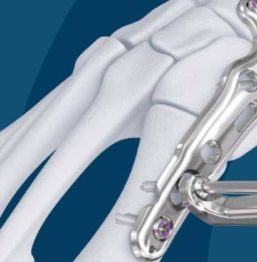
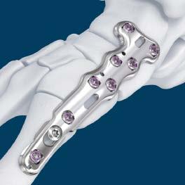
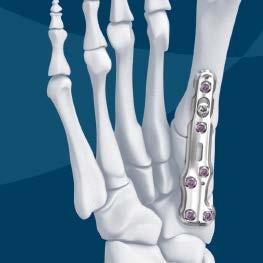
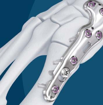
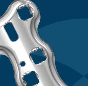
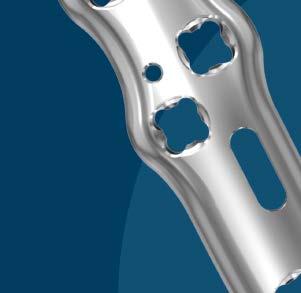
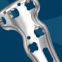
8 3.5 mm Variable Angle LCP Medial Column Fusion Plates 3.5 mm VA LCP Medial Column Fusion Plates are designed with advanced stabilization capabilities for fusion applications, specifically Charcot foot or severe arthritis. Plates are available for applications including dorsomedial, medial, medial with talus extension, and plantar. Innovative compression instrumentation provides the ability to independently compress each joint through the plate using forceps, 2.8 mm wires, and posts. Compression slots provide the ability to achieve 4 mm 6 mm of compression through the plate Variable Angle Locking Technology provides the ability to adapt screw trajectory to varied patient anatomy and to target specific areas of cortical bone. 3.5 mm VA LCP Medial Column Fusion Plate, 78 mm Designed for arthrodesis of the first naviculocuneiform and tarsometatarsal joints 3.5 mm VA LCP Medial Column Fusion Plate, 95 mm, Talus Extension Designed for arthrodesis of the talonavicular, first naviculocuneiform and tarsometatarsal joints 2 DePuy Synthes Trauma 3.5 mm Variable Angle LCP Medial Column Fusion Plates Surgical Technique


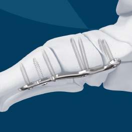
9 3.5 mm Variable Angle LCP Medial Column Fusion Plates 3.5 mm VA LCP Medial Column Fusion Plantar Plate, 78 mm Designed for arthrodesis of the first naviculocuneiform and tarsometatarsal Plantar application for increased biomechanical strength, allowing the plate to be applied on the tension side of the medial column* *References: 1. Vincent James Sammarco, Julie Chevillet. The role of internal fixation in surgery of the Charcot foot and the evolution of super-construct techniques. Current Orthopaedic Practice Volume 21, Number 3, May/June Joshua P. Nadaud, Lew C. Schon. Chronic Charcot Midfoot Reconstruction. Techniques in Foot & Ankle Surgery. Volume 9, Number 3, September naohiro Shibuya, Lacey D. Clawson, Monica R. Agarwal. Suspension and Dynamic Compression of the Medial Column 3.5 mm Variable Angle LCP Medial Column Fusion Plates Surgical Technique DePuy Synthes Trauma 3


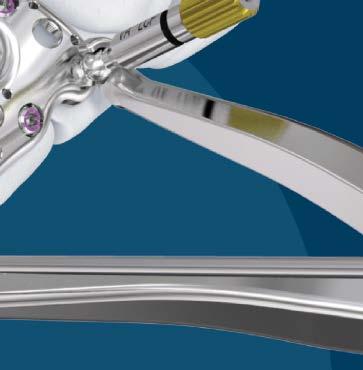
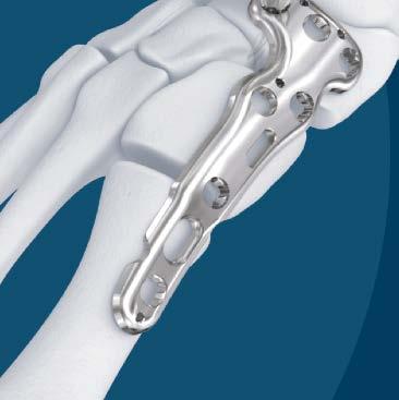
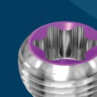
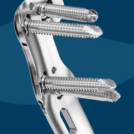
10 3.5 mm Variable Angle LCP Medial Column Fusion Plates Variable Angle Locking Screws are designed to sit flush within the plate holes* to reduce the likelihood of soft-tissue irritation. 3.5 mm Variable Angle Locking Screw holes accept 3.5 mm Variable Angle Locking screws, 3.5 mm Low-Profile Cortex screws, and 3.5 mm Cortex screws. 3.5 mm Locking screws can be inserted at nominal angle Elongated screw holes accept 3.5 mm Low-profile Cortex or 3.5 mm Cortex screws Plates are pre-contoured, with the ability to contour the plate shaft further, depending on the deformity and patient anatomy. Plates feature 1.6 mm K-wires holes for preliminary plate placement. * Variable Angle Locking screws sit flush within the plate when inserted at nominal angle. 4 DePuy Synthes Trauma 3.5 mm Variable Angle LCP Medial Column Fusion Plates Surgical Technique


11 Compression System Compression Forceps Maintain tactile compression during screw insertion using the speed lock mechanism Can be used within the plate through the compression slots or elongated screw hole in the shaft of the plate Compression Wires 2.8 mm diameter, 200 mm overall length Thread lengths from 10 mm to 60 mm in 5 mm increments Allow the plate to be drawn to the bone facilitating preliminary plate fixation Cobalt chromium alloy material that is stiffer than conventional stainless steel* 3.5 mm Compression/Distraction Post for VA Locking Screw Hole Threads into Variable Angle plate holes Use with compression forceps StarDrive recess in head for insertion into plate and final tightening using torque limiter (2.5 Nm) *Cobalt chromium stiffness value: 186 GPa; Stainless Steel stiffness value: 243 MPa 3.5 mm Variable Angle LCP Medial Column Fusion Plates Surgical Technique DePuy Synthes Trauma 5
12


13 3.5 mm VarIable angle locking technique 3.5 mm Variable angle locking technique
14
 insertion Instruments 03.127.002 2.8 mm Variable Angle Double Drill guide with Cone 310.288 2.8 mm Drill Bit, quick coupling, 165 mm or 03.127.004 2.")


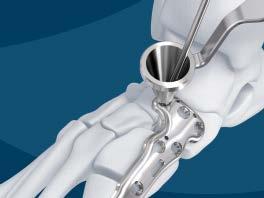
15 3.5 mm Variable Angle Locking Technique Drill for Variable Angle Locking screws: Conical (variable angle) 1 Drill for variable angle locking screws A. Conical (off nominal-axis) insertion Instruments mm Variable Angle Double Drill guide with Cone mm Drill Bit, quick coupling, 165 mm or mm Variable Angle Spherical Drill guide, long mm Calibrated Drill Bit, quick coupling, 250 mm, 95 mm calibration To insert the Variable Angle Locking screw off the nominal axis, insert the cone-shaped side of drill guide in the desired variable angle locking screw hole in the plate. The funnel of the drill guide allows a drilling angle within a 30 cone. When drilling off-axis, the drill guide should remain in place and the drill bit may be aimed in any direction within the cone. Verify the drill bit angle and depth under radiographic imaging to ensure the desired angle has been achieved. If necessary, drill at a different angle and verify again under imaging. Precaution: Avoid excessive re-drilling, especially in poor bone quality. 3.5 mm Variable Angle LCP Medial Column Fusion Plates Surgical Technique DePuy Synthes Trauma 7

. To do so, drill to the desired depth and verify that the plastic stop sits on the drill guide.")
16 3.5 mm Variable Angle Locking Technique Optional Technique The 2.8 mm Variable Angle Spherical Drill Guide can be used to drill for a 3.5 mm Variable Angle Locking Screw. Gently press the spherical tip of the VA drill guide into the variable angle locking hole to ensure the lip of the drill guide stops on the edge of the variable angle locking hole to prevent drilling beyond 15 degrees. Toggle the drill guide to the desired angulation and drill. Note: The screw hole depth can be measured off of the 2.8 mm Calibrated Drill Bit, 250 mm ( ) when using 2.8 mm Spherical Drill Guide ( ). To do so, drill to the desired depth and verify that the plastic stop sits on the drill guide. Remove the drill bit and read the indicated drill depth below the plastic stop. 8 DePuy Synthes Trauma 3.5 mm Variable Angle LCP Medial Column Fusion Plates Surgical Technique

17 3.5 mm Variable Angle Locking Technique Drill for Variable Angle Locking screws: Coaxial (fixed angle) B. Coaxial (fixed angle) insertion Instruments mm Variable Angle Double Drill Guide with Cone mm Drill Bit, quick coupling, 165 mm Additionally available mm Fixed Angle Drill Guide Variable angle locking screws can be inserted into the plate in line with the predefined screw trajectory. Drill to the desired depth. Verify the drill bit depth under radiographic imaging. 3.5 mm Variable Angle LCP Medial Column Fusion Plates Surgical Technique DePuy Synthes Trauma 9

18 3.5 mm Variable Angle Locking Technique USE THE DEPTH GAUGE TO MEASURE FOR THE CORRECT SCREW LENGTH Instrument Depth Gauge for Small Screws Additionally available Percutaneous Depth Gauge for 2.7 mm screws Percutaneous Depth Gauge Adaptor for 3.5 mm screws Use the depth gauge to measure for the correct screw length. 11 DePuy Synthes Trauma 3.5 mm Variable Angle LCP Medial Column Fusion Plates Surgical Technique
19 3.5 mm Variable Angle Locking Technique Insert Variable Angle Locking Screws 2 Insert variable angle locking screws Instruments Handle with Quick Coupling StarDrive Screwdriver Shaft, quick coupling, T15 Insert the correct length variable angle locking screw. The Variable Angle Locking screws can be inserted manually or with power. For manual insertion, use the StarDrive Screwdriver Shaft and handle with quick coupling. Initial insertion of Variable Angle Locking screws may be done using power equipment. Do not lock the screws with power tools. Confirm screw position and length prior to final tightening. Final tightening must be done manually with the torque limiter. Precaution: Do not engage the screw head with the plate hole while inserting under power. Screw engagement and final locking must be done manually with the torque limiter Do not use the torque limiting handle for screw removal 3.5 mm Variable Angle LCP Medial Column Fusion Plates Surgical Technique DePuy Synthes Trauma 11
20 3.5 mm Variable Angle Locking Technique Lock Variable Angle Locking Screws 3 Lock Variable Angle screws Instruments Nm Torque Limiting Handle with Quick Coupling StarDrive Screwdriver Shaft, T15 Use the torque limiter for final tightening of Variable Angle Locking screws. The use of the torque limiter is mandatory when engaging the screws into variable angle locking holes to ensure the appropriate amount of torque is applied. Confirm screw position and length prior to final tightening. Precaution: Do not lock the screws to the plate under power. Screw engagement and final tightening must be done manually with the torque limiting attachment or handle: 2.5 Nm torque limiting handle for 3.5 mm Do not use the torque limiters for screw removal 11 DePuy Synthes Trauma 3.5 mm Variable Angle LCP Medial Column Fusion Plates Surgical Technique


21 3.5 mm Va lcp medial column fusion PlateS 3.5 mm Va lcp medial fusion Plates
22
23 Preparation Required sets mm VA LCP Medial Column Fusion Instrument and Implant Set or mm VA LCP Medial Column Fusion Plate Module Set / Ankle Trauma Set Optional sets / Small Fragment LCP Instrument and Implant set for 3.5 mm Cortex screws General Instrument Set Joint Preparation Instrument Set 3.5 mm Variable Angle LCP Medial Column Fusion Plates Surgical Technique DePuy Synthes Trauma 11


24 3.5 mm VA LCP Medial Column Fusion Plates 1 Position patient Place the patient in the supine position. Place a bump under the contralateral hip to facilitate visualization of the medial side. 2 Approach Make a medial utility incision 1 cm below the medial malleolus, from the navicular down to the first metatarsal. When inserting the 3.5 mm VA LCP Medial Column Fusion Plantar Plate, make an incision at the junction of the plantar and medial skin, right above the abductor and inferior border of the first metatarsal. Note: Take care to avoid the tibialis anterior tendon. 11 DePuy Synthes Trauma 3.5 mm Variable Angle LCP Medial Column Fusion Plates Surgical Technique

25 3.5 mm VA LCP Medial Column Fusion Plates 3 Prepare joint surfaces Expose and prepare all the joints for fusion. Anatomically align the tarsometatarsal axis. If necessary, correct deformities with bone resection or bone graft. The corrections should be performed to the estimated final shape of the foot. K-wires used to temporarily hold joints in place must be placed to avoid interference with final plate placement. 3.5 mm Variable Angle LCP Medial Column Fusion Plates Surgical Technique DePuy Synthes Trauma 11


26 3.5 mm VA LCP Medial Column Fusion Plates Additionally available Distraction Forceps mm Compression Wires mm length, 20 mm 40 mm thread or Bone Spreader, 8 mm tip width, one tooth, 210 mm Additionally available set Joint Preparation Instrument Set The Joint Preparation set chisels can be used to help prepare the bone for fusion. Distraction forceps and 1.6 mm Compression wires, or a Bone Spreader can be used to open the joint for joint preparation. Note: Reference the Orthopaedic Foot Instrument Technique Guide for the Joint Preparation technique steps and product assembly. 11 DePuy Synthes Trauma 3.5 mm Variable Angle LCP Medial Column Fusion Plates Surgical Technique
27 3.5 mm VA LCP Medial Column Fusion Plates 4 Contour plate (optional) Instruments bending Iron for 2.7 mm and 3.5 mm plates bending Iron for 2.7 mm and 3.5 mm plates Additionally available Plate-Bending Press The Medial Column Fusion Plates may require additional contouring in the shaft depending on patient anatomy and the desired correction of the medial arch. The plate should only be bent in between the Variable Angle Locking screw holes. Precaution: Excessive contouring may weaken the plate. 3.5 mm Variable Angle LCP Medial Column Fusion Plates Surgical Technique DePuy Synthes Trauma 11

28 3.5 mm VA LCP Medial Column Fusion Plates 5 Position plate Instrument mm Kirschner Wire with Trocar Point, 150 mm Remove any bony prominences if needed. Provisionally fix the plate to the bone with 1.6 mm K-wires. Confirm proper plate positioning prior to screw insertion. Precaution: Care should be taken to protect the anterior tibial tendon during plate application to maintain its integrity, as it inserts on the plantar medial aspect of the foot. The 3.5 mm VA LCP Medial Column Fusion Plate, 78 mm is designed to span the navicular, cuneiform, and first metatarsal. The 3.5 mm VA LCP Medial Column Fusion Plate, 95 mm is designed to span the talus, navicular, cuneiform, and first metatarsal. 11 DePuy Synthes Trauma 3.5 mm Variable Angle LCP Medial Column Fusion Plates Surgical Technique

29 3.5 mm VA LCP Medial Column Fusion Plates The 3.5 mm VA LCP Medial Column Fusion Plantar Plate, 78 mm is designed to span the navicular, cuneiform, and first metatarsal. Note: Osteophytes may need to be removed to facilitate placement of the plantar plate. 3.5 mm Variable Angle LCP Medial Column Fusion Plates Surgical Technique DePuy Synthes Trauma 11
30 3.5 mm VA LCP Medial Column Fusion Plates 6 Insert screws and independently compress each joint To insert 3.5 mm Variable Angle Locking screws, follow the technique as described on page 7. If using the compression technique to independently compress each joint, follow the technique specified on the following pages for each plate: A) Medial Column Fusion Plate, 78 mm page 21 B) medial Column Fusion Plate, Talus Extension, page mm C) Medial Column Fusion Plantar Plate, 78 mm page 30 Instruments for Compression Technique mm Compression Wires, 200 mm length, 15 mm 40 mm thread lengths Large Compression Forceps mm Compression/Distraction Post for VA Locking Hole Nm Torque Limiting Handle with Quick Coupling StarDrive Screwdriver Shaft, quick coupling, T Handle with Quick Coupling 22 DePuy Synthes Trauma 3.5 mm Variable Angle LCP Medial Column Fusion Plates Surgical Technique

31 3.5 mm VA LCP Medial Column Fusion Plates a) 3.5 mm VA LCP Medial Column Fusion Plate, 78 mm To compress the naviculocuneiform joint: a 1) Insert a 3.5 mm VA Locking screw into the navicular portion of the plate in the lateral hole following the Variable Angle Locking screw insertion technique on page mm Variable Angle LCP Medial Column Fusion Plates Surgical Technique DePuy Synthes Trauma 22


32 3.5 mm VA LCP Medial Column Fusion Plates a) 3.5 mm VA LCP Medial Column Fusion Plate, 78 mm a 2) Insert a compression post into the navicular portion of the plate in the medial or center hole. Precaution: The compression post needs to be inserted and locked using the 2.5 Nm torque limiter. Using a wire driver, insert a 2.8 mm compression wire into the compression slot in the cuneiform portion of the plate. Insert the wire as far distal as anatomy permits to maximize compression. Precaution: Bicortical fixation is recommended To minimize stripping of the wire threads, wire insertion should proceed slowly when the spherical stop nears the plate. Control the insertion for tactile confirmation of compression between the wire, the plate, and the bone Remove K-wires before compressing. a 3) Compress the joint using the compression forceps. Thread the speed nut counterclockwise so the forceps are in their open position. Place the compression forceps into position, with the tips around the spheres of the compression wire and post. Compress by squeezing the handles. Precaution: Do not exert excessive force as this may cause the compression wire to strip out of the bone. To lock the device, thread the speed nut clockwise while maintaining pressure on the forceps. Note: The total amount of compression that can be achieved through the compression slot is 6 mm. 22 DePuy Synthes Trauma 3.5 mm Variable Angle LCP Medial Column Fusion Plates Surgical Technique


33 3.5 mm VA LCP Medial Column Fusion Plates a) 3.5 mm VA LCP Medial Column Fusion Plate, 78 mm a 4) Insert a 3.5 mm VA Locking screw in the proximal hole of the cuneiform portion of the plate following the Variable Angle Locking screw insertion technique on page 7. Remove the compression forceps and 2.8 mm compression wire. Leave the compression post in the current position. 3.5 mm Variable Angle LCP Medial Column Fusion Plates Surgical Technique DePuy Synthes Trauma 22

34 3.5 mm VA LCP Medial Column Fusion Plates a) 3.5 mm VA LCP Medial Column Fusion Plate, 78 mm To compress the first tarsometatarsal joint: a 5) Insert a 2.8 mm compression wire into the elongated screw hole in the metatarsal portion of the plate. Insert the wire as far distal as anatomy permits to maximize compression. Reference a2 for specific technique. Remove K-wires before compressing. a 6) Compress the joint using the compression forceps. Reference a3 for specific technique. Note: The elongated screw hole can achieve up to 4 mm of compression. 22 DePuy Synthes Trauma 3.5 mm Variable Angle LCP Medial Column Fusion Plates Surgical Technique


35 3.5 mm VA LCP Medial Column Fusion Plates a) 3.5 mm VA LCP Medial Column Fusion Plate, 78 mm a 7) Insert a 3.5 mm VA Locking screw in either open hole in the metatarsal portion of the plate following the Variable Angle Locking screw insertion technique on page 7. Remove the 2.8 mm compression wire and the compression post. Precaution: The compression posts should be removed using the StarDrive Screwdriver Shaft and Handle with quick coupling. Do not remove the posts with the torque limiter. Insert additional screws into the open holes of the plate as needed. Precaution: It is recommended to fill three 3.5 mm Variable Angle Locking screw holes in the navicular portion of the plate, and two screw holes per additional segment.* *Testing on file at DePuy Synthes Companies, April mm Variable Angle LCP Medial Column Fusion Plates Surgical Technique DePuy Synthes Trauma 22


36 3.5 mm VA LCP Medial Column Fusion Plates b) 3.5 MM VA LCP Medial Column Fusion Plate, 95 mm, TALUS EXTENSION To compress the naviculocuneiform joint: b 1) Insert a 3.5 mm VA Locking screw into the navicular portion of the plate in the lateral hole following the Variable Angle Locking screw insertion technique on page 7. b 2) Insert a compression post in the open hole in the navicular portion of the plate. Precaution: The compression post needs to be inserted and locked using the 2.5 Nm torque limiter. Using a wire driver, insert a 2.8 mm compression wire into the compression slot in the cuneiform portion of the plate. Reference a2 for specific technique. Precaution: Bicortical fixation is recommended. Remove K-wires before compressing. 22 DePuy Synthes Trauma 3.5 mm Variable Angle LCP Medial Column Fusion Plates Surgical Technique
 Insert a 3.")
37 3.5 mm VA LCP Medial Column Fusion Plates b) 3.5 MM VA LCP Medial Column Fusion Plate, 95 mm, TALUS EXTENSION b 3) Compress the joint using the compression forceps. Reference a3 for specific technique. Note: The total amount of compression that can be achieved through the compression slot is 6 mm. b 4) Insert a 3.5 mm VA Locking screw in the most proximal hole within cuneiform portion of the plate following the Variable Angle Locking screw insertion technique on page 7. Remove the compression forceps and compression wire. Leave the compression post in the current position. 3.5 mm Variable Angle LCP Medial Column Fusion Plates Surgical Technique DePuy Synthes Trauma 22
38 3.5 mm VA LCP Medial Column Fusion Plates B) 3.5 MM VA LCP MEDIAL COLUMN FUSION PLATE, 95 MM, TALUS EXTENSION To compress the first tarsometatarsal joint: b 5) Insert a 2.8 mm compression wire into the elongated screw hole within the metatarsal portion of the plate. Reference a2 for specific technique. Remove K-wires before compressing. b 6) Compress the joint using compression forceps. Reference a3 for specific technique. Note: The elongated screw hole can achieve up to 4 mm of compression. b 7) Insert a 3.5 mm VA Locking screw into an open hole within the metatarsal portion of the plate following the Variable Angle Locking screw insertion technique on page 7. Remove the compression forceps and the 2.8 mm compression wire. Leave the compression post in the current position. 22 DePuy Synthes Trauma 3.5 mm Variable Angle LCP Medial Column Fusion Plates Surgical Technique

39 3.5 mm VA LCP Medial Column Fusion Plates B) 3.5 MM VA LCP MEDIAL COLUMN FUSION PLATE, 95 MM, TALUS EXTENSION To compress the talonavicular joint: b 8) Insert a 2.8 mm compression wire into the compression slot within the talus portion of the plate. Reference a2 for specific technique. Remove K-wires before compressing. b 9) Compress the joint using compression forceps. Reference a3 for specific technique. Note: The total amount of compression that can be achieved through the compression slot is 4 mm. b 10) Insert a 3.5 mm VA Locking screw into the lateral screw hole within the talus portion of the plate following the Variable Angle Locking screw insertion technique on page 7. Remove the compression forceps, 2.8 mm compression wire, and compression post. Insert additional screws into the open holes of the plate as needed following the VA Locking screw insertion technique on page 7. Precaution: It is recommended to fill two 3.5 mm VA Locking screw holes per segment.* *Testing on file at DePuy Synthes Companies, April mm Variable Angle LCP Medial Column Fusion Plates Surgical Technique DePuy Synthes Trauma 22


40 3.5 mm VA LCP Medial Column Fusion Plates c) 3.5 mm VA LCP Medial Column Fusion Plantar Plate, 78 mm To compress the naviculocuneiform joint: c 1) Insert a 3.5 mm VA Locking screw into the lateral hole in the navicular portion of plate following the Variable Angle Locking screw insertion technique on page 7. c 2) Insert a compression post into the open hole in the navicular portion of the plate. Precaution: The compression post needs to be inserted and locked using the 2.5 Nm torque limiter. Using a wire driver, insert a 2.8 mm compression wire into the compression slot within the cuneiform portion of the plate. Reference a2 for specific technique. Precaution: Bicortical fixation is recommended. Remove K-wires before compressing. 33 DePuy Synthes Trauma 3.5 mm Variable Angle LCP Medial Column Fusion Plates Surgical Technique
 Insert a 3.")

41 3.5 mm VA LCP Medial Column Fusion Plates c) 3.5 mm VA LCP Medial Column Fusion Plantar Plate, 78 mm c 3) Compress the joint using the compression forceps. Reference a3 for specific technique. Note: The total amount of compression that can be achieved through the compression slot is 6 mm. c 4) Insert a 3.5 mm VA Locking screw in the proximal hole within the cuneiform portion of the plate following the Variable Angle Locking screw insertion technique on page 7. Remove the compression forceps and the compression wire. Leave the compression post in the current position. 3.5 mm Variable Angle LCP Medial Column Fusion Plates Surgical Technique DePuy Synthes Trauma 33
 Insert a 2.")
 Compress the joint using the compression forceps. Reference a3 for specific technique.")
42 3.5 mm VA LCP Medial Column Fusion Plates c) 3.5 mm VA LCP Medial Column Fusion Plantar Plate, 78 mm To compress the first tarsometatarsal joint: c 5) Insert a 2.8 mm compression wire into the elongated screw hole within the metatarsal portion of the plate. Reference a2 for specific technique. Remove K-wires before compressing. c 6) Compress the joint using the compression forceps. Reference a3 for specific technique. Note: The elongated screw hole can achieve up to 4 mm of compression. 33 DePuy Synthes Trauma 3.5 mm Variable Angle LCP Medial Column Fusion Plates Surgical Technique


43 3.5 mm VA LCP Medial Column Fusion Plates c) 3.5 mm VA LCP Medial Column Fusion Plantar Plate, 78 mm c 7) Insert a 3.5 mm VA Locking screw in the metatarsal portion of the plate following the Variable Angle Locking screw insertion technique on page 7. Remove the compression wire and compression post. Precaution: The compression posts should be removed using the StarDrive Screwdriver Shaft and Handle with quick coupling. Do not remove the posts with the torque limiter. Insert additional screws into the open holes of the plate as needed. Precaution: It is recommended to fill two Variable Angle Locking screw holes per segment.* *Testing on file at DePuy Synthes Companies, April mm Variable Angle LCP Medial Column Fusion Plates Surgical Technique DePuy Synthes Trauma 33


44 3.5 mm VA LCP Medial Column Fusion Plates 7 Lock Variable Angle Locking Screws Lock the 3.5 mm Variable Angle Locking screws manually with the 2.5 Nm torque limiting handle, as described in the Variable Angle Locking Technique on page 12. Confirm fixation and positioning of the plate and screws under fluoroscopic imaging. 33 DePuy Synthes Trauma 3.5 mm Variable Angle LCP Medial Column Fusion Plates Surgical Technique
45 3.5 mm VA LCP Medial Column Fusion Plates Implant removal Instruments StarDrive Screwdriver Shaft, T Handle with Quick Coupling Optional set Screw Removal Set Optional instrument Conical Extraction Screw, for 2.7 mm and 3.5 mm Cortex Screws If implant removal is desired, unlock all screws manually from the plate using the proper screwdriver shaft and handle. Then remove the screws completely from the bone. Precaution: Do not use the torque limiters for screw removal. If the screws cannot be removed with the screwdriver, insert the conical extraction screw with the left-handed thread into the screwhead using the handle with quick coupling, and loosen the screw by turning counterclockwise. 3.5 mm Variable Angle LCP Medial Column Fusion Plates Surgical Technique DePuy Synthes Trauma 33
46


47 ImPlantS and InStrumentS Implants and Instruments
48
49 ScreWS for the 3.5 mm VarIable angle lcp medial column fusion PlateS The 3.5 mm Variable Angle LCP Medial Column Fusion plates accept the following screws: 3.5 mm Variable Angle Locking Screws threaded, rounded head locks securely into the variable angle locking holes locked screws allow unicortical screw fixation and load transfer to the near cortex used with 2.8 mm drill bit t15 StardrIVe recess Self-tapping color coded for easier identification Screws in set: 12 mm 50 mm ( ) additionally available: 10 mm 95 mm ( ) 3.5 mm Low-profile Cortex Screws for use in locking, non-locking, or combi holes used to provide compression or neutral fixation low-profile head used with 2.5 mm drill bit t15 StardrIVe recess Self-tapping tip Screws in set: 12 mm 50 mm ( ) additionally available: 10 mm 110 mm ( ) 3.5 mm Variable Angle LCP Medial Column Fusion Plates Surgical Technique DePuy Synthes Trauma 37
50 Screws for the 3.5 mm Variable angle lcp medial column fusion Plates 3.5 mm Locking Screws* only for axial insertion in the variable angle locking holes threaded, conical head locks securely into the variable angle locking holes used with 2.8 mm drill bit t15 StardrIVe recess Self-tapping tip additionally available: 10 mm 95 mm ( ) 3.5 mm Cortex Screws* for use in locking, non-locking, or combi holes used to provide compression or neutral fixation used with 2.5 mm drill bit Self-tapping tip available with t15 StardrIVe or Small hexagonal recess additionally available: StardrIVe recess 10 mm 150 mm ( ) hexagonal recess 10 mm 110 mm ( ) *also available. 38 DePuy Synthes Trauma 3.5 mm Variable Angle LCP Medial Column Fusion Plates Surgical Technique

 02.211.420 Right 95 02.211.421 Left 95 3.")

51 IMPLANTs 3.5 mm VA LCP Medial Column Fusion Plate* Right Left Stainless Steel Right/Left Size (mm) Right Left mm VA LCP Medial Column Fusion Plate, Talus Extension* Right Left Stainless Steel Right/Left Size (mm) Right Left mm VA LCP Medial Column Fusion Plantar Plate* Right Left Stainless Steel Right/Left Size (mm) Right Left 78 * available non sterile or sterile packed. Add S to catalog number to order sterile product. 3.5 mm Variable Angle LCP Medial Column Fusion Plates Surgical Technique DePuy Synthes Trauma 33

52 Instruments for 3.5 mm Variable Angle LCP Medial Column Fusion Plates For 3.5 mm Variable Angle Locking and Cortex Screws mm Drill Bit, quick coupling, 165 mm mm Calibrated Drill Bit, quick coupling, 250 mm, 95 mm calibration mm Variable Angle Double Drill Guide with cone mm Fixed Angle Drill Guide mm Variable Angle Spherical Drill Guide, Long mm Drill Bit, quick coupling, 180 mm mm Drill Bit, quick coupling, 110 mm mm/2.5 mm Double Drill Sleeve 44 DePuy Synthes Trauma 3.5 mm Variable Angle LCP Medial Column Fusion Plates Surgical Technique


 03.118.015 15 03.118.020 20 03.118.025 25 03.")
53 Instruments for 3.5 mm Variable Angle LCP Medial Column Fusion Plates Depth Gauge for small screws Handle with quick coupling StarDrive Screwdriver Shaft quick coupling, T Nm Torque Limiting Handle with quick coupling mm Kirschner Wire with trocar point, 150 mm Large Compression Forceps 2.8 mm Compression Wires, 200 mm thread length (mm) mm Variable Angle LCP Medial Column Fusion Plates Surgical Technique DePuy Synthes Trauma 44
54 Instruments for 3.5 mm Variable Angle LCP Medial Column Fusion Plates mm Compression/Distraction Post for VA locking hole bending Irons for 2.7 mm and 3.5 mm plates for 2.7 mm and 3.5 mm plates also available Percutaneous Depth Gauge for 2.7 mm screws Percutaneous Depth Gauge Adaptor for 3.5 mm screws Large Distraction Forceps 44 DePuy Synthes Trauma 3.5 mm Variable Angle LCP Medial Column Fusion Plates Surgical Technique


55 General Instruments bone Spreader, 8 mm beak width, one tooth, 210 mm length hohmann Retractor, 8 mm width, narrow tip, 148 mm length Sharp Hook-Small Taper bending Irons for 2.7 mm and 3.5 mm plates for 2.7 mm and 3.5 mm plates Bending/Cutting Pliers Universal Bending Pliers Stagbeetle Forceps, 125 mm 3.5 mm Variable Angle LCP Medial Column Fusion Plates Surgical Technique DePuy Synthes Trauma 44
56 general Instruments Periosteal elevators mm curved blade, round edge mm curved blade, straight edge mm curved blade, round edge reduction forceps with ratchet points mm mm mm reduction forceps with serrated Jaw ratchet, 144 mm also available hohmann retractor, 8 mm round tip, 215 mm hohmann retractor, 8 mm narrow tip, 215 mm 44 DePuy Synthes Trauma 3.5 mm Variable Angle LCP Medial Column Fusion Plates Surgical Technique


57 3.5 mm VA LCP Medial Column Fusion Instrument and Implant Set Stainless Steel ( ) Graphic Case Graphic Case, Full Length, 2 High tray for 3.5 mm VA LCP Medial Column Fusion Plates and Instruments Screw Rack for 3.5 mm Variable Angle Locking and 3.5 mm Cortex, Low-Profile Head Screws tray for Compression/Distraction Forceps and Instruments Support Posts for Screw Rack for Modular Graphic Cases Instruments Tray Handle with quick coupling mm Fixed Angle Drill Guide mm Variable Angle Double Drill Guide with cone mm Variable Angle Spherical Drill Guide, Long Nm Torque Limiting Handle with quick coupling* mm Kirschner Wire with trocar point, 150 mm mm Drill Bit, quick coupling, 180 mm, 2 ea mm Drill Bit, quick coupling, 165 mm, 2 ea mm Drill Bit, quick coupling, 110 mm, 2 ea mm/2.5 mm Double Drill Sleeve StarDrive Screwdriver Shaft, quick coupling, T15, 2 ea Depth Gauge for small screws bending Irons for 2.7 mm and 3.5 mm plates for 2.7 mm and 3.5 mm plates * recalibration of the Torque Limiter: DePuy Synthes Companies recommends annual servicing and inspection by the original manufacturer. The Torque Limiter has to be sent to your DePuy Synthes repair center annually for calibration. The user accepts the responsibility for this annual calibration. For detailed cleaning and sterilization instructions, please refer to: In Canada, the cleaning and sterilization instructions will be provided with the Loaner shipments. 3.5 mm Variable Angle LCP Medial Column Fusion Plates Surgical Technique DePuy Synthes Trauma 44
 03.")
58 3.5 mm VA LCP Medial Column Fusion Instrument and Implant Set Stainless Steel ( ) Tray Large Compression Forceps mm Compression/Distraction Post for VA Locking Hole, 2 ea mm Calibrated Drill Bit, quick coupling, 250 mm, 95 mm calibration 2.8 mm Compression Wires, 200 mm, thread length (mm) Screw Rack Screw Forceps Implants Tray mm VA LCP Medial Column Fusion Plate* Stainless Steel Right/Left Size (mm) Right Left mm VA LCP Medial Column Fusion Plate, Talus Extension* Stainless Steel Right/Left Size (mm) Right Left mm VA LCP Medial Column Fusion Plantar Plate* Stainless Steel Right/Left Size (mm) Right Left 78 *Available nonsterile or sterile-packed. Add S to catalog number to order sterile product. 44 DePuy Synthes Trauma 3.5 mm Variable Angle LCP Medial Column Fusion Plates Surgical Technique
 Steel (mm) 02.127.112 12 02.127.132 32 02.")
59 3.5 mm VA LCP Medial Column Fusion Instrument and Implant Set Stainless Steel ( ) Screw Rack mm Variable Angle Locking Screws, self-tapping, with T15 StarDrive recess* mm, 3 ea., mm, 2 ea. Stainless Length Stainless Length Steel (mm) Steel (mm) mm Cortex Screws, Low Profile Head, self-tapping, T15 StarDrive recess, 2 ea. Stainless Length Stainless Length Steel (mm) Steel (mm) mm Variable Angle LCP Medial Column Fusion Plates Surgical Technique DePuy Synthes Trauma 44
 02.211.416 Right 78 02.")
60 3.5 mm VA LCP Medial Column Fusion Plate Module Set Stainless Steel ( ) Graphic Case Module Shell module Bin for 3.5 mm VA LCP Medial Column Fusion Plates Auxiliary Bin, Full Length, 1/2 Height label pack for 3.5 mm VA LCP Medial Column Fusion Plate Set Implants Module Bin mm VA LCP Medial Column Fusion Plate* Stainless Steel Right/Left Size (mm) Right Left mm VA LCP Medial Column Fusion Plate, Talus Extension* Stainless Steel Right/Left Size (mm) Right Left mm VA LCP Medial Column Fusion Plantar Plate* Stainless Steel Right/Left Size (mm) Right Left 78 *Available nonsterile or sterile-packed. Add S to catalog number to order sterile product. 44 DePuy Synthes Trauma 3.5 mm Variable Angle LCP Medial Column Fusion Plates Surgical Technique

61 General Instrument Set ( ) Graphic Case Graphic Case, Full Length, 1 High General Instrument Tray, Full Length Label pack for General Instrument Set Instruments hohmann Retractor, 8 mm width, narrow tip, 148 mm, 4 ea bone Spreader, 8 mm tip width, one tooth, 210 mm length Sharp Hook-Small Taper bending Irons for 2.7 mm and 3.5 mm plates for 2.7 mm and 3.5 mm plates Bending/Cutting Pliers Universal Bending Pliers, 2 ea Stagbeetle Forceps, 125 mm Periosteal elevators mm curved blade, round edge mm curved blade, straight edge mm curved blade, round edge reduction Forceps with points, ratchet mm mm mm reduction Forceps with serrated jaw ratchet, 144 mm 3.5 mm Variable Angle LCP Medial Column Fusion Plates Surgical Technique DePuy Synthes Trauma 44
62
63
64 CAUTION: Federal Law Restricts These Devices To Sale By Or On The Order Of A Physician. Some devices listed in this technique guide may not have been licensed in accordance with Canadian law and may not be for sale in Canada. Please contact your sales consultant for items approved for sale in Canada. Manufactured or distributed in the United States by: Synthes, Inc Goshen Parkway West Chester, PA Telephone: (610) To order: (800) Legal manufacturer in Canada: Synthes (Canada) Ltd Meadowpine Boulevard Mississauga, Ontario L5N 6P9 Telephone: (905) To order: (800) Fax: (905) DePuy Synthes Trauma, a division of DOI All rights reserved. DSUS/TRM/0814/0193 8/14 DV
VA LCP MEDIAL COLUMN FUSION PLATES 3.5
 VA LCP MEDIAL COLUMN FUSION PLATES 3.5 Instruments and Implants approved by the AO Foundation. This publication is not intended for distribution in the USA. SURGICAL TECHNIQUE Image intensifier control
Low Bend Distal Tibia Plates
 Part of the DePuy Synthes Locking Compression Plate (LCP ) System 3.5 mm LCP Low Bend Medial Distal Tibia Plates Surgical Technique Table of Contents Introduction 3.5 mm LCP Low Bend Medial Distal Tibia
Technique Guide. 3.5 mm LCP Low Bend Medial Distal Tibia Plates. Part of the Synthes locking compression plate (LCP) system.
 system.") Technique Guide 3.5 mm LCP Low Bend Medial Distal Tibia Plates. Part of the Synthes locking compression plate (LCP) system. Table of Contents Introduction 3.5 mm LCP Low Bend Medial Distal Tibia Plates
Technique Guide 3.5 mm LCP Low Bend Medial Distal Tibia Plates. Part of the Synthes locking compression plate (LCP) system. Table of Contents Introduction 3.5 mm LCP Low Bend Medial Distal Tibia Plates
3.5 mm LCP Extra-articular Distal Humerus Plate
 Part of the DePuy Synthes Locking Compression Plate (LCP ) System 3.5 mm LCP Extra-articular Distal Humerus Plate Surgical Technique Table of Contents Introduction 3.5 mm LCP Extra-articular Distal Humerus
3.5 mm LCP Olecranon Plates
 Part of the DePuy Synthes Locking Compression Plate (LCP ) System 3.5 mm LCP Olecranon Plates Surgical Technique Table of Contents Introduction 3.5 mm LCP Olecranon Plates 2 AO Principles 3 Indications
Technique Guide. 3.5 mm LCP Low Bend Medial Distal Tibia Plate Aiming Instruments. Part of the 3.5 mm LCP Percutaneous Instrument System.
 Technique Guide 3.5 mm LCP Low Bend Medial Distal Tibia Plate Aiming Instruments. Part of the 3.5 mm LCP Percutaneous Instrument System. Table of Contents Introduction 3.5 mm LCP Low Bend Medial Distal
Technique Guide. 3.5 mm LCP Olecranon Plates. Part of the Synthes locking compression plate (LCP) system.
 system.") Technique Guide 3.5 mm LCP Olecranon Plates. Part of the Synthes locking compression plate (LCP) system. Table of Contents Introduction 3.5 mm LCP Olecranon Plates 2 AO Principles 3 Indications 3 Clinical
Technique Guide 3.5 mm LCP Olecranon Plates. Part of the Synthes locking compression plate (LCP) system. Table of Contents Introduction 3.5 mm LCP Olecranon Plates 2 AO Principles 3 Indications 3 Clinical
3.5 mm LCP Low Bend Medial Distal Tibia Plate Aiming Instruments
 Part of the 3.5 mm LCP 3.5 mm LCP Low Bend Medial Distal Tibia Plate Aiming Instruments Surgical Technique TABLE OF CONTENTS INTRODUCTION 3.5 mm LCP Low Bend Medial Distal Tibia Plate 2 Aiming Instruments
LCP Low Bend Medial Distal Tibia Plates 3.5 mm. Anatomic plates with low profile head for intra- and extraarticular fractures.
 LCP Low Bend Medial Distal Tibia Plates 3.5 mm. Anatomic plates with low profile head for intra- and extraarticular fractures. Surgical Technique This publication is not intended for distribution in the
Long Volar Plates for Diaphyseal-Metaphyseal Radius Fractures LCP. Dia-Meta Volar Distal Radius Plates. Surgical Technique
 Long Volar Plates for Diaphyseal-Metaphyseal Radius Fractures LCP Dia-Meta Volar Distal Radius Plates Surgical Technique Table of Contents Introduction LCP Dia-Meta Volar Distal Radius Plates 2 AO Principles
2.7 mm/3.5 mm LCP Distal Fibula Plate
 Part of the DePuy Synthes Locking Compression Plate (LCP ) System 2.7 mm/3.5 mm LCP Distal Fibula Plate Surgical Technique Table of Contents Introduction 2.7 mm/3.5 mm LCP Distal Fibula Plates 2 AO Principles
2.4 mm Variable Angle LCP Volar Rim Distal Radius Plates
 For Fragment-Specific Fracture Fixation With Variable Angle Locking Technology 2.4 mm Variable Angle LCP Volar Rim Distal Radius Plates Surgical Technique Table of Contents Introduction 2.4 mm Variable
3.5 mm LCP Hook Plate
 Part of the DePuy Synthes Locking Compression Plate (LCP ) System 3.5 mm LCP Hook Plate Surgical Technique Table of Contents Introduction 3.5 mm LCP Hook Plate 2 AO Principles 4 Indications 5 Clinical
Technique Guide. 2.4 mm Variable Angle LCP Distal Radius System. For fragment-specific fracture fixation with variable angle locking technology.
 Technique Guide 2.4 mm Variable Angle LCP Distal Radius System. For fragment-specific fracture fixation with variable angle locking technology. Table of Contents Introduction 2.4 mm Variable Angle LCP
Part of the DePuy Synthes Locking Compression Plate (LCP ) System. 3.5 mm LCP Medial Proximal Tibia Plates
 System. 3.5 mm LCP Medial Proximal Tibia Plates") Part of the DePuy Synthes Locking Compression Plate (LCP ) System 3.5 mm LCP Medial Proximal Tibia Plates Surgical Technique Table of Contents Introduction 3.5 mm LCP Medial Proximal Tibia Plates 2 AO
Part of the DePuy Synthes Locking Compression Plate (LCP ) System 3.5 mm LCP Medial Proximal Tibia Plates Surgical Technique Table of Contents Introduction 3.5 mm LCP Medial Proximal Tibia Plates 2 AO
LCP Medial Distal Tibia Plate, without Tab. The Low Profile Anatomic Fixation System with Angular Stability and Optimal Screw Orientation.
 LCP Medial Distal Tibia Plate, without Tab. The Low Profile Anatomic Fixation System with Angular Stability and Optimal Screw Orientation. Technique Guide LCP Small Fragment System Table of Contents Introduction
2.7 mm/3.5 mm Variable Angle LCP. Ankle Trauma System
 Part of the DePuy Synthes Variable Angle Locking Compression Plate (VA LCP ) System 2.7 mm/3.5 mm Variable Angle LCP Ankle Trauma System Surgical Technique Table of Contents Introduction 2.7 mm/3.5 mm
Technique Guide. 3.5 mm LCP Periarticular Proximal Humerus Plate. Part of the Synthes locking compression plate (LCP) system.
 system.") Technique Guide 3.5 mm LCP Periarticular Proximal Humerus Plate. Part of the Synthes locking compression plate (LCP) system. Table of Contents Introduction 3.5 mm LCP Proximal Humerus Plate 2 AO Principles
Technique Guide 3.5 mm LCP Periarticular Proximal Humerus Plate. Part of the Synthes locking compression plate (LCP) system. Table of Contents Introduction 3.5 mm LCP Proximal Humerus Plate 2 AO Principles
Technique Guide. 2.7 mm/3.5 mm LCP Distal Fibula Plates. Part of the Synthes locking compression plate (LCP) system.
 system.") Technique Guide 2.7 mm/3.5 mm LCP Distal Fibula Plates. Part of the Synthes locking compression plate (LCP) system. Table of Contents Introduction 2.7 mm/3.5 mm LCP Distal Fibula Plates 2 AO Principles
Technique Guide 2.7 mm/3.5 mm LCP Distal Fibula Plates. Part of the Synthes locking compression plate (LCP) system. Table of Contents Introduction 2.7 mm/3.5 mm LCP Distal Fibula Plates 2 AO Principles
6.5 mm midfoot fusion bolt
 6.5 mm midfoot fusion bolt For intramedullary fixation of the medial column of the foot SurgIcal technique Table of Contents Introduction 6.5 mm Midfoot Fusion Bolt 2 AO Principles 4 Indications 5 Surgical
3.5 mm LCP Anterolateral Distal Tibia Plates
 Part of the DePuy Synthes Locking Compression Plate (LCP ) System 3.5 mm LCP Anterolateral Distal Tibia Plates Surgical Technique Table of Contents Introduction 3.5 mm LCP Anterolateral Distal Tibia Plates
The Locking Calcaneal Plate Instrument and Implant Sets
 Part of the DePuy Synthes Locking Compression Plate (LCP ) System The Locking Calcaneal Plate Instrument and Implant Sets Surgical Technique Table of Contents Introduction Locking Calcaneal Plate 2 AO
LCP Anterolateral Distal Tibia Plate 3.5. The low profile anatomic fixation system with optimal plate placement and angular stability.
 LCP Anterolateral Distal Tibia Plate 3.5. The low profile anatomic fixation system with optimal plate placement and angular stability. Technique Guide LCP Small Fragment System Table of Contents Introduction
3.5 mm LCP Distal Humerus Plates
 Part of the DePuy Synthes Locking Compression Plate (LCP ) System 3.5 mm LCP Distal Humerus Plates Surgical Technique Table of Contents Introduction 3.5 mm LCP Distal Humerus Plates 2 AO Principles 4 Indications
VA-LCP Anterior Clavicle Plate. The anatomically precontoured fixation system with angular stability for clavicle shaft and lateral clavicle.
 Technique Guide VA-LCP Anterior Clavicle Plate. The anatomically precontoured fixation system with angular stability for clavicle shaft and lateral clavicle. Table of Contents Introduction VA-LCP Anterior
VA LOCKING CALCANEAL PLATES 2.7
 VA LOCKING CALCANEAL PLATES 2.7 Instruments and Implants approved by the AO Foundation. This publication is not intended for distribution in the USA. SURGICAL TECHNIQUE Image intensifier control Warning
Compact Distal Radius System. Consolidated solution for Distal Radius Plating Systems
 Compact Distal Radius System. Consolidated solution for Distal Radius Plating Systems Designed to allow customized system configurations Variable Angle (VA) and Locking Compression Plate (LCP ) options
3.5 mm Locking Attachment Plate
 For Treatment of Periprosthetic Fractures 3.5 mm Locking Attachment Plate Surgical Technique Table of Contents Introduction 3.5 mm Locking Attachment Plate 2 Indications 4 Surgical Technique Preparation
3.5 mm LCP Distal Tibia T-Plates
 Part of the DePuy Synthes Locking Compression Plate (LCP ) System 3.5 mm LCP Distal Tibia T-Plates Surgical Technique Table of Contents Introduction 3.5 mm LCP Distal Tibia T-Plates 2 AO Principles 4 Indications
Technique Guide. TomoFix Osteotomy System. A comprehensive plating system for stable fixation of osteotomies around the knee.
 Technique Guide TomoFix Osteotomy System. A comprehensive plating system for stable fixation of osteotomies around the knee. Table of Contents Introduction TomoFix Osteotomy System 2 AO Principles 4 Indications
3.5 mm LCP Superior Anterior Clavicle Plates
 Part of the DePuy Synthes Locking Compression Plate (LCP ) System 3.5 mm LCP Superior Anterior Clavicle Plates Surgical Technique Table of Contents Introduction 3.5 mm LCP Superior Anterior Clavicle Plates
LCP Anterolateral Distal Tibia Plate 3.5. The low profile anatomic fixation system with optimal plate placement and angular stability.
 LCP Anterolateral Distal Tibia Plate 3.5. The low profile anatomic fixation system with optimal plate placement and angular stability. Technique Guide LCP Small Fragment System Table of Contents Introduction
3.5 mm LCP Clavicle Hook Plates
 Part of the Synthes Locking Compression Plate (LCP ) System 3.5 mm LCP Clavicle Hook Plates Surgical Technique Table of Contents Introduction 3.5 mm LCP Clavicle Hook Plates 2 AO Principles 4 Indications
2.4 mm LCP Radial Head Plates. Part of the Synthes LCP Distal Radius Plate System.
 2.4 mm LCP Radial Head Plates. Part of the Synthes LCP Distal Radius Plate System. Technique Guide Instruments and Implants approved by the AO Foundation Table of Contents Introduction 2.4 mm LCP Radial
3.5 MM VA-LCP PROXIMAL TIBIA PLATE SYSTEM
 3.5 MM VA-LCP PROXIMAL TIBIA PLATE SYSTEM Part of the DePuy Synthes Variable Angle Periarticular Plating System SURGICAL TECHNIQUE TABLE OF CONTENTS INTRODUCTION 3.5 mm VA-LCP Proximal Tibial Plate 2 AO
modular ClaVICle PlaTe system
 modular ClaVICle PlaTe system 3.7 mm/3.3 mm VA LCP Anterior Clavicle Plates and 3.3 mm Superior Anterior Clavicle Plates surgical TeChnIque Table of Contents Introduction Modular Clavicle Plate System
2.4 mm Variable Angle LCP Dorsal Distal Radius Plate
 For Fragment-Specific Fracture Fixation With Variable Angle (VA) Locking Technology 2.4 mm Variable Angle LCP Dorsal Distal Radius Plate Surgical Technique Table of Contents Introduction 2.4 mm VA LCP
2.4 mm Variable Angle Locking Intercarpal Fusion System
 For Partial Wrist Arthrodesis With Variable Angle Locking Technology 2.4 mm Variable Angle Locking Intercarpal Fusion System Surgical Technique Table of Contents Introduction 2.4 mm Variable Angle Locking
2.7 mm/3.5 mm VA-LCP Anterior Clavicle Plates. Part of the Synthes modular clavicle plate system.
 2.7 mm/3.5 mm VA-LCP Anterior Clavicle Plates. Part of the Synthes modular clavicle plate system. Technique Guide Instruments and implants approved by the AO Foundation Table of Contents Introduction 2.7
Variable Angle LCP Volar Rim Distal Radius Plate 2.4. For fragment-specific fracture fixation with variable angle locking technology.
 Technique Guide Variable Angle LCP Volar Rim Distal Radius Plate 2.4. For fragment-specific fracture fixation with variable angle locking technology. Image intensifier control Warning This description
LCP Medial Proximal Tibial Plate 3.5. Part of the Synthes small fragment Locking Compression Plate (LCP) system.
 system.") LCP Medial Proximal Tibial Plate 3.5. Part of the Synthes small fragment Locking Compression Plate (LCP) system. Technique Guide This publication is not intended for distribution in the USA. Instruments
LCP Medial Proximal Tibial Plate 3.5. Part of the Synthes small fragment Locking Compression Plate (LCP) system. Technique Guide This publication is not intended for distribution in the USA. Instruments
Technique Guide. 6.5 mm Midfoot Fusion Bolt. For intramedullary fixation of the medial column of the foot.
 Technique Guide 6.5 mm Midfoot Fusion Bolt. For intramedullary fixation of the medial column of the foot. Table of Contents Introduction 6.5 mm Midfoot Fusion Bolt 2 AO Principles 4 Indications 5 Surgical
3.5 mm LCP Superior and Superior Anterior Clavicle Plates. Part of the Synthes modular clavicle plate system.
 3.5 mm LCP Superior and Superior Anterior Clavicle Plates. Part of the Synthes modular clavicle plate system. Technique Guide Instruments and implants approved by the AO Foundation Table of Contents Introduction
Small Fragment Locking Compression Plate (LCP ) System
 System") Stainless Steel and Titanium Small Fragment Locking Compression Plate (LCP ) System Surgical Technique Table of Contents Introduction Small Fragment Locking Compression Plate (LCP) System 2 AO Principles
Stainless Steel and Titanium Small Fragment Locking Compression Plate (LCP ) System Surgical Technique Table of Contents Introduction Small Fragment Locking Compression Plate (LCP) System 2 AO Principles
Technique Guide. LCP Proximal Femoral Hook Plate 4.5/5.0. Part of the LCP Periarticular Plating System.
 Technique Guide LCP Proximal Femoral Hook Plate 4.5/5.0. Part of the LCP Periarticular Plating System. Table of Contents Introduction Features and Benefits 2 AO ASIF Principles 4 Indications 5 Surgical
OBSOLETED. LCP Medial Distal Tibia Plate, without Tab. The Low Profile Anatomic Fixation System with Angular Stability and Optimal Screw Orientation.
 LCP Medial Distal Tibia Plate, without Tab. The Low Profile Anatomic Fixation System with Angular Stability and Optimal Screw Orientation. Surgical Technique LCP Small Fragment System This publication
VA-LCP Ankle Trauma System 2.7/3.5. Our most comprehensive ankle plating system.
 VA-LCP Ankle Trauma System 2.7/3.5. Our most comprehensive ankle plating system. Surgical Technique This publication is not intended for distribution in the USA. Instruments and implants approved by the
Technique Guide. Small Fragment Locking Compression Plate (LCP) System. Stainless steel and titanium.
 System. Stainless steel and titanium.") Technique Guide Small Fragment Locking Compression Plate (LCP) System. Stainless steel and titanium. Table of Contents Introduction Small Fragment Locking Compression Plate (LCP) System 2 AO Principles
Technique Guide Small Fragment Locking Compression Plate (LCP) System. Stainless steel and titanium. Table of Contents Introduction Small Fragment Locking Compression Plate (LCP) System 2 AO Principles
LCP Superior Clavicle Plate. The anatomically precontoured fixation system with angular stability for clavicle shaft and lateral clavicle.
 LCP Superior Clavicle Plate. The anatomically precontoured fixation system with angular stability for clavicle shaft and lateral clavicle. Surgical Technique This publication is not intended for distribution
LCP Superior Clavicle Plate. The anatomically precontoured fixation system with angular stability for clavicle shaft and lateral clavicle.
 Technique Guide LCP Superior Clavicle Plate. The anatomically precontoured fixation system with angular stability for clavicle shaft and lateral clavicle. Table of Contents Introduction LCP Superior Clavicle
4.5 mm LCP Medial Proximal Tibia Plates
 Part of the DePuy Synthes LCP Periarticular Plating System 4.5 mm LCP Medial Proximal Tibia Plates Surgical Technique Table of Contents Introduction 4.5 mm LCP Medial Proximal Tibia Plates 2 AO Principles
DOUBLE/TRIPLE PELVIC OSTEOTOMY PLATES For Treating Coxofemoral Joint Instability and Subluxation in Immature Dogs
 DOUBLE/TRIPLE PELVIC OSTEOTOMY PLATES For Treating Coxofemoral Joint Instability and Subluxation in Immature Dogs Instruments and implants approved by the AO Foundation. This publication is not intended
2.4 mm Variable Angle LCP Volar Extra-Articular Distal Radius System. For fragment-specific fracture fixation with variable angle locking technology.
 2.4 mm Variable Angle LCP Volar Extra-Articular Distal Radius System. For fragment-specific fracture fixation with variable angle locking technology. Surgical Technique This publication is not intended
Technique Guide. LCP Distal Fibula Plates. Part of the Synthes locking compression plate (LCP) system.
 system.") Technique Guide LCP Distal Fibula Plates. Part of the Synthes locking compression plate (LCP) system. Table of Contents Introduction LCP Distal Fibula Plates 2 AO Principles 4 Indications 5 Surgical Technique
Technique Guide LCP Distal Fibula Plates. Part of the Synthes locking compression plate (LCP) system. Table of Contents Introduction LCP Distal Fibula Plates 2 AO Principles 4 Indications 5 Surgical Technique
Instrument and Implant for wrist fracture
 Instrument and Implant for wrist fracture Jansri Janpanya Product specialist The Bangkok Unitrade Co,.ltd. Objectives Type of LCP for distal radius Fx. The new LCP design for distal radius Fx. Have knowledge
2.4 mm Variable Angle LCP Volar Extra-Articular Distal Radius System. For fragment-specific fracture fixation with variable angle locking technology.
 Technique Guide 2.4 mm Variable Angle LCP Volar Extra-Articular Distal Radius System. For fragment-specific fracture fixation with variable angle locking technology. Table of Contents Introduction 2.4
LCP Anterior Ankle Arthrodesis Plates. Part of the Synthes Locking Compression Plate (LCP) System.
 System.") LCP Anterior Ankle Arthrodesis Plates. Part of the Synthes Locking Compression Plate (LCP) System. Technique Guide Instruments and implants approved by the AO Foundation Table of Contents Introduction
LCP Anterior Ankle Arthrodesis Plates. Part of the Synthes Locking Compression Plate (LCP) System. Technique Guide Instruments and implants approved by the AO Foundation Table of Contents Introduction
Technique Guide. 4.5 mm LCP Proximal Tibia Plates. Part of the Synthes LCP Periarticular Plating System.
 Technique Guide 4.5 mm LCP Proximal Tibia Plates. Part of the Synthes LCP Periarticular Plating System. Table of Contents Introduction 4.5 mm LCP Proximal Tibia Plates 2 AO Principles 4 Indications 5 Surgical
LCP Medial Proximal Tibial Plate 4.5/5.0. Part of the Synthes LCP periarticular plating system.
 LCP Medial Proximal Tibial Plate 4.5/5.0. Part of the Synthes LCP periarticular plating system. Technique Guide This publication is not intended for distribution in the USA. Instruments and implants approved
LCP DISTAL TIBIA PLATE
 LCP DISTAL TIBIA PLATE Instruments and implants approved by the AO Foundation. This publication is not intended for distribution in the USA. SURGICAL TECHNIQUE Image intensifier control This description
VA-LCP Ankle Trauma System 2.7/3.5. Our most comprehensive ankle plating system.
 VA-LCP Ankle Trauma System 2.7/3.5. Our most comprehensive ankle plating system. Surgical Technique This publication is not intended for distribution in the USA. Instruments and implants approved by the
2.4 mm Variable Angle LCP Intercarpal Fusion System. Variable angle locking technology for mediocarpal partial arthrodesis.
 2.4 mm Variable Angle LCP Intercarpal Fusion System. Variable angle locking technology for mediocarpal partial arthrodesis. Technique Guide Instruments and implants approved by the AO Foundation Table
LCP Proximal Radius Plates 2.4. Plates for radial head rim and for radial head neck address individual fracture patterns of the proximal radius.
 Technique Guide LCP Proximal Radius Plates 2.4. Plates for radial head rim and for radial head neck address individual fracture patterns of the proximal radius. Table of Contents Introduction LCP Proximal
VA-LCP Anterior Clavicle Plate. The anatomically precontoured fixation system with angular stability for clavicle shaft and lateral clavicle.
 VA-LCP Anterior Clavicle Plate. The anatomically precontoured fixation system with angular stability for clavicle shaft and lateral clavicle. Surgical Technique This publication is not intended for distribution
Distal Radius Plate Instrument and Implant Set. Discontinued December 2017 DSUS/TRM/0916/1063(1)
") Distal Radius Plate Instrument and Implant Set Surgical Technique Discontinued December 2017 DSUS/TRM/0916/1063(1) The Distal Radius Plates Indications For fixation of fractures and osteotomies, including
Distal Radius Plate Instrument and Implant Set Surgical Technique Discontinued December 2017 DSUS/TRM/0916/1063(1) The Distal Radius Plates Indications For fixation of fractures and osteotomies, including
LCP Superior Anterior Clavicle Plate. The anatomically precontoured fixation system with angular stability for clavicle shaft and lateral clavicle.
 LCP Superior Anterior Clavicle Plate. The anatomically precontoured fixation system with angular stability for clavicle shaft and lateral clavicle. Surgical Technique This publication is not intended for
3.5 mm LCP Tibial Plateau Sets. Configuration options.
 3.5 mm LCP Tibial Plateau Sets. Configuration options. Multiple trays Multiple levels Multiple options 3.5 mm LCP Tibial Plateau Sets The tibial plateau trays allow the hospital to build a custom - ized
Surgical Technique. Proximal Humerus Locking Plate
 Surgical Technique Proximal Humerus Locking Plate PERI-LOC Upper Extremity Locked Plating System 3.5mm & 4.5mm Proximal Humerus Locking PlatesCatalog Infor Table of Contents Introduction.........................................................2
LCP Distal Fibula Plates. Part of the Synthes locking compression plate (LCP) system.
 system.") LCP Distal Fibula Plates. Part of the Synthes locking compression plate (LCP) system. Surgical Technique This publication is not intended for distribution in the USA. Instruments and implants approved
LCP Distal Fibula Plates. Part of the Synthes locking compression plate (LCP) system. Surgical Technique This publication is not intended for distribution in the USA. Instruments and implants approved
Technique Guide. PHILOS and PHILOS Long. The anatomic fixation system for the proximal humerus.
 Technique Guide PHILOS and PHILOS Long. The anatomic fixation system for the proximal humerus. Table of Contents Introduction PHILOS and PHILOS Long 2 AO Principles 4 Indications 5 Surgical Technique
Pediatric LCP Plate System. For osteotomies and fracture fixation of the proximal and distal femur.
 Pediatric LCP Plate System. For osteotomies and fracture fixation of the proximal and distal femur. Angular stability Intraoperative correction and flexibility Universal design Indications The Pediatric
2.7 mm/3.5 mm LCP Distal Fibula Plate System. Part of the Synthes locking compression plate (LCP) system.
 system.") 2.7 mm/3.5 mm LCP Distal Fibula Plate System. Part of the locking compression plate (LCP) system. Anatomically contoured Multiple screw options for fixedangle support Coaxial holes minimize screw head
2.7 mm/3.5 mm LCP Distal Fibula Plate System. Part of the locking compression plate (LCP) system. Anatomically contoured Multiple screw options for fixedangle support Coaxial holes minimize screw head
2.4 mm LCP Volar Column Distal Radius Plates. Part of the 2.4 mm LCP Distal Radius System.
 2.4 mm LCP Volar Column Distal Radius Plates. Part of the 2.4 mm LCP Distal Radius System. 12 anatomically shaped volar plates Multiple screw options for fixedangle support to articular surface Combi holes
Thoracolumbar Spine Locking Plate (TSLP) System. A low-profile plating system for anterior stabilization of the thoracic and lumbar spine.
 System. A low-profile plating system for anterior stabilization of the thoracic and lumbar spine.") Thoracolumbar Spine Locking Plate (TSLP) System. A low-profile plating system for anterior stabilization of the thoracic and lumbar spine. Technique Guide Instruments and implants approved by the AO Foundation
Thoracolumbar Spine Locking Plate (TSLP) System. A low-profile plating system for anterior stabilization of the thoracic and lumbar spine. Technique Guide Instruments and implants approved by the AO Foundation
Pediatric LCP Hip Plate. For osteotomy and trauma applications in the proximal femur.
 Pediatric LCP Hip Plate. For osteotomy and trauma applications in the proximal femur. Angular stability Intraoperative correction and flexibility Universal design Pediatric LCP Hip Plate The Pediatric
Cannulated Pediatric Osteotomy System (CAPOS)
") A Single System of Osteotomy Blade Plates and Cannulated Instrumentation Cannulated Pediatric Osteotomy System (CAPOS) Surgical Technique Table of Contents Introduction Cannulated Pediatric Osteotomy System
A Single System of Osteotomy Blade Plates and Cannulated Instrumentation Cannulated Pediatric Osteotomy System (CAPOS) Surgical Technique Table of Contents Introduction Cannulated Pediatric Osteotomy System
Variable Angle LCP Locking Technology
 BUILDING ON SUCCESS Variable Angle LCP Locking Technology The Evolution of Plating Technology Round Screw Hole Dynamic Compression Plate Limited-Contact Dynamic Compression Less Invasive Stabilization
Technique Guide. LCP Posterior Medial Proximal Tibial Plate 3.5. Part of the Synthes small fragment LCP system.
 Technique Guide LCP Posterior Medial Proximal Tibial Plate 3.5. Part of the Synthes small fragment LCP system. Table of Contents Introduction LCP Posterior Medial Proximal Tibial Plate 3.5 2 AO Principles
Variable Angle LCP Forefoot/Midfoot System 2.4/2.7. Procedure specific plates for osteotomies, arthrodeses and fractures of the foot.
 Variable Angle LCP Forefoot/Midfoot System 2.4/2.7. Procedure specific plates for osteotomies, arthrodeses and fractures of the foot. Compression technology Variable angle locking technology Anatomic and
LCP Periprosthetic System. Part of the Synthes locking compression plate (LCP) system.
 system.") LCP Periprosthetic System. Part of the Synthes locking compression plate (LCP) system. 4.5 mm LCP curved broad plates 5.0 mm periprosthetic locking screws Orthopaedic cable components LCP Periprosthetic
LCP Periprosthetic System. Part of the Synthes locking compression plate (LCP) system. 4.5 mm LCP curved broad plates 5.0 mm periprosthetic locking screws Orthopaedic cable components LCP Periprosthetic
LCP Distal Tibia Plate
 Surgical Technique LCP Locking Compression Plate Original Instruments and Implants of the Association for the Study of Internal Fixation AO/ASIF Table of contents Indications 3 Implants/Instruments 5 Surgical
3.5 mm Clavicle Hook Plates
 A Single Solution for Lateral Clavicle Fractures and Acromioclavicular Joint Dislocations 3.5 mm Clavicle Hook Plates Surgical Technique Discontinued December 2017 DSUS/TRM/1016/1126(1) Table of Contents
LCP Distal Fibula Plates. Part of the Synthes locking compression plate (LCP) system.
 system.") LCP Distal Fibula Plates. Part of the Synthes locking compression plate (LCP) system. Surgical Technique This publication is not intended for distribution in the USA. Instruments and implants approved
LCP Distal Fibula Plates. Part of the Synthes locking compression plate (LCP) system. Surgical Technique This publication is not intended for distribution in the USA. Instruments and implants approved
4.5 mm LCP Condylar Plate Aiming Instruments
 Part of the LCP Periarticular Aiming Instrument System (Large) 4.5 mm LCP Condylar Plate Aiming Instruments Surgical Technique Table of Contents Introduction 4.5 mm LCP Condylar Plate Aiming Instruments
Technique Guide. Small Fragment Locking Compression Plate (LCP) System. Stainless Steel and Titanium.
 System. Stainless Steel and Titanium.") Technique Guide Small Fragment Locking Compression Plate (LCP) System. Stainless Steel and Titanium. Table of Contents Introduction Small Fragment Locking Compression Plate (LCP) System 2 AO Principles
Technique Guide Small Fragment Locking Compression Plate (LCP) System. Stainless Steel and Titanium. Table of Contents Introduction Small Fragment Locking Compression Plate (LCP) System 2 AO Principles
Olecranon Osteotomy Nail. For simple fractures and osteotomies of the olecranon.
 Olecranon Osteotomy Nail. For simple fractures and osteotomies of the olecranon. Technique Guide Discontinued June 2016; AVAILABLE FOR IMPLANT REMOVAL PURPOSES ONLY DSEM/TRM/0517/0843 Table of Contents
Mandible External Fixator II. Provides treatment for fractures of the maxillofacial area.
 Mandible External Fixator II. Provides treatment for fractures of the maxillofacial area. Technique Guide This publication is not intended for distribution in the USA. Instruments and implants approved
Surgical Technique. Anterolateral and Medial Distal Tibia Locking Plates
 Surgical Technique Anterolateral and Medial Distal Tibia Locking Plates PERI-LOC Periarticular Locked Plating System Anterolateral and Medial Distal Tibia Locking Plates Surgical Technique Contents Product
LCP Distal Femur Plates. Shape based on Distal Femur LISS Plate design.
 LCP Distal Femur Plates. Shape based on Distal Femur LISS Plate design. Preshaped plate Round threaded holes in plate head Combi holes in plate shaft LCP Distal Femur Plates Plates The LCP Distal Femur
COMPACT ANKLE FRACTURE SYSTEM
 COMPACT ANKLE FRACTURE SYSTEM Consolidated ankle fracture solution for surgery centers SURGICAL TECHNIQUE TABLE OF CONTENTS INTRODUCTION Compact Ankle Fracture System 2 AO Principles 8 Indications 9 Clinical
Surgical Technique. Clavicle Locking Plate
 Surgical Technique Clavicle Locking Plate PERI-LOC Locked Plating System Clavicle Locking Plate Surgical Technique Table of Contents Introduction...2 Indications...3 Plate Features...3 Patient Positioning...4
low ProfIle neuro PlaTIng system
 low ProfIle neuro PlaTIng system surgical TeChnIque Table of Contents Introduction Low Profile Neuro Cranial Plating System 2 Surgical Technique Technique 5 Product Information Low Profile Neuro Plates
Periarticular Aiming Arm Instruments for LCP Proximal Tibial Plate 4.5/5.0. Part of the LCP Periarticular Aiming Arm Instrument System (large).
.") Technique Guide Periarticular Aiming Arm Instruments for LCP Proximal Tibial Plate 4.5/5.0. Part of the LCP Periarticular Aiming Arm Instrument System (large). Image intensifier control Warning This description
Technique Guide Periarticular Aiming Arm Instruments for LCP Proximal Tibial Plate 4.5/5.0. Part of the LCP Periarticular Aiming Arm Instrument System (large). Image intensifier control Warning This description
LCP Condylar Plate 4.5/5.0. Part of the LCP Periarticular Plating System.
 LCP Condylar Plate 4.5/5.0. Part of the LCP Periarticular Plating System. Surgical Technique This publication is not intended for distribution in the USA. Instruments and implants approved by the AO Foundation.
Large Fragment LCP Instrument and Implant Set
 Part of the DePuy Synthes Locking Compression Plate (LCP ) System Large Fragment LCP Instrument and Implant Set Surgical Technique Table of Contents Introduction Large Fragment LCP Instrument and Implant
LCP Anterolateral Distal Tibia Plate 3.5. The low profile anatomic fixation system with optimal plate placement and angular stability.
 LCP Anterolateral Distal Tibia Plate 3.5. The low profile anatomic fixation system with optimal plate placement and angular stability. Surgical Technique LCP Small Fragment System This publication is not
4.5 mm VA-LCP. Part of the Variable Angle Periarticular Plating System
 4.5 mm VA-LCP Curved Condylar Plate Part of the Variable Angle Periarticular Plating System Surgical Technique Table of Contents Introduction 4.5 mm VA-LCP Curved Condylar Plates 2 4.5 mm VA-LCP Curved
LCP Proximal Radius Plates 2.4. Plates for radial head rim and for radial head neck address individual fracture patterns of the proximal radius.
 LCP Proximal Radius Plates 2.4. Plates for radial head rim and for radial head neck address individual fracture patterns of the proximal radius. Surgical Technique This publication is not intended for
Technique Guide. Locking Attachment Plate. For treatment of periprosthetic fractures.
 Technique Guide Locking Attachment Plate. For treatment of periprosthetic fractures. Table of Contents Introduction Locking Attachment Plate 2 Indications 4 Surgical Technique Patient Positioning 5 Preparation
Surgical Technique. Olecranon Locking Plate
 Surgical Technique Olecranon Locking Plate PERI-LOC Locked Plating System Olecranon Locking Plate Surgical Techniquealog Infor Table of Contents Introduction...2 Indications...3 Plate Features...3 Patient