Mandible External Fixator II
|
|
|
- Ariel Bridges
- 5 years ago
- Views:
Transcription


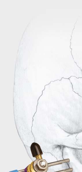
1 Provides Treatment for Fractures of the Mandible Mandible External Fixator II Surgical Technique
2 Table of Contents Introduction Mandible External Fixator II 2 AO Principles 4 Indications 5 MRI Information 6 Surgical Technique Fixation Using Schanz Screws 7 Optional Technique to Implant Schanz Screws 18 Optional Technique Fixation Using Kirschner Wires 21 Alternate Frame Configurations 25 Product Information Instruments 28 Set List 31 Mandible External Fixator II Surgical Technique DePuy Synthes 1
 Accepts a 2.5 mm/4.0 mm Schanz screw, 2.")
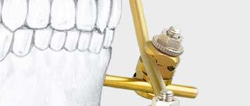
3 Mandible External Fixator II The Mandible External Fixator II allows the surgeon to create a rigid construct using three basic components: 4.0 mm rods, 2.0 mm and 2.5 mm Kirschner wires and/or 2.5 mm/4.0 mm Schanz screws, and adjustable snap-on clamps. Features Lightweight single-phase system Adjustable throughout application Adjustable Clamp ( ) Accepts a 2.5 mm/4.0 mm Schanz screw, 2.5 mm Kirschner wire, or 4.0 mm rod on each end of the clamp Snap-on design allows clamp placement between previously applied clamps Connects two rods in any orientation, for greater versatility of Schanz screw/kirschner wire placement and frame construction Maintains rod position during frame assembly and fracture reduction Adjustable Parallel Pin Clamp ( ) Allows two Kirschner wires to be implanted in the same small bone fragment Snap-on design allows clamp placement between previously applied clamps Allows greater versatility of Schanz screw/kirschner wire placement and frame construction Top portion of the clamp accepts a 2.5 mm Kirschner wire and 4.0 mm rod, and the bottom portion of the clamp accepts two 2.0 mm or 2.5 mm Kirschner wires 2.0 mm and 2.5 mm Kirschner Wires ( and ) For fixation of fragments that may be too small for 4.0 mm Schanz Screws. 2 DePuy Synthes Mandible External Fixator II Surgical Technique

4 Mandible External Fixator II 2.5 mm/4.0 mm Titanium Schanz Screws ( ) For Symphysis, Body and Ramus two thread lengths per region Self-drilling threads allow direct screw implantation, or predrilling if desired Shoulder prevents over-insertion Biocompatible titanium alloy* 4.0 mm Titanium Pre-Bent Rods ( ) Pre-bent to shape of mandible Available in four sizes Can be contoured to match patient anatomy * Ti-6Al-7Nb Mandible External Fixator II Surgical Technique DePuy Synthes 3

5 AO Principles AO PRINCIPLES In 1958, the AO formulated four basic principles, which have become the guidelines for internal fixation. 1,2 In 1958, the AO formulated four basic principles, which have become the guidelines for internal fixation 1, 2. 4_Priciples_03.pdf :08 Anatomic Anatomic reduction reduction Fracture Fracture reduction reduction and and fixation fixation to to restore restore anatomical anatomical relationships. relationships. 1 2 Stable Stable fixation fixation Fracture Fracture fixation fixation providing providing absolute absolute or relative or relative stability, stability, as required as by the required patient, by the the injury, patient, and the the injury, personality and the personality of the fracture. of the fracture. Early, active mobilization Early Early and and safe safe mobilization and rehabilitation of of the injured part and and the the patient as as a whole. 4 3 Preservation Preservation of of blood blood supply supply Preservation Preservation of of the the blood blood supply supply to to soft soft tissues tissues and and bone bone by by gentle reduction gentle reduction techniques techniques and and careful careful handling. handling. 1. Müller ME, Allgöwer M, Schneider R, Willenegger H. Manual of Internal Fixation. 3rd ed. Berlin, Heidelberg, New York: Springer-Verlag; Rüedi TP, RE Buckley, CG Moran. AO Principles of Fracture Management. 1 Müller ME, M Allgöwer, R Schneider, H Willenegger. Manual of Internal 2nd ed. Stuttgart New York: Thieme; Fixation. 3rd ed. Berlin Heidelberg New York: Springer Rüedi TP, RE Buckley, CG Moran. AO Principles of Fracture Management. 2nd ed. Stuttgart, New York: Thieme DePuy Synthes Mandible External Fixator II Surgical Technique

 Fractures associated")


6 Indications The Mandible External Fixator II is intended to stabilize and provide treatment for fractures of the maxillofacial area, including: Severe open mandibular fractures Highly comminuted closed fractures Nonunions and delayed unions (especially associated with infection) Fractures associated with infection Tumor resections Facial deformity corrections Gunshot wounds Panfacial fractures Burn maintenance Bone grafting defects Photo courtesy of Daniel Buchbinder, DMD, MD Chief, Division of Maxillofacial Surgery, Continuum Cancer Centers of New York Mandible External Fixator II Surgical Technique DePuy Synthes 5
7 MRI Safety Information Non-clinical testing has demonstrated that the Mandible External Fixator is MR Conditional. A patient with this device can be safely scanned in an MR system meeting the following conditions: Static magnetic field of 1.5 or 3 Tesla (T) Maximum spatial gradient field of 2,410 Gauss/cm (24.1 T/m) Maximum MR system reported, whole body averaged specific absorption rate (SAR) of 2 W/kg (at Normal Operating Mode) Do not place any radio frequency (RF) transmit coils over the external fixation frame All components of the Mandible External Fixator must be properly identified as MR Conditional Under the scan conditions defined above, the Mandible External Fixator is expected to produce a maximum temperature rise of less than 2 C after 15 minutes of continuous scanning. 6 DePuy Synthes Mandible External Fixator II Surgical Technique

8 Fixation Using Schanz Screws Special Note: The Mandible External Fixator II offers the option of using an adjustable parallel pin clamp with two Kirschner wires for fragments that are too small to accept two Schanz screws, eg, as in the condylar region. If the patient presents with such a fragment, consult Fixation Using Kirschner Wires on page 20 for proper technique. 1 Patient preparation Place the patient in maxillomandibular fixation when appropriate. 2 Identify appropriate rods Identify the appropriate rod or combination of rods for fixation. In most cases, the pre-bent rods will not need additional contouring. If contouring is needed, follow steps 3 4, otherwise proceed to step 5. 3 Contour the rod template Instrument Rod Template Contour the rod template to match the patient s bony anatomy. Precaution: The rod should be positioned approximately one fingerbreadth away from the patient s skin, evenly throughout the entire length of the rod. Mandible External Fixator II Surgical Technique DePuy Synthes 7


9 Fixation Using Schanz Screws 4 Contour the rod(s) Instrument mm Rod Bender Using the 4.0 mm rod bender, contour the selected pre-bent rod to match the rod template. Increase bend Decrease bend 5 Verify fit and screw location Verify the fit of the pre-bent rod on the patient. Identify the desired Schanz screw locations for the first and last screw (furthest from the defect on the proximal and distal side) and mark accordingly. Note: A minimum of two Schanz screws per segment (two screws in greatest segment and two in other segments) is recommended to ensure adequate stability. Optimal location of Schanz screws will place one screw 10 mm distal and another screw 10 mm proximal to the defect. 8 DePuy Synthes Mandible External Fixator II Surgical Technique


10 Fixation Using Schanz Screws 6 Make small stab incision Prepare for Schanz screw insertion by making a small stab incision at the first marked screw location. 7 Dissect soft tissue Instruments mm Obturator Drill Guide/Cannula Drill Sleeve Handle Thread the cannula into the drill sleeve handle. Insert the 4.0 mm obturator into the handle/cannula assembly. Bluntly dissect the soft tissue and pass the cannula and obturator through the stab incision to the bone. Remove the obturator, leaving the cannula in place. Mandible External Fixator II Surgical Technique DePuy Synthes 9


11 Fixation Using Schanz Screws 8 Implant Schanz screw Instrument Schanz Screw Rapid Driver Select the appropriate anatomic Schanz screw. The anatomic Schanz screws are self-drilling, but if predrilling is desired, refer to the optional technique on pages Secure the rapid driver into the Jacobs chuck of a power drill, then load the anatomic Schanz screw into the rapid driver. Using the cannula as a guide, drive the Schanz screw into the mandible down to the stop. Pull on the drill to disengage the rapid driver from the implanted Schanz screw. Note: DePuy Synthes recommends using the Compact Air Drive II or an equivalent drill with an operating speed of approximately 900 RPM. Precautions: Drill speed rate should never exceed 1,800 rpm, particularly in dense, hard bone. Higher drill speed rates can result in: thermal necrosis of the bone, soft tissue burns, an oversized hole, which can lead to reduced pullout force, increased ease of the screws stripping in bone, suboptimal fixation, and/or the need for emergency screws. Always irrigate during drilling to avoid thermal damage to the bone. Irrigate and apply suction for removal of debris potentially generated during implantation or removal. Handle devices with care and dispose of worn bone cutting instruments in an approved sharps container. Warning: Instruments and screws may have sharp edges or moving joints that may pinch or tear user s glove or skin. 10 DePuy Synthes Mandible External Fixator II Surgical Technique

12 Fixation Using Schanz Screws 8 Implant Schanz screw continued Instruments Schanz Screw Rapid Driver Ratcheting Screwdriver Handle If the Schanz screw requires additional tightening, load the other Schanz screw rapid driver into the ratcheting screwdriver handle. Engage the exposed end of the Schanz screw and ratchet the handle clockwise until the Schanz screw is fully inserted. Pull back on the handle to disengage the Schanz screw. Optional technique Instruments Adaptor for Ratcheting Screwdriver Handle Ratcheting Screwdriver Handle Combination Wrench Socket Wrench Load the adaptor for ratcheting screwdriver handle into the ratcheting screwdriver handle. Slide the adaptor over the exposed end of the Schanz screw. Hand-tighten the thumbscrew on the adaptor to capture the Schanz screw. Using the combination wrench or socket wrench, tighten the thumbscrew at least 1/4 turn. Use the ratcheting screwdriver handle to complete the implantation of the Schanz screw. Mandible External Fixator II Surgical Technique DePuy Synthes 11

13 Fixation Using Schanz Screws 9 Implant second Schanz screw Position and insert the second Schanz screw on the opposite side of the defect and furthest from the defect, as outlined in steps DePuy Synthes Mandible External Fixator II Surgical Technique


14 Fixation Using Schanz Screws 10 Assemble the rigid construct Instrument Adjustable Clamp Snap adjustable clamps onto both Schanz screws and rod. Reduce the fracture and tighten the clamp nuts. Verify the correct alignment before proceeding. Mandible External Fixator II Surgical Technique DePuy Synthes 13

15 Fixation Using Schanz Screws 11 Add third clamp Attach a third clamp to the rod approximately 10 mm proximal or distal to the defect. Insert the cannula into the clamp, as shown. Angle the cannula and clamp for the desired screw placement. Mark the incision site. Temporarily rotate the cannula and clamp upward to minimize obstruction while making the incision. Make a small incision and bluntly dissect the soft tissue. Rotate the clamp and cannula to the original position. Insert the obturator and pass the cannula through the incision to the bone. Remove the obturator. Tighten the clamp, securing the cannula to the rod. Precaution: Do not overtighten the clamp, as this will result in damage to the cannula. 14 DePuy Synthes Mandible External Fixator II Surgical Technique


16 Fixation Using Schanz Screws 12 Implant third Schanz screw Insert an anatomic Schanz screw as outlined in steps 6 8. Loosen the clamp slightly and slide the cannula off the Schanz screw. With the clamp capturing the Schanz screw and the rod, tighten the clamp nut. Mandible External Fixator II Surgical Technique DePuy Synthes 15


17 Fixation Using Schanz Screws 13 Complete the construct Note: A minimum of two Schanz screws is required on each side of the defect. Insert all remaining Schanz screws to complete the frame, as outlined in steps DePuy Synthes Mandible External Fixator II Surgical Technique
18 Fixation Using Schanz Screws 14 Verify reduction and adjust Verify reduction and alignment. If adjustment is needed, loosen the clamp nuts, manipulate the mandible, and retighten the clamps. Precaution: Pin sites should be meticulously cared for to avoid pin-tract infection. Schanz screws may be surrounded with antiseptic-coated foam sponges in an effort to avoid infection. A pin-site care procedure should be reviewed with the patient. 15 Trim Schanz screws and rod (optional) Optional Instruments * Cutter (small) * Rod Cutter (large) Protective Caps or Trim the Schanz screws or rod, if necessary, using the cutter (small) or the rod cutter (large). Place protective caps over the cut ends. Warning: Instruments and screws may have sharp edges or moving joints that may pinch or tear user s glove or skin. 16 Implant Removal For removal of the construct, follow steps 8 through 13 in reverse order to untighten and remove all clamps, then remove the frame and/or connection rods and subsequently remove the Schanz Screws, using the appropriate instrumentation. *Also available. Mandible External Fixator II Surgical Technique DePuy Synthes 17


19 Optional Technique to Implant Schanz Screws 1 Predrill the bone Instruments mm/4.0 mm Stepped Drill Bit * ComPact Air Drive II Use the 1.8 mm/4.0 mm stepped drill bit through the cannula to drill into the bone. Note: DePuy Synthes recommends using the ComPact Air Drive II or an equivalent drill with an operating speed of approximately 900 RPM. Precautions: Drill speed rate should never exceed 1,800 rpm, particularly in dense, hard bone. Higher drill speed rates can result in: thermal necrosis of the bone, soft tissue burns, an oversized hole, which can lead to reduced pullout force, increased ease of the screws stripping in bone, suboptimal fixation, and/or the need for emergency screws. Always irrigate during drilling to avoid thermal damage to the bone. Irrigate and apply suction for removal of debris potentially generated during implantation or removal. Handle devices with care and dispose of worn bone cutting instruments in an approved sharps container. 2 Use measuring device Instrument Measuring Device Insert the measuring device through the cannula and hook the lingual cortex of the mandible. Record the number indicated on the measuring device. Precaution: The reading from the device does not represent the bone thickness. Remove the measuring device from the cannula. *Also available. 18 DePuy Synthes Mandible External Fixator II Surgical Technique


20 Optional Technique to Implant Schanz Screws 3 Select and measure Schanz screw Precaution: Select the appropriate anatomic Schanz screw for the patient s bony anatomy. Slide the selected Schanz screw into the cannulated end of the measuring device and align the tip of the Schanz screw with the number recorded in step 2. 4 Load Schanz screw Instruments Adaptor for Ratcheting Screwdriver Handle Ratcheting Screwdriver Handle Combination Wrench Socket Wrench Slide the adaptor over the exposed end of the Schanz screw. Hand-tighten the thumbscrew on the adaptor to secure the Schanz screw. Using the combination wrench or socket wrench, tighten the thumbscrew at least 1/4 turn. Load the adaptor for ratcheting screwdriver handle into the ratcheting screwdriver handle. Mandible External Fixator II Surgical Technique DePuy Synthes 19


21 Optional Technique to Implant Schanz Screws 5 Implant Schanz screw With clockwise rotation, insert the Schanz screw through the cannula until the adaptor contacts the cannula, ensuring proper implant depth. 6 Remove adaptor from implanted Schanz screw Remove the adaptor from the Schanz screw by loosening the thumbscrew with either the combination wrench or the socket wrench. Remove the cannula. 20 DePuy Synthes Mandible External Fixator II Surgical Technique
 and mark accordingly. 2 Prepare to implant first Kirschner wire Instruments 03.305.021 Kirschner Wire Drill Guide 03.305.022 2.")

22 Optional Technique Fixation Using Kirschner Wires 1 Identify location of first Kirschner wire Identify the desired location for the first Kirschner wire (either 2.0 mm or 2.5 mm) and mark accordingly. 2 Prepare to implant first Kirschner wire Instruments Kirschner Wire Drill Guide mm/2.5 mm Obturator Make a small stab incision at the marked site. Insert the 2.0 mm/2.5 mm obturator into the Kirschner wire drill guide and dissect through the soft tissue to the small bone fragment. Remove the obturator, leaving the drill guide in place. Mandible External Fixator II Surgical Technique DePuy Synthes 21


23 Optional Technique Fixation Using Kirschner Wires 3 Implant first Kirschner wire Using a wire driver, implant the Kirschner wire through the drill guide into the bone. Disengage the wire driver from the implanted Kirschner wire. 4 Prepare to implant second Kirschner wire Slide the drill guide off the implanted Kirschner wire. Slide the shorter adjacent tube over the wire. The first Kirschner wire may have to be trimmed to make room for the second wire. 22 DePuy Synthes Mandible External Fixator II Surgical Technique


24 Optional Technique Fixation Using Kirschner Wires 5 Implant second Kirschner wire Identify the desired location for the second Kirschner wire and mark accordingly. Insert a second wire using the same technique as in steps 2 and 3. 6 Build the construct Upon implantation of the second Kirschner wire, disengage it from the wire driver. Slide the drill guide off the wire. Snap an adjustable parallel pin clamp to the Kirschner wires, and connect the clamp to the adjoining construct. Mandible External Fixator II Surgical Technique DePuy Synthes 23


25 Optional Technique Fixation Using Kirschner Wires 7 Tighten the rigid construct Instruments Combination Wrench Socket Wrench The adjustable parallel pin clamp can be tightened with either the combination wrench or the socket wrench. 8 Trim wires and apply protective caps Instruments Protective Caps, for 2.0 mm Kirschner Wires Protective Caps, for 2.5 mm Kirschner Wires Trim the Kirschner wires and place protective caps over cut ends. 24 DePuy Synthes Mandible External Fixator II Surgical Technique


26 Alternative Frame Configurations One-Half Frame As applied on an infected angle fracture. Items to build construct Quantity Adjustable Clamps mm Titanium Pre-Bent Rod, 1 One-Half Mandible mm/4.0 mm Titanium Schanz Screws, for Ramus mm/4.0 mm Titanium Schanz Screws, for Body Modular Carbon Fiber Frame As applied on an infected angle fracture. The modular carbon fiber frame provides improved x-ray imaging. Items to build construct Quantity Adjustable Clamps mm Carbon Fiber Rods, 2 60 mm mm Carbon Fiber Rod, 1 80 mm mm/4.0 mm Titanium Schanz Screws, for Ramus mm/4.0 mm Titanium Schanz Screws, for Body Mandible External Fixator II Surgical Technique DePuy Synthes 25


27 Alternative Frame Configurations Double-Stacked Frame As applied on a resected mandible. Items to build construct Quantity Adjustable Clamps mm Titanium Pre-Bent Rod, 1 One-Half Mandible mm Titanium Pre-Bent Rod, 1 Full Mandible with Ramus mm/4.0 mm Titanium Schanz Screws, for Ramus mm/4.0 mm Titanium Schanz Screws, for Body mm/4.0 mm Titanium Schanz Screw, for Symphysis Modular Frame As applied on a comminuted fracture. A modular frame can be created to accommodate the fracture location. Items to build construct Quantity Adjustable Clamps mm Titanium Pre-Bent Rod, 1 One-Half Mandible mm Titanium Pre-Bent Rod, 1 Full Mandible mm/4.0 mm Titanium Schanz Screws, for Ramus mm/4.0 mm Titanium Schanz Screws, for Body mm/4.0 mm Titanium Schanz Screws, for Symphysis 26 DePuy Synthes Mandible External Fixator II Surgical Technique


28 Alternative Frame Configurations Frame incorporating an Adjustable Parallel Pin Clamp As applied on a mandible with a fragment that is too small to accept two Schanz screws. Items to build construct Quantity Adjustable Clamps Adjustable Parallel Pin Clamp mm Titanium Pre-Bent Rod, 1 Full Mandible with Ramus mm/4.0 mm Titanium Schanz Screws, for Ramus mm/4.0 mm Titanium Schanz Screws, for Body mm/4.0 mm Titanium Schanz Screws, for Symphysis mm Kirschner Wires Protective Caps, for 2.0 mm 2 Kirschner Wires Mandible External Fixator II Surgical Technique DePuy Synthes 27

29 Instruments mm/4.0 mm Stepped Drill Bit Measuring Device mm Rod Bender Rod Template Drill Guide for 2.0 mm/2.5 mm Kirschner Wires mm/2.5 mm Obturator for Mandible External Fixator II 28 DePuy Synthes Mandible External Fixator II Surgical Technique
30 Instruments Schanz Screw Rapid Driver for Mandible External Fixator II mm Obturator for Mandible External Fixator II Drill Guide/Cannula, extra long Adaptor for Ratcheting Screwdriver Handle Mandible External Fixator II Surgical Technique DePuy Synthes 29


31 Instruments Ratcheting Screwdriver Handle Combination Wrench Socket Wrench Drill Sleeve Handle 30 DePuy Synthes Mandible External Fixator II Surgical Technique

32 Mandible External Fixator II Set ( ) Graphic Case Mandible External Fixator II Set Graphic Case Base Mandible External Fixator II Set Graphic Case Lid Schanz Screws 2.5 mm/4.0 mm Titanium Schanz Screws, self-drilling, 3 ea for Ramus, 8 mm thread length for Ramus, 10 mm thread length for Body, 14 mm thread length for Body, 18 mm thread length for Symphysis, 20 mm thread length for Symphysis, 22 mm thread length Wire (10/pkg.) mm Kirschner Wire with Thread, 150 mm, trocar point, 15 mm thread length mm Kirschner Wire with Thread, 150 mm, trocar point, 15 mm thread length Rods 4.0 mm Titanium Pre-Bent Rods, 2 ea Full mandible with ramus Full mandible Three-quarter mandible One-half mandible 4.0 mm Carbon Fiber Rods, 2 ea mm mm mm mm Clamp Adjustable Clamp, 8 ea Adjustable Parallel Pin Clamp, 2 ea. Protective Caps (10/pkg.) for 2.0 mm Kirschner wires for 2.5 mm Kirschner wires for 4.0 mm bars and rods Note: For additional information, please refer to package insert. For detailed cleaning and sterilization instructions, please refer to or sterilization instructions, if provided. Mandible External Fixator II Surgical Technique DePuy Synthes 31
33 Mandible External Fixator II Set ( ) Instruments mm/4.0 mm Stepped Drill Bit, Stryker J-latch, with 24 mm stop, 2 ea Measuring Device, for Mandible External Fixator mm Rod Bender, for Mandible External Fixator Rod Template, for 4.0 mm rods, for Mandible External Fixator Drill Guide for 2.0 mm/2.5 mm Kirschner Wires mm/2.5 mm Obturator Schanz Screw Rapid Driver, 2 ea mm Obturator Drill Guide/Cannula, extra long, for 4.0 mm Schanz Screws, 2 ea Adaptor for Ratcheting Screwdriver Handle Ratcheting Screwdriver Handle Combination Wrench, 7 mm width across flats Socket Wrench, 7 mm width across flats Drill Sleeve Handle Also Available* S Mandible External Fixator, 500 mm Rod Kit, sterile (includes one straight 500 mm Titanium Rod and one 500 mm Rod Template) Cutter (small) Rod Cutter (large) 4.0 mm Carbon Fiber Rods mm mm mm mm Compact Air Drive II *These items will not fit in the Mandible External Fixator II graphic case. Available nonsterile and sterile-packed. Delete S from catalog number for nonsterile product. 32 DePuy Synthes Mandible External Fixator II Surgical Technique


34 Mandible External Fixator Set ( ) Graphic Case Mandible External Fixator Set Graphic Case Base Mandible External Fixator Set Graphic Case Lid Instruments mm Obturator, for Mandible External Fixator Drill Guide/Cannula, short, 2 ea Drill Guide/Cannula, long, 2 ea mm/4.0 mm Stepped Drill Bit, Stryker J-latch, with 24 mm stop, 2 ea Measuring Device, for Mandible External Fixator mm Rod Bender, for Mandible External Fixator Cannulated T-Handle, for Mandible External Fixator Rod Template, for 4.0 mm rods, for Mandible External Fixator Combination Wrench, 7 mm width across flats Socket Wrench, 7 mm width across flats Drill Sleeve Handle Implants mm Kirschner Wire with Thread, 150 mm, trocar point, 15 mm thread length, 1 pkg. of mm/4.0 mm Titanium Schanz Screws, blunt tip, 3 ea mm mm mm 2.5 mm/4.0 mm Titanium Self-Drilling Schanz Screws, 3 ea mm mm mm Fixation Material Adjustable Clamp, 8 ea Protective Caps, for 2.5 mm Kirschner wires, 1 pkg. of Protective Caps, for 4.0 mm bars and rods, 1 pkg. of mm Titanium Pre-Bent Rods, 2 ea Full mandible with ramus Full mandible Three-quarter mandible One-half mandible 4.0 mm Carbon Fiber Rods, 2 ea mm mm mm Also Available S Mandible External Fixator, 500 mm Rod Kit, sterile (includes one straight 500 mm titanium rod and one 500 mm rod template) Cutter (small) Rod Cutter (large) mm/2.5 mm Titanium Self-Drilling Schanz Screw, 20 mm thread length, 80 mm Note: For additional information, please refer to package insert. For detailed cleaning and sterilization instructions, please refer to or sterilization instructions, if provided. Mandible External Fixator II Surgical Technique DePuy Synthes 33
35 Limited Warranty and Disclaimer: DePuy Synthes products are sold with a limited warranty to the original purchaser against defects in workmanship and materials. Any other express or implied warranties, including warranties of merchantability or fitness, are hereby disclaimed. Please also refer to the package insert(s) or other labeling associated with the devices identified in this surgical technique for additional information. CAUTION: Federal Law restricts these devices to sale by or on the order of a physician. Some devices listed in this surgical technique may not have been licensed in accordance with Canadian law and may not be for sale in Canada. Please contact your sales consultant for items approved for sale in Canada. Not all products may currently be available in all markets. Manufactured or distributed by: Synthes USA Products, LLC 1302 Wrights Lane East West Chester, PA Synthes USA, LLC 1101 Synthes Avenue Monument, CO To order (USA): To order (Canada): Note: For recognized manufacturer, refer to the product label. DePuy Synthes All rights reserved. DSUS/CMF/1016/0625 3/17 DV
Mandible External Fixator II. Provides treatment for fractures of the maxillofacial area.
 Mandible External Fixator II. Provides treatment for fractures of the maxillofacial area. Technique Guide This publication is not intended for distribution in the USA. Instruments and implants approved
Mandible External Fixator II. Provides treatment for fractures of the maxillofacial area.
 Mandible External Fixator II. Provides treatment for fractures of the maxillofacial area. Lightweight single-phase system Adjustable throughout application Rigid construct using three basic components
Mandible External Fixator II
 Provides treatment for fractures of the mandible Mandible External Fixator II Surgical Technique Image intensifier control This description alone does not provide sufficient background for direct use of
Mini External Fixator
 Stabilize the Phalanges and Metacarpals Mini External Fixator Surgical Technique Table of Contents Introduction Mini External Fixator 2 Indications 4 Surgical Technique Technique Overview 5 Product Information
Low Profile Neuro Plating System. Surgical Technique
 Low Profile Neuro Plating System Surgical Technique TABLE OF CONTENTS INTRODUCTION Low Profile Neuro Plating System 2 SURGICAL TECHNIQUE Technique 5 PRODUCT INFORMATION Low Profile Neuro Plates 10 Low
3.5 mm LCP Extra-articular Distal Humerus Plate
 Part of the DePuy Synthes Locking Compression Plate (LCP ) System 3.5 mm LCP Extra-articular Distal Humerus Plate Surgical Technique Table of Contents Introduction 3.5 mm LCP Extra-articular Distal Humerus
3.5 mm Clavicle Hook Plates
 A Single Solution for Lateral Clavicle Fractures and Acromioclavicular Joint Dislocations 3.5 mm Clavicle Hook Plates Surgical Technique Discontinued December 2017 DSUS/TRM/1016/1126(1) Table of Contents
3.5 mm LCP Olecranon Plates
 Part of the DePuy Synthes Locking Compression Plate (LCP ) System 3.5 mm LCP Olecranon Plates Surgical Technique Table of Contents Introduction 3.5 mm LCP Olecranon Plates 2 AO Principles 3 Indications
Lag Screw Device Intended for symphyseal fracture fixation of the mandible
 Lag Screw Device Intended for symphyseal fracture fixation of the mandible SUrgicaL TecHNiqUe Lag Screw Device Intended for symphyseal fracture fixation of the mandible Simplifies the lag screw fixation
Low Bend Distal Tibia Plates
 Part of the DePuy Synthes Locking Compression Plate (LCP ) System 3.5 mm LCP Low Bend Medial Distal Tibia Plates Surgical Technique Table of Contents Introduction 3.5 mm LCP Low Bend Medial Distal Tibia
Part of the DePuy Synthes Locking Compression Plate (LCP ) System. 3.5 mm LCP Medial Proximal Tibia Plates
 System. 3.5 mm LCP Medial Proximal Tibia Plates") Part of the DePuy Synthes Locking Compression Plate (LCP ) System 3.5 mm LCP Medial Proximal Tibia Plates Surgical Technique Table of Contents Introduction 3.5 mm LCP Medial Proximal Tibia Plates 2 AO
Part of the DePuy Synthes Locking Compression Plate (LCP ) System 3.5 mm LCP Medial Proximal Tibia Plates Surgical Technique Table of Contents Introduction 3.5 mm LCP Medial Proximal Tibia Plates 2 AO
low ProfIle neuro PlaTIng system
 low ProfIle neuro PlaTIng system surgical TeChnIque Table of Contents Introduction Low Profile Neuro Cranial Plating System 2 Surgical Technique Technique 5 Product Information Low Profile Neuro Plates
3.5 mm Locking Attachment Plate
 For Treatment of Periprosthetic Fractures 3.5 mm Locking Attachment Plate Surgical Technique Table of Contents Introduction 3.5 mm Locking Attachment Plate 2 Indications 4 Surgical Technique Preparation
For Small-Statured Adults and Pediatric Patients. Medium External Fixator Basic Modular Frame
 For Small-Statured Adults and Pediatric Patients Medium External Fixator Basic Modular Frame Surgical Technique MRI Information DePuy Synthes Medium External Fixation devices are labeled MR Conditional
2.0 mm Mandible Locking Plate System
 Advanced Plating System for Trauma, Microvascular Reconstruction, and Orthognathic Surgery 2.0 mm Mandible Locking Plate System Surgical Technique TABLE OF CONTENTS INTRODUCTION 2.0 mm Mandible Locking
3.5 mm LCP Clavicle Hook Plates
 Part of the Synthes Locking Compression Plate (LCP ) System 3.5 mm LCP Clavicle Hook Plates Surgical Technique Table of Contents Introduction 3.5 mm LCP Clavicle Hook Plates 2 AO Principles 4 Indications
Long Volar Plates for Diaphyseal-Metaphyseal Radius Fractures LCP. Dia-Meta Volar Distal Radius Plates. Surgical Technique
 Long Volar Plates for Diaphyseal-Metaphyseal Radius Fractures LCP Dia-Meta Volar Distal Radius Plates Surgical Technique Table of Contents Introduction LCP Dia-Meta Volar Distal Radius Plates 2 AO Principles
Large External Fixator Modular Knee Bridge
 Using Multi-pin Clamps Large External Fixator Modular Knee Bridge Surgical Technique Large External Fixator Modular Knee Bridge DePuy Synthes Large External Fixation devices are labeled MR Conditional
The Locking Calcaneal Plate Instrument and Implant Sets
 Part of the DePuy Synthes Locking Compression Plate (LCP ) System The Locking Calcaneal Plate Instrument and Implant Sets Surgical Technique Table of Contents Introduction Locking Calcaneal Plate 2 AO
3.5 mm LCP Low Bend Medial Distal Tibia Plate Aiming Instruments
 Part of the 3.5 mm LCP 3.5 mm LCP Low Bend Medial Distal Tibia Plate Aiming Instruments Surgical Technique TABLE OF CONTENTS INTRODUCTION 3.5 mm LCP Low Bend Medial Distal Tibia Plate 2 Aiming Instruments
2.7 mm/3.5 mm LCP Distal Fibula Plate
 Part of the DePuy Synthes Locking Compression Plate (LCP ) System 2.7 mm/3.5 mm LCP Distal Fibula Plate Surgical Technique Table of Contents Introduction 2.7 mm/3.5 mm LCP Distal Fibula Plates 2 AO Principles
3.5 mm LCP Hook Plate
 Part of the DePuy Synthes Locking Compression Plate (LCP ) System 3.5 mm LCP Hook Plate Surgical Technique Table of Contents Introduction 3.5 mm LCP Hook Plate 2 AO Principles 4 Indications 5 Clinical
Cannulated Pediatric Osteotomy System (CAPOS)
") A Single System of Osteotomy Blade Plates and Cannulated Instrumentation Cannulated Pediatric Osteotomy System (CAPOS) Surgical Technique Table of Contents Introduction Cannulated Pediatric Osteotomy System
A Single System of Osteotomy Blade Plates and Cannulated Instrumentation Cannulated Pediatric Osteotomy System (CAPOS) Surgical Technique Table of Contents Introduction Cannulated Pediatric Osteotomy System
3.5 mm LCP Distal Humerus Plates
 Part of the DePuy Synthes Locking Compression Plate (LCP ) System 3.5 mm LCP Distal Humerus Plates Surgical Technique Table of Contents Introduction 3.5 mm LCP Distal Humerus Plates 2 AO Principles 4 Indications
For Fast and Stable Fixation of the Sternum. Sternal ZIPFIX. System. Surgical Technique
 For Fast and Stable Fixation of the Sternum Sternal ZIPFIX System Surgical Technique TABLE OF CONTENTS INTRODUCTION Sternal ZIPFIX System 2 AO Principles 7 Indications and Contraindications 8 SURGICAL
orthodontic Bone anchor (oba) SYStem
 SYStem") orthodontic Bone anchor (oba) SYStem Skeletal implants for the orthodontic movement of teeth SurgIcal technique Table of Contents Introduction Orthodontic Bone Anchor (OBA) System 2 Indications and Contraindications
orthodontic Bone anchor (oba) SYStem Skeletal implants for the orthodontic movement of teeth SurgIcal technique Table of Contents Introduction Orthodontic Bone Anchor (OBA) System 2 Indications and Contraindications
4.5 mm LCP Medial Proximal Tibia Plates
 Part of the DePuy Synthes LCP Periarticular Plating System 4.5 mm LCP Medial Proximal Tibia Plates Surgical Technique Table of Contents Introduction 4.5 mm LCP Medial Proximal Tibia Plates 2 AO Principles
Medium External Fixator Humeral Shaft Frame. Modular frame for upper extremity use.
 Medium External Fixator Humeral Shaft Frame. Modular frame for upper extremity use. Technique Guide Part of the Medium External Fixation System MRI Information Synthes Medium External Fixation devices
Femur Frame. The Distraction Osteogenesis Ring System
 Femur Frame The Distraction Osteogenesis Ring System Surgical Technique Table of Contents Introduction The Distraction Osteogenesis Ring System 2 MRI Information 4 AO Principles 5 Indications 6 Surgical
MatrixORTHOGNATHIC TM Plating System
 Specialized Implants and Instruments for Orthognathic Surgery MatrixORTHOGNATHIC TM Plating System Surgical Technique TABLE OF CONTENTS INTRODUCTION MatrixORTHOGNATHIC Plating System 2 Features and Benefits
Technique Guide. Compact 2.0 LOCK Mandible. The locking system for the mandible.
 Technique Guide Compact 2.0 LOCK Mandible. The locking system for the mandible. Table of Contents Introduction Compact 2.0 LOCK Mandible 2 AO Principles 4 Indications and Contraindications 5 Surgical
For the Treatment of Wrist Fractures. Small External Fixator Nonspanning Wrist Frame
 For the Treatment of Wrist Fractures Small External Fixator Nonspanning Wrist Frame Surgical Technique Small External Fixator Nonspanning Wrist Frame DePuy Synthes Small External Fixation devices are labeled
Large External Fixator Modular Knee Bridge. Using multi-pin clamps.
 Large External Fixator Modular Knee Bridge. Using multi-pin clamps. Technique Guide Part of the Large External Fixation System Large External Fixator Modular Knee Bridge Technique Overview Insert Schanz
Medium External Fixator Pediatric Femoral Shaft Frame. Using medium multi-pin clamps.
 Medium External Fixator Pediatric Femoral Shaft Frame. Using medium multi-pin clamps. Technique Guide Part of the Medium External Fixation System MRI Information Synthes Medium External Fixation devices
For Distal Femur Fractures. 95º Condylar Plate. Quick Reference Chart
 For Distal Femur Fractures 95º Condylar Plate Quick Reference Chart 95 Condylar Plate. Quick reference chart for distal femur fractures. Insert guide wires Fix condylar fragments with 6.5 mm cancellous
Titanium Wire With Barb and Needle
 For Canthal Tendon Procedures Titanium Wire With Barb and Needle Surgical Technique Table of Contents Introduction Titanium Wire With Barb and Needle 2 Indications 2 Surgical Technique Preoperative Planning
2.4 mm Variable Angle LCP Volar Rim Distal Radius Plates
 For Fragment-Specific Fracture Fixation With Variable Angle Locking Technology 2.4 mm Variable Angle LCP Volar Rim Distal Radius Plates Surgical Technique Table of Contents Introduction 2.4 mm Variable
MatrixNEURO Cranial Plating System
 The Next Generation Cranial Plating System MatrixNEURO Cranial Plating System Surgical Technique TABLE OF CONTENTS INTRODUCTION MatrixNEURO Cranial Plating System 2 MatrixNEURO Reconstruction Mesh 6 MatrixNEURO
LCP DISTAL TIBIA PLATE
 LCP DISTAL TIBIA PLATE Instruments and implants approved by the AO Foundation. This publication is not intended for distribution in the USA. SURGICAL TECHNIQUE Image intensifier control This description
Large Distractor Femur
 Fracture Reduction and Provisional Stabilization Large Distractor Femur Surgical Technique Table of Contents Introduction Standard Femoral Distraction 2 Large Distractor System 4 Surgical Technique Prepare
The Distraction Osteogenesis Ring System
 Nonarticular Tibia Frame The Distraction Osteogenesis Ring System Surgical Technique Table of Contents Introduction The Distraction Osteogenesis Ring System 2 MRI Information 4 AO Principles 5 Indications
Large External Fixator Traveling Traction
 Used to Correct Angular Deformity Large External Fixator Traveling Traction Surgical Technique Large External Fixator Traveling Traction DePuy Synthes Large External Fixation devices are labeled MR Conditional
MATrIxMANDIBLE. PrEFOrMED reconstruction PLATES. SUrgICAL TEChNIqUE
 MATrIxMANDIBLE PrEFOrMED reconstruction PLATES Preshaped ed to the mandibular anatomy SUrgICAL TEChNIqUE Table of Contents Introduction MatrixMANDIBLE Preformed Reconstruction Plates 2 AO Principles 5
Medium External Fixator Delta Frame Ankle Bridge
 For Small-Statured Adults Medium External Fixator Delta Frame Ankle Bridge Surgical Technique MRI Information DePuy Synthes Medium External Fixation devices are labeled MR Conditional according to the
Distal Radius Plate Instrument and Implant Set. Discontinued December 2017 DSUS/TRM/0916/1063(1)
") Distal Radius Plate Instrument and Implant Set Surgical Technique Discontinued December 2017 DSUS/TRM/0916/1063(1) The Distal Radius Plates Indications For fixation of fractures and osteotomies, including
Distal Radius Plate Instrument and Implant Set Surgical Technique Discontinued December 2017 DSUS/TRM/0916/1063(1) The Distal Radius Plates Indications For fixation of fractures and osteotomies, including
LCP Low Bend Medial Distal Tibia Plates 3.5 mm. Anatomic plates with low profile head for intra- and extraarticular fractures.
 LCP Low Bend Medial Distal Tibia Plates 3.5 mm. Anatomic plates with low profile head for intra- and extraarticular fractures. Surgical Technique This publication is not intended for distribution in the
2.4 mm Variable Angle LCP Volar Extra-Articular Distal Radius System. For fragment-specific fracture fixation with variable angle locking technology.
 2.4 mm Variable Angle LCP Volar Extra-Articular Distal Radius System. For fragment-specific fracture fixation with variable angle locking technology. Surgical Technique This publication is not intended
LOW PROFILE NEURO. This publication is not intended for distribution in the USA. SURGICAL TECHNIQUE
 LOW PROFILE NEURO This publication is not intended for distribution in the USA. SURGICAL TECHNIQUE TABLE OF CONTENTS INTRODUCTION Low Profile Neuro Plating System 2 Intended Use, Indications, Contraindications
4.5 mm LCP Condylar Plate Aiming Instruments
 Part of the LCP Periarticular Aiming Instrument System (Large) 4.5 mm LCP Condylar Plate Aiming Instruments Surgical Technique Table of Contents Introduction 4.5 mm LCP Condylar Plate Aiming Instruments
The Distraction Osteogenesis Ring System
 Nonarticular tibia frame The Distraction Osteogenesis Ring System Surgical Technique Image intensifier control This description alone does not provide sufficient background for direct use of DePuy Synthes
Titanium Distal Femoral Nail System
 For Retrograde Insertion Titanium Distal Femoral Nail System Surgical Technique Table of Contents Introduction Titanium Distal Femoral Nail System 2 AO Principles 4 Indications 5 Clinical Cases 6 Surgical
3.5 mm LCP Anterolateral Distal Tibia Plates
 Part of the DePuy Synthes Locking Compression Plate (LCP ) System 3.5 mm LCP Anterolateral Distal Tibia Plates Surgical Technique Table of Contents Introduction 3.5 mm LCP Anterolateral Distal Tibia Plates
The Distraction Osteogenesis Ring System
 Intra-articular Distal Tibia Frame The Distraction Osteogenesis Ring System Surgical Technique Table of Contents Introduction The Distraction Osteogenesis Ring System 2 MRI Information 4 AO Principles
2.4 mm Variable Angle Locking Intercarpal Fusion System
 For Partial Wrist Arthrodesis With Variable Angle Locking Technology 2.4 mm Variable Angle Locking Intercarpal Fusion System Surgical Technique Table of Contents Introduction 2.4 mm Variable Angle Locking
matrixwave tm mmf expand your possibilities A novel system that expands and compresses to achieve maxillomandibular fixation
 matrixwave tm mmf expand your possibilities A novel system that expands and compresses to achieve maxillomandibular fixation Designed for adaptability, versatility, and patient comfort MatrixWAVE TM MMF
3.5 mm LCP Superior Anterior Clavicle Plates
 Part of the DePuy Synthes Locking Compression Plate (LCP ) System 3.5 mm LCP Superior Anterior Clavicle Plates Surgical Technique Table of Contents Introduction 3.5 mm LCP Superior Anterior Clavicle Plates
Large External Fixator Delta Frame Ankle Bridge. Using pin clamps with outrigger posts.
 Large External Fixator Delta Frame Ankle Bridge. Using pin clamps with outrigger posts. Technique Guide Part of the Large External Fixation System Large External Fixator Delta Frame Ankle Bridge Technique
Large External Fixator Delta Frame Ankle Bridge. For staged fixation of the distal tibia.
 Large External Fixator Delta Frame Ankle Bridge. For staged fixation of the distal tibia. Technique Guide Part of the Large External Fixation System Large External Fixator Delta Frame Ankle Bridge Technique
3.5 mm LCP Distal Tibia T-Plates
 Part of the DePuy Synthes Locking Compression Plate (LCP ) System 3.5 mm LCP Distal Tibia T-Plates Surgical Technique Table of Contents Introduction 3.5 mm LCP Distal Tibia T-Plates 2 AO Principles 4 Indications
Trochanter Stabilization Plate for DHS Implants
 Extends DHS Plate Construct to Help Stabilize Greater Trochanter Trochanter Stabilization Plate for DHS Implants Surgical Technique Table of Contents Introduction Trochanter Stabilization Plate for DHS
Titanium Sternal Fixation System
 For Stable Internal Fixation of the Sternum Titanium Sternal Fixation System Surgical Technique Table of Contents Introduction Titanium Sternal Fixation System 2 AO Principles 5 Indications 6 Surgical
Technique Guide. Midface Distractor System. For the temporary stabilization and gradual lengthening of the cranial or midfacial bones.
 Technique Guide Midface Distractor System. For the temporary stabilization and gradual lengthening of the cranial or midfacial bones. Table of Contents Introduction Midface Distractor System 2 Indications
Technique Guide. 3.5 mm LCP Olecranon Plates. Part of the Synthes locking compression plate (LCP) system.
 system.") Technique Guide 3.5 mm LCP Olecranon Plates. Part of the Synthes locking compression plate (LCP) system. Table of Contents Introduction 3.5 mm LCP Olecranon Plates 2 AO Principles 3 Indications 3 Clinical
Technique Guide 3.5 mm LCP Olecranon Plates. Part of the Synthes locking compression plate (LCP) system. Table of Contents Introduction 3.5 mm LCP Olecranon Plates 2 AO Principles 3 Indications 3 Clinical
Large External Fixator Pelvic Frame
 For Treatment of Unstable Pelvic Ring Injuries Large External Fixator Pelvic Frame Surgical Technique LARGE EXTERNAL FIXATOR PELVIC FRAME DePuy Synthes Companies of Johnson & Johnson Large External Fixation
Small External Fixator Nonspanning Wrist Frame. For the treatment of wrist fractures.
 Small External Fixator Nonspanning Wrist Frame. For the treatment of wrist fractures. Technique Guide Part of the Small External Fixation System Small External Fixator Nonspanning Wrist Frame When to use
Olecranon Osteotomy Nail. For simple fractures and osteotomies of the olecranon.
 Olecranon Osteotomy Nail. For simple fractures and osteotomies of the olecranon. Technique Guide Discontinued June 2016; AVAILABLE FOR IMPLANT REMOVAL PURPOSES ONLY DSEM/TRM/0517/0843 Table of Contents
Low-Profile Wrist Fixator. For stabilization of fractures of the distal radius.
 Low-Profile Wrist Fixator. For stabilization of fractures of the distal radius. Technique Guide Part of the External Fixation System Low-Profile Wrist Fixator Indications Intended for stabilization of
LCP Proximal Tibial Plate 4.5/5.0 with Periarticular Aiming Arm Instruments
 LCP Proximal Tibial Plate 4.5/5.0 with Periarticular Aiming Arm Instruments Surgical Technique This publication is not intended for distribution in the USA. Instruments and implants approved by the AO
Large Fragment LCP Instrument and Implant Set
 Part of the DePuy Synthes Locking Compression Plate (LCP ) System Large Fragment LCP Instrument and Implant Set Surgical Technique Table of Contents Introduction Large Fragment LCP Instrument and Implant
Compact 2.0 LOCK Mandible. The locking system for the mandible.
 Compact 2.0 LOCK Mandible. The locking system for the mandible. Surgical Technique This publication is not intended for distribution in the USA. Instruments and implants approved by the AO Foundation.
Technique Guide. 3.5 mm LCP Low Bend Medial Distal Tibia Plates. Part of the Synthes locking compression plate (LCP) system.
 system.") Technique Guide 3.5 mm LCP Low Bend Medial Distal Tibia Plates. Part of the Synthes locking compression plate (LCP) system. Table of Contents Introduction 3.5 mm LCP Low Bend Medial Distal Tibia Plates
Technique Guide 3.5 mm LCP Low Bend Medial Distal Tibia Plates. Part of the Synthes locking compression plate (LCP) system. Table of Contents Introduction 3.5 mm LCP Low Bend Medial Distal Tibia Plates
LCP Metaphyseal Plates. For extra-articular fractures.
 LCP Metaphyseal Plates. For extra-articular fractures. Surgical Technique This publication is not intended for distribution in the USA. Instruments and implants approved by the AO Foundation. Image intensifier
PROXIMAL FEMORAL NAIL REMOVAL SET
 PROXIMAL FEMORAL NAIL REMOVAL SET for PFN, TFN and PFNA/PFNA-II Instruments and Implants approved by the AO Foundation. This publication is not intended for distribution in the USA. SURGICAL TECHNIQUE
Orthodontic Bone Anchor (OBA) System
 System") Skeletal implants for the orthodontic movement of the teeth Orthodontic Bone Anchor (OBA) System Surgical Technique Image intensifier control This description alone does not provide sufficient background
Skeletal implants for the orthodontic movement of the teeth Orthodontic Bone Anchor (OBA) System Surgical Technique Image intensifier control This description alone does not provide sufficient background
Small Fragment Locking Compression Plate (LCP ) System
 System") Stainless Steel and Titanium Small Fragment Locking Compression Plate (LCP ) System Surgical Technique Table of Contents Introduction Small Fragment Locking Compression Plate (LCP) System 2 AO Principles
Stainless Steel and Titanium Small Fragment Locking Compression Plate (LCP ) System Surgical Technique Table of Contents Introduction Small Fragment Locking Compression Plate (LCP) System 2 AO Principles
Technique Guide. 3.5 mm LCP Low Bend Medial Distal Tibia Plate Aiming Instruments. Part of the 3.5 mm LCP Percutaneous Instrument System.
 Technique Guide 3.5 mm LCP Low Bend Medial Distal Tibia Plate Aiming Instruments. Part of the 3.5 mm LCP Percutaneous Instrument System. Table of Contents Introduction 3.5 mm LCP Low Bend Medial Distal
2.4 mm Variable Angle LCP Dorsal Distal Radius Plate
 For Fragment-Specific Fracture Fixation With Variable Angle (VA) Locking Technology 2.4 mm Variable Angle LCP Dorsal Distal Radius Plate Surgical Technique Table of Contents Introduction 2.4 mm VA LCP
Technique Guide. The Distraction Osteogenesis Ring System. Nonarticular tibia frame.
 Technique Guide The Distraction Osteogenesis Ring System. Nonarticular tibia frame. Table of Contents Introduction The Distraction Osteogenesis Ring System 2 AO Principles 4 Indications 5 Surgical Technique
Compact Midface. The systematic solution for craniomaxillofacial indications and displacement osteotomies.
 Compact Midface. The systematic solution for craniomaxillofacial indications and displacement osteotomies. Surgical Technique This publication is not intended for distribution in the USA. Instruments and
TOMOFIX Medial Distal Femur Plate
 For Closed-Wedge Varus Femoral Osteotomies TOMOFIX Medial Distal Femur Plate Surgical Techniquee TABLE OF CONTENTS INTRODUCTION TOMOFIX Medial Distal Femur Plate 2 AO Principles 4 Indications 5 SURGICAL
Femoral Neck System. Surgical Technique
 Femoral Neck System Surgical Technique Image intensifier control This description alone does not provide sufficient background for direct use of DePuy Synthes products. Instruction by a surgeon experienced
Technique Guide. The Distraction Osteogenesis Ring System. Intra-articular distal tibia frame.
 Technique Guide The Distraction Osteogenesis Ring System. Intra-articular distal tibia frame. Table of Contents Introduction The Distraction Osteogenesis Ring System 2 MRI Information 4 AO Principles 5
Small External Fixator Wrist Spanning Frame. For the treatment of wrist fractures.
 Small External Fixator Wrist Spanning Frame. For the treatment of wrist fractures. Technique Guide Part of the Small External Fixation System Small External Fixator Wrist Spanning Frame When to use The
LCP Condylar Plate 4.5/5.0. Part of the LCP Periarticular Plating System.
 LCP Condylar Plate 4.5/5.0. Part of the LCP Periarticular Plating System. Surgical Technique This publication is not intended for distribution in the USA. Instruments and implants approved by the AO Foundation.
DOUBLE/TRIPLE PELVIC OSTEOTOMY PLATES For Treating Coxofemoral Joint Instability and Subluxation in Immature Dogs
 DOUBLE/TRIPLE PELVIC OSTEOTOMY PLATES For Treating Coxofemoral Joint Instability and Subluxation in Immature Dogs Instruments and implants approved by the AO Foundation. This publication is not intended
LCP Distal Fibula Plates. Part of the Synthes locking compression plate (LCP) system.
 system.") LCP Distal Fibula Plates. Part of the Synthes locking compression plate (LCP) system. Surgical Technique This publication is not intended for distribution in the USA. Instruments and implants approved
LCP Distal Fibula Plates. Part of the Synthes locking compression plate (LCP) system. Surgical Technique This publication is not intended for distribution in the USA. Instruments and implants approved
ANGLED BLADE PLATES FOR ADULTS
 ANGLED BLADE PLATES FOR ADULTS Instruments and implants approved by the AO Foundation. This publication is not intended for distribution in the USA. SURGICAL TECHNIQUE Image intensifier control This description
LCP Proximal Radius Plates 2.4. Plates for radial head rim and for radial head neck address individual fracture patterns of the proximal radius.
 LCP Proximal Radius Plates 2.4. Plates for radial head rim and for radial head neck address individual fracture patterns of the proximal radius. Surgical Technique This publication is not intended for
Thoracolumbar Anterior Compression (TAC) System. Allows distraction, compression, and lateral fixation of the lower thoracic and lumbar spine.
 System. Allows distraction, compression, and lateral fixation of the lower thoracic and lumbar spine.") Thoracolumbar Anterior Compression (TAC) System. Allows distraction, compression, and lateral fixation of the lower thoracic and lumbar spine. Technique Guide Instruments and implants approved by the AO
Thoracolumbar Anterior Compression (TAC) System. Allows distraction, compression, and lateral fixation of the lower thoracic and lumbar spine. Technique Guide Instruments and implants approved by the AO
VA LOCKING CALCANEAL PLATES 2.7
 VA LOCKING CALCANEAL PLATES 2.7 Instruments and Implants approved by the AO Foundation. This publication is not intended for distribution in the USA. SURGICAL TECHNIQUE Image intensifier control Warning
Distal Radius Sterile Kit. Optimization And Efficiency You Can Rely On
 Distal Radius Sterile Kit Optimization And Efficiency You Can Rely On Introducing The Distal Radius Sterile Kit Improved OR Workflow The Distal Radius Sterile Kit provides high-quality single-use implants
3.5 MM VA-LCP PROXIMAL TIBIA PLATE SYSTEM
 3.5 MM VA-LCP PROXIMAL TIBIA PLATE SYSTEM Part of the DePuy Synthes Variable Angle Periarticular Plating System SURGICAL TECHNIQUE TABLE OF CONTENTS INTRODUCTION 3.5 mm VA-LCP Proximal Tibial Plate 2 AO
Titanium Cannulated Lateral Entry Femoral Recon Nail
 Expert Nailing System Titanium Cannulated Lateral Entry Femoral Recon Nail Surgical Technique Table of Contents Introduction Titanium Cannulated Lateral Entry 2 Femoral Recon Nail Expert System AO Principles
VA-LCP Anterior Clavicle Plate. The anatomically precontoured fixation system with angular stability for clavicle shaft and lateral clavicle.
 Technique Guide VA-LCP Anterior Clavicle Plate. The anatomically precontoured fixation system with angular stability for clavicle shaft and lateral clavicle. Table of Contents Introduction VA-LCP Anterior
Technique Guide. LCP Proximal Femoral Hook Plate 4.5/5.0. Part of the LCP Periarticular Plating System.
 Technique Guide LCP Proximal Femoral Hook Plate 4.5/5.0. Part of the LCP Periarticular Plating System. Table of Contents Introduction Features and Benefits 2 AO ASIF Principles 4 Indications 5 Surgical
MATRIXRIBTM. Stable fixation of normal and osteoporotic ribs SURGICAL TECHNIQUE
 MATRIXRIBTM Stable fixation of normal and osteoporotic ribs SURGICAL TECHNIQUE TABLE OF CONTENTS INTRODUCTION MatrixRIB 2 AO Principles 4 Indications 5 SURGICAL TECHNIQUE Patient Positioning 5 Plating
Periarticular Aiming Arm Instruments for LCP Proximal Tibial Plate 4.5/5.0. Part of the LCP Periarticular Aiming Arm Instrument System (large).
.") Technique Guide Periarticular Aiming Arm Instruments for LCP Proximal Tibial Plate 4.5/5.0. Part of the LCP Periarticular Aiming Arm Instrument System (large). Image intensifier control Warning This description
Technique Guide Periarticular Aiming Arm Instruments for LCP Proximal Tibial Plate 4.5/5.0. Part of the LCP Periarticular Aiming Arm Instrument System (large). Image intensifier control Warning This description
Mini External Fixator.
 Mini External Fixator. Assembly and Surgical Technique This publication is not intended for distribution in the USA. Instruments and implants approved by the AO Foundation. Image intensifier control Warning
Technique Guide. 2.7 mm/3.5 mm LCP Distal Fibula Plates. Part of the Synthes locking compression plate (LCP) system.
 system.") Technique Guide 2.7 mm/3.5 mm LCP Distal Fibula Plates. Part of the Synthes locking compression plate (LCP) system. Table of Contents Introduction 2.7 mm/3.5 mm LCP Distal Fibula Plates 2 AO Principles
Technique Guide 2.7 mm/3.5 mm LCP Distal Fibula Plates. Part of the Synthes locking compression plate (LCP) system. Table of Contents Introduction 2.7 mm/3.5 mm LCP Distal Fibula Plates 2 AO Principles
Sterile-Packaged Large External Fixator Kits. For treatment of long bone and pelvic fractures that require external fixation.
 Sterile-Packaged Large External Fixator Kits. For treatment of long bone and pelvic fractures that require external fixation. Large External Fixator Ankle Frame Kit Large External Fixator Trauma Kit Large