ACR Accreditation Update in MRI
|
|
|
- Darleen Pitts
- 6 years ago
- Views:
Transcription

 Ron Price")

1 ACR Accreditation Update in MRI Whole Body Systems Extremity (MSK) Ron Price Vanderbilt University Medical Center Nashville, TN Dedicated Breast
2 MRI Accreditation Update 1. ACR MRI Accreditation: Overview, History and Role of the Medical Physicist 2. CMS/MIPPA Requirements 3. Other Accreditation Organizations 4. ACR MRI Application Specifics (Whole-body modular, Extremity and Breast) 5. Revised ACR MRI Quality Control Manual (Radiologist, Technologist and Medical Physicist Sections) 6. Accreditation and MRI Safety
3 ACR MRI Accreditation Program History Goals of the ACR MRI Accreditation Program are to set quality standards for best practice and to help continuously improve the quality of patient care. Primary components of the ACR program are the evaluation of: 1. Qualifications of all personnel (Physicians, Physicists and Technologists) 2. Equipment performance 3. Effectiveness of quality control measures 4. Quality of clinical images ACR Milestones 1996: Voluntary Whole-body/Cardiac MRI accreditation with Large QA phantom 2008: Modular program (Head, Spine, MSK, Body, MRA, Cardiac) 2008: Small phantom for dedicated extremity systems. (Note: Not required for MSK module accreditation) 2010: Breast MRI Accreditation (Mammography program not MRI program ) 2012: Required site visit once during each 3-year accreditation cycle

4 Role of the Medical Physicist ACR Technical Standard for Diagnostic Medical Physics Performance Monitoring of Magnetic Resonance Imaging (MRI) Equipment (revised 2009, Resolution 10) I. Qualifications and Responsibilities of Personnel II. System Performance Characteristics to be Monitored A. Acceptance Testing B. Annual Equipment Performance Testing C. Quality Control and Safety Program D. Written Survey Reports and Follow-up Procedures MRI Accreditation Program Requirements (7/2/13) ACR Guidance Document for Safe MR Practices: AAPM Report No. 100
5 ACR Annual Performance Tests (Red indicate new requirements.) Technologist QC (Weekly) Medical Physicist/MR Scientist (Annually) 1 Table Positioning, Setup and Scanning X X 2 Center (Central) Frequency X X 3 Transmitter Gain or Attenuation X X 4 Geometric Accuracy X X 5 High-Contrast Spatial Resolution X X 6 Low-Contrast Detectability X X 7 Artifact Evaluation X X 8 Hardcopy (Film) QC (if applicable) X X 9 Visual Checklist X X 10 Percent Signal Ghosting (PSG) X 11 Image Intensity Uniformity (PIU) X 12 Magnetic Field Homogeneity X 13 Slice Position Accuracy X 14 Slice Thickness Accuracy X 15 Radiofrequency Coil Checks (SNR for all coils used clinically) X 16 Soft Copy (Monitor) QC (Luminance, uniformity and SMTE) X 17 MR Safety Program Assessment X As part of annual testing, physicist must repeat and evaluate the weekly Tech QC measurements as well as the sequences required for accreditation submission. Note: Interslice RF cross-talk test has been eliminated.
6 CMS/MIPPA Requirements The Centers for Medicare and Medicaid Services (CMS/MIPPA)* requires that all facilities providing Advanced Diagnostic Imaging (ADI)** services that are billed under Part B of the Medicare Physician Fee Schedule must be accredited by one of the four CMS approved accreditation organizations by January 1, Accreditation requirements specifically do not apply to the physician s image interpretation. American College of Radiology (ACR) Joint Commission (JC) Intersocietal Accreditation Commission (IAC) RadSite (RS) (2013) * MIPPA: Medicare Improvements for Patients and Providers Act ** ADI: MRI, CT and Nuclear Medicine/PET
")


7 (Extended to 2015) (Very similar to current ACR standards.)
8 Accreditation Areas Cardiovascular MRI Breast MRI Body MRI (Chest, abdomen, pelvis, extremity) Musculoskeletal MRI Neurological MRI MRA Key Features 1. Performance referenced to manufacturer s specifications 2. Medical Physicist qualifications not specified 3. No specific phantom identified 4. Safety Assessment Required
 Areas of Accreditation Neurologic")
 MR Spectroscopy Key Features 1.")

9 (MAP v 2.1) Areas of Accreditation Neurologic Musculoskeletal Body Cardiovascular Breast Imaging MR Angiography (MRA) MR Spectroscopy Key Features 1. Personnel qualifications and performance guidelines similar to ACR. 2. Phantom images may use either ACR or MagPhan SMR Safety Assessment ACR Large Phantom
10 The ACR MRI accreditation program requires: 20% 1. Sites to provide breast MR imaging 20% 20% 20% 20% 2. Magnets to be 1.0T 3. A verification site visit every year 4. CMS accreditation 5. Annual system performance testing 10
11 The ACR MRI Accreditation program requires: 1. Sites to provide breast MR imaging 2. Magnets to be 1.0T 3. A verification site visit every year 4. CMS accreditation 5. Annual system performance testing Reference: ACR MRI Accreditation Requirements
12 ACR Accreditation Application: No Significant Changes The accreditation process consists of two phases: Phase 1: Account Activation (Must be completed online.) Phase 2: Application (Image submission either online or mailed CDs.) For the modular whole-body and extremity magnets the Full Application requires: Phantom and Clinical Images Physicist s Equipment Performance Report for each magnet (< 1 year) and last quarter QC documents Note: At the present time the Breast MRI application does not require phantom images but does require the Physicist s Equipment Performance Report and QC documents.


13 #3 ACR Account Activation #1 When completed and accepted, you will receive an indicating that your online testing package is available. Testing Package Window #2


14
15 Phantom Images Required for Accreditation Application: No Change Large Phantom Sagittal Localizer TR/TE = 200/20 ms, 25 cm FOV, 256 x 256, 1@20 mm, 1 NEX, 0:56 ACR TI Axial Series TR/TE = 500/20 ms, 25 cm FOV, 256 x 256, 11@5 mm slices, 1 NEX, 2:16 ACR T2 Axial Series TR/TE1/TE2 = 2000/20/80 ms, 25 cm FOV, 256 x 256, 11@5 mm slices, 1 NEX, 8:56 (same locations as for ACR T1 series) Site T1 Brain Series (11@5 mm slices) Site T2 Brain Series (11@5 mm slices) Small Phantom Sagittal Localizer TR/TE = 200/20 ms, 12 FOV, 152/192, 1@20 mm, 1 NEX, 0:32 ACR TE Axial Series TR/TE = 500/20 ms, 12 FOV, 152/192, 7@5 mm slices, 1 NEX, 1:16 ACR T2 Axial Series TE/TE = 2000/80 ms, 12 FOV, 152/192, 7@5 mm slices, 1 NEX, 5:04 Site T1 Knee Series (7@5 mm slices) Site T2 Knee Series (7@5 mm slices) 15

16 Large and Small Phantom Test Guidance Document Available at 16


17 Clinical Examination Choices by Module Typical requirements: 4-6 exams per scanner depending upon the number of modules. Exams must include a specialty exam.
18 At the present time there is no specific ACR MRI phantom. For Breast MRI Accreditation, the Medical Physicist/MR Scientist has the added responsibility of choosing the phantom to be used for the weekly QA measurements and determining the specifics of the QC program. Currently, the ACR Breast MRI Accreditation application does not require phantom images. However because of the specific and detailed requirements for the clinical image acquisition parameters, the Medical Physicist has an important roll in the submission process to confirm that the images meet the technical requirements.


19 Note: Application now only requires submission of a biopsy-proven CA patient. There is currently no specific requirement for phantom images with the breast MRI application. However, the ACR does require submission of most recent Medical Physics report and phantom QA measurements with a phantom. The specifics of the QA program and the phantom is to be determined by the Medical Physicist.


20 Breast Weekly QC Measurements
21 ACR Breast Accreditation Clinical Images (review Dicom header) The Medical Physicist will need to help the site determine some of the required information, e.g. slice thickness, phase and frequency-encoding steps and FOV.

22 Revised ACR MRI QC Manual Minor changes from the 2004 version. Changes primarily for clarification with more detail on testing procedures for both large and small phantoms. There are added optional methods for SNR and field homogeneity and there is a greater emphasis on MRI safety*. Updated version to be released in Electronic with FAQs and annual update *Kanal E, Barkovich AJ, Bell C, et al. ACR guidance document on MR safe practice: J Magn Reson Imaging. 2013;37(3):
23 MR Technologist s Section Technologist weekly/daily QC tests: New (large and small phantoms) 1. Center frequency 2. Table positioning 3. Setup and scanning 4. Geometric accuracy 5. High-contrast resolution 6. Low-contrast detectability 7. Artifact analysis 8. Laser camera QC (if applicable) 9. Visual checklist 10. Laser Light Alignment 11. Ensure Universal-Standard Precautions for infection control are followed
24 Weekly Visual Inspection Check patient table, patient communication, patient panic button, table movement, laser alignment and all light indicators If the table positioning system functions properly and the center of the sagittal image is within ± 2 mm of the central grid structure on the phantom, enter YES in column 2 Table OK? of the Data Form for Weekly MRI Equipment Quality Control. Check RF room integrity (doors contacts and windows) Check emergency cart, safety lights, signage, equipment for MR compatibility and all patient monitors Check all RF coils for damage and cable integrity
25 The ACR MRI accreditation program requires the technologist to make a weekly assessment of: 20% 1. Slice thickness accuracy 20% 20% 20% 20% 2. Percent image uniformity 3. Low-contrast detectability 4. Magnetic field homogeneity 5. Five-gauss line location 10
26 The ACR MRI accreditation program requires the technologist s to make a weekly assessment of: 1. Slice thickness accuracy 2. Percent image uniformity 3. Low-contrast detectability 4. Magnetic field homogeneity 5. Five-gauss line location Reference: ACR website ACR MRI Quality Control Manual Technologist s Section
27 Establishing Action Limits for Technologist s QC Specific action limits are the responsibility of the medical physicist but must be at least as restrictive as the ACR recommended guidelines. How to start? 1. Service engineer should run all vendor tests to assure system is performing to vendor specifications 2. Establish baseline during acceptance testing (AAPM Report 100) 3. Collect weekly QC data for at least 10 days Central frequency Transmitter gain / attenuation Geometric accuracy High contrast resolution Low contrast resolution 4. Record as Baseline in Technologist s QC notebook
28 Acceptance Testing: Image Performance (Similar accreditation annual performance tests.) 1. Static Magnetic Field: Uniformity and Drift 2. RF System 3. Gradient System 4. System measurements Slice thickness and position accuracy Signal-to-Noise Ratio (SNR) Percent Image Uniformity (PIU) Percent Signal Ghosting (PSG) High-contrast spatial resolution Low-contrast detectability 5. Advanced MR System Tests Ultrafast (EPI) Tests (N/2 ghosting and spatial distortion) Spectroscopy Tests (VOI position accuracy and spectral quality)


 scanners Small Phantom ACR Large Phantom: 190")
29 QC Phantom Selection The selection of the phantom used for routine QC is the responsibility of the medical physicist. The phantom must be capable of providing assessment of the JC/ACR/IAC/RS required parameters and will typically dependent upon the type of scanner: Whole body scanners Large Phantom Extremity (Breast) scanners Small Phantom ACR Large Phantom: 190 mm ACR Small Phantom: 100 mm
30 Establishing QC Action Limits (Determined by the medical physicist. Must be as restrictive as ACR guidelines.) 1. Central frequency expressed in ppm (typically ± 1.5 ppm) ( T ~ 96 Hz or determined by the medical physicist) 2. Transmitter Gain or Attenuation (determined by medical physicist) 3. Geometric Accuracy ( ± 2 mm) 4. High-Contrast Resolution (at least 1mm) 5. Low-Contrast Detectability (determined by medical physicist) 6. Artifacts (any artifacts should be noted and image saved) Common approach: Determine mean and standard deviation (SD). May need to use ± 2SD depending upon the system.
31 Annual Magnetic Field Homogeneity Testing (Optional methods described in the revised manual.) Spectral FWHM with large sphere (Only global sensitivity) Phase-Difference Method (Provides planar map image) Phase-Map Method (Provides planar map image) Bandwidth-Difference Method (Global sensitivity) (Chen, et al Med. Phys. 33 (11), Note: only sensitive along frequency axis.) Alternative: For systems that do not allow any of these methods. One may use the service engineer s most recent shim report (< 6 month).
 Spheres are provided by some vendors and should")

 available.")
32 Magnetic Field Homogeneity Spectral FWHM Method (Same) FWHM(Hz) Spheres are provided by some vendors and should be used for the homogeneity tests. The sphere should be placed at the field isocenter. The homogeneity should be specified for the largest diameter of the spherical volume (DSV) available. (Hz)) FWHM(ppm) = FWHM (Hz)/63.87 B o (T)


33 TE1 =10ms Phase-Difference Method TE2 =20ms D f Phase Image 1 Phase Image 2 Difference Image DB 0 = (Df / g)/(te 1 -TE 2 ), where the DB 0 is in mt, Df is the phase difference in radians, g is the gyromagnetic ratio and the TE values are in units of seconds. Use either a 3D Gradient Echo sequence or repeat the measurements for each orthogonal plane. The phase-difference method provides a spatial map of the field homogeneity within the chosen plane. Note: Consult system manufacturer to determine the units used for the value of the phase pixels (e.g. radiansx1000)

34 Phase-Map Method (Revised Manual) Gradient Echo Sequence: TE ~ 1/resonance frequency (ppm) (e.g T = 1/63 Hz = 15.6 ms) 3T 3T TE = 10 ms TE = 10 ms ~ 0.8 ppm/transition TE = 20 ms TE = 20 ms ~ 0.4 ppm/transition The field homogeneity (DB 0 ) is determined by counting the number of transitions and then multiplying by the ppm/transition for the specific TE.


35 Bandwidth-Difference Method* (Revised Manual) Axial Sagittal Coronal Note: The BW-difference method is sensitive to homogeneity in the frequency-encode direction only and thus should be repeated for all three orthogonal axes at largest DSV. * Chen, et al Med. Phys. 33 (11), 2006.
36 The ACR MRI accreditation program requires that field homogeneity be assessed: 20% 1. As part of the weekly QA 20% 20% 20% 20% 2. When a new coil is purchased 3. Only at the time of installation 4. At each annual performance testing 5. By the vendor s service engineer 10
37 The ACR MRI accreditation program requires that field homogeneity be assessed: 1. As part of the weekly QA 2. When a new coil is purchased 3. Only at the time of installation 4. At each annual performance testing 5. By the vendor s service engineer Reference: ACR MRI Accreditation Requirements
, 1 NEX) Localizer #1 #7 #8 #5 #9 #10 #11 Localizer: Geometric Accuracy (z) #1) Slice thickness")
 #8-11) Low contrast object detectability (LCD), and slice position (in #11) Images courtesy of E.F.")
38 ACR Large Phantom Analysis Five sequences: ACR T1, Dual-Echo T2, and Site T1 and T2 (SE 50/500 and SE 20-80/2000 ms, 25 cm, 256X256, multi-slice (11 at 5mm), 1 NEX) Localizer #1 #7 #8 #5 #9 #10 #11 Localizer: Geometric Accuracy (z) #1) Slice thickness and position, geometric accuracy, high contrast resolution #5) Geometric accuracy (x,y) #7) Percent image uniformity (PIU), Percent signal ghosting (PSG) #8-11) Low contrast object detectability (LCD), and slice position (in #11) Images courtesy of E.F. Jackson, PhD
 b. NEMA Method 4: (SNR = 0.655 X Mean/s air ) 2.")
39 Volume Coil SNR (two SNR methods in revised manual) (Two SNR methods in revised manual: Single-image or Image-difference) Assess for all coils used clinically with a uniform phantom. 1. Single-Image Methods Note: Intensity correction algorithms should be off a. ACR 2004 Original: (SNR = Mean Signal/s air ) b. NEMA Method 4: (SNR = X Mean/s air ) 2. Image-Difference Method Image-Difference Method: NEMA Method 1 NEMA MS : Determination of Signal-to-Noise Ratio in Diagnostic Magnetic Resonance Images (Method 4) Note: No background subtraction and NEMA (4) X for Rician noise correction.
40 For Single-image methods, image intensity correction should be off e.g. SCIC, CLEAR and PURE. The intensity correction algorithm will significantly affect the background noise (s air ) estimate and thus the calculated SNR. s air s air With intensity correction Without intensity correction
, One Physics Ellipse, College Park, MD.")
41 Image-Difference Method (NEMA Method 1*) 2 corrects for error propagation. AAPM REPORT NO. 100: Acceptance Testing and Quality Assurance Procedures for Magnetic Resonance Imaging Facilities (2010), One Physics Ellipse, College Park, MD. *NEMA MS : Determination of Signal-to-Noise Ratio in Diagnostic Magnetic Resonance Images (Method 1)

42 Gradient Non-linearity Correction Effect on PSG Window and level to make sure ROIs are in the background noise. (Warping of image space due to gradient nonlinearity corrections may affect ROI location.) 42

43 Gradient Distortion Correction Effect on Spatial Resolution Without Correction With Correction
 Magnify by 2-4")

44 High Contrast Spatial Resolution ACR T1 and T mm Turn off gradient distortion correction algorithm (if possible) Magnify by 2-4 X Use UL for horizontal resolution and LR for vertical resolution Must be able to resolve 1.0 mm holes vertically and horizontally Set WW and WL for visualization UL LR Slice 1 44 Criterion: 1.0 mm
45 When measuring SNR in multi-element volume coils, it is recommended that: 20% 1. Only the ACR phantom be used 20% 20% 20% 20% 2. Intensity-correction algorithms be used 3. Largest available image matrix be used 4. Be performed by the service engineer 5. Each individual element be tested 10
46 When measuring SNR in multi-element volume coils, it is recommended that: 1. Only the ACR phantom be used 2. Intensity-correction algorithms be used 3. Largest available image matrix be used 4. Be performed by the service engineer 5. Each individual element be tested AAPM REPORT NO. 100: Acceptance Testing and Quality Assurance Procedures for Magnetic Resonance Imaging Facilities (2010), One Physics Ellipse, College Park, MD.
, One Physics Ellipse, College")
47 Volume Coil PIU and PSG : (2014 ACR Manual) Percent Image Uniformity: PIU (same) Percent Signal Ghosting: PSG (same) AAPM REPORT NO. 100: Acceptance Testing and Quality Assurance Procedures for Magnetic Resonance Imaging Facilities (2010), One Physics Ellipse, College Park, MD.
 Slice thickness and position,")
 PIU, ghosting (PSG) #6-7) LCD")
 ACR")
 5) Site T2 (knee) 3) ACR T2 SE (80/2000) FOV")
48 ACR Small Phantom (Extremity Systems) Sag localizer: Geometric accuracy #1) Slice thickness and position, geometric accuracy, high contrast resolution #3) Geometric accuracy #5) PIU, ghosting (PSG) #6-7) LCD Sag Loc Sag Loc #3 #1 #5 #6 #7 1 sag 20 mm slice 7 axial 5mm slices w/ 3mm gap Five Sequences 1) ACR Sagittal (20/200) 4) Site T1 (knee) 2) ACR T1 SE (20/500) 5) Site T2 (knee) 3) ACR T2 SE (80/2000) FOV 12 cm 192 x 152 matrix
49 ACR Guidelines for Phantom Scans Large Phantom (FOV = 25 cm, 256X256) ACR Limits: Unchanged Dimensional accuracy (Sagittal) 148 ± 2 mm Dimensional accuracy (Axial) 190 ± 2 mm Slice Thickness 5 ± 0.7 mm Slice Position 5mm Image Uniformity (PIU) 87.5% (< 3T) 82.0% (3T) Percent Signal Ghosting 2.5% High-contrast Resolution 1 mm Low-contrast Detectability Score 9 (<3T) 37 (3T) Small Phantom (FOV = 12 cm, 152X192) ACR Limits: Unchanged Dimensional accuracy (Sagittal) 100 ± 2 mm Dimensional accuracy (Axial) 100 ± 2 mm Slice Thickness 5 ± 0.7 mm Slice Position 5mm Image Uniformity (PIU) 87.5% (< 3T) Percent Signal Ghosting 2.5% High-contrast Resolution 0.8 mm Low-contrast Detectability Score 9 (<3T)


50 Surface Coil SNR Measurements: Changes Original manual recommendation was to use phantom geometry that best matched the coil and to measure the Maximum SNR. In order to improve year-to-year reproducibility, recommendation is to measure the Mean SNR and to use the largest ROI s possible for both signal and background. Maximum SNR Mean SNR


51 Testing Coil Arrays The revised ACR MRI Manual recommends that if possible the images from each coil element be reconstructed and evaluated individually to check for malfunctioning elements. This is increasingly important with high-density arrays. A single SNR and/or uniformity measurement often will not detect a single bad element (or even a few bad elements). Some scanners provide an easy option, selectable by the technologist or other operator, to allow for the reconstruction and display of the image from each element. On other systems, service or research mode access is required. PIU = 93% Dead Coil Element in 8-channel array Images Courtesy of Ed Jackson
 Specifications: 1.")
/(L max + L min ) 4.")
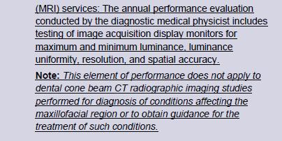
52 Soft Copy Displays (no changes) Four Tests: 1. Max and Min luminance (L max and L min ) 2. Luminance uniformity 3. Resolution using SMPTE pattern 4. Spatial accuracy (SMPTE) Specifications: 1. Max luminance (WL/WW = min): > 90 Cd/m 2 2. Min luminance: < 1.2 Cd/m 2 3. Uniformity: % difference = 200* (L max - L min )/(L max + L min ) 4. Resolution: display bar pattern of 100% contrast 5. Spatial accuracy: lines straight within +/- 5mm
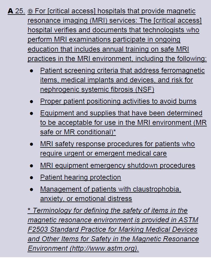
53 ACR Medical Physicist s Site Safety Assessment (Checklist) Site Access Restrictions (MR Zones) Documented MR Safety Education/Training for all personnel Patient and non MR Personnel Screening Pediatric Patient Policy Designated MR Safety Officer Disaster Policy Quench Policy Cryogen Safety Policy Acoustic Noise Policy Pregnancy Policy Contrast Agent Safety Policy Sedation Policy Thermal Burns Policy Emergency Code Procedures Device and Object Screening and designation of MR Safe/MR Conditional status Procedures for Reporting MR Safety Incidents or Adverse Incidents Patient Communication Infection Control Criteria for Compliance (All new section) 1. Written policies are present and are being reviewed and updated on a regular basis. 2. Facility has appropriate signage and methods of controlled access. Documentation of regular MR safety training for each facility staff member


54 Joint Commission MRI Safety Performance Standards
55 As part of the annual performance testing, the medical physicist is expected to perform all of the following except: 20% 1. Site safety assessment 20% 20% 20% 20% 2. Repeat of weekly QA tests 3. Static-field gradient measurement 4. Magnetic field homogeneity test 5. SNR for all clinical coils 10
56 As part of the annual equipment performance testing, the medical physicist is expected to perform all of the following except:. 1. Site safety assessment 2. Repeat of weekly QA tests 3. Static-field gradient measurement 4. Magnetic field homogeneity test 5. SNR for all clinical coils Reference: ACR MRI Accreditation Requirements I/Requirements.pdf
57 Conclusion and Comments The revised ACR MRI Quality Control Manual has relatively minor changes from the 2004 version. Specific tests are basically the same but with more options and better descriptions. There is a increased emphasis on MRI safety and infection control to minimize patient risk. An attempt was made to embrace NEMA standards, Joint Commission recommendations and AAPM Report 100. Currently the revised manual does not identify a specific method for testing parallel imaging. However, when a generally accepted method is identified, it will be incorporated into the electronic manual by means of an annual update.
ACR MRI Accreditation Program. ACR MRI Accreditation Program Update. Educational Objectives. ACR accreditation. History. New Modular Program
 ACR MRI Accreditation Program Update Donna M. Reeve, MS, DABR, DABMP Department of Imaging Physics University of Texas M.D. Anderson Cancer Center Educational Objectives Present requirements of the new
ACR MRI Accreditation: Medical Physicist Role in the Application Process
 ACR MRI Accreditation: Medical Physicist Role in the Application Process Donna M. Reeve, MS, DABR, DABMP Department of Imaging Physics University of Texas M.D. Anderson Cancer Center Educational Objectives
MRI FAQs. After reading these documents and checking your protocols, you can apply online here: https://acredit.acr.org.
 MRI FAQs Application - General Q. My facility plans to apply for ACR MRI Accreditation, where do I start? A. Start by reading the following documents, available on the ACR website: The Diagnostic Modality
MR QA/QC for MRgRT. Rick Layman, PhD, DABR Department of Radiology July 13, 2015
 MR QA/QC for MRgRT Rick Layman, PhD, DABR Department of Radiology July 13, 2015 The Ohio State University Comprehensive Cancer Center Arthur G. James Cancer Hospital and Richard J. Solove Research Institute
Nuts and Bolts of MRI Physics Tests or Selling Preparation H. Robert A. Bell, PhD. Learning Objectives
 Nuts and Bolts of MRI Physics Tests or Selling Preparation H Robert A. Bell, PhD (858) 759-0150 bell3660@sbcglobal.net Robert A. Bell, PhD Since 1987 RAB has provided independent consulting services to
AAPM Annual Meeting. ACR Accreditation Update in CT
 AAPM Annual Meeting ACR Accreditation Updates in CT, Ultrasound, Mammography and MRI: ACR Accreditation Update in CT Michael McNitt-Gray, PhD, DABR, FAAPM Professor, Department of Radiological Sciences
ACR Breast MRI Accreditation Program - DRAFT
 ACR Breast MRI Accreditation Program - DRAFT Donna M. Reeve, MS, DABR, DABMP Department of Imaging Physics Educational Objectives Provide an overview of the ACR Breast MRI Accreditation Program (BMRAP)
ACR Accreditation Toolkit for Validation Site Surveys
 ACR Accreditation Toolkit for Validation Site Surveys The ACR will be performing unannounced validation site surveys as part of the accreditation process. This checklist is designed to assist you in gathering
Implementation of the 2012 ACR CT QC Manual in a Community Hospital Setting BRUCE E. HASSELQUIST, PH.D., DABR, DABSNM ASPIRUS WAUSAU HOSPITAL
 Implementation of the 2012 ACR CT QC Manual in a Community Hospital Setting BRUCE E. HASSELQUIST, PH.D., DABR, DABSNM ASPIRUS WAUSAU HOSPITAL Conflict of Interest Disclaimer Employee of Aspirus Wausau
CT Quality Control Manual FAQs
 CT Quality Control Manual FAQs General Question: How often will the QC Manual be updated and how will those updates be communicated? Answer: The ACR CT Physics Subcommittee will review any comments, issues
BICOE Stereotactic Breast Biopsy and Breast Ultrasound Accreditation. Introduction. Educational Objectives
 BICOE Stereotactic Breast Biopsy and Breast Ultrasound Accreditation William Geiser, MS DABR Senior Medical Physicist MD Anderson Cancer Center Houston, Texas wgeiser@mdanderson.org 1 Introduction Objectives
Routine Quality Assurance Cookbook
 This Cookbook is a companion guide to the AIUM Routine Quality Assurance (QA) for Diagnostic Ultrasound Equipment document, which outlines the basic QA requirements for AIUM-accredited practices. The Guide
The American College of Radiology Breast MRI Accreditation Program: Frequently Asked Questions (Updated: October 4, 2017)
") The American College of Radiology Breast MRI Accreditation Program: Frequently Asked Questions (Updated: October 4, 2017) To navigate in this document, either click a section in the Table of Contents or
The American College of Radiology Breast MRI Accreditation Program: Frequently Asked Questions (Updated: October 4, 2017) To navigate in this document, either click a section in the Table of Contents or
BICOE Breast Imaging Center of Excellence. What is it? - Requirements. National Mammography Database. What do you get? ACR Accreditation in:
 BICOE Breast Imaging Center of Excellence What is it? - Requirements William Geiser, MS DABR Senior Medical Physicist MD Anderson Cancer Center Houston, Texas wgeiser@mdanderson.org ACR Accreditation in:
Ultrasound Accreditation and Regulatory Compliance from A to Z
 Jim Rickner presents: Ultrasound Accreditation and Regulatory Compliance from A to Z Sponsored by: Conquest Imaging Wednesday, September 28, 2016, 2:00pm ET PRESENTER: JIM RICKNER, CONQUEST IMAGING Jim
Please follow all instructions carefully.
 Nuclear Medicine Accreditation Program 1891 Preston White Drive, Reston VA 20191-4397 NUCLEAR MEDICINE TESTING INSTRUCTIONS General Instructions Please follow all instructions carefully. The enclosed labels
BICOE Stereotactic Breast Biopsy and Breast Ultrasound Accreditation. Introduction. Educational Objectives
 BICOE Stereotactic Breast Biopsy and Breast Ultrasound Accreditation William Geiser, MS DABR Senior Medical Physicist MD Anderson Cancer Center Houston, Texas wgeiser@mdanderson.org 1 Introduction Objectives
Hands-On Ultrasound Physics and Quality Control Workshop. Outline
 Slide 1 Hands-On Ultrasound Physics and Quality Control Workshop Z. F. Lu, PhD Radiology Department Columbia University New York, NY R. L. Kruger, PhD Radiology Department Marshfield Clinic Marshfield,
Update on MQSA and Mammography Accreditation
 MQSA - Who s Who Update on MQSA and Mammography Accreditation The Law: Mammography Quality Standards Act (MQSA) The Regulator: US Food and Drug Administration (FDA) Priscilla F. Butler, M.S. Senior Director,
X-Ray & CT Physics / Clinical CT
 Computed Tomography-Basic Principles and Good Practice X-Ray & CT Physics / Clinical CT INSTRUCTORS: Dane Franklin, MBA, RT (R) (CT) Office hours will be Tuesdays from 5pm to 6pm CLASSROOM: TIME: REQUIRED
ULTRASOUND QA SOLUTIONS. Ensure Accurate Screening, Diagnosis & Monitoring DOPPLER FLOW PHANTOMS MULTI-PURPOSE PHANTOMS TRANSDUCER TEST PHANTOMS
 ULTRASOUND QA SOLUTIONS Ensure Accurate Screening, Diagnosis & Monitoring DOPPLER FLOW PHANTOMS MULTI-PURPOSE PHANTOMS TRANSDUCER TEST PHANTOMS INNOVATORS IN ADVANCED ULTRASOUND TECHNIQUES Gammex is the
ULTRASOUND QA SOLUTIONS. Ensure Accurate Screening, Diagnosis and Monitoring DOPPLER FLOW PHANTOMS MULTI-PURPOSE PHANTOMS TRAINING PHANTOMS
 ULTRASOUND QA SOLUTIONS Ensure Accurate Screening, Diagnosis and Monitoring DOPPLER FLOW PHANTOMS MULTI-PURPOSE PHANTOMS TRAINING PHANTOMS INNOVATORS IN ADVANCED ULTRASOUND TECHNIQUES Gammex is the only
Current Ultrasound Quality Control Recommendations and Techniques
 Current Ultrasound Quality Control Recommendations and Techniques Evan J. Boote, Ph.D. University of Missouri-Columbia Thank you for your interest - today I hope to provide some basic recommendations for
Kish chakrabarti, Ph.D. Senior Physicist CDRH/FDA
 Facility Certification Extension Requirements, Quality Assurance and Medical Physicists role for Hologic Selenia Dimensions Digital Breast Tomosynthesis (DBT) System Kish chakrabarti, Ph.D. Senior Physicist
The American College of Radiology Breast Ultrasound Accreditation Program: Frequently Asked Questions (Revised: July 31, 2017)
") The American College of Radiology Breast Ultrasound Accreditation Program: Frequently Asked Questions (Revised: July 31, 2017) Table of Contents APPLICATION - GENERAL... 1 MOVED FACILITIES AND UNITS...
The American College of Radiology Breast Ultrasound Accreditation Program: Frequently Asked Questions (Revised: July 31, 2017) Table of Contents APPLICATION - GENERAL... 1 MOVED FACILITIES AND UNITS...
Overview. ACR Accreditation Update in Mammography. New ACR Activities. Requirements Today. What s New For Tomorrow. ACR: Recognized by FDA and CMS
 ACR Accreditation Update in Mammography Eric Berns, PhD University of Colorado Hospital Denver Health Medical Center Denver, CO *No financial disclosures to report Overview New ACR Activities Requirements
Debbie Childs RDMS, RVT Sonographer Murphy Medical Center Murphy, NC
 Debbie Childs RDMS, RVT Sonographer Murphy Medical Center Murphy, NC Worked at Murphy Medical Center as a sonographer for 18 years Registered in Abdomen, OB/GYN, Breast, & Vascular Ultrasound ACR Accredited
ULTRASOUND QA SOLUTIONS. Ensure Accurate Screening, Diagnosis & Monitoring DOPPLER FLOW PHANTOMS MULTI-PURPOSE PHANTOMS TRANSDUCER TEST PHANTOMS
 ULTRASOUND QA SOLUTIONS Ensure Accurate Screening, Diagnosis & Monitoring DOPPLER FLOW PHANTOMS MULTI-PURPOSE PHANTOMS TRANSDUCER TEST PHANTOMS INNOVATORS IN ADVANCED ULTRASOUND TECHNIQUES Gammex is the
Mammography Quality Control: A Refresher
 Mammography Quality Control: A Refresher MATT WAIT, MS, DABR Objectives Attendees will re-familiarize themselves with the purpose of quality assurance and quality control Attendees will re-familiarize
PET/MR. Are You Ready?? Derek Lee, BS, CNMT, PET
 PET/MR Are You Ready?? Derek Lee, BS, CNMT, PET Allow Myself to Introduce Myself I ve been around a while: 23+ years in Nuclear Medicine and counting PET & PET/CT for 13 years and counting Currently working
American College of Radiology CT Accreditation Program. Testing Instructions
 American College of Radiology CT Accreditation Program Testing Instructions (Revised January 6, 2017) This guide provides all of the instructions necessary for clinical tests, phantom tests and general
MAMMOGRAPHY QC: MORE THAN PHANTOM IMAGES. Stephanie Schofield RTR QC Technologist Nova Scotia Health Authority
 1 MAMMOGRAPHY QC: MORE THAN PHANTOM IMAGES Stephanie Schofield RTR QC Technologist Nova Scotia Health Authority Disclaimer 2 I have nothing to disclose. Outline 3 QC tests for the mammography technologist
KNEE ALIGNMENT SYSTEM (KAS) MRI Protocol
 MRI Protocol") KNEE ALIGNMENT SYSTEM (KAS) MRI Protocol Sample referral sticker Referral Sticker Insert here Corin 17 Bridge Street Pymble NSW Australia 2073 P: +61 (0)2 9497 7400 F: +61 (0)2 9497 7498 E: KAS.customerservice@coringroup.com
KNEE ALIGNMENT SYSTEM (KAS) MRI Protocol Sample referral sticker Referral Sticker Insert here Corin 17 Bridge Street Pymble NSW Australia 2073 P: +61 (0)2 9497 7400 F: +61 (0)2 9497 7498 E: KAS.customerservice@coringroup.com
NONE. Disclosures. Accreditation Update
 ACR Ultrasound Accreditation: Requirements and Pitfalls Presented to: American Association of Physicists in Medicine Presented by: Jennifer Walter RDMS,RVT, RT(R) ACR Quality & Safety August 03, 2016 Disclosures
Medical Diagnostic Imaging
 Medical Diagnostic Imaging Laboratories Medical Diagnostic Imaging Lab Name Location Person in Charge Programs Served Courses Served Patient Care and Management (2) Introduction to MDI Radiographic Technique
RECENT ADVANCES IN CLINICAL MR OF ARTICULAR CARTILAGE
 In Practice RECENT ADVANCES IN CLINICAL MR OF ARTICULAR CARTILAGE By Atsuya Watanabe, MD, PhD, Director, Advanced Diagnostic Imaging Center and Associate Professor, Department of Orthopedic Surgery, Teikyo
3/22/2017. MR Protocol Review Clinical Opportunities for Physicists. AAPM Newsletter March/April Technical to Clinical Transition - How?
 AAPM Newsletter March/April 2017 Clinical Opportunities for Physicists Anshuman Panda, Ph.D. AAPM Spring Clinical Meeting New Orleans, LA March 2017 No conflict of interest to declare Physics Summit on
Quality Assurance of Ultrasound Imaging in Radiation Therapy. Zuofeng Li, D.Sc. Murty S. Goddu, Ph.D. Washington University St.
 Quality Assurance of Ultrasound Imaging in Radiation Therapy Zuofeng Li, D.Sc. Murty S. Goddu, Ph.D. Washington University St. Louis, Missouri Typical Applications of Ultrasound Imaging in Radiation Therapy
354 Korean J Radiol 9(4), August 2008
, August 2008") Review of Failed CT Phantom Image Evaluations in 2005 and 2006 by the CT Accreditation Program of the Korean Institute for Accreditation of Medical Image Hye Jung Park, MD 1 Seung Eun Jung, MD 1, 2 Young
Review of Failed CT Phantom Image Evaluations in 2005 and 2006 by the CT Accreditation Program of the Korean Institute for Accreditation of Medical Image Hye Jung Park, MD 1 Seung Eun Jung, MD 1, 2 Young
Outline. QA/QC of Ultrasound Imagers: Basic Physics, Procedures and Experiences. Frame Rate Limitation. US Imaging Range Equation.
 QA/QC of Ultrasound Imagers: Basic Physics, Procedures and Experiences Zheng F. Lu, PhD Radiology Department Columbia University Email: zfl1@columbia.edu Outline General overview of basic ultrasound physics
Computed tomography Acceptance testing and dose measurements
 Computed tomography Acceptance testing and dose measurements Jonas Andersson Medical Physicist, Ph.D. Department of Radiation Sciences University Hospital of Norrland, Umeå Sweden Contents The Computed
PROPHECY. Preoperative Navigation Guides ANKLE CT SCAN PROTOCOL
 PROPHECY Preoperative Navigation Guides ANKLE CT SCAN PROTOCOL 90 FIGURE 1 Examples FIGURE 1 Examples of neutral ankle positioning. PROPHECY Ankle CT Scan Protocol PROPHECY INBONE and PROPHECY INFINITY
REGULATION: QUALITY ASSURANCE PROGRAMS FOR MEDICAL DIAGNOSTIC X-RAY INSTALLATIONS N.J.A.C. 7:28-22
 REGULATION: QUALITY ASSURANCE PROGRAMS FOR MEDICAL DIAGNOSTIC X-RAY INSTALLATIONS N.J.A.C. 7:28-22 New Jersey Department of Environmental Protection Bureau of X-ray Compliance PO Box 420, Mail Code 25-01
ESTABLISHING DRLs in PEDIATRIC CT. Keith Strauss, MSc, FAAPM, FACR Cincinnati Children s Hospital University of Cincinnati College of Medicine
 ESTABLISHING DRLs in PEDIATRIC CT Keith Strauss, MSc, FAAPM, FACR Cincinnati Children s Hospital University of Cincinnati College of Medicine CT Dose Indices CTDI INTRODUCTION CTDI 100, CTDI w, CTDI vol
Herlev radiation oncology team explains what MRI can bring
 Publication for the Philips MRI Community Issue 46 2012/2 Herlev radiation oncology team explains what MRI can bring The radiotherapy unit at Herlev University Hospital investigates use of MRI for radiotherapy
Tissue-engineered medical products Evaluation of anisotropic structure of articular cartilage using DT (Diffusion Tensor)-MR Imaging
-MR Imaging") Provläsningsexemplar / Preview TECHNICAL REPORT ISO/TR 16379 First edition 2014-03-01 Tissue-engineered medical products Evaluation of anisotropic structure of articular cartilage using DT (Diffusion Tensor)-MR
Provläsningsexemplar / Preview TECHNICAL REPORT ISO/TR 16379 First edition 2014-03-01 Tissue-engineered medical products Evaluation of anisotropic structure of articular cartilage using DT (Diffusion Tensor)-MR
Ultrasound Accreditation Program Requirements
 Ultrasound Accreditation Program Requirements OVERVIEW... 2 MANDATORY ACCREDITATION TIME REQUIREMENTS... 2 PERSONNEL QUALIFICATIONS... 2 PHYSICIAN QUALIFICATIONS... 3 TECHNOLOGIST QUALIFICATIONS... 4 QUALITY
MRI to fit your planning. Philips Panorama HFO Oncology Configuration
 MRI to fit your planning Philips Panorama HFO Oncology Configuration MR Imaging that fits Philips Panorama HFO Oncology Configuration allows radiation oncologists to take full advantage of MRI s excellent
The American College of Radiology Digital Mammography QC Manual: Frequently Asked Questions (Revised 12/12/2018; new and updated items in red)
") The American College of Radiology Digital Mammography QC Manual: Frequently Asked Questions (Revised 12/12/2018; new and updated items in red) Table of Contents General... 1 Applicability... 3 Transitioning
The American College of Radiology Digital Mammography QC Manual: Frequently Asked Questions (Revised 12/12/2018; new and updated items in red) Table of Contents General... 1 Applicability... 3 Transitioning
created by high-voltage devices Examples include medical and dental x-rays, light, microwaves and nuclear energy
 What is radiation? Radiation is energy emitted from a source, that travels through space and can penetrate matter. Listed below are two types that we are exposed to and contribute to our overall radiation
8/3/2016. Diagnostic Ultrasound Imaging Quality Assurance. Purpose. Information on US QA. James A. Zagzebski 1, Ph.D. Zheng Feng Lu 2, Ph.D.
 Diagnostic Ultrasound Imaging Quality Assurance James A. Zagzebski 1, Ph.D. Zheng Feng Lu 2, Ph.D. 1 Dept. of Medical Physics University of Wisconsin, Madison 2 Dept. Of Radiology, University of Chicago,
Overview. ACR Update on FFDM Accreditation. New ACR Activities. QC Today. QC Tomorrow. ACR Breast Imaging Centers of Excellence BICOE
 ACR Update on FFDM Accreditation Overview New ACR Activities Eric Berns, PhD University of Colorado Hospital Denver Health Medical Center Denver, CO QC Today QC Tomorrow *No financial disclosures to report
A quality control program for MR-guided focused ultrasound ablation therapy
 JOURNAL OF APPLIED CLINICAL MEDICAL PHYSICS, VOLUME 3, NUMBER 2, SPRING 2002 A quality control program for MR-guided focused ultrasound ablation therapy Tao Wu* and Joel P. Felmlee Department of Radiology,
A. DeWerd. Michael Kissick. Larry. Editors. The Phantoms of Medical. and Health Physics. Devices for Research and Development.
 Larry Editors A. DeWerd Michael Kissick The Phantoms of Medical and Health Physics Devices for Research and Development ^ Springer Contents 1 Introduction to Phantoms of Medical and Health Physics 1 1.1
Preparing for Medical Physics Components of the ABR Core Examination
 Preparing for Medical Physics Components of the ABR Core Examination The ABR core examination for radiologists contains material on medical physics. This content is based on the medical physics that is
TG-128: Quality Assurance for Prostate Brachytherapy Ultrasound
 TG-128: Quality Assurance for Prostate Brachytherapy Ultrasound STEVEN SUTLIEF DOUG PFEIFFER (HEATHER PIERCE, WENGZHENG FENG, JIM KOFLER) AAPM ANNUAL MEETING 2010 Educational Objectives To describe the
High Field MR of the Spine
 Department of Radiology University of California San Diego 3T for MR Applications Advantages High Field MR of the Spine Increased signal-to-noise Better fat suppression Increased enhancement with gadolinium
1.5 Tesla and 3 Tesla Magnetic Resonance Imaging (MRI) Guidelines for the Senza System (IPG1000 and IPG1500)
 Guidelines for the Senza System (IPG1000 and IPG1500)") 1.5 Tesla and 3 Tesla Magnetic Resonance Imaging (MRI) Guidelines for the Senza System (IPG1000 and IPG1500) ONLY NEVRO CORP. All questions or concerns about Nevro products should be forwarded to: Nevro
1.5 Tesla and 3 Tesla Magnetic Resonance Imaging (MRI) Guidelines for the Senza System (IPG1000 and IPG1500) ONLY NEVRO CORP. All questions or concerns about Nevro products should be forwarded to: Nevro
Introduction. The goal of TRUS QA is to ensure your system can do all of this accurately.
 TRUS QA Workshop Introduction The goals of using TRUS in prostate brachytherapy Visualize the prostate Need the US to penetrate deeply enough Need sufficient grey scale resolution to be able to visualize
How Much Tesla Is Too Much?
 How Much Tesla Is Too Much? Johnny U. V. Monu, MB, BS; MSc Professor of Radiology and Orthopedics University of Rochester School of Medicine Rochester, New York Historical Timeline Clinical Imaging 1970
HISTORY OF MQSA AND ACR
 HISTORY OF MQSA AND ACR DEBORAH THAMES R.T. (R)(M)(QM) WHY MQSA? In the United States, there was a lack of standards in mammography imaging. Reporting Imaging Type of imaging screening/diagnostic Equipment
1.5 Tesla and 3 Tesla Magnetic Resonance Imaging (MRI) Guidelines for the Senza System (IPG1000 and IPG1500)
 Guidelines for the Senza System (IPG1000 and IPG1500)") 1.5 Tesla and 3 Tesla Magnetic Resonance Imaging (MRI) Guidelines for the Senza System (IPG1000 and IPG1500). ONLY NEVRO CORP. All questions or concerns about Nevro products should be forwarded to: Nevro
1.5 Tesla and 3 Tesla Magnetic Resonance Imaging (MRI) Guidelines for the Senza System (IPG1000 and IPG1500). ONLY NEVRO CORP. All questions or concerns about Nevro products should be forwarded to: Nevro
Hands-on. Physics Workshop. 2-day Workshop June 1-2, 2018 Ann Arbor, MI. Program Director: Brian Fowlkes, PhD
 Hands-on Ultrasound Physics Workshop 2-day Workshop June 1-2, 2018 Ann Arbor, MI Program Director: Brian Fowlkes, PhD Faculty: Paul Carson, PhD Mitch Goodsitt, PhD Oliver Kripfgans, PhD Sandra Larson,
Cover Page. The handle holds various files of this Leiden University dissertation.
 Cover Page The handle http://hdl.handle.net/1887/35124 holds various files of this Leiden University dissertation. Author: Wokke, Beatrijs Henriette Aleid Title: Muscle MRI in Duchenne and Becker muscular
AHLA. UU. Diagnostic Imaging Services. Thomas W. Greeson Reed Smith LLP Falls Church, VA
 AHLA UU. Diagnostic Imaging Services Thomas W. Greeson Reed Smith LLP Falls Church, VA Institute on Medicare and Medicaid Payment Issues March 25-27, 2015 AHLA Institute on Medicare and Medicaid Payment
Echelon Oval provides a robust suite of leading musculoskeletal imaging capabilities for detailed assessment of all anatomy for your most challenging
 Echelon Oval provides a robust suite of leading musculoskeletal imaging capabilities for detailed assessment of all anatomy for your most challenging cases. Hitachi Medical Systems America, Inc. 1959 Summit
FreeWave upgrade enables faster, more powerful imaging
 FreeWave upgrade enables faster, more powerful imaging Western Neurological Associates upgrades Intera 1.5T to Release 2.5 with FreeWave, and sees a significant improvement in image quality and efficiency.
Multicenter Trial, Phase I Assessment of 2-D FFDM Versus Combo of 2-D and 3-D Tomosynthesis in Breast Cancer Screening
 ACRIN PA 4006: Comparison of Full Field Digital Mammography with Digital Breast Tomosynthesis Image Acquisition in Relation to Screening Call-Back Rate Imaging Manual Multicenter Trial, Phase I Assessment
MQSA Physicist Qualifications. Jon J. Erickson, Ph.D., DABR
 2027 North 36 th Street Saint Joseph, Mo 64506 (86) 390-90 (800) 306-4477 MQSA Physicist Qualifications Jon J. Erickson, Ph.D., DABR Contents Missouri Mammography Physicist Initial Qualifications Certificate
CT Scanning Protocol For V2R Guided Surgery Solutions
 CT Scanning Protocol For V2R Guided Surgery Solutions 2 V2R CT Scanning Protocol \\ Contents Contents General requirements... 3 V2R Dual Scan Protocol... 5 V2R Single Scan Protocol... 8 Overview... 10
Descriptions of NDT Projects Fall 2004 October 31, 2004
 Descriptions of NDT Projects Fall 2004 October 31, 2004 Introduction There are two separate NDT labs in Magister: ULTRA for ultrasound and EDDY for eddy current. Both labs are equipped with mechanical
STRUCTURED EDUCATION REQUIREMENTS EFFECTIVE: JANUARY 1, 2016
 Computed Tomography The purpose of structured education is to provide the opportunity for individuals to develop mastery of discipline-specific knowledge that, when coupled with selected clinical experiences,
RADIATION PROTECTION IN DIAGNOSTIC AND INTERVENTIONAL RADIOLOGY. L19: Optimization of Protection in Mammography
 IAEA Training Material on Radiation Protection in Diagnostic and Interventional Radiology RADIATION PROTECTION IN DIAGNOSTIC AND INTERVENTIONAL RADIOLOGY L19: Optimization of Protection in Mammography
Acknowledgements. QA Concerns in MR Brachytherapy. Learning Objectives. Traditional T&O. Traditional Summary. Dose calculation 3/7/2015
 Acknowledgements QA Concerns in MR Brachytherapy Robert A. Cormack Dana Farber Cancer Institute & Brigham and Women s Hospital No financial conflicts of interest I may mention use of devices in ways that
Q-Sense fmri in the MRI environment
 Q-Sense fmri in the MRI environment An evaluation at the Erwin L. Hahn Institute for Magnetic Resonance Imaging, and at the Institute of Diagnostic and Interventional Radiology and Neuroradiology, Essen
Update on the ACR FFDM QC Manual
 Update on the ACR FFDM QC Manual Eric Berns, PhD University of Colorado Hospital Denver Health Medical Center Denver, CO AAPM 2011 Vancouver, Canada 9000 8500 8000 7500 7000 6500 6000 5500 5000 4500 4000
MRI PEDIATRIC PROTOCOLS (Updated 6/19/2018)
") MRI PEDIATRIC PROTOCOLS (Updated 6/19/2018) *Please get or let us know where radiologist can review plain films. *For Texas Orthopedics and other Docs requesting only MSK section read for their pediatric
MRI PEDIATRIC PROTOCOLS (Updated 6/19/2018) *Please get or let us know where radiologist can review plain films. *For Texas Orthopedics and other Docs requesting only MSK section read for their pediatric
Why Talk About Technique? MRI of the Knee:
 Why Talk About Technique? MRI of the Knee: Part 1 - Imaging Techniques Mark Anderson, M.D. University of Virginia Health Sciences Center Charlottesville, Virginia Always had an interest teach our fellows
Highmark Privileging Application. 1. Complete this application if you provide any diagnostic
 Highmark Privileging Application INSTRUCTIONS 1. Complete this application if you provide any diagnostic imaging services. 2. Please complete a separate application for each physical location where imaging
ACCREDITATION CASE REVIEW 2: US AND US-GUIDED BIOPSY CRITERIA
 ACCREDITATION CASE REVIEW 2: US AND US-GUIDED BIOPSY CRITERIA Stamatia Destounis, MD, FSBI, FACR Elizabeth Wende Breast Care, LLC Clinical Professor University of Rochester School of Medicine and Dentistry
Effect of intravenous contrast medium administration on prostate diffusion-weighted imaging
 Effect of intravenous contrast medium administration on prostate diffusion-weighted imaging Poster No.: C-1766 Congress: ECR 2015 Type: Authors: Keywords: DOI: Scientific Exhibit J. Bae, C. K. Kim, S.
Supplementary information Detailed Materials and Methods
 Supplementary information Detailed Materials and Methods Subjects The experiment included twelve subjects: ten sighted subjects and two blind. Five of the ten sighted subjects were expert users of a visual-to-auditory
Visualization strategies for major white matter tracts identified by diffusion tensor imaging for intraoperative use
 International Congress Series 1281 (2005) 793 797 www.ics-elsevier.com Visualization strategies for major white matter tracts identified by diffusion tensor imaging for intraoperative use Ch. Nimsky a,b,
R. Edward Hendrick, PhD, FACR. Breast MRI. Fundamentals and Technical Aspects
 Breast MRI R. Edward Hendrick, PhD, FACR Breast MRI Fundamentals and Technical Aspects R. Edward Hendrick, PhD, FACR Research Professor and Director, Breast Imaging Research (retired) Lynn Sage Comprehensive
Clinical Applications
 C H A P T E R 16 Clinical Applications In selecting pulse sequences and measurement parameters for a specific application, MRI allows the user tremendous flexibility to produce variations in contrast between
Ingenia MR-RT. MR Systems. The comprehensive MR-sim solution to fit your planning
 Ingenia MR-RT MR Systems The comprehensive MR-sim solution to fit your planning Table of contents Experience the difference MRI makes 3 A comprehensive MR-sim solution 4 Position with precision 6 Tailored
Low Dose CT Lung Screening: What is Technically Required?
 Low Dose CT Lung Screening: What is Technically Required? COMP/CCPM Annual Scientific Meeting Ottawa, Ontario July 15 th, 2017 Yogesh Thakur, PhD, MCCPM Medical Physicist Lead and Regional RSO (X-Ray)
Dental ConeBeam Computed Tomography (CBCT) X-ray Systems
 X-ray Systems") Dental ConeBeam Computed Tomography (CBCT) X-ray Systems PROPOSED REVISIONS TO 4732.XXXX, 1.0 4732.#### DENTAL CONEBEAM COMPUTED TOMOGRAPHY (CBCT) X-RAY SYSTEMS; STATIONARY AND MOBILE. Subpart 1. Applicability.
Dental ConeBeam Computed Tomography (CBCT) X-ray Systems PROPOSED REVISIONS TO 4732.XXXX, 1.0 4732.#### DENTAL CONEBEAM COMPUTED TOMOGRAPHY (CBCT) X-RAY SYSTEMS; STATIONARY AND MOBILE. Subpart 1. Applicability.
CLINICAL RADIATION SCIENCES (CLRS)
") Clinical Radiation Sciences (CLRS) 1 CLINICAL RADIATION SCIENCES (CLRS) CLRS 101. Introduction to Clinical Radiologic Sciences. 1 Hour. Semester course; 1 lecture hour. 1 credit. Presentation and discussion
Clinical Radiation Sciences (CLRS) 1 CLINICAL RADIATION SCIENCES (CLRS) CLRS 101. Introduction to Clinical Radiologic Sciences. 1 Hour. Semester course; 1 lecture hour. 1 credit. Presentation and discussion
Q. Where can I find out if my state currently requires stereotactic breast biopsy accreditation?
 The American College of Radiology Stereotactic Breast Biopsy Accreditation Program: Frequently Asked Questions (Revised: September 7, 2017; updated questions in red) Table of Contents Application - General...
The first MR with IQ. Philips Intera 1.5T With SmartExam
 The first MR with IQ Philips Intera 1.5T With SmartExam Smart Exam reproducibility 2 consistency, and efficiency Philips Intera 1.5T gives you the image quality, patient comfort and range of clinical applications
Personalized Solutions. MRI Protocol for PSI and Signature Guides
 Personalized Solutions MRI Protocol for PSI and Signature Guides 2 Personalized Solutions MRI Protocol for PSI and Signature Guides Purpose and Summary This protocol is applicable for the Zimmer Biomet
Prepublication Requirements
 Issued Prepublication Requirements Standards Revisions for Organizations Providing Fluoroscopy Services The Joint Commission has approved the following revisions for prepublication. While revised requirements
SOMATOM Drive System Owner Manual Dosimetry and imaging performance report
 www.siemens.com/healthcare SOMATOM Drive System Owner Manual Dosimetry and imaging performance report Table of contents 1 Dosimetry and imaging performance report 5 1.1 Dose information 5 1.1.1 General
Magnetic Resonance Imaging
 Magnetic Resonance Imaging The purpose of structured education is to provide the opportunity for individuals to develop mastery of discipline-specific knowledge that, when coupled with selected clinical
Goals and Objectives MRI (First Year)
") Goals and Objectives MRI (First Year) 1. Protocol cases, in consultation with the attending, to assure that the MRI 2. Describe basic physics concepts used in generating clinical MR images and identify
Goals and Objectives MRI (First Year) 1. Protocol cases, in consultation with the attending, to assure that the MRI 2. Describe basic physics concepts used in generating clinical MR images and identify
Musculoskeletal Imaging at 3T with Simultaneous Use of Multipurpose Loop Coils
 Clinical Orthopedic Imaging Musculoskeletal Imaging at 3T with Simultaneous Use of Multipurpose Loop Coils Elena Ferrer 1 ; Rafael Coronado Santos 2 1 Radiology Department, Clínica Creu Blanca, Barcelona,
NEURO PROTOCOLS MRI NEURO PROTOCOLS (SIEMENS SCANNERS)
") Page 1 NEURO PROTOCOLS Brain Stroke Brain Brain with contrast Brain for seizures Brain for MS Brain for Pineal gland Sella FAST Scan for hydrocephalus MRA/MRV Brain MRA carotids 8 th nerve Cranial nerves
Page 1 NEURO PROTOCOLS Brain Stroke Brain Brain with contrast Brain for seizures Brain for MS Brain for Pineal gland Sella FAST Scan for hydrocephalus MRA/MRV Brain MRA carotids 8 th nerve Cranial nerves
WHAT DOES THE BRAIN TELL US ABOUT TRUST AND DISTRUST? EVIDENCE FROM A FUNCTIONAL NEUROIMAGING STUDY 1
 SPECIAL ISSUE WHAT DOES THE BRAIN TE US ABOUT AND DIS? EVIDENCE FROM A FUNCTIONAL NEUROIMAGING STUDY 1 By: Angelika Dimoka Fox School of Business Temple University 1801 Liacouras Walk Philadelphia, PA
Sensitivity and Specificity in Detection of Labral Tears with 3.0-T MRI of the Shoulder
 Magee and Williams MRI for Detection of Labral Tears Musculoskeletal Imaging Clinical Observations C M E D E N T U R I C L I M G I N G JR 2006; 187:1448 1452 0361 803X/06/1876 1448 merican Roentgen Ray