The many faces of extranodal lymphoma
|
|
|
- Gwendolyn McCormick
- 6 years ago
- Views:
Transcription
1 The many faces of extranodal lymphoma Frank Pameijer Departments of Radiology and Radiation Oncology University Medical Center Utrecht

2 Special thanks to Ilona M Schmalfuss, MD University of Florida Gainesville, USA For supplying material used in this lecture
3 The many faces.. Let s look at 5 patients with pathology confirmed extranodal head&neck lymphoma
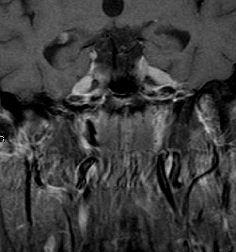
4 Middle-aged male Referred otalgia right side T1 STIR
5 Elderly lady Bilateral orbital swelling, visual disturbance
6 50 year old male Nasal stuffiness
7 Middle-aged female Bilateral parotid swelling, dry eyes
8 Female patient Enlarging thyroid mass
9 Purpose To present the spectrum of imaging features of extra-nodal H&N lymphoma Contrast this with other diseases in comparable locations
10 Extranodal lymphoma H&N region: 2nd most frequent site (after GI tract) DD from other malignancies difficult because of overlapping clinical presentations and imaging manifestations => Diagnosis should be considered in any extranodal H&N mass
11 Extranodal (H&N) Lymphoma General imaging features CT: Isodense to muscle, homogeneous enhancement MR: T1: isointense to muscle, T2: variable, +Gd: homogeneous enhancement, ADC: restriction ++ Pet-CT: Usually high metabolic activity (depends on cell type and lymphoma grade)
12 Let s go back to the cases More systematically
13 Extranodal H&N Lymphoma Lymphatic Extralymphatic
14 Extranodal H&N Lymphoma Lymphatic Waldeyer s ring Extralymphatic
15 Extranodal H&N Lymphoma Lymphatic Waldeyer s ring Extralymphatic Orbit Upper respiratory tract (sinonasal) Salivary glands Thyroid
16 Extranodal H&N Lymphoma 80% additional nodal involvement Enhanced CT / MR (usually) non-necrotic nodes SCC
17 Waldeyer s ring Anatomical term describing a circle of lymphoid tissue in the naso- and oropharynx Nasopharynx
18 Waldeyer s ring Anatomical term describing a circle of lymphoid tissue in the naso- and oropharynx Nasopharynx Tonsillar fossa

19 Waldeyer s ring Anatomical term describing a circle of lymphoid tissue in the naso- and oropharynx Nasopharynx Tonsillar fossa Tongue Base


20 Large nasopharyngeal mass No destruction / infiltration DWI: restriction ++ Associated adenopathy b=1000 Nasopharyngeal lymphoma
21 Middle-aged man Referred otalgia right side T1 STIR Tonsillar fossa lymphoma

 Pre")
22 Tonsillar fossa lymphoma T2 (STIR) Pre RT Post RT


23 Companion case Female, 64 year; swelling right neck Tonsillar fossa SCC with ipsilateral lymph node metastasis

24 Lymphoma SCC
25 Patient with swallowing complaints Exofytic mass base of tongue (BOT), no ventral infiltration Ipsilateral non-necrotic adenopathy Proven BOT lymphoma


26 Companion case Exofytic BOT mass with left-sided ventral infiltration Bilateral non-homogeneous adenopathy BOT SCC with bilateral lymph node metastases


27 Lymphoma SCC
28 Waldeyer s ring DD: between lymphoma & SCC Primary tumor Usually impossible Lymph nodes Lymphoma usually non-necrotic SCC usually necrotic!
 Salivary glands")
29 Extranodal H&N Lymphoma Lymphatic Waldeyer s ring Extralymphatic Orbit Upper respiratory tract (sinonasal) Salivary glands Thyroid
30 Orbital lymphoma Extraocular Intraocular Exceedingly rare!
31 Extraocular lymphoma Conjunctival involvement (25%) Without conjunctival involvement (75%)

32 Conjunctival involvement Salmon red patch swollen conjunctiva


33 Extraocular lymphoma Conjunctival involvement T1 T1 + Gd
34 Extraocular lymphoma Without conjunctival involvement (75%) Any structure may be affected In decreasing order of frequency DD Lacrimal gland conal compartment (i.e. extraocular muscles) intraconal compartment (retrobulbar Dx) Idiopathic inflammatory pseudotumor Graves s eye disease Other orbital malignancies
35 Extraocular lymphoma Clinical presentation Palpable (usually painless) mass Exophthalmos Ptosis Diplopia and/or abnormal eye movement Usually preserved vision


36 Swelling left eye T1 T1Gd DWI b=1000 DD: Lacrimal gland malignancy PET: multifocal Dx! Lacrimal gland lymphoma
37 Elderly lady Bilateral orbital swelling, visual disturbance Bilateral lacrimal gland lymphoma
38 Conal involvement Infiltrating mass of right inferior rectus (proven lymphoma) Medially extending beyond muscle boundary DD: Inflammatory pseudotumor Graves s eye disease
39 Companion case Enlargement of inferior & lateral rectus muscles Respecting muscle boundaries Proven Graves s eye disease
40 Lymphoma Graves s


41 Intraconal (retrobulbar) involvement T1 T1FS Gd Smooth intraconal mass, homogeneous enhancement Melting around optic nerve Proven extraorbital lymphoma
42 CT Loss of vision left eye
43 Loss of vision left eye CT T1 T1+Gd

44 Loss of vision left eye CT T1 T1+Gd DWI b=1000 Smooth intraocular mass left eye with intraconal extension. MR DWI: restriction DD: Inflammatory pseudotumor Proven intraocular lymphoma (very rare!)
45 Extranodal H&N Lymphoma Lymphatic Waldeyer s ring Extralymphatic Orbit Upper respiratory tract (sinonasal) Salivary glands Thyroid
46 Sinonasal lymphoma Two distinct subgroups DD B-cell lymphomas: less aggressive NK-T-cell lymphomas: destructive! (indistinguishable from SCC) (other) Malignancies; SCC! Infection Sinusitis, invasive fungal rhinosinusitis Granulomatous diseases Polyangiitis, sarcoid

47 Adult male: rhinosinusitis, B-symptoms Non aggressive mucosal thickening DD: sinusitis Biopsy: B-cell lymphoma


48 Companion case Female patient: rhinosinusitis Non aggressive mucosal thickening Nasoseptal / palatal defect Biopsy: granulomatosis with polyangiitis (Wegener)

49 Lymphoma Wegener s
50 50 year old male Nasal stuffiness

 Carcinoma")
51 50 year old male nasal stuffiness Biopsy: lymphoma (NK-T-cell) Carcinoma look-alike!!
52 Extranodal H&N Lymphoma Lymphatic Waldeyer s ring Extralymphatic Orbit Upper respiratory tract (sinonasal) Salivary glands (80% parotid gland) Thyroid
53 Parotid lymphoma Primary Parotid parenchyma Rare MALT lymphoma Mucosa Associated Lymphoid Tissue Increased incidence in Sjögren s syndrome! Secondary * Within intraparotid lymph nodes Systemic Dx * : i.e. nodal disease!
54 Female patient Bilateral parotid swelling, dry eyes Sjögren s disease & (MALT) lymphoma


55 Companion case Female patient: Sjögren s disease MALT lymphoma lymph nodes

 -")
56 Companion case: Female 85 years Progressive swelling right cheek Transspatial mass (Waldeyer s / masticator / parotid space) - Proven lymphoma


57 Same patient Post chemotherapy (3x CHOP) Complete radiological & metabolic response


58 Secondary Parotid Lymphoma Bilateral disease! Multiple intra- and peri-parotid nodes Associated Level I nodes R. Hermans


59 Companion case Male patient; bilateral parotid&neck swelling Multiple intraparotid lesions & neck adenopathy DD: Lymphoepithelial cysts in HIV Infection!
60 Extranodal H&N Lymphoma Lymphatic Waldeyer s ring Extralymphatic Orbit Upper respiratory tract (sinonasal) Salivary glands Thyroid
61 Primary thyroid gland lymphoma F > M, typically (peak years) 80% associated with chronic lymphocytic thyroiditis (Hashimoto s) DD: Other thyroid malignancies, esp. anaplastic thyroid carcinoma Presentation: enlarging mass +/- lymphadenopathy Imaging: CT/MR: infiltrating pattern with invasion of surrounding structures (trachea and esophagus) Pet-CT: usually hypermetabolic
62 Female patient Enlarging thyroid mass Markedly enlarged thyroid gland; left > right Extension into left tracheoesophageal groove Marked FDG avidity Proven thyroid lymphoma


63 Elderly patient Rapidly enlarging thyroid mass Diffuse enlarged thyroid, associated lymphadenopathy Infiltration of right TE-groove & trachea Proven thyroid lymphoma R. Hermans
64 Summary: extranodal lymphoma Indeed: it has many faces Lymphatic Waldeyer s ring Extralymphatic Orbit Upper respiratory tract (sinonasal) Salivary glands Thyroid


65 Key points Lymphoma should be considered in any extranodal head and neck mass


66 Key points Waldeyer s ring is the most common site of extranodal head and neck lymphoma Nasofarynx Tonsil Base of tongue
67 Key points Associated lymph nodes in extranodal lymphoma (usually) are non-necrotic
")

68 Key points Tissue sampling is (almost) always neccessary to establish the diagnosis J.A. Castelijns
69 The many faces of extranodal lymphoma Frank Pameijer
70 Question? Can perineural spread occur with lymphoma? Answer:

71 Question? Can perineural spread occur with lymphoma? Answer: YES


72 Extensive PNS along V2, V3 in lymphoma


73 Secondary bone involvement R. Hermans Permeative
Imaging: When to get MRI, CT or PET-CT?
 Imaging: When to get MRI, CT or PET-CT? Alina Uzelac, D.O. Assistant Clinical Professor Neuroradiology UCSF Department of Radiology and Biomedical Imaging San Francisco General Hospital Overview CT MRI
Carcinoma of Unknown Primary site (CUP) in HEAD & NECK SURGERY
 in HEAD & NECK SURGERY") Carcinoma of Unknown Primary site (CUP) in HEAD & NECK SURGERY SEARCHING FOR THE PRIMARY? P r o f J P P r e t o r i u s H e a d : C l i n i c a l U n i t C r i t i c a l C a r e U n i v e r s i t y O f
Carcinoma of Unknown Primary site (CUP) in HEAD & NECK SURGERY SEARCHING FOR THE PRIMARY? P r o f J P P r e t o r i u s H e a d : C l i n i c a l U n i t C r i t i c a l C a r e U n i v e r s i t y O f
Management of Neck Metastasis from Unknown Primary
 Management of Neck Metastasis from Unknown Primary.. Definition Histologic evidence of malignancy in the cervical lymph node (s) with no apparent primary site of original tumour Diagnosis after a thorough
Case Scenario 1. 7/13/12 Anterior floor of mouth biopsy: Infiltrating squamous cell carcinoma, not completely excised.
 Case Scenario 1 7/5/12 History A 51 year old white female presents with a sore area on the floor of her mouth. She claims the area has been sore for several months. She is a current smoker and user of
Case Scenario. 7/13/12 Anterior floor of mouth biopsy: Infiltrating squamous cell carcinoma, not completely excised.
 Case Scenario 7/5/12 History A 51 year old white female presents with a sore area on the floor of her mouth. She claims the area has been sore for several months. She is a current smoker and user of alcohol.
AJCC Staging of Head & Neck Cancer (7 th edition, 2010) -LIP & ORAL CAVITY-
 -LIP & ORAL CAVITY-") TX: primary tumor cannot be assessed T0: no evidence of primary tumor Tis: carcinoma in situ. T1: tumor is 2 cm or smaller AJCC Staging of Head & Neck Cancer (7 th edition, 2010) -LIP & ORAL CAVITY- T2:
TX: primary tumor cannot be assessed T0: no evidence of primary tumor Tis: carcinoma in situ. T1: tumor is 2 cm or smaller AJCC Staging of Head & Neck Cancer (7 th edition, 2010) -LIP & ORAL CAVITY- T2:
Ranbir Singh Sandhu Gillian Lieberman, MD. Orbital Lymphoma. Ranbir Singh Sandhu, University College London Medical School Gillian Lieberman MD, BIDMC
 Orbital Lymphoma Ranbir Singh Sandhu, University College London Medical School Gillian Lieberman MD, BIDMC 1 Our Patient Clinical Picture This topic will be presented in the context of our patient. She
Head & Neck Clinical Sub Group. Network Agreed Imaging Guidelines for UAT and Thyroid Cancer. Measure Nos: 11-1C-105i & 11-1C-106i
 Greater Manchester, Lancashire & South Cumbria Strategic Clinical Network & Senate Head & Neck Clinical Sub Group Network Agreed Imaging Guidelines for UAT and Thyroid Cancer Measure Nos: 11-1C-105i &
C. Douglas Phillips, MD FACR Director of Head and Neck Imaging Weill Cornell Medical Center NewYork Presbyterian Hospital
 C. Douglas Phillips, MD FACR Director of Head and Neck Imaging Weill Cornell Medical Center NewYork Presbyterian Hospital Objectives Review basics of head and neck imaging Discuss our spatial approach
6 th Reprint Handbook Pages AJCC 7 th Edition
 6 th Reprint Handbook Pages AJCC 7 th Edition AJCC 7 th Edition Errata for 6 th Reprint Table 1 Handbook No Significant Staging Clarifications for 6 th Reprint AJCC 7 th Edition Errata for 6 th Reprint
Imaging Technique. Ultrasound Imaging of the Salivary Glands. Parotid Gland. The Major Salivary Glands. Parotid Gland: Stenson s Duct.
 Ultrasound Imaging of the Salivary Glands Edward G. Grant MD Professor & Chairman, Dept of Radiology USC Keck School of Medicine edgrant@usc.edu edgrant@usc.edu Imaging Technique Linear array transducer
ACHIEVING EXCELLENCE IN ABSTRACTING: LYMPHOMA
 ACHIEVING EXCELLENCE IN ABSTRACTING: LYMPHOMA ACHIEVING EXCELLENCE IN ABSTRACTING LYMPHOMA Recoding Audit Performed in 2009 260 cases audited 17 data items audited per case 4420 possible discrepancies
Orbital Tumors - A Clinico Pathological Study
 Orbital Tumors - A Clinico Pathological Study Radha. J. DO, Ani Sreedhar. MS. Little Flower Hospital, Angamaly, Kerala ORIGINAL ARTICLES Abstract: Aim. To study the clinical and histopathological profiles
The following images were all acquired using a CTI Biograph
 Positron Emission Tomography/ Computed Tomography Imaging of Head and Neck Tumors: An Atlas Michael M. Graham, MD, PhD, and Yusuf Menda, MD Department of Radiology, University of Iowa, Iowa City, IA. Address
Orbital facia. Periororbital facia Orbital septum Bulbar facia Muscular facia
 Anatomy Orbital facia Periororbital facia Orbital septum Bulbar facia Muscular facia Physiology of symptoms 1) Proptosis ( exophthalmos) Pseudoproptosis Axial Non axial Pulsating Positional Intermittent
A CASE OF PRIMARY THYROID LYMPHOMA. Prof Dr.Dilek Gogas Yavuz Marmara University School of Medicine Endocrinology and Metabolism Istanbul, Turkey
 A CASE OF PRIMARY THYROID LYMPHOMA Prof Dr.Dilek Gogas Yavuz Marmara University School of Medicine Endocrinology and Metabolism Istanbul, Turkey 38 year old female She recognized a mass in her right neck
42 yr old male with h/o Graves disease and prior I 131 treatment presents with hyperthyroidism and undetectable TSH. 2 hr uptake 20%, 24 hr uptake 50%
 Pinhole images of the neck are acquired in multiple projections, 24hrs after the oral administration of approximately 200 µci of I123. Usually, 24hr uptake value if also calculated (normal 24 hr uptake
Unknown Cases from the Participants
 Unknown Cases from the Participants Case 1: 1 Case 1: Case 1: DDX? Answer on next slide Case 1: MS V5 Neuropathy Case 2: Case 2: 76 year old woman Ultrasound for multinodular goiter finds suspicious nodule
Evaluation of Neck Mass. Disclosure. Learning Objectives 3/24/2014. Karen T. Pitman MD, FACS Banner MDACC, Gilbert AZ. Nothing to disclose
 Evaluation of Neck Mass Karen T. Pitman MD, FACS Banner MDACC, Gilbert AZ Nothing to disclose Disclosure Learning Objectives 1. Describe a systematic method to evaluate a patient with a neck mass 2. Select
Evaluation of Head and Neck Masses in Adults
 Evaluation of Head and Neck Masses in Adults Kristi Chang, MD Associate Professor Department of Otolaryngology-Head and Neck Surgery University of Iowa Hospitals and Clinics Annual Refresher Course for
FINE NEEDLE ASPIRATION OF ENLARGED LYMPH NODE: Metastatic squamous cell carcinoma
 Case Scenario 1 HNP: A 70 year old white male presents with dysphagia. The patient is a current smoker, current user of alcohol and is HPV positive. A CT of the Neck showed mass in the left pyriform sinus.
FACULTY OF MEDICINE SIRIRAJ HOSPITAL
 Neck Dissection Pornchai O-charoenrat MD, PhD Division of Head, Neck and Breast Surgery Department of Surgery FACULTY OF MEDICINE SIRIRAJ HOSPITAL Introduction Status of the cervical lymph nodes is the
objectives Pitfalls and Pearls in PET/CT imaging Kevin Robinson, DO Assistant Professor Department of Radiology Michigan State University
 objectives Pitfalls and Pearls in PET/CT imaging Kevin Robinson, DO Assistant Professor Department of Radiology Michigan State University To determine the regions of physiologic activity To understand
Malignant Non-Hodgkin's Lymphomas of the Nose and Paranasal Sinuses
 Mcd, J. Malaysia Vol. 43 No. 1 March 1988 Malignant Non-Hodgkin's Lymphomas of the Nose and Paranasal Sinuses JOHN TAN, MBBS, FRCSEd HUSAIN SAID, FRCS (Edin and Glasgow) S.M. CHONG DCP(London), MRCPath,
C. Douglas Phillips MD FACR Director of Head and Neck Imaging Weill Cornell Medical College/NewYork-Presbyterian Hospital
 C. Douglas Phillips MD FACR Director of Head and Neck Imaging Weill Cornell Medical College/NewYork-Presbyterian Hospital Disclosures Neither I nor any family members have any pertinent financial relations
Pathology of Selected Head and Neck Lesions. Adel Assaad MD Department of Pathology
 Pathology of Selected Head and Neck Lesions Adel Assaad MD Department of Pathology 1 NOSE Infections 2 Zygomycosis (Mucormycosis) Opportunistic infection caused by "bread mold fungi," including Rhizopus,
OUTLINE. Regulation of Thyroid Hormone Production Common Tests to Evaluate the Thyroid Hyperthyroidism - Graves disease, toxic nodules, thyroiditis
 THYROID DISEASE OUTLINE Regulation of Thyroid Hormone Production Common Tests to Evaluate the Thyroid Hyperthyroidism - Graves disease, toxic nodules, thyroiditis OUTLINE Hypothyroidism - Hashimoto s thyroiditis,
CLINICAL MEDICATION POLICY
 CLINICAL MEDICATION POLICY Policy Name: Opdivo (nivolumab) injection Policy Number: Approved By: Medical Management, Clinical Pharmacy Products: Highmark Health Options Application: All participating hospitals
Lecture 07. Lymphatic's of Head & Neck. By: Dr Farooq Amanullah Khan PMC
 Lecture 07 Lymphatic's of Head & Neck By: Dr Farooq Amanullah Khan PMC Dated: 28.11.2017 Lymphatic Vessels Of the 800 lymph nodes in the human body, 300 are in the Head & neck region. The lymphatic vessels
SALIVARY GLAND DISEASES. Omar alnoubani MD,MRCS
 SALIVARY GLAND DISEASES Omar alnoubani MD,MRCS Salivary Glands Overview Parotid gland Sublingual gland Submandibular gland Salivary glands - Types 3 Major Salivary Glands Parotid Submandibular Sublingual
Physician to Physician AJCC 8 th Edition. Head and Neck. Summary of Changes. AJCC Cancer Staging Manual, 7 th Ed. Head and Neck Chapters
 Physician to Physician Head and Neck William M. Lydiatt, MD Chair of Surgery Nebraska Methodist Hospital Clinical Professor of Surgery, Creighton University Validating science. Improving patient care.
TEDDY PET/CT AT THE U OF U READY FOR THE UNCLEAR MEDICINE? Case #1. Case #1 60 y/o male with MGUS, PET/CT to evaluate for active multiple myeloma
 PET/CT AT THE U OF U READY FOR THE UNCLEAR MEDICINE? STOP DICTATING SO LOUD OR I WILL EAT YOU TEDDY Case #1 60 y/o male with MGUS, PET/CT to evaluate for active multiple myeloma Case #1 1.5 cm midline
Salivary Gland Imaging. Mary Scanlon MD FACR October 2016
 Salivary Gland Imaging Mary Scanlon MD FACR October 2016 Objectives Recognize normal and abnormal anatomy Discuss work up, management and differential diagnosis of commonly referred clinical scenarios
Volumi di trattamento del cavo orale
 SIMPOSIO: Neoplasie del cavo orale Volumi di trattamento del cavo orale F. Miccichè ! DICHIARAZIONE Relatore: Francesco Miccichè Come da nuova regolamentazione della Commissione Nazionale per la Formazione
FDG PET/CT in Lung Cancer Read with the experts. Homer A. Macapinlac, M.D.
 FDG PET/CT in Lung Cancer Read with the experts Homer A. Macapinlac, M.D. Patient with suspected lung cancer presents with left sided chest pain T3 What is the T stage of this patient? A) T2a B) T2b C)
Modalities of Radiation
 Modalities of Radiation Superficial radiotherapy Orthovoltage Megavoltage Photons Electrons Brachytherapy Interstitial Moulds When to refer? The vast majority of skin cancers will be managed without any
Management of unknown primary with neck node metastasis: Current evidence
 Management of unknown primary with neck node metastasis: Current evidence Dr. Pooja Nandwani Patel Associate Professor Dept. of Radiation Oncology GCRI, Ahmedabad Introduction- Approach to Topic What is
FOLLICULARITY in LYMPHOMA
 FOLLICULARITY in LYMPHOMA Reactive Follicular Hyperplasia Follicular Hyperplasia irregular follicles Follicular Hyperplasia dark and light zones Light Zone Dark Zone Follicular hyperplasia MIB1 Follicular
Radiology- Pathology Conference 4/29/2012. Lymph Nodes. John McGrath
 Radiology- Pathology Conference 4/29/2012 Lymph Nodes John McGrath 1 Presentation material is for education purposes only. All rights reserved. 2012 URMC Radiology Page 1 of 24 Case 1: 51 year-old male
Shadow because the air
 Thyroid Ultrasound Thyroid US examination needs: 1. high frequency transducer 2. extended patient's neck 3. check all the neck area because the swelling could be in areas other than the thyroid such as
CLINICAL PRESENTATION AND RADIOLOGY QUIZ QUESTION
 Donald L. Renfrew, MD Radiology Associates of the Fox Valley, 333 N. Commercial Street, Suite 100, Neenah, WI 54956 4/30/2011 Radiology Quiz of the Week # 18 Page 1 CLINICAL PRESENTATION AND RADIOLOGY
AJCC Cancer Staging 8 th edition. Lip and Oral Cavity Oropharynx (p16 -) and Hypopharynx Larynx
 and Hypopharynx Larynx") AJCC Cancer Staging 8 th edition Lip and Oral Cavity Oropharynx (p16 -) and Hypopharynx Larynx AJCC 7 th edition Lip and Oral cavity Pharynx Larynx KEY CHANGES Skin of head and neck (Vermilion of the lip)
AJCC Cancer Staging 8 th edition Lip and Oral Cavity Oropharynx (p16 -) and Hypopharynx Larynx AJCC 7 th edition Lip and Oral cavity Pharynx Larynx KEY CHANGES Skin of head and neck (Vermilion of the lip)
Lymphomas of the Head and Neck: CT Findings at Initial
 665 Lymphomas of the Head and Neck: CT Findings at Initial Presentation Ya-Yen Lee Pamela Van Tassel Craig Nauert Luceil B. North Baa-Shan Jing CT findings were reviewed in 68 patients with untreated head
Head and Neck Case Studies
 Head and Neck Case Studies John Chaplin & Nick McIvor www.headneck.co.nz Head and Neck lumps every lump must have a diagnosis Working diagnosis» +/- investigations Review» +/- investigations auckland head
Lymphoma Read with the experts
 Lymphoma Read with the experts Marc Seltzer, MD Associate Professor of Radiology Geisel School of Medicine at Dartmouth Director, PET-CT Course American College of Radiology Learning Objectives Recognize
a mimicker of Wegener s Granulomatosis
 a mimicker of Wegener s Granulomatosis Combined Meeting October 2009 a story of 2 ladies Madam JA 56 year-old Madam RH 36 year-old Madam JA 56 year-old Apr 2008 May Jun Jul Aug Sept Oct Nov 2008 Madam
Radiology Pathology Conference
 Radiology Pathology Conference Sharlin Johnykutty,, MD, Cytopathology Fellow Sara Majewski, MD, Radiology Resident Friday, August 28, 2009 Presentation material is for education purposes only. All rights
Case Studies in the Skull Base
 Case Studies in the Skull Base Amy C Tsai, MD Neuroradiology Fellow Department of Radiology and Imaging Sciences University of Utah Health Sciences Center Salt Lake City, Utah, USA No disclosures related
Chapter 13: Mass in the Neck. Raymond P. Wood II:
 Chapter 13: Mass in the Neck Raymond P. Wood II: In approaching the problem of a mass in the neck, one immediately encounters the fact that there are normally palpable masses in the neck (eg, almost all
NASOPHARYNX MALIGNANT NEOPLASM MOHAMMED ALESSA MBBS, FRCSC ASSISTANT PROFESSOR, CONSULTANT OTOLARYNGOLOGY, HEAD & NECK SURGRY KING SAUD UNIVERSITY
 NASOPHARYNX MALIGNANT NEOPLASM MOHAMMED ALESSA MBBS, FRCSC ASSISTANT PROFESSOR, CONSULTANT OTOLARYNGOLOGY, HEAD & NECK SURGRY KING SAUD UNIVERSITY Epidemiology Anatomy Histopathology Clinical presentation
Nasopharyngeal Carcinoma. Rusty Stevens, MD Christopher Rassekh, MD
 Nasopharyngeal Carcinoma Rusty Stevens, MD Christopher Rassekh, MD Introduction Rare in the US, more common in Asia High index of suspicion required for early diagnosis Nasopharyngeal malignancies SCCA
EVERYTHING YOU WANTED TO KNOW ABOUT. Robin Billet, MA, CTR, Head & Neck CTAP Member May 9, 2013
 EVERYTHING YOU WANTED TO KNOW ABOUT. Robin Billet, MA, CTR, Head & Neck CTAP Member May 9, 2013 Head and Neck Coding and Staging Head and Neck Coding and Staging Anatomy & Primary Site Sequencing and MPH
Cystic carcinoma of the neck
 Case Report Brunei Int Med J. 2010; 6 (1): 56-60 Cystic carcinoma of the neck Prathibha Parampalli SUBRHAMANYA, Ghazala KAFEEL, Hla OO, Pemasiri Upali TELISINGHE, Department of Pathology, RIPAS Hospital,
PET SCANS: THE WHO, WHEN AND WHY & HOW TO GET REIMBURSED
 PET SCANS: THE WHO, WHEN AND WHY & HOW TO GET REIMBURSED Cecelia E. Schmalbach, MD, FACS Associate Professor of Surgery Head & Neck Surgery Otolaryngology Residency Program Director No Financial Disclosures
Case Scenario #1 Larynx
 Case Scenario #1 Larynx 56 year old white female who presented with a 2 month history of hoarseness treated with antibiotics, but with no improvement. In the last 3 weeks, she has had a 15 lb weight loss,
Clinical indications for positron emission tomography
 Clinical indications for positron emission tomography Oncology applications Brain and spinal cord Parotid Suspected tumour recurrence when anatomical imaging is difficult or equivocal and management will
( Rosai Dorfman disease)
") Sinus histiocytosis with massive lymphadenopathy ( Rosai Dorfman disease) BIBI SHAIN SHAMSIAN Rosai Dorfman disease Rosai-Dorfman Disease (RDD) or Sinus Histiocytosis with massive lymhadenopathy (SHML):
Sinus histiocytosis with massive lymphadenopathy ( Rosai Dorfman disease) BIBI SHAIN SHAMSIAN Rosai Dorfman disease Rosai-Dorfman Disease (RDD) or Sinus Histiocytosis with massive lymhadenopathy (SHML):
LYMPHATIC DRAINAGE IN THE HEAD & NECK
 LYMPHATIC DRAINAGE IN THE HEAD & NECK Like other parts of the body, the head and neck contains lymph nodes (commonly called glands). Which form part of the overall Lymphatic Drainage system of the body.
Evaluation of Whole-Field and Split-Field Intensity Modulation Radiation Therapy (IMRT) Techniques in Head and Neck Cancer
 Techniques in Head and Neck Cancer") 1 Charles Poole April Case Study April 30, 2012 Evaluation of Whole-Field and Split-Field Intensity Modulation Radiation Therapy (IMRT) Techniques in Head and Neck Cancer Abstract: Introduction: This study
1 Charles Poole April Case Study April 30, 2012 Evaluation of Whole-Field and Split-Field Intensity Modulation Radiation Therapy (IMRT) Techniques in Head and Neck Cancer Abstract: Introduction: This study
INFECTION. HIV Infection DWI
 HIV Infection INFECTION DWI Fig Axial CT and MRI images show multiple enlarged lymph nodes in the neck as well as in the parotid gland bilaterally. These nodes were suppurative with positive diffusion.
gg4-related inflammatory pseudotumour of the trigeminal nerve: imaging findings and clinical features
 gg4-related inflammatory pseudotumour of the trigeminal nerve: imaging findings and clinical features Poster No.: C-2603 Congress: ECR 2013 Type: Scientific Exhibit Authors: Y. Kawamura, Y. Kikuchi, I.
Is there a role of CT in the evaluation of Proptosis
 International Journal of scientific research and management (IJSRM) Volume 3 Issue 4 Pages 2662-2666 2015 \ Website: www.ijsrm.in ISSN (e): 2321-3418 Is there a role of CT in the evaluation of Proptosis
Neck Imaging Reporting and Data System: An Atlas of NI-RADS Categories for Head and Neck Cancer
 Neck Imaging Reporting and Data System: An Atlas of NI-RADS Categories for Head and Neck Cancer Bethany Cavazuti Patricia Hudgins Tanya Rath Char Branstetter Kristen Baugnon Amanda Corey Ashley Aiken Disclosures
Experience with malignant tumours of the maxillary sinus in the Department of Otolaryngology Universiti Kebangsaan Malaysia, Kuala Lumpur
 Med. J. Malaysia Vol. 44 No. 1 March 1989 Experience with malignant tumours of the maxillary sinus in the Department of Otolaryngology Universiti Kebangsaan Malaysia, Kuala Lumpur S. Lokman, MD (UKMalaysia)
Thyroid nodules - medical and surgical management. Endocrinology and Endocrine Surgery Manchester Royal Infirmary
 Thyroid nodules - medical and surgical management JRE Davis NR Parrott Endocrinology and Endocrine Surgery Manchester Royal Infirmary Thyroid nodules - prevalence Thyroid nodules common, increase with
QUIZZES WITH ANSWERS FOR COLLECTING CANCER DATA: PHARYNX
 QUIZZES WITH ANSWERS FOR COLLECTING CANCER DATA: PHARYNX MP/H Quiz 1. A patient presented with a prior history of squamous cell carcinoma of the base of the tongue. The malignancy was originally diagnosed
From GTV to CTV: A Critical Step Towards Cure. Kenneth Hu, MD Associate Professor New York University Langone Medical Center June 21, 2017
 From GTV to CTV: A Critical Step Towards Cure Kenneth Hu, MD Associate Professor New York University Langone Medical Center June 21, 2017 Head and Neck Cancer Model for Understanding CTV Expansion Radiation
category cm0. Category will ensure it T1 melanoma. 68 Retinoblastoma
 AJCC 8 th Edition Chapter 1 Principles of Cancer Staging: Node Status Not Required in Rare Circumstances Clinical Staging, cn Category For some cancer sites in which lymph node involvement is rare, patients
NICE guideline Published: 10 February 2016 nice.org.uk/guidance/ng36
 Cancer of the upper aerodigestive e tract: assessment and management in people aged 16 and over NICE guideline Published: 10 February 2016 nice.org.uk/guidance/ng36 NICE 2018. All rights reserved. Subject
Perineural Tumor Spread. In Head & Neck Cancer
 Head and Neck Imaging Conference University of Perineural Tumor Spread In Head & Neck Cancer Philip Chapman MD University of Alabama, Birmingham OBJECTIVES: 1. Define (PNTS) 2. Distinguish from pathologic
13/02/1440 بسم ا هلل ا لرحمن ا لر حيم
 بسم ا هلل ا لرحمن ا لر حيم 1 Slowly progressive versus rapidly progressive proptosis by Ali M ISMAIL professor of ophthalmology @SOHAG U H Occuloplastic fellow @NNUH Occuloplastic fellow @Cambridge UH
NCCN GUIDELINES ON PROTON THERAPY (AS OF 4/23/18) BONE (Version , 03/28/18)
 BONE (Version , 03/28/18)") BONE (Version 2.2018, 03/28/18) NCCN GUIDELINES ON PROTON THERAPY (AS OF 4/23/18) Radiation Therapy Specialized techniques such as intensity-modulated RT (IMRT); particle beam RT with protons, carbon ions,
BONE (Version 2.2018, 03/28/18) NCCN GUIDELINES ON PROTON THERAPY (AS OF 4/23/18) Radiation Therapy Specialized techniques such as intensity-modulated RT (IMRT); particle beam RT with protons, carbon ions,
Head and Neck Cancer How to recognize it in your office
 Head and Neck Cancer How to recognize it in your office Peter M Hunt, MD, FACS Associates in ENT/Head & Neck Surgery Director CHI Memorial Head & Neck and Melanoma Centers of Excellence September 8, 2018
Adenoid Cystic Carcinoma Minor Salivary Gland Origin
 Adenoid Cystic Carcinoma Minor Salivary Gland Origin Educational Session Presenter: Smith JA Supervisors: Palme CE, Gupta R Content Case report Imaging Primary Therapy Surgery Adjuvant Therapy Radiotherapy
Oral cancer: Prognosis & Treatment. Dr. Hani Al Sheikh Radhi
 Oral cancer: Prognosis & Treatment Dr. Hani Al Sheikh Radhi Prognostic factors in Oral caner TNM staging T stage N stage M stage Site Histological Factors Vascular & Perineural Invasion Surgical Margins
A Rough look at the tonsils and adenoids, for Bonny Peppa!
 A Rough look at the tonsils and adenoids, for Bonny Peppa! tonsils (two oval masses in the back of the throat) Lymphoid organs include: adenoids (two glands located at the back of the nasal passage) appendix
AJCC update Disclosures. AJCC TNM staging system. Objectives:
 Disclosures AJCC update 2018 Remy Lobo, MD remylobo@med.umich.edu remy.lobo@hsc.utah.edu No relevant disclosures Information is based on the 8 th AJCC manual Amin MB, Edge SB, Greene FL et al, eds. AJCC
LEUKAEMIA and LYMPHOMA. Dr Mubarak Abdelrahman Assistant Professor Jazan University
 LEUKAEMIA and LYMPHOMA Dr Mubarak Abdelrahman Assistant Professor Jazan University OBJECTIVES Identify etiology and epidemiology for leukemia and lymphoma. Discuss common types of leukemia. Distinguish
Head and Neck Squamous Subtypes
 1 Head and Neck Squamous Subtypes Adel K. El-Naggar, M.D., Ph.D. The University of Texas MD Anderson Cancer Center, Houston, Texas HNSCC 5 th -6 th most common cancer 400,000/year 50% mortality Considerable
QUIZZES WITH ANSWERS FOR COLLECTING CANCER DATA: PHARYNX
 QUIZZES WITH ANSWERS FOR COLLECTING CANCER DATA: PHARYNX MP/H Quiz 1. A patient presented with a prior history of squamous cell carcinoma of the base of the tongue. The malignancy was originally diagnosed
Case Studies In the Orbit
 Case Studies In the Orbit Ethan Neufeld MD Department of Radiology and Imaging Sciences University of Utah Health Sciences Center Salt Lake City, Utah, USA Case Studies In the Orbit Learning objectives:
MedStar Health, Inc. POLICY AND PROCEDURE MANUAL
 MedStar Health, Inc. POLICY AND PROCEDURE MANUAL MP.138.MH Oral Maxillofacial Prosthesis This policy applies to the following lines of business: MedStar Employee (Select) MedStar MA DSNP CSNP MedStar CareFirst
PILOT STUDY OF CONCURRENT CHEMO-RADIOTHERAPY FOR ADVANCED NASOPHARYNGEAL CARCINOMA (Forum for Nuclear Cooperation in Asia)
") PILOT STUDY OF CONCURRENT CHEMO-RADIOTHERAPY FOR ADVANCED NASOPHARYNGEAL CARCINOMA (Forum for Nuclear Cooperation in Asia) Dr. Miriam Joy C. Calaguas Dept. of Radiation Oncology St. Luke s Medical Center
PILOT STUDY OF CONCURRENT CHEMO-RADIOTHERAPY FOR ADVANCED NASOPHARYNGEAL CARCINOMA (Forum for Nuclear Cooperation in Asia) Dr. Miriam Joy C. Calaguas Dept. of Radiation Oncology St. Luke s Medical Center
Dr Nick McIvor. Dr John Chaplin. Head & Neck Surgeon Auckland City Hospital Auckland. Auckland Head & Neck Surgeon Gillies Hospital Auckland
 Dr Nick McIvor Head & Neck Surgeon Auckland City Hospital Auckland Dr John Chaplin Auckland Head & Neck Surgeon Gillies Hospital Auckland 14:00-14:55 WS #148: Case Studies of Lumps in the Neck 15:05-16:00
The International Federation of Head and Neck Oncologic Societies. Current Concepts in Head and Neck Surgery and Oncology
 The International Federation of Head and Neck Oncologic Societies Current Concepts in Head and Neck Surgery and Oncology www.ifhnos.net The International Federation of Head and Neck Oncologic Societies
Salivary Glands. The glands are found in and around your mouth and throat. We call the major
 Salivary Glands Where Are Your Salivary Glands? The glands are found in and around your mouth and throat. We call the major salivary glands the parotid, submandibular, and sublingual glands. They all secrete
! Women greater than men (4:1)» Typical of other autoimmune diseases
» Typical of other autoimmune diseases") 1 2 3 4 : Overview and Diagnosis Suzanne K. Freitag, M.D. Director, Ophthalmic Plastic Surgery Massachusetts Eye and Ear Infirmary Harvard Medical School! I have no financial disclosures. Learning Objectives!
1 2 3 4 : Overview and Diagnosis Suzanne K. Freitag, M.D. Director, Ophthalmic Plastic Surgery Massachusetts Eye and Ear Infirmary Harvard Medical School! I have no financial disclosures. Learning Objectives!
Q&A Session Collecting Cancer Data: Hematopoietic and Lymphoid Neoplasms Thursday, November 6, 2014
 Q&A Session Collecting Cancer Data: Hematopoietic and Lymphoid Neoplasms Thursday, November 6, 2014 Q: If polycythemia ruba vera (PRV) or essential thrombocythemia (ET) is diagnosed by peripheral smear,
Swelling in the head and neck. Bernhard Schuknecht. Order of business. Choice of diagnostic technique
 Swelling in the head and neck next speaker: Bernhard Schuknecht Bernhard Schuknecht MRI Medical Radiological Institute Zurich Switzerland ESHNR Sept 24-26 2015 Krakow Depends on Choice of diagnostic technique
Disclosures. HPV and Head and Neck Cancer NONE 5/8/2018
 Bill Lydiatt, MD EMBA Chair Department of Surgery Methodist Hospital Clinical Professor of Surgery Creighton University HPV and Head and Neck Cancer Disclosures NONE 1 OVERVIEW Traditional Head and Neck
OPHTHALMIC PATHOLOGY SPECIALTY CONFERENCE
 DISCLOSURE STATEMENT OPHTHALMIC PATHOLOGY SPECIALTY CONFERENCE No financial disclosures No off-label usage Diva Regina Salomão, M.D. Professor of Laboratory Medicine and Pathology Mayo Clinic College of
Imaging Work-Up of a Neck Mass - Adults & Children
 Disclosures Imaging Work-Up of a Neck Mass - Adults & Children I have nothing to disclose Christine M Glastonbury MBBS Professor of Radiology & Biomedical Imaging Otolaryngology-Head & Neck Surgery and
Neck Dissection. Asst Professor Jeeve Kanagalingam MA (Cambridge), BM BCh (Oxford), MRCS (Eng), DLO, DOHNS, FRCS ORL-HNS (Eng), FAMS (ORL)
, BM BCh (Oxford), MRCS (Eng), DLO, DOHNS, FRCS ORL-HNS (Eng), FAMS (ORL)") Neck Dissection Asst Professor Jeeve Kanagalingam MA (Cambridge), BM BCh (Oxford), MRCS (Eng), DLO, DOHNS, FRCS ORL-HNS (Eng), FAMS (ORL) History radical neck Henry Butlin proposed enbloc removal of upper
Neck Dissection Asst Professor Jeeve Kanagalingam MA (Cambridge), BM BCh (Oxford), MRCS (Eng), DLO, DOHNS, FRCS ORL-HNS (Eng), FAMS (ORL) History radical neck Henry Butlin proposed enbloc removal of upper
PITUITARY PARASELLAR LESIONS. Kim Learned, MD
 PITUITARY PARASELLAR LESIONS Kim Learned, MD DIFFERENTIALS Pituitary Sella Clivus, Sphenoid Sinus Suprasellar Optic chiasm, Hypothalamus, Circle of Willis Parasellar Cavernous Sinus Case 1 17 YEAR-OLD
Exercise 15: CSv2 Data Item Coding Instructions ANSWERS
 Exercise 15: CSv2 Data Item Coding Instructions ANSWERS CS Tumor Size Tumor size is the diameter of the tumor, not the depth or thickness of the tumor. Chest x-ray shows 3.5 cm mass; the pathology report
Lymphatic System Disorders
 Lymphatic System Disorders Lymphomas Malignant neoplasms involving lymphocyte proliferation in lymph nodes Specific causes not identified // Higher risk in adults who received radiation during childhood
Perineural Tumor Spread (PNS) Perineural Tumor Spread (PNS) PNS Anatomic Considerations. Perineural Tumor Spread-Imaging
 Perineural Tumor Spread (PNS) PNS Anatomic Considerations. Perineural Tumor Spread-Imaging") Imaging of Perineural Tumor Spread in Head and Neck Cancer Lawrence E. Ginsberg, MD Departments of Diagnostic Radiology and Head and Neck Surgery University of Texas M.D. Anderson Cancer Center Houston,
Imaging of Perineural Tumor Spread in Head and Neck Cancer Lawrence E. Ginsberg, MD Departments of Diagnostic Radiology and Head and Neck Surgery University of Texas M.D. Anderson Cancer Center Houston,
MALIGNANT TUMOURS OF THE JAWS
 MALIGNANT TUMOURS OF THE JAWS MALIGNANT TUMOURS OF THE JAWS Squamous cell carcinoma Osteogenic sarcoma Chondrosarcoma Fibrosarcoma Malignant lymphomas (incl. Burkitt s) Multiple myeloma Ameloblastoma Secondary
Surgical Margins in Transoral Robotic Surgery for Oropharyngeal Squamous Cell Carcinoma
 Surgical Margins in Transoral Robotic Surgery for Oropharyngeal Squamous Cell Carcinoma Consensus update and recommendations, 2018 Head and Neck Steering Committee P. Gorphe *, F. Nguyen, Y. Tao, P. Blanchard,