Zebras in Dermatopathology
|
|
|
- Ruby Lynch
- 5 years ago
- Views:
Transcription


1 Gonzalo De Toro, MD Puerto Montt - Chile Disclosure of Relevant Financial Relationships. Dr. Gonzalo de Toro declares he has no conflict(s) of interest to disclose. CASE 1 Zebras in Dermatopathology. Relatively rare entities that may be missed in a skin biopsy with serious consequences Clinical History A 30-year-old woman presented to the Puerto Montt Hospital in August 2012 by progressive dyspnea on exertion. Also, she was seeking cosmetic treatment of multiple facial papules that had been gradually increasing in number over the past three years. Clinical History She had a medical history of epilepsy, hypothyroidism and a surgical procedure in 2009, for a skin colored firm, plaque over her forehead. A descriptive diagnosis was made, referring fibrosis, sebaceous hyperplasia and follicular dilatation. At that time, she was diagnosed as having facial acne vulgaris and was prescribed topical antibiotics. However, no improvement was noticed. 1


2 2


")


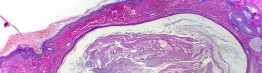
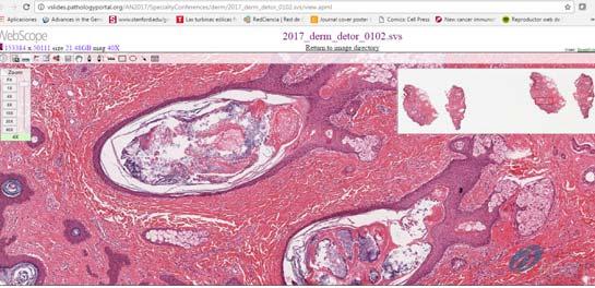
3 Differential Diagnosis Cystic Acne Follicular dilatation Round to oval collections of laminated keratin, sebum, and bacteria Surrounding perifollicular inflammatory infiltrate Favre-Racouchot síndrome (nodular elastosis with comedons and cyst) Dilated and keratin filled follicles Small cysts Solar elastosis Nevus comedonicus Large grouped, dilated follicular ostia devoid of hair shafts Small cysts, cystic invaginations, and occasionally large cysts may be seen Torrelo A, Hadj-Rabia S, Colmenero I, Piston R, Sybert VP, Hilari-Carbonell H, et al. Folliculocystic and collagen hamartoma of tuberous sclerosis complex. J Am Acad Dermatol. 2012;66:


4 What about our patient? 4
 in 2012 by Torrelo et al.")


5 FOLLICULOCYSTIC AND COLLAGEN HAMARTOMA OF TUBEROUS SCLEROSIS COMPLEX FOLLICULOCYSTIC AND COLLAGEN HAMARTOMA OF TUBEROUS SCLEROSIS COMPLEX Folliculocystic and collagen hamartoma was suggested as a new type of complex hamartoma related with tuberous sclerosis complex (TSC) in 2012 by Torrelo et al. They reported six cases of complex hamartomas showing thick collagen deposition, concentric perifollicular fibrosis, and keratin-filled infundibular cysts, and that clinically appeared as a large thickened plaque studded with multiple comedo-like openings and keratin-containing cysts. FOLLICULOCYSTIC AND COLLAGEN HAMARTOMA OF TUBEROUS SCLEROSIS COMPLEX Among the six patients, five had central nervous system and/or cardiac manifestations of TSC, and had a diagnosis of TSC according to the currently accepted diagnostic criteria; therefore, it seems that the two disorders are causally related. Histopathology Perifollicular fibrosis Comedo-like dilatations Abnormal hair follicles Collagen deposition Infundibular cyst formation. Infundibular cyst formation is not observed in shagreen patches of TSC and other TSC-unrelated collagen nevi; thus, these characteristic histopathologic features are recognized to form a distinctive hamartomatous skin lesion 5

.")

.")

, Gingival")
 and ashleaf macules.")

6 Tuberous sclerosis complex The prevalence of TSC is estimated at 1/10,000, and about twothirds of the cases are sporadic with no family history due to new mutational events. TSC is due to inactivating mutations in either of two genes, TSC1 (on chromosome 9q34) or TSC2 (on chromosome 16p13.3). The TSC genes TSC1 and TSC2 encode proteins, termed hamartin and tuberin respectively, that form a functional complex. TSC1/TSC2 functions as a GTPase-Activating Protein(GAP) towards Rheb, which is a major regulator of the mammalian target of rapamycin (mtor). In the absence of either TSC1 or TSC2, high levels of Rheb-GTP lead to constitutive activation of mtor raptor signalling, thereby leading to enhanced and deregulated protein synthesis and cell growth. Cutaneous findings of TSC Facial angiofibromas, Periungual fibromas (Koenen's tumor), Gingival fibromas, Shagreen patches (plaque of collagenoma), Fibrous plaque of the forehead, Folliculocystic and collagen hamartoma (still not a criteria) and ashleaf macules. In terms of abundant collagen deposition in the dermis of TSC, folliculocystic and collagen hamartoma might be considered an atypical type of shagreen patches. Clinical History: CASE 2 A 57-year-old woman without medical history presents to the dermatology clinic with a two months history of multiples violaceous and painful subcutaneous nodules on her left leg. In the last 2 week, she developed a 5 cm ulcer on her left calf. A biopsy was taken from that area. Additionally, she is referring a mild rhinorrhea starting the last month. 6


7 7


8 CD20 CD3 CD8 CD4 CD30 8
9 CD56 Ki-67 EBER ISH EXTRANODAL NK/T CELL LYMPHOMA, EXTRANASAL TYPE NASAL MUCOSA 9
10 CD3 CD4 CD8 CD56 10
11 Extranodal NK/T cell lymphoma, extranasal type Non-Hodgkin lymphoma presents in extranodal sites in about 40% of patients. Extranodal lymphoma of the paranasal sinuses is a rare clinical entity seen only in 5 8% of extranodal lymphomas of the head and neck. Nasal NK/T cell lymphma (NKTCL), which is a subtype of peripheral T cell lymphoma, constitutes about 1.4% of all lymphomas. Extranodal NKTCL nasal type is an NK cell-derived neoplasm. It usually affects the aerodigestive tract (e.g., nose, oropharynx, and larynx), but skin, gastrointestinal, and testis involvement can occur. It is a locally destructive tumor mainly affecting the midface, hence its old name lethal midline granuloma Epidemiology NKTCL is seen more in Asian and Latin-American countries. It has a male-to-female ratio of 2: 1 to 3: 1 and mainly affects patients in their 60s. EBV also has a role in the development of this neoplasm. Clinical Features In general, NKTCL presents with nonspecific symptoms. Weight loss, fever, night sweats, and anemia are usually only encountered in late stages. The nasal variety can cause nasal obstruction, epistaxis, rhinorrhea, extensive midfacial structure, involvement of the orbit causing proptosis, and, occasionally, the hard palate. Clinically, it can be divided into nasal and extranasal types; the nasal variety commonly presents with nasal obstruction, but also it can cause epistaxis, extensive involvement of the midfacial structure, involvement of the orbit causing proptosis, and, occasionally, the hard palate. In extranasal variety, skin is the most common site of involvement, but it can affect the gastrointestinal system, spleen, and testes. Muscles and adrenal glands are rarely involved. The extranasal type usually disseminates early in the course of the disease, but most were found to have occult nasal primaries. Nasal panendoscopy should be done irrespective of the primary site of presentation. 11

12 Morphology Immunohistochemistry Histologically, the neoplasm shows ulceration; angiocentric and angiodestructive growth is seen with areas of necrosis and lymphocytic infiltration with irregular nuclei on the surface of the epithelium and subepithelium, which is called polymorphic reticulosis. The tumor tends to cause coagulative necrosis in the tissue, so a large biopsy should be taken. The immunophenotypes of these tumors are CD2+, CD56+, and cytoplasmic CD3ε+. Cytotoxic molecules (Granzyme B, TIA-1, and Perforin) are positive, and often show negative expression of T cell antigen (e.g., CD4, CD5, and surface CD3) and negative for B cell marker CD20. DDx Blastic plasmacytoid dendritic cell neoplasm (BPDCN): CD123 (+) Leukemia cutis Quantification of Plasma EBV DNA level by PCR correlates with the disease status of the patient, and can be used as a marker for tumor load; a high titer indicates extensive disease, poor response to therapy, and poor survival. Serial EBV DNA can be used to monitor the response to treatment. Many patients with NKTCL are hepatitis B carriers, and antiviral prophylaxis should be given during chemotherapy. Prognosis The prognosis of the disease is variable; it is generally poor, with a 30% 5- year survival rate, but it recently increased to 71% due to utilizing intensive therapy like up-front radiotherapy. Multiple factors affect the prognosis: age of the patient, stage of disease, EBV DNA level, number of extranodal sites, LDH level, and regional lymphadenopathy. Extranasal NKTCL in general has a worse prognosis than nasal type, as patients tend to have B symptoms (fever, night sweats, and weight loss) and involvement of the lymph nodes. Treatment A combination of radiotherapy and chemotherapy is the best modality of treatment, especially for the early stages, because of the high recurrence rate when radiotherapy alone is used. Regimens like SMILE (Dexamethasone, Methotrexate with Leucovorin, Ifosfamide, L-asparaginase, and Etoposide) can be used; it has an 86% response rate (RR). If radio-therapy is used, the RR increases to 89.7% and the response is durable. 12


13 Important Information Regarding CME/SAMs The Online CME/Evaluations/SAMs claim process will only be available on the USCAP website until September 30, No claims can be processed after that date! After September 30, 2017 you will NOT be able to obtain any CME or SAMs credits for attending this meeting. 13
3/27/2017. Pulmonary Pathology Specialty Conference. Disclosure of Relevant Financial Relationships. Clinical History:
 Pulmonary Pathology Specialty Conference Saul Suster, M.D. Medical College of Wisconsin Disclosure of Relevant Financial Relationships USCAP requires that all planners (Education Committee) in a position
Evening specialty conference: Liver
 Evening specialty conference: Liver Joseph Misdraji, M.D. Disclosure of Relevant Financial Relationships Disclosure of Relevant Financial Relationships USCAP requires that all planners (Education Committee)
Primary Cutaneous CD30-Positive T-cell Lymphoproliferative Disorders
 Primary Cutaneous CD30-Positive T-cell Lymphoproliferative Disorders Definition A spectrum of related conditions originating from transformed or activated CD30-positive T-lymphocytes May coexist in individual
a mimicker of Wegener s Granulomatosis
 a mimicker of Wegener s Granulomatosis Combined Meeting October 2009 a story of 2 ladies Madam JA 56 year-old Madam RH 36 year-old Madam JA 56 year-old Apr 2008 May Jun Jul Aug Sept Oct Nov 2008 Madam
Hepatic Lymphoma Diagnosis An Algorithmic Approach
 Hepatic Lymphoma Diagnosis An Algorithmic Approach Ryan M. Gill, M.D., Ph.D. University of California, San Francisco PLEASE TURN OFF YOUR CELL PHONES Disclosure of Relevant Financial Relationships USCAP
Diagnosing TSC by Making Clinical Connections
 Diagnosing TSC by Making Clinical Connections TSC = tuberous sclerosis complex. Diagnosing tuberous sclerosis complex: MORE CLUES Definite Diagnosis of Tuberous Sclerosis Complex (TSC) Possible Diagnosis
Follicular Lymphoma: the WHO
 Follicular Lymphoma: the WHO and the WHERE? Yuri Fedoriw, MD Associate Professor of Pathology and Laboratory Medicine Director of Hematopathology University of North Carolina Chapel Hill, NC Disclosure
Molecular Pathology of Lymphoma (Part 1) Rex K.H. Au-Yeung Department of Pathology, HKU
 Rex K.H. Au-Yeung Department of Pathology, HKU") Molecular Pathology of Lymphoma (Part 1) Rex K.H. Au-Yeung Department of Pathology, HKU Lecture outline Time 10:00 11:00 11:15 12:10 12:20 13:15 Content Introduction to lymphoma Review of lymphocyte biology
Molecular Pathology of Lymphoma (Part 1) Rex K.H. Au-Yeung Department of Pathology, HKU Lecture outline Time 10:00 11:00 11:15 12:10 12:20 13:15 Content Introduction to lymphoma Review of lymphocyte biology
Disclosure of Relevant Financial Relationships
 Squamous entities of the thyroid: Reactive to Neoplastic Michelle D. Williams Associate Professor Dept of Pathology, Head & Neck Section University of Texas MD Anderson Cancer Center Disclosure of Relevant
Methotrexate-associated Lymphoproliferative Disorders
 Methotrexate-associated Lymphoproliferative Disorders Definition A lymphoid proliferation or lymphoma in a patient immunosuppressed with methotrexate, typically for treatment of autoimmune disease (rheumatoid
Extranodal natural killer T-cell lymphoma, nasal type, presenting as periorbital cellulitis: A case report
 CASE REPORT Miller et al. 1 PEER REVIEWED OPEN ACCESS Extranodal natural killer T-cell lymphoma, nasal type, presenting as periorbital cellulitis: A case report Katherine Miller, Amir Kamran, Anna Koget
Extranodal natural killer/t-cell lymphoma with long-term survival and repeated relapses: does it indicate the presence of indolent subtype?
 VOLUME 47 ㆍ NUMBER 3 ㆍ September 2012 THE KOREAN JOURNAL OF HEMATOLOGY ORIGINAL ARTICLE Extranodal natural killer/t-cell lymphoma with long-term survival and repeated relapses: does it indicate the presence
Non-Hodgkin lymphomas (NHLs) Hodgkin lymphoma )HL)
 Hodgkin lymphoma )HL)") Non-Hodgkin lymphomas (NHLs) Hodgkin lymphoma )HL) Lymphoid Neoplasms: 1- non-hodgkin lymphomas (NHLs) 2- Hodgkin lymphoma 3- plasma cell neoplasms Non-Hodgkin lymphomas (NHLs) Acute Lymphoblastic Leukemia/Lymphoma
Non-Hodgkin lymphomas (NHLs) Hodgkin lymphoma )HL) Lymphoid Neoplasms: 1- non-hodgkin lymphomas (NHLs) 2- Hodgkin lymphoma 3- plasma cell neoplasms Non-Hodgkin lymphomas (NHLs) Acute Lymphoblastic Leukemia/Lymphoma
Teleconference on Tuberous Sclerosis Complex (TSC) Research October 28 th, 2008 and November 11 th, 2008
 Research October 28 th, 2008 and November 11 th, 2008") Wong 1 Teleconference on Tuberous Sclerosis Complex (TSC) Research October 28 th, 2008 and November 11 th, 2008 Sponsored by the Tuberous Sclerosis Alliance Faculty Participants: Elizabeth Henske, MD,
Wong 1 Teleconference on Tuberous Sclerosis Complex (TSC) Research October 28 th, 2008 and November 11 th, 2008 Sponsored by the Tuberous Sclerosis Alliance Faculty Participants: Elizabeth Henske, MD,
Case 1 PLEASE TURN OFF YOUR CELL PHONES 3/28/2017. Disclosure of Relevant Financial Relationships. Disclosure of Relevant Financial Relationships
 PLEASE TURN OFF YOUR CELL PHONES Disclosure of Relevant Financial Relationships USCAP requires that all planners (Education Committee) in a position to influence or control the content of CME disclose
Infections and nonmicrobial inflammatory stimuli can cause leukocytosis (as seen in Lab 1) as well as lymph node enlargement (lymphadenopathy).
 as well as lymph node enlargement (lymphadenopathy).") LAB 5: LYMPHOID TISSUE AND SKIN The focus of this week s lab will be pathology of the lymphoid tissue and skin. The lymphoid organs include the thymus, spleen, and lymph nodes. Abnormalities in the lymph
LAB 5: LYMPHOID TISSUE AND SKIN The focus of this week s lab will be pathology of the lymphoid tissue and skin. The lymphoid organs include the thymus, spleen, and lymph nodes. Abnormalities in the lymph
NK/T cell lymphoma Recent advances. Y.L Kwong University Department of Medicine Queen Mary Hospital
 NK/T cell lymphoma Recent advances Y.L Kwong University Department of Medicine Queen Mary Hospital Natural killer cell lymphomas NK cell lymphomas are mainly extranodal lymphomas Clinical classification
Dermatopathology: The tumor is composed of keratinocytes which show atypia, increase mitoses and abnormal mitoses.
 Squamous cell carcinoma (SCC): A common malignant tumor of keratinocytes arising in the epidermis, usually from a precancerous condition: 1- UV induced actinic keratosis, usually of low grade malignancy.
Clinical History. 29 yo woman with polyhydramnios Cardiac mass at fetal ultrasound At 35 weeks, newborn died 30 minutes after delivery
 CASE 1 a Clinical History 29 yo woman with polyhydramnios Cardiac mass at fetal ultrasound At 35 weeks, newborn died 30 minutes after delivery Interface between tumor and normal myocardium Smaller well-demarcated
NK/T cell lymphoma Hong Kong Experience with L-Asparaginase containing regimens. Y.L. Kwong University Department of Medicine Queen Mary Hospital
 NK/T cell lymphoma Hong Kong Experience with L-Asparaginase containing regimens Y.L. Kwong University Department of Medicine Queen Mary Hospital Management of NK cell malignancies Principles 1. Accurate
Herczegfalvi Ágnes. Tuberous sclerosis. Case history. SE. II. sz. Gyermekklinika. Budapest, Febr
 Herczegfalvi Ágnes Tuberous sclerosis Case history SE. II. sz. Gyermekklinika Budapest, Febr. 2016. Case history 1. GD. DOB: 05-03-1989 Pre and perinatal history: normal His development was normal till
A Unique Case of Nasal NK/T Cell Lymphoma with Frequent Remission and Relapse Showing Different Histological Features During 12 Years of Follow Up
 J Clin Exp Hematopathol Vol. 50, No. 1, May 2010 Case Study A Unique Case of Nasal NK/T Cell Lymphoma with Frequent Remission and Relapse Showing Different Histological Features During 12 Years of Follow
Lymphatic System Disorders
 Lymphatic System Disorders Lymphomas Malignant neoplasms involving lymphocyte proliferation in lymph nodes Specific causes not identified // Higher risk in adults who received radiation during childhood
Blastic NK-Cell Leukemia / Lymphoma
 * * Blastic NK-Cell Leukemia / Lymphoma A Case Report Chun-Ming Lin Shu-Hui Wang Tseng-tong Kuo* Ching-Chi Chi Hsin-Chun Ho Hong-Shang Hong Blastic natural killer (NK) cell lymphoma / leukemia is a rare
Samer Ghosn, MD Associate professor, Derpartment of Dermatology American University of Beirut Medical Center. Follicular lesions
 Samer Ghosn, MD Associate professor, Derpartment of Dermatology American University of Beirut Medical Center Follicular lesions Introduction Follicular lesions are important to recognize: For proper management
Review Article Extranodal Natural-Killer/T-Cell Lymphoma, Nasal Type
 Hindawi Publishing Corporation Advances in Hematology Volume 2010, Article ID 627401, 5 pages doi:10.1155/2010/627401 Review Article Extranodal Natural-Killer/T-Cell Lymphoma, Nasal Type Harinder Gill,
7 Omar Abu Reesh. Dr. Ahmad Mansour Dr. Ahmad Mansour
 7 Omar Abu Reesh Dr. Ahmad Mansour Dr. Ahmad Mansour -Leukemia: neoplastic leukocytes circulating in the peripheral bloodstream. -Lymphoma: a neoplastic process in the lymph nodes, spleen or other lymphatic
Burkitt lymphoma. Sporadic Endemic in Africa associated with EBV Translocations involving MYC gene on chromosome 8
 Heme 8 Burkitt lymphoma Sporadic Endemic in Africa associated with EBV Translocations involving MYC gene on chromosome 8 Most common is t(8;14) Believed to be the fastest growing tumor in humans!!!! Morphology
Benign versus Cancerous Lesions How to tell the difference FMF 2014 Christie Freeman MD, CCFP, DipPDerm, MSc
 1 Benign versus Cancerous Lesions How to tell the difference FMF 2014 Christie Freeman MD, CCFP, DipPDerm, MSc Benign lesions Seborrheic Keratoses: Warty, stuck-on Genetics and birthdays Can start in late
SKIN SONOGRAPHY IN CHILDREN. CRISTIAN J. GARCIA MD Santiago, Chile
 SKIN SONOGRAPHY IN CHILDREN CRISTIAN J. GARCIA MD Santiago, Chile I HAVE NO DISCLOSURES OBJECTIVES RELEVANCE OF SKIN LESIONS IN CHILDREN ROLEN OF THE RADIOLOGIST CLINICAL CORRELATION US TECHNIQUE NORMAL
Conflicts. Objectives. University of Texas Health Science Center at San Antonio. Pediatrics Grand Rounds 24 August Pediatric Dermatology 101
 Pediatric Dermatology 101 John C. Browning, MD, FAAD, FAAP Conflicts Investigator: ViroXis Advisor: ViroXis Advisory Board: TopMD Speaker: Galderma Objectives Understand the meaning and importance of cutaneous
Case Report Nevus Lipomatosus Superficialis with a Folliculosebaceous Component: Report of 2 Cases
 SAGE-Hindawi Access to Research Pathology Research International Volume 2011, Article ID 105973, 4 pages doi:10.4061/2011/105973 Case Report Nevus Lipomatosus Superficialis with a Folliculosebaceous Component:
Pimples and Boils!! Dr Nathan Harvey Anatomical Pathology, PathWest
 Pimples and Boils!! Dr Nathan Harvey Anatomical Pathology, PathWest Overview & Learning Objectives Review the cardinal signs/symptoms of acute inflammation Review the histological features of acute inflammation
Dermatopathology. Dr. Rafael Botella Estrada. Hospital La Fe de Valencia
 Dermatopathology Dr. Rafael Botella Estrada. Hospital La Fe de Valencia DERMATOPATHOLOGY CASE CHALLENGE: RECOGNIZING MIMIS AND MASQUERADERS Rosalie Elenitsas. University of Pennsylvania Spectrum Lupus
HIDRADENITIS SUPPURATIVA
 Print Close Window Note: Large images and tables on this page may necessitate printing in landscape mode. Copyright 2004-2005 The McGraw-Hill Companies. All rights reserved. Fitzpatrick Color Atlas, 5e
Contents. vii. Preface... Acknowledgments... v xiii
 Contents Preface... Acknowledgments... v xiii SECTION I 1. Introduction... 3 Knowledge-Based Diagnosis... 4 Systematic Examination of the Lymph Node... 7 Cell Type Identification... 9 Cell Size and Cellularity...
Tuberous Sclerosis Complex
 Tuberous Sclerosis Complex A successful transition from the bench to the bedside Mary Kay Koenig, MD The University of Texas Medical School at Houston Children s Memorial Hermann Hospital University of
Lymphoma/CLL 101: Know your Subtype. Dr. David Macdonald Hematologist, The Ottawa Hospital
 Lymphoma/CLL 101: Know your Subtype Dr. David Macdonald Hematologist, The Ottawa Hospital Function of the Lymph System Lymph Node Lymphocytes B-cells develop in the bone marrow and influence the immune
Michi Shinohara MD Associate Professor University of Washington/Seattle Cancer Care Alliance Dermatology, Dermatopathology
 Michi Shinohara MD Associate Professor University of Washington/Seattle Cancer Care Alliance Dermatology, Dermatopathology Agenda Overview of cutaneous T and B- cell lymphomas Diagnosis, Staging, Prognosis
Case Presentation. Maha Akkawi, MD, Fatima Obeidat, MD, Tariq Aladily, MD. Department of Pathology Jordan University Hospital Amman, Jordan
 Case Presentation Maha Akkawi, MD, Fatima Obeidat, MD, Tariq Aladily, MD Department of Pathology Jordan University Hospital Amman, Jordan The 25th Annual Congress of the ADIAP The 8/11/2013 1 5th International
3/24/2017 DENDRITIC CELL NEOPLASMS: HISTOLOGY, IMMUNOHISTOCHEMISTRY, AND MOLECULAR GENETICS. Disclosure of Relevant Financial Relationships
 DENDRITIC CELL NEOPLASMS: HISTOLOGY, IMMUNOHISTOCHEMISTRY, AND MOLECULAR GENETICS Jason L. Hornick, M.D., Ph.D. Director of Surgical Pathology and Immunohistochemistry Brigham and Women s Hospital Professor
Lymphoma: What You Need to Know. Richard van der Jagt MD, FRCPC
 Lymphoma: What You Need to Know Richard van der Jagt MD, FRCPC Overview Concepts, classification, biology Epidemiology Clinical presentation Diagnosis Staging Three important types of lymphoma Conceptualizing
Appendageal skin tumors
 Appendageal skin tumors Ibrahim Khalifeh, M.D. Associate Professor Department of Pathology American University of Beirut Medical Center Beirut, Lebanon Appendageal tumors Neoplasms whose differentiation
88-year-old Female with Lymphadenopathy. Faizi Ali, MD
 88-year-old Female with Lymphadenopathy Faizi Ali, MD Clinical History A 88-year-old caucasian female presented to our hospital with the complaints of nausea, vomiting,diarrhea, shortness of breath and
Recent diagnostic and therapeutic innovations of T-cell-lymphoma. Prof. Nossrat Firusian, Recklinghausen, Germany
 Recent diagnostic and therapeutic innovations of T-cell-lymphoma Prof. Nossrat Firusian, Recklinghausen, Germany NODAL Angioimmunoblastic T-cell Lymphoma Peripheral T-cell-Lymphoma Anaplastic Large-cell-Lymphoma
3/28/2017. Disclosure of Relevant Financial Relationships. GU Evening Subspecialty Case Conference. Differential Diagnosis:
 GU Evening Subspecialty Case Conference Rajal B. Shah, M.D. VP, Medical Director, Urologic Pathology Miraca Life Sciences, Irving, Texas Clinical Associate Professor of Pathology Baylor College of Medicine,
Disclosures. Parathyroid Pathology. Objectives. The normal parathyroid 11/10/2012
 Disclosures Parathyroid Pathology I have nothing to disclose Annemieke van Zante MD/PhD Assistant Professor of Clinical Pathology Associate Chief of Cytopathology Objectives 1. Review the pathologic features
Challenging Cases in Dermatopathology. Rosalie Elenitsas, M.D. Professor of Dermatology Director, Dermatopathology University of Pennsylvania
 Challenging Cases in Dermatopathology Rosalie Elenitsas, M.D. Professor of Dermatology Director, Dermatopathology University of Pennsylvania DISCLOSURE OF RELATIONSHIPS WITH INDUSTRY Rosalie Elenitsas
3/22/2017. Disclosure of Relevant Financial Relationships. Disclosure of Relevant Financial Relationships. Grading G1. Grading. Ki67 index V.
 Disclosure of Relevant Financial Relationships USCAP requires that all planners (Education Committee) in a position to influence or control the content of CME disclose any relevant financial relationship
Overview of Cutaneous Lymphomas: Diagnosis and Staging. Lauren C. Pinter-Brown MD, FACP Health Sciences Professor of Medicine and Dermatology
 Overview of Cutaneous Lymphomas: Diagnosis and Staging Lauren C. Pinter-Brown MD, FACP Health Sciences Professor of Medicine and Dermatology Definition of Lymphoma A cancer or malignancy that comes from
Anaplastic Large Cell Lymphoma (of T cell lineage)
") Anaplastic Large Cell Lymphoma (of T cell lineage) Definition T-cell lymphoma comprised of large cells with abundant cytoplasm and pleomorphic, often horseshoe-shaped nuclei CD30+ Most express cytotoxic
Anaplastic Large Cell Lymphoma (of T cell lineage) Definition T-cell lymphoma comprised of large cells with abundant cytoplasm and pleomorphic, often horseshoe-shaped nuclei CD30+ Most express cytotoxic
MECHANISMS OF HUMAN DISEASE: LABORATORY SESSIONS LYMPHOMA. April 16, 2008
 MECHANISMS OF HUMAN DISEASE: LABORATORY SESSIONS LYMPHOMA April 16, 2008 FACULTY COPY GOAL: Learn the appearance of normal peripheral blood elements and lymph nodes. Recognize abnormal peripheral blood
Indolent Lymphomas. Dr. Melissa Toupin The Ottawa Hospital
 Indolent Lymphomas Dr. Melissa Toupin The Ottawa Hospital What does indolent mean? Slow growth Often asymptomatic Chronic disease with periods of relapse (long natural history possible) Incurable with
Acute Hepatitis in a Patient with NASH and Elevation of CMV-IgM
 Acute Hepatitis in a Patient with NASH and Elevation of CMV-IgM Uta Drebber, MD Disclosure of Relevant Financial Relationships USCAP requires that all faculty in a position to influence or control the
Neurocutaneous Syndromes. Phakomatoses
 Neurocutaneous Syndromes Phakomatoses Financial Disclosures I have NO SIGNIFICANT FINANCIAL, GENERAL, OR OBLIGATION INTERESTS TO REPORT Neurocutaneous Syndomes Definition Entities Diagnosis/ Presentation
3/23/2017. Disclosure of Relevant Financial Relationships. Pitfalls in Immunohistochemistry in Hematopathology: CD20 and CD3 Can Let Me Down?!
 Pitfalls in Immunohistochemistry in Hematopathology: CD20 and CD3 Can Let Me Down?! Judith A. Ferry Massachusetts General Hospital Disclosure of Relevant Financial Relationships USCAP requires that all
A 40-year old male with follicular papule and pustule at central face area for 3 months
 A 40-year old male with follicular papule and pustule at central face area for 3 months GMS- Neg AFB-Neg Fite stain - neg HISTOPATHOLOGICAL DIFFERENTIAL DIAGNOSIS CASEOUS GRANULOMA INFECTION -MYCOBACTERIUM
21/07/2017. Hobnail endothelial cells are not the same as epithelioid endothelial cells
 UPDATE IN CUTANEOUS VASCULAR S DERMATOPATHOLOGY SESSION BELFAST PATHOLOGY JUNE 21/2017 Dr E Calonje St John s Institute of Dermatology, London, United Kingdom THE FAMILY OF VASCULAR S WITH EPITHELIOID
Experience with malignant tumours of the maxillary sinus in the Department of Otolaryngology Universiti Kebangsaan Malaysia, Kuala Lumpur
 Med. J. Malaysia Vol. 44 No. 1 March 1989 Experience with malignant tumours of the maxillary sinus in the Department of Otolaryngology Universiti Kebangsaan Malaysia, Kuala Lumpur S. Lokman, MD (UKMalaysia)
Case Report Bifocal Presentation of Primary Testicular Extranasal NK/T-Cell Lymphoma: A Case Report and Review of the Literature
 Case Reports in Oncological Medicine Volume 2013, Article ID 267389, 4 pages http://dx.doi.org/10.1155/2013/267389 Case Report Bifocal Presentation of Primary Testicular Extranasal NK/T-Cell Lymphoma:
LYMPHOMA Joginder Singh, MD Medical Oncologist, Mercy Cancer Center
 LYMPHOMA Joginder Singh, MD Medical Oncologist, Mercy Cancer Center Lymphoma is cancer of the lymphatic system. The lymphatic system is made up of organs all over the body that make up and store cells
3. Histopathology. 1. Introduction. 2. Case History. Volume 6 Issue 4, April Licensed Under Creative Commons Attribution CC BY
 Spiradenocylindroma with Trichoepithelioma A Collision Tumor with Multiple Differentiation R. Lavanya 1, S. K. Sridevi 2, P. Viswanathan 3, P. V. S.Prasad 4 1 II nd Year Post Graduate, Department of Pathology,
Cowden Syndrome PTEN Hamartoma Tumor Syndrome. ACCME/Disclosure. 1. Background. Outline
 MASSACHUSETTS GENERAL HOSPITAL HARVARD MEDICAL SCHOOL PATHOLOGY Cowden Syndrome PTEN Hamartoma Tumor Syndrome ACCME/Disclosure Vania Nosé, MD, PhD Professor of Pathology Director of Anatomic Pathology
NASAL SEPTUM ADENOID CYSTIC CARCINOMA: A CASE REPORT
 NASAL SEPTUM ADENOID CYSTIC CARCINOMA: A CASE REPORT Shu-Yu Tai, 1 Chen-Yu Chien, 2 Chih-Feng Tai, 2,4 Wen-Rei Kuo, 2,4 Wan-Ting Huang, 3 and Ling-Feng Wang 2,4 Departments of 1 Family Medicine, 2 Otolaryngology
SESSION 1: GENERAL (BASIC) PATHOLOGY CONCEPTS Thursday, October 16, :30am - 11:30am FACULTY COPY
 PATHOLOGY CONCEPTS Thursday, October 16, :30am - 11:30am FACULTY COPY") SESSION 1: GENERAL (BASIC) PATHOLOGY CONCEPTS Thursday, October 16, 2008 9:30am - 11:30am FACULTY COPY GOAL: Describe the basic morphologic (structural) changes which occur in various pathologic conditions.
SESSION 1: GENERAL (BASIC) PATHOLOGY CONCEPTS Thursday, October 16, 2008 9:30am - 11:30am FACULTY COPY GOAL: Describe the basic morphologic (structural) changes which occur in various pathologic conditions.
Incidence. Bimodal age incidence 15-40, >55 years Childhood form (0-14) more common in developing countries M:F=1.5:1; in all subtypes except NS
 more common in developing countries M:F=1.5:1; in all subtypes except NS") Hodgkin Lymphoma Hodgkin Lymphoma 30% of all lymphomas Absolute incidence unchanged Arise in lymph node, cervical region Neoplastic tissues usually contain a small number of tumor cells Incidence Bimodal
Hodgkin Lymphoma Hodgkin Lymphoma 30% of all lymphomas Absolute incidence unchanged Arise in lymph node, cervical region Neoplastic tissues usually contain a small number of tumor cells Incidence Bimodal
FOLLICULARITY in LYMPHOMA
 FOLLICULARITY in LYMPHOMA Reactive Follicular Hyperplasia Follicular Hyperplasia irregular follicles Follicular Hyperplasia dark and light zones Light Zone Dark Zone Follicular hyperplasia MIB1 Follicular
IN THE NAME OF GOD Dr. Kheirandish Oral and maxillofacial pathology
 IN THE NAME OF GOD Dr. Kheirandish Oral and maxillofacial pathology ORAL FOCAL MUCINOSIS Uncommon Tumorlike Cutaneous myxoid cyst Overproduction of hyaluronic acid by firoblasts Young adults Female Gingiva
LEUKAEMIA and LYMPHOMA. Dr Mubarak Abdelrahman Assistant Professor Jazan University
 LEUKAEMIA and LYMPHOMA Dr Mubarak Abdelrahman Assistant Professor Jazan University OBJECTIVES Identify etiology and epidemiology for leukemia and lymphoma. Discuss common types of leukemia. Distinguish
CPC. Chutika Srisuttiyakorn, M.D. Kobkul Aunhachoke, M.D. Phramongkutklao Hospital Bangkok, Thailand
 CPC Chutika Srisuttiyakorn, M.D. Kobkul Aunhachoke, M.D. Phramongkutklao Hospital Bangkok, Thailand A 53 year-old woman with fever, facial swelling and rashes on face, trunk and upper extremities for 3
Cutaneous features of TSC when the diagnosis is written on your face. Dr Fiona Browne Consultant Dermatologist
 Cutaneous features of TSC when the diagnosis is written on your face Dr Fiona Browne Consultant Dermatologist Tuberous sclerosis complex Hypomelanotic macules Angiofibromas Ungal fibromas Shagreen patch
Case Report A Rare Cutaneous Adnexal Tumor: Malignant Proliferating Trichilemmal Tumor
 Case Reports in Medicine Volume 2015, Article ID 742920, 4 pages http://dx.doi.org/10.1155/2015/742920 Case Report A Rare Cutaneous Adnexal Tumor: Malignant Proliferating Trichilemmal Tumor Omer Alici,
Diseases of the breast (1 of 2)
") Diseases of the breast (1 of 2) Introduction A histology introduction Normal ducts and lobules of the breast are lined by two layers of cells a layer of luminal cells overlying a second layer of myoepithelial
Diseases of the breast (1 of 2) Introduction A histology introduction Normal ducts and lobules of the breast are lined by two layers of cells a layer of luminal cells overlying a second layer of myoepithelial
Catholic University of Louvain, St - Luc University Hospital Head and Neck Oncology Programme. Anatomopathology. Pathology 1 Sept.
 Anatomopathology Pathology 1 Anatomopathology Biopsies Frozen section Surgical specimen Peculiarities for various tumor site References Pathology 2 Biopsies Minimum data, which should be given by the pathologist
Desmoplastic Melanoma R/O BCC. Clinical Information. 74 y.o. man with lesion on left side of neck r/o BCC
 R/O BCC Sabine Kohler, M.D. Professor of Pathology and Dermatology Dermatopathology Service Stanford University School of Medicine Clinical Information 74 y.o. man with lesion on left side of neck r/o
Indolent Lymphomas: Current. Dr. Laurie Sehn
 Indolent Lymphomas: Current Dr. Laurie Sehn Why does indolent mean? Slow growth Often asymptomatic Chronic disease with periods of relapse (long natural history possible) Incurable with current standard
What s new on the horizon in T-cell lymphoma Elaine S Jaffe National Cancer Institute, Bethesda MD
 What s new on the horizon in T-cell lymphoma Elaine S Jaffe National Cancer Institute, Bethesda MD WHO classification: where are we today? Of 12 monographs planned for 4 th Edition Bluebook series, only
GOBLET CELL CARCINOID. Hanlin L. Wang, MD, PhD University of California Los Angeles
 GOBLET CELL CARCINOID Hanlin L. Wang, MD, PhD University of California Los Angeles Disclosure of Relevant Financial Relationships USCAP requires that all planners (Education Committee) in a position to
GOBLET CELL CARCINOID
 GOBLET CELL CARCINOID Hanlin L. Wang, MD, PhD University of California Los Angeles Disclosure of Relevant Financial Relationships USCAP requires that all planners (Education Committee) in a position to
Benign and malignant epithelial lesions: Seborrheic keratosis: A common benign pigmented epidermal tumor occur in middle-aged or older persons more
 Benign and malignant epithelial lesions: Seborrheic keratosis: A common benign pigmented epidermal tumor occur in middle-aged or older persons more common on the trunk; but extremities, head and neck are
Glistening, Skin-Colored Nodule
 To Print: Click your browser's PRINT button. NOTE: To view the article with Web enhancements, go to: http://www.medscape.com/viewarticle/436334 Medscape Dermatology Clinic Glistening, Skin-Colored Nodule
الفتوي الاصفر الحبيبوم = Xanthogranuloma_Juvenile JUVENILE XANTHOGRANULOMA 1 / 9
 JUVENILE XANTHOGRANULOMA 1 / 9 Clinical Findings CUTANEOUS LESIONS JXG is a benign, self-healing disorder that is characterized by asymptomatic yellowish papulonodular lesions of the skin and other organs
2. Sézary syndrome (SS)
") Go Back to the Top To Order, Visit the Purchasing Page for Details Clinical images are available in hardcopy only. Clinical images are available in Clinical images are available in d e f g h i j Fig..36-2
Go Back to the Top To Order, Visit the Purchasing Page for Details Clinical images are available in hardcopy only. Clinical images are available in Clinical images are available in d e f g h i j Fig..36-2
Lách
 Lách Lách Lách Lách Splenogonadal fusion. Splenic tissue is attached to testicular tissue. Pseudocyst (false or secondary cyst). A, Outer aspect. Pseudocyst (false or secondary cyst). B, Inner surface.
A CASE OF PRIMARY THYROID LYMPHOMA. Prof Dr.Dilek Gogas Yavuz Marmara University School of Medicine Endocrinology and Metabolism Istanbul, Turkey
 A CASE OF PRIMARY THYROID LYMPHOMA Prof Dr.Dilek Gogas Yavuz Marmara University School of Medicine Endocrinology and Metabolism Istanbul, Turkey 38 year old female She recognized a mass in her right neck
Case Report Intravascular NK/T-cell lymphoma in the testis: a novel case report and review of the literature
 Int J Clin Exp Pathol 2017;10(9):9793-9797 www.ijcep.com /ISSN:1936-2625/IJCEP0031597 Case Report Intravascular NK/T-cell lymphoma in the testis: a novel case report and review of the literature Hong Yu
SEBACEOUS NEOPLASMS. Dr. Prachi Saraogi Clinical Fellow in Dermatology
 SEBACEOUS NEOPLASMS Dr. Prachi Saraogi Clinical Fellow in Dermatology Sebaceous neoplasms Sebaceous adenoma (Benign) Sebaceous carcinoma (Malignant) SEBACEOUS ADENOMA Benign tumours composed of incompletely
CASE year old male with a PET avid nodule in the left adrenal gland
 CASE 1 55 year old male with a PET avid nodule in the left adrenal gland Case 1 Adrenal gland parenchyma partly replaced by a spindle cell tumour with mild nuclear pleomorphism Atypical mitoses present
Pigmented lesions of the Oral cavity
 Oral medicine أ.م.د احسان عبد هللا كميل Pigmented lesions of the Oral cavity Pigmented oral lesions are a large group of disorders in which the dark or brown color is the essential clinical characteristic.
Photo Quiz Self-Test Your Diagnostic Acumen
 Do You Know Your Nevi? Case 1: The parents of a 3-year-old girl seek medical evaluation of the nodules on their daughter s back. The lesions have been present since birth and have grown with the child.
Disorders of Cell Growth & Neoplasia. Histopathology Lab
 Disorders of Cell Growth & Neoplasia Histopathology Lab Paul Hanna April 2010 Case #84 Clinical History: 5 yr-old, West Highland White terrier. skin mass from axillary region. has been present for the
Pathology of Hematopoietic and Lymphoid tissue
 CONTENTS Pathology of Hematopoietic and Lymphoid tissue White blood cells and lymph nodes Quantitative disorder of white blood cells Reactive lymphadenopathies Infectious lymphadenitis Tumor metastasis
Plexiform Tumor of the Orbit
 Plexiform Tumor of the Orbit Anat Stemmer-Rachamimov, MD Department of Pathology Massachusetts General Hospital Harvard Medical School Disclosure of Relevant Financial Relationships USCAP requires that
Antonella Tosti Fredric Brandt Endowed Professor of Dermatology & Cutaneous Surgery
 Dermoscopy in the evaluation and treatment of hair loss Antonella Tosti Fredric Brandt Endowed Professor of Dermatology & Cutaneous Surgery DISCLOSURE OF RELATIONSHIPS WITH INDUSTRY Antonella Tosti, MD
HODGKIN LYMPHOMA DR. ALEJANDRA ZARATE OSORNO HOSPITAL ESPAÑOL DE MEXICO
 HODGKIN LYMPHOMA DR. ALEJANDRA ZARATE OSORNO HOSPITAL ESPAÑOL DE MEXICO HODGKIN LYMPHOMA CLASSIFICATION Lukes & Butler Rye WHO-2016 Linphocytic and/or histiocytic Nodular & diffuse Nodular Sclerosis Lymphocyte
Unusual cutaneous presentation of a T-cell lymphoproliferation
 Department of Pathology and Cytology University Hospital Centre Zagreb, Croatia Unusual cutaneous presentation of a T-cell lymphoproliferation Snjezana Dotlic, Stefan Dojcinov, Leticia Quintanilla-Fend
Carcinoma of Unknown Primary site (CUP) in HEAD & NECK SURGERY
 in HEAD & NECK SURGERY") Carcinoma of Unknown Primary site (CUP) in HEAD & NECK SURGERY SEARCHING FOR THE PRIMARY? P r o f J P P r e t o r i u s H e a d : C l i n i c a l U n i t C r i t i c a l C a r e U n i v e r s i t y O f
Carcinoma of Unknown Primary site (CUP) in HEAD & NECK SURGERY SEARCHING FOR THE PRIMARY? P r o f J P P r e t o r i u s H e a d : C l i n i c a l U n i t C r i t i c a l C a r e U n i v e r s i t y O f
Objectives. 1. Recognizing benign skin lesions. 2.Know which patients will likely need surgical intervention.
 The Joy of Pediatric Skin Dr. Claire Sanger University of Kentucky Plastic & Reconstructive Surgery Objectives 1. Recognizing benign skin lesions 2.Know which patients will likely need surgical intervention.
PRIMARY GASTRIC LYMPHOMA: CASE REPORT WITH REVIEW OF LITERATURE
 PRIMARY GASTRIC LYMPHOMA: CASE REPORT WITH REVIEW OF LITERATURE Rana K. Sherwani, *Kafil Akhtar, Noorin Zaidi, Anjum Ara Department of Pathology, J.N. Medical College, Aligarh Muslim University, Aligarh,