Multifunctional Regulatory T Cells from Human Umbilical Cord Blood and the Role of Tumor Necrosis Factor in Immune Homeostasis
|
|
|
- Coral Stevenson
- 5 years ago
- Views:
Transcription

1 Loyola University Chicago Loyola ecommons Master's Theses Theses and Dissertations 2017 Multifunctional Regulatory T Cells from Human Umbilical Cord Blood and the Role of Tumor Necrosis Factor in Immune Homeostasis Alexander Nelson Loyola University Chicago, anelson9@luc.edu Recommended Citation Nelson, Alexander, "Multifunctional Regulatory T Cells from Human Umbilical Cord Blood and the Role of Tumor Necrosis Factor in Immune Homeostasis" (2017). Master's Theses This Thesis is brought to you for free and open access by the Theses and Dissertations at Loyola ecommons. It has been accepted for inclusion in Master's Theses by an authorized administrator of Loyola ecommons. For more information, please contact ecommons@luc.edu. This work is licensed under a Creative Commons Attribution-Noncommercial-No Derivative Works 3.0 License. Copyright 2017 Alexander Nelson
2 LOYOLA UNIVERSITY CHICAGO MULTIFUNCTIONAL REGULATORY T CELLS FROM HUMAN UMBILICAL CORD BLOOD AND THE ROLE OF TUMOR NECROSIS FACTOR IN IMMUNE HOMEOSTASIS A THESIS SUBMITTED TO THE FACULTY OF THE GRADUATE SCHOOL IN CANDIDACY FOR THE DEGREE OF MASTER OF SCIENCE PROGRAM IN INFECTIOUS DISEASE AND IMMUNOLOGY BY ALEXANDER J NELSON CHICAGO, ILLINOIS AUGUST, 2017
3 Copyright by Alexander J Nelson, 2017 All rights reserved.
4 TABLE OF CONTENTS LIST OF FIGURES ABSTRACT v vi CHAPTER ONE: INTRODUCTION Immune Tolerance 1 Regulatory T Cells 2 Foxp3 3 Mechanisms of suppression 5 Regulatory T cell subsets 6 Chemokine Receptors and T Cell Trafficking 8 Fetal Tolerance and Neonatal Immunity 9 Tumor Necrosis Factor Biology 11 TNF signaling 12 TNF in human disease 12 CHAPTER TWO: MATERIALS AND METHODS Mononuclear Cell Isolation and Cell Purification 14 Treg Induction 14 Antibodies 15 Flow Cytometry 15 Suppression Assay 15 Survival Assay 16 Statistical Analysis 16 CHAPTER THREE: PROPENSITY FOR TREG DIFFERENTIATION BY NAÏVE T CELLS FROM HUMAN UMBILICAL CORD BLOOD Introduction 17 The Role of TCR Signaling in Treg Generation 18 Plasticity of Cord Blood Naïve T Cells in Response to Polarizing Cytokines 20 Discussion 23 CHAPTER FOUR: PHENOTYPE OF CORD BLOOD TREGS IN COMPARISON TO ADULT PERIPHERAL BLOOD TREGS Introduction 26 Cord Blood Tregs Express Chemokine Receptors and are Distinct from Adult ttregs 27 Cord Blood Tregs Express Distinct Surface Antigens 28 Cord Blood Tregs Express Distinct Cytokine Profile 31 Discussion 34 Chemokine receptor expression 34 Surface antigen phenotype 35 Cytokine profile 37 iii
5 CHAPTER FIVE: FUNCTION OF TUMOR NECROSIS FACTOR SIGNALING IN TREGS Introduction 41 Effect of TNF on Treg Differentiation 41 Effect of TNF on Suppressive Activity of Tregs 43 Adult Foxp3+ Tregs Express Membrane TNF 44 Function of Treg-derived TNF 48 Discussion 51 CHAPTER SIX: GENERAL DISCUSSION 54 REFERENCE LIST 58 VITA 76 iv
6 LIST OF FIGURES Figure 1. Decreasing anti-cd3 stimulation reduces Foxp3 expression 19 Figure 2. Effect of anti-cd3 concentration on T cell proliferation 20 Figure 3. Treg differentiation in response to Th17 and Th1 polarizing cytokines 22 Figure 4. Chemokine receptor expression by cord blood and adult Tregs 28 Figure 5. Surface antigen expression by cord blood and adult Tregs 30 Figure 6. Distinct cytokine profile of cord blood Tregs 32 Figure 7. Effect of TNF inhibition on cord blood Treg generation 42 Figure 8. Effect of TNF inhibition on suppressive activity of cord blood and adult Tregs 44 Figure 9. Expression of TNF by human Foxp3+ Tregs 46 Figure 10. Treg survival in response to TNF inhibition 50 Figure 11. Effect of TNF neutralization on apoptosis and cell proliferation 51 v
7 ABSTRACT Regulatory T cells (Tregs) are required to suppress inflammation and prevent autoimmunity. During fetal development Tregs are crucial to maintain tolerance between mother and child. After birth, neonates require tolerance to avoid harmful immune responses to foreign antigens in food and allow colonization with commensal microbes. We demonstrate a propensity for T cells in human umbilical cord blood to differentiate into Tregs in response to antigen receptor stimulation ex vivo. Cord blood-derived Tregs potently suppress T cell proliferation, but also produce pro-inflammatory cytokines known to activate innate immune responses. These results suggest that antigen exposure during early life results in development of T cells with both regulatory and effector functions. Surprisingly, we observe expression of tumor necrosis factor (TNF) by cord blood and adult Tregs. We show a role for autocrine TNF signaling in survival of Tregs, suggesting an important function for TNF in immune tolerance and homeostasis. vi
8 CHAPTER ONE INTRODUCTION Immune Tolerance The concept of immune tolerance was first demonstrated in 1945 by Ray Owen, who observed that dizygotic twin cattle shared red blood cells in the placenta which persisted into adult life 1. The absence of immune response to the foreign red blood cells in the blood of cattle suggested that immune tolerance could be generated in response to foreign cells acquired before birth. In 1953 Billingham, Brent, and Medawar demonstrated that mice inoculated in utero with cells from a different strain of mouse were tolerant to skin grafts from the same strain in adult life 2. Importantly, this study found that induction of tolerance for foreign tissues was dependent on the timing of exposure. Mice that were injected with foreign cells after birth demonstrated a lack of tolerance to skin grafts, suggesting that the developing immune system was somehow biased toward the acquisition of tolerance. These studies laid the groundwork for the concept of acquired immunological tolerance, for which Burnet and Medawar won the 1960 Nobel Prize in Physiology or Medicine 3. In 1961, Miller discovered the role of the thymus in generation of immune tolerance 4, which led Burnet to hypothesize that immune tolerance was maintained via the elimination of self-reactive lymphocytes 3. In the late 1980s, Marrack and colleagues demonstrated that tolerance to self-antigens was the result of removal of autoreactive T cells during thymic selection, a process now called clonal deletion 5. Removal of autoreactive T cells in the thymus is now known to be mediated by 1
9 2 the transcription factor AIRE, or autoimmune regulator, which is expressed in the thymus 6. AIRE drives medullary thymic epithelial cells (mtecs) to express self-antigens from peripheral tissues, resulting in apoptosis of T cells that bind to these proteins and thus preventing T cell responses to self-antigens in the periphery 7. A second mechanism for immune tolerance was described by Nossal, who discovered the presence of mature, autoreactive B lymphocytes in circulation which failed to respond to antigen stimulation 8. Clonal anergy, as this new mechanism came to be called, was similarly described in T cells by Jenkins and Schwartz, who observed that the absence of costimulatory signals during antigen stimulation resulted in unresponsiveness upon subsequent stimulation 9. These studies demonstrate anergy as another means by which tolerance can be maintained during thymocyte development in the thymus. Clonal deletion and anergy maintain tolerance by preventing peripheral immune responses by autoreactive lymphocytes. However, until 1970 an active mechanism for peripheral tolerance had not been identified 10. Gershon and Kondo discovered that T cells were able to suppress immune responses in addition to mediating them, identifying a distinct population of T cells initially referred to as suppressor T cells 11. However, due to difficulty in definitive identification of this new cell population and a lack of evidence of their role in disease, studies of suppressor T cells waned during the 1980s 10. Despite this, however, studies of autoimmune disease revealed a population of T cells that were responsible for maintenance of self-tolerance 12. Regulatory T Cells Beginning in 1969, a series of experiments revealed a population of T cells that inhibited the development and progression of autoimmune disease in mice and rats 10. Nishizuka and
10 3 Sakakura demonstrated that thymectomized neonatal mice developed autoimmune destruction of ovaries 13. Further, other groups showed that thymectomy of adult rats in conjunction with radiation resulted in autoimmune thyroiditis and type 1 diabetes 14, 15. Importantly, syngeneic transfer of T cells, in particular CD4+ T cells, from normal animals abrogated disease 15. These studies provided evidence for the hypothesis that the thymus produces CD4+ T cells with the ability to suppress T-cell mediated autoimmune disease, prompting investigations aimed at determining a way to identify these suppressive T cells 10. In 1995, Sakaguchi et al. discovered that the suppressive population of CD4+ T cells expressed the IL-2 receptor CD Depletion of CD4+CD25+ T cells resulted in the development of organ-specific autoimmune disease and reconstitution of this group of cells prevented the onset of symptoms, suggesting that expression of CD25 could be used to identify suppressive T cells Furthermore, several studies demonstrated the functional importance of CD25 expression as a receptor for IL-2. IL-2- deficient mice developed autoimmunity and exhibited a paucity of CD4+CD25+ cells while maintaining a normal composition and number of T cells 19. IL-2 was also identified as a growth factor for CD4+CD25+ suppressive T cells, as antibody-mediated neutralization of endogenous IL-2 reduced CD4+CD25+ T cell numbers and resulted in onset of autoimmune disease 20. Foxp3. Identification of the transcription factor forkhead box P3, or Foxp3, in 2001 provided a crucial phenotypic and functional marker for regulatory T cells (Tregs), as they came to be called 21. The discovery came from investigations into the cause of the fatal lymphoproliferative disorder of scurfy mice, and identified a single gene mutation in the X-chromosome responsible for a loss of Foxp3 expression 22. Subsequently, a mutation in Foxp3 was also identified as the
11 4 cause of the human disease IPEX (immune dysregulation, polyendocrinopathy, enterophathy, X- linked syndrome), a fatal disease characterized by excessive inflammation and multi-system autoimmunity 23. The symptoms observed in scurfy mice and IPEX patients closely resembled those of mice deficient in CD4+CD25+ T cells, giving rise to the hypothesis that mutations in Foxp3 depleted Tregs and resulted in excessive lymphoproliferation and inflammation 24. By 2003, several studies supported the role of Foxp3 in generation and suppressive function of Tregs. Foxp3 expression was confirmed in CD4+CD25+ T cells with suppressive function, and retroviral transduction of Foxp3 induced suppressive function and Treg phenotype in naïve T cells 25. Furthermore, experiments in mice demonstrated the requirement of Foxp3 in Treg development, solidifying Foxp3 as the regulatory T cell lineage-specifying transcription factor 26, 27. While the precise function of Foxp3 in suppressive activity of Tregs remains poorly understood, whole genome analyses in mice indicate Foxp3 acts as both a transcriptional activator and repressor, acting in concert with other transcription factors to influence expression of hundreds of genes 28. Specifically, Foxp3 is known to form complexes with NFAT, repressing expression of IL-2 and upregulating Treg surface markers CD25 and cytotoxic T-lymphocyteassociated antigen-4 (CTLA-4) 29, 30. Interaction of Foxp3 with AML1 (acute myeloid leukemia 1 or Runx1) has also been tied to suppression of IL-2 and interferon-gamma production while activating suppressive activity of Tregs and upregulating glucocorticoid-induced TNF-receptorfamily-related protein (GITR) 31. Importantly, while Foxp3 expression in mice is limited to Tregs, human T cells transiently express low levels of Foxp3 during T cell receptor (TCR) stimulation in contrast to stable expression in Tregs 32.
12 5 Mechanisms of Suppression. Despite extensive studies on the function of Tregs in vivo and in vitro, the mechanisms of by which Tregs suppress effector T cell proliferation and inflammation remain poorly understood. Furthermore, several studies suggest Tregs not only directly suppress T cell responses, but also alter the activity of antigen-presenting cells (APCs) as an indirect means of modulating immune responses 33. To date, many mechanisms of Treg-mediated suppression have been proposed, falling broadly into two classes: contact-dependent and contact-independent 34. Importantly, while Treg suppression requires antigen stimulation via TCR activation, activated Tregs can suppress in an antigen-nonspecific manner referred to as bystander suppression 33. Therefore, Tregs can modulate immune responses by T cells independent of TCR specificity. Initial investigations into Treg suppression revealed that in vivo CTLA-4 blockade resulted in development of autoimmune disease in mice and abrogated in vitro suppression of T cell proliferation 35, 36. Subsequent studies revealed that CTLA-4 on Tregs inhibited T cell activation by competitively binding B7 molecules on APCs, preventing costimulatory signal transduction via CD28 on T cells 37, 38. Thus, Tregs can prevent activation and proliferation of conventional T cells in a contact-dependent manner. Direct cytotoxicity by Tregs has also been observed, demonstrating that under some conditions Tregs are capable of killing a variety of target cells to control immune responses by expression of granzyme A and perforin 39. Production of cyclic adenosine monophosphate (camp) by Tregs is implicated in contact-dependent suppression, particularly in vitro 35. A potent inhibitor of proliferation and IL-2 production, camp has been shown to transport across cell membranes through gap junctions formed between Tregs and conventional T cells 40. Moreover, the suppressive activity of Tregs is abrogated in the presence
13 6 of a camp antagonist as well as by the use of a gap junction inhibitor 40. While in vitro studies suggest contact-dependent mechanisms are the dominant form of Treg suppression, several disease models implicate the production of immunosuppressive cytokines are important for Treg function in vivo. Studies of inflammation in rodents demonstrate the requirement of interleukin 10 (IL-10) and transforming growth factor beta (TGF-β) production by Tregs in control of immune pathology 41, 42. Tregs produce both soluble and membrane-bound TGF-β, and notably blockade of TGF-β in vitro mildly reduces suppression of both human and mouse T cell proliferation 43, 44. Control of intestinal inflammation also requires IL-10 and TGF-β production by Tregs 45. Taken together, these studies demonstrate the complex nature of Treg suppression. Tregs employ a variety of mechanisms to maintain tolerance and downregulate inflammation, and the exact manner of suppression is likely dependent on many factors including the type of immune response, tissue site, and cell types involved. Furthermore, recent evidence reveals that Tregs are not a homogeneous population, but rather a complex group of distinct subsets with specific functions. Regulatory T cell subsets. The majority of Tregs are produced in the thymus (ntregs/ttregs), however Tregs can also be generated in peripheral tissues (ptregs) and induced in vitro from naïve T cells (itregs) 46. During development in the thymus, T cells reactive against self-antigens expressed on mtecs are either deleted by apoptosis, become anergic, or become Foxp3+ Tregs, which leave the thymus and enter peripheral tissues to maintain self-tolerance 17, 47. Additionally, mature, naïve T cells in the periphery can become Foxp3+ Tregs when stimulated with high affinity cognate antigen in the presence of TGF-β and IL-2 48, 49. Similarly, naïve T cells can be
14 7 induced to express Foxp3 and acquire suppressive function in cell culture by TCR stimulation in the presence of TGF-β and IL Recent studies in mice and humans elucidated a heterogeneous population of CD8+CD25+Foxp3+ T cells with functional suppressive activity 50, 51. While the role of CD8+ Tregs in immune homeostasis remains unclear, patients with asthma and inflammatory bowel disease display a paucity of CD8+ Tregs 52, 53. Therefore, CD8+ Tregs may play distinct roles in maintenance of immune homeostasis, perhaps preferentially controlling specific types of immune responses. CD4+ T cells with suppressive activity in the absence of Foxp3 expression have been identified in mice, including IL-10-producing Tr1 cells and TGF-β-secreting Th3 cells However, the suppressive activity of Foxp3- Treg subsets is limited to immunosuppressive cytokine secretion, and in the case of Th3 cells, driving peripheral Foxp3+ Treg induction through the expression of TGF-β 56. Due to the wide variety of immune responses required to defend against pathogens, the diversity of Treg function and phenotype is not surprising. Indeed, recent reports indicate that Foxp3+ Tregs exist in distinct populations with precise anti-inflammatory roles 57. Naïve T cells differentiate into effector subsets based on TCR stimulation in the presence of a specific cytokine environment and can be distinguished based on the cytokines and transcription factors they express 58. Interleukin 12 (IL-12) drives Th1 differentiation, resulting in interferon-gamma (IFN-γ) production and expression of the transcription factor T-bet Similarly, Th2 cells require interleukin 4 (IL-4) and express IL-4 and GATA-3, while Th17 cells are induced by TGF-β and interleukin 6 (IL-6) and express interleukin 17 (IL-17) and RARrelated orphan receptor gamma (RORγt) Several studies in mice and humans describe upregulation or activation of Th-associated transcription factors in Foxp3+ Tregs during
15 8 regulation of Th1, Th2, or Th17 immune responses, suggesting that Tregs differentiate into specialized subsets to modulate these responses 57, Furthermore, pro-inflammatory cytokine production is associated with Th-like Treg subsets, despite the maintenance of suppressive activity and stable Foxp3 expression 57, To describe these subsets of Foxp3+ Tregs in humans and mice, the terms Th1-like, Th2-like, and Th17-like have been posited to refer to Tregs with Th-associated phenotype or function 57. An ongoing difficulty in the study of Treg biology, particularly in vivo, is the lack of known surface markers to distinguish between subsets. However, recent studies of human Tregs demonstrate that the surface phenotype of Treg subsets corresponds to their effector counterparts, providing a means by which to identify and isolate specific populations 57, 73. Chemokine Receptors and T cell Trafficking In addition to the expression of cytokines and transcription factors, effector T cell subsets express different chemokine receptors which, along with adhesion molecules, mediate the ability to migrate to peripheral tissues and sites of inflammation 74. Mature, naïve T cells exit the thymus and primarily migrate to the blood and secondary lymphoid tissues, where they encounter antigen presented by APCs and become activated 75. Subsequently, effector T cells are generated which mount a short-lived immune response in peripheral tissues while long-lived memory T cells migrate to tissues to participate in immune surveillance 74. Differential expression of chemokine receptors on T cell subsets mediate migration to inflamed tissues as well as trafficking to lymph nodes 76 In this way, chemokine receptor expression directs specific types of T cells to appropriate tissues and controls the type of immune response mounted against a particular pathogen. For this reason, expression of chemokine receptors can be used not only
16 9 to distinguish types of T cells, but also which tissues they localize to. Th1 cells, for example, preferentially express CXC chemokine receptor 3 (CXCR3) in contrast to Th2 cells which express CC chemokine receptor 3 (CCR3) and CCR4 77, 78. Th17 cells, on the other hand, express CCR4 and CCR6 which mediate migration to sites of Th17 inflammation and, in particular, entry into the central nervous system (CNS) in animal models of immune-mediated neuroinflammation 79, 80. CCR10, in combination with CCR4 and CCR6 is associated with skinhoming T cells, illustrating the importance of complex chemokine receptor signaling in determining the migratory ability of T cells 81. Finally, CCR7 confers the ability to migrate to secondary lymphoid tissue and its expression is associated with naïve and central memory T cells 74. Like conventional T cells, Tregs express chemokine receptors which control tissue localization and migration in response to inflammation 82. Maintenance of immune homeostasis in peripheral tissues is dependent upon colocalization of Tregs with effector T cells, meaning that Tregs respond to chemokines in the same manner as conventional T cells 83, 84. Furthermore, ex vivo analysis of chemokine receptor expression by human Foxp3+ Tregs reveals the existence of distinct subsets which closely resemble effector T cell subsets despite in vitro suppressive activity 57. Together, these studies suggest Tregs are functionally specialized to regulate immune responses by localizing with effector T cells in vivo. Fetal Tolerance and Neonatal Immunity For humans and other placental mammals, pregnancy presents a significant immunological challenge for both the maternal immune system and the developing fetus. The maternal immune system must tolerate foreign antigens the fetus inherits from the paternal genome while the developing fetus must remain tolerant to maternal antigens that cross the
17 10 placenta 85, 86. While development of T cells in mice is delayed, in the human fetus T cells can be detected in peripheral lymphoid tissue by 10 weeks of gestation, necessitating tolerance early in pregnancy 87. Tregs are critical to maintaining tolerance at the fetomaternal interface, as antigens are exchanged between mother and fetus throughout pregnancy 88. In the mother, T cells do not differentiate to pathogenic effector cells, but rather preferentially differentiate to Tregs and expand in response to fetal antigens Notably, maternal Tregs specific for fetal antigen persist after delivery and rapidly expand upon subsequent pregnancy 89. The importance of Tregs during pregnancy is illustrated by cases of deficient Treg expansion, which result in spontaneous abortion and preecalmpsia The fetus also depends upon Tregs to maintain tolerance in response to maternal antigens and cells that cross the fetomaternal interface 86. Although T cells are present in fetal lymphoid tissues early during development, the fetal adaptive immune system was long considered immature, and therefore unresponsive to antigen 95, 96. However, as early as 1945, seminal experiments demonstrated that antigens encountered in utero induced long-lasting tolerance, suggesting an active immunosuppressive response 1, 2. Furthermore, the fetal immune system is highly responsive to stimulation under some conditions, and mounts adaptive immune responses in response to certain pathogens 97, 98. Recent studies demonstrate that a high frequency of Tregs in fetal lymphoid tissues are responsible for dominant, tolerant responses observed during antigen exposure in utero 86, 99. By the time of birth, frequencies of peripheral blood Tregs are comparable to adult, however naïve T cells in neonates and infants display an enhanced propensity to differentiate to Tregs rather than conventional T cells 100. In agreement, studies of human umbilical cord blood demonstrate elevated Treg generation in response to antigen stimulation, suggesting an intrinsic
18 11 bias toward tolerance that persists into early life 101. Birth presents an immense challenge to the newborn, requiring maintenance of tolerance to avoid damaging responses to environmental antigens and commensals while also necessitating protective immune responses against pathogens 102. For this reason, neonatal immunity is dependent upon plasticity to adapt to the complex demands of early life 102. In addition, newborns mount immune responses differently from adults, including the well-documented bias toward Th2 responses and absence of Th1 responses 103, 104. Despite a downregulation of antibacterial Th1 responses, newborns display enhanced production of interleukin 8 (IL-8) by CD4+ and CD8+ T cells, a cytokine known to potently activate and recruit antimicrobial neutrophils 105. In contrast, IL-8 production by T cells in adults is rare, perhaps representing a unique adaptation of the neonatal immune system to early life 105. Infectious disease remains a significant threat to newborns and neonatal infection results in high morbidity and mortality even in the developed world 106. Despite this, neonatal immunity remains poorly understood. Understanding the balance between tolerance and inflammation is critical to preventing newborn mortality and improving outcomes of vaccination in early life 102. Tumor Necrosis Factor Biology Tumor necrosis factor (TNF) was first discovered in 1975 and named for its ability to induce necrosis of tumors in vitro 107. Not long after, studies in mice demonstrated efficacy against both human and murine tumors in vivo 108. Expression of TNF was identified in monocytes, macrophages, and activated T cells 109, 110. Recombinant TNF was quickly moved into clinical trials, where minimal anti-tumor effects were far outweighed by massive side effects and toxicity 111. Subsequently, studies in mice led to the observation that immunization against
19 12 TNF protected mice from lethal effects of endotoxin, demonstrating for the first time that TNF mediated inflammation 112. Over the past 30 years, a wide range of activities have been attributed to TNF, revealing diverse functions and solidifying TNF as perhaps the most pleiotropic cytokine known today 111. TNF signaling. TNF is produced as a 26 kda type II transmembrane protein by macrophages, monocytes, microglia, and activated NK and T cells 113. The membrane form of TNF (mtnf) arranges in homotrimers and is released to the soluble form (stnf) via proteolytic cleavage by the metalloprotease TNF alpha converting enzyme (TACE) 114, 115. Soluble TNF circulates in blood and elicits potent responses at distant sites while mtnf signals locally through cell-cell contact 111. Both forms of TNF bind two transmembrane receptors with distinct functions; TNF receptor 1 (TNFR1 or CD120a) contains a death domain and induces cell cycle arrest and apoptosis while TNF receptor 2 (TNFR2 or CD120b) promotes cell growth and survival 116. Importantly, the membrane form of TNF signals more potently than stnf via TNFR Expression of TNFR1 is ubiquitous, expressed at low levels in most cell types and tissues, while TNFR2 expression is limited to lymphoid and myeloid cells 118. mtnf can also signal back to the TNF-expressing cell when ligated with TNFR, resulting in phosphorylation of intracellular domains and activating nuclear factor-kb (NF-κB) to modulate cytokine responses in a process called reverse signaling 119, 120. TNF in human disease. The role of TNF in inflammation is well established, and inhibition of TNF in treatment of inflammatory diseases is common and efficacious 121. Monoclonal antibodies or soluble
20 13 receptors against TNF are particularly effective in patients with rheumatoid arthritis (RA) and inflammatory bowel disease 122, 123. However, some patients treated with TNF inhibitors experience the onset or exacerbation of inflammatory diseases including psoriasis, uveitis, and Crohn s disease 124. In addition, studies of inflammatory bowel disease in mice demonstrate that colonic inflammation is paradoxically enhanced in TNF knockout animals 125. Furthermore, treatment of multiple sclerosis (MS) patients with anti-tnf therapy resulted in exacerbation of disease symptoms 126. These unexpected complications suggest that TNF mediates antiinflammatory effects as well as promoting inflammation. TNF is reported to inhibit the function of Foxp3, thus reducing suppressive function of Tregs isolated from RA patients 127, 128. However, another study reports that suppressive activity of Tregs is not affected by TNF 129. In addition, TNF promotes proliferation of Tregs and enhances Treg differentiation by suppressing Th17 generation 130, 131. These contradictory results suggest that TNF signaling in Tregs is complex and probably dependent upon experimental approach. Tregs express TNFR2 and require TNFR2 signaling for stability of Foxp3 expression and suppressive activity Furthmore, mtnf preferentially signals via TNFR2, indicating that contract-dependent TNF signaling may be crucial to immunosuppressive responses 117. The impact of TNF on Treg function and stability remains controversial. Therefore, the role of mtnf on Treg biology must be elucidated in order to understand paradoxical responses to anti- TNF therapeutics in human disease.
21 CHAPTER TWO MATERIALS AND METHODS Mononuclear Cell Isolation and Cell Purification Umbilical cord blood samples were collected from healthy volunteer donors into citrate phosphate dextrose solution. Adult samples were collected from healthy volunteer donors into heparin solution. Mononuclear cells were isolated by density gradient centrifugation using Lymphocyte Separation Medium (Corning). Red blood cells were lysed using ACK lysis buffer (Gibco). Adult Tregs and CD4+CD25- T cells were isolated from mononuclear cells using EasySep Human CD4+CD127lowCD25+ Regulatory T Cell Isolation Kit (Stem Cell Technologies). For mouse experiments, T cells were isolated from spleens of C57BL/6 mice and enriched for CD4+ cells using mouse CD4+ T cell enrichment kit (Stem Cell). Naïve T cells for suppression assay were obtained by FACS sorting for CD3+CD45RA+ and APCs by sorting for CD3- cells. Treg Induction Umbilical cord blood mononuclear cells were stimulated with α-cd3 (200 ng/ml unless otherwise stated) in the presence of IL-2 (10 ng/ml) in RPMI 1640 (Hyclone) supplemented with 10% fetal calf serum. Cells were cultured for 14 days with change of media every 2-3 days, maintaining IL-2 concentration throughout culture duration. Tregs were analyzed on day 14 14
22 15 after stimulation. For Th1 polarization cultures, IL-12 (20 ng/ml) was maintained in culture medium. For Th17 polarization cultures, IL-1β (20 ng/ml) and IL-6 (20 ng/ml) were maintained in culture medium. Recombinant human cytokines were obtained from Peprotech. Antibodies Antibodies used for flow cytometry were anti-cd4, CD8, CD25, Foxp3, CCR4, CCR6, CCR7, CCR10, CXCR3, TNFR2, TIM-3, CD27, CD26, OX40, CD31, IL-2, GM-CSF, IFN-γ, TNF-α, and IL-8 from Biolegend. Anti-TNFR1 was from Bio-Rad. Functional antibodies for cell culture were anti-cd3 (OKT3), TNF-α (Mab11), IL-2, and CD28 from Biolegend. Antibodies against mouse CD4, CD25, Foxp3, and TNF were obtained from Biolegend. Flow Cytometry Single cell suspensions were prepared in phosphate buffered saline with 0.5% fetal calf serum and 0.01% sodium azide. Staining was performed according to standard protocols. Prior to surface staining, cells were blocked with Human TruStain FcX blocking solution (Biolegend). Foxp3 staining was performed using Foxp3 Fix/Perm buffer set (Biolegend). For cytokine staining, cells were restimulated for 4 hours using phorbol myristate acetate (PMA; 50 ng/ml) and ionomycin (1 µm; Sigma Aldrich) in the presence of monensin (1 µm; Biolegend) in RPMI 1640 (Hyclone) supplemented with 10% fetal calf serum. For surface TNF-α analysis, stimulation was performed without monensin. Data were collected on FACS Canto II and LSR Fortessa (BD Biosciences) and analyzed by Flowjo software (Tree Star). Suppression Assay Adult Treg cells were enriched by magnetic sorting as described above. Naïve T cells and APCs were FACS sorted as previously described. Cord blood assays used total cord blood
23 16 from Treg induction cultures. Prior to stimulation, Tregs were labeled with Cell Trace Violet (Thermo Fisher) and naïve T cells labeled with CFSE (Thermo Fisher). 100,000 Tregs, 100,000 naïve T cells, and 50,000 APCs were added to a 96-well tissue culture-treated plate and stimulated with 5 µg/ml anti-cd3 for 5 days. Data collected on FACS Canto II. Survival Assay Adult Tregs were isolated from peripheral blood and isolated by magnetic sorting as previously described. Cells were labeled with CFSE and 100,000 cells added to each well of 96- well non-tissue culture-treated plate coated with anti-cd3. Costimulation was provided by 5 µg/ml anti-cd28. IL-2 (10 ng/ml) was provided to control wells only and all other conditions were treated with low endotoxin azide-free (LEAF) grade anti-il-2 antibody (2 µg/ml). LEAF grade anti-tnf or isotype control LEAF migg1 (25 µg/ml) were added as indicated. After 5 days, cells were counted in triplicate in 0.1% trypan blue solution in PBS. Average cell number was calculated from triplicate values and divided by cell number of untreated control to generate plotted data. Apoptosis was assessed by staining with annexin V (BD Biosciences) and measured by flow cytometry. Statistical Analysis Statistical analysis was performed using GraphPad Prism software. For TNF and IFN-y expression were analyzed by student s t-test (p<0.05). Treg survival assay was analyzed by paired t-test (p<0.05).
24 CHAPTER THREE PROPENSITY FOR TREG GENERATION BY CORD BLOOD NAÏVE T CELLS Introduction Umbilical cord blood and adult blood display a similar frequency of Tregs, however naïve T cells from cord blood display an enhanced propensity for Treg differentiation when stimulated ex vivo in the presence of IL-2 (Lee et al., manuscript in preparation). Tregs can be generated from naïve T cells in peripheral tissues or in cell culture through TCR stimulation in the presence of TGF-β and IL Previous work demonstrates that CD14+CD36hi monocytes in cord blood provide TGF-β and retinoic acid to naïve T cells, resulting in the majority of CD4 and CD8 T cells differentiating to Foxp3+ Tregs when stimulated in the presence of exogenous IL-2 (Lee et al., manuscript in preparation). Recognition of cognate antigen in the context of MHC is critical for T cell activation and differentiation. Furthermore, many factors determine the fate of T cells during activation including affinity of TCR for antigen in the context of MHC, the extent of TCR ligation, costimulatory signals provided by antigen presenting cells (APCs), and the local cytokine environment 58, 136. In order to understand the in vivo role of cord blood Tregs, it is first important to gain an understanding for the factors that influence differentiation of naïve T cells from cord blood. For these reasons, we tested the impact of TCR stimulation strength and T lineage-polarizing cytokines on the differentiation of naïve T cells from cord blood. 17
25 18 The Role of TCR Signaling in Treg Generation Outside the thymus, TCR ligation is required for differentiation of Tregs 46. Numerous studies indicate affinity of antigen is an important determinant for T cell differentiation, and low dose, high affinity antigen is recognized as a potent driver of Treg generation in vivo 137, 138. Furthermore, the extent of TCR ligation is known to determine differentiation of naïve T cells into specific lineages of effector T cells 136. However, the precise role of TCR ligation in Treg differentiation remains poorly understood. To avoid the requirement for specific antigen, naïve T cells in cord blood were stimulated with monoclonal antibodies against CD3, the signaling domain of TCR. In this manner, APCs to provide costimulatory signals and cytokines as would occur in vivo. However, the extent of TCR ligation as modeled by binding of anti-cd3 presented by APCs may impact the fate of T cells. Specifically, we aimed to address whether the extent of TCR ligation in ex vivo stimulated T cells in cord blood affects their differentiation into Tregs. To determine this, several doses of anti-cd3 were tested and differentiation of cord blood T cells measured by flow cytometry. Human T cells transiently express low levels of Foxp3 upon antigen stimulation, so cells were expanded with change of media for 14 days to ensure that cells staining positive for Foxp3 were stable Tregs 32. We found that decreasing concentrations of anti-cd3 antibody reduced total Foxp3 expression in both CD4 and CD8 subsets, indicating that less TCR stimulation decreases T cell differentiation to Foxp3+ cells (Fig. 1). To ensure that changes in Foxp3 expression were not due to lack of TCR stimulation at low concentrations of anti-cd3, total cell numbers were calculated at the end of 14 day culture period. Below 200 ng/ml anti-cd3, a two-fold reduction in cell number was found (Fig. 2).
26 19 Together, these data indicate that higher doses of TCR stimulation promote Foxp3 expression. Notably, even at low doses of stimulation the majority of CD4 and CD8 T cells express Foxp3. Figure 1. Decreasing anti-cd3 reduces Foxp3 expression. Cord blood T cells stimulated with decreasing concentrations of anti-cd3 and stained for Foxp3 after 14 days. Data plotted are means of Foxp3+ frequency from 11 donors. Error bars indicate standard error of the mean.

27 20 Figure 2. Effect of anti-cd3 concentration on T cell proliferation. Total cell number calculated 14 days after anti-cd3 stimulation of naïve T cells from cord blood. Data are means of 11 donors. Error bars indicate standard deviation. Plasticity of Cord Blood Naïve T Cells in Response to Polarizing Cytokines Differentiation of naïve T cells into effector lineages is dependent upon local cytokine milieu during activation 58. In the presence of IL-2, naïve T cells from cord blood upregulate Foxp3 and differentiate to functionally suppressive Tregs. Previous work demonstrates that the addition of exogenous IL-4 potently blocks Treg differentiation by cord blood T cells (Lee et al., manuscript in preparation). However, the plasticity of naïve T cells from cord blood in response to other lineage-polarizing cytokines remains unresolved. For this reason, we investigated the impact of Th1 and Th17 polarizing conditions on cord blood Treg differentiation.
28 21 Cord blood T cells readily upregulate Foxp3 and become Tregs in response to ex vivo stimulation, a propensity that is maintained in the infant throughout the first year of life 139. Like Treg differentiation, generation of inflammatory Th17 cells is dependent on TGF-β 140. However, while IL-2 promotes Foxp3 expression, Th17 differentiation is driven by the presence of inflammatory cytokines. Specifically, IL-6 and IL-1β are known to drive RORγt and IL-17 expression, which are key identifiers of inflammatory Th17 cells 141, 142. Cord blood T cells fail to proliferate in the absence of IL-2 (data not shown), so the capacity for Th17 differentiation was tested by adding recombinant IL-6 and IL-1β to naïve T cells in the presence of exogenous IL-2. If Th17 differentiation occurred, we expected to see a decrease in Foxp3+CD25+ cells. After 14 days, Treg phenotype was assessed by flow cytometry after staining for Foxp3 and CD25. Addition of Th17-polarizing cytokines had no effect on the expression of these markers by CD4+ or CD8+ cells, indicating that Treg differentiation was not affected in either subset (Fig. 3). Furthmore, Th17 differentiation was assessed by intracellular staining for RORγt, revealing no difference between Treg-induced (IL-2 alone) and Th17-induced (IL-2+IL-1β+IL- 6) conditions (data not shown). To test for IL-17 expression, Treg and Th17-induced cells were restimulated with PMA/ionomycin and culture supernatants assessed for soluble IL-17 by cytometric bead array. IL-17 was not detected in either condition, confirming the absence of Th17 differentiation by cord blood T cells (data not shown).

29 22 Figure 3. Treg differentiation in response to Th17 and Th1 polarizing cytokines. Cord blood mononuclear cells stimulated with anti-cd3 in the presence of indicated cytokines for 14 days. Data shown are representative of 3 donors. Data generated by size gating on live cells, then gating on CD4+ and CD8+ groups. To test the plasticity of cord blood naïve T cells in response to Th1 polarizing conditions in vitro, recombinant human IL-12 and IL-2 were added to cord blood mononuclear cells at the time of stimulation and maintained throughout culture duration. Differentiation of Tregs was assessed by the expression of Foxp3 and CD25 using flow cytometry. After 10 days in culture, cessation of growth became evident in cells treated with IL-12, and by day 14 after stimulation cells exhibited widespread death. Flow cytometric analysis revealed markedly reduced expression of Foxp3 and CD25 in both CD4+ and CD8+ populations, indicating a reduction in
30 23 Treg differentiation in IL-12-treated samples (Fig. 3). Alternatively, reduction in Foxp3 expression in live-gated cell groups may be due to loss of expression by apoptotic cells. Discussion Th1 helper cells are critical for immune responses against intracellular bacteria. IL-12 and IL-2 drive Th1 differentiation, upregulating T-bet expression and the signature cytokine IFN-γ, which activates macrophages to kill intracellular bacteria 58, 60. Appropriate immune responses to intracellular bacteria require differentiation of naïve T cells in response to polarizing cytokines, thus inducing expansion of Th1 effector cells in response to infection. Studies in mice and human cord blood naïve T cells demonstrate reduced Th1 differentiation compared with adult naïve T cells and a bias towards production of IL-4, IL-5, and IL-13 rather than IFN-γ A bias toward Th2 immunity combined with a deficiency in cell-mediated immunity make newborns susceptible to viral and bacterial infections and limit efficacy of vaccination against intracellular pathogens 146. Furthermore, differentiation of Tregs rather than effector T cells may contribute to impaired immune responses to pathogens and vaccines. For these reasons, understanding the extent to which cord blood naïve T cells differentiate to Tregs in the presence of T helper-polarizing cytokines is crucial to improving outcomes for neonates in response to infectious disease. We demonstrate that Th1-polarizing conditions reduce Foxp3 expression and Treg development in vitro. Furthermore, Th1-polarizing cytokines resulted in cell death and reduced expansion in response to TCR stimulation. Th1 cytokines may promote cell death or attenuation of proliferation in newborns, contributing to the paucity of Th1 immunity in early life. This possibility, however has not been directly tested. Future work will
31 24 determine whether reduction in Foxp3 during Th1-polarization is associated with upregulation in Th1 cytokines and T-bet expression. In contrast, Th17-polarization had no effect on Treg generation. IL-2 is shown to block Th17 differentiation, so the possibility remains that IL-2 addition inhibited Th17 generation 147. Without IL-2, however, cord blood T cells fail to proliferate. In agreement with our results, Th17 cells are not readily detected in the peripheral blood of infants and only begin to differentiate after the first few months of life 139. The propensity for Treg generation observed in cord blood and neonates may block Th17 differentiation, resulting in a tolerant, rather than proinflammatory, immune status. The effect of antigen dose was modeled by stimulation with variable concentrations of anti-cd3, revealing a relationship between antigen dose and Treg differentiation. We observe a correlation between concentration of anti-cd3 and frequency of cells expressing Foxp3, suggesting that the extent of TCR ligations impacts cell fate during stimulation. Interestingly, even the lowest doses of anti-cd3 resulted in Foxp3 expression in a large fraction of cells, demonstrating the propensity for Treg generation by cord blood T cells. The role of antigen affinity on Treg generation, however, was not tested. Anti-CD3 stimulates TCR signaling, and therefore concentration of anti-cd3 affects the amount of TCR complexes activated, but does not directly provide any information about antigen affinity in Treg generation. Together, these results demonstrate a propensity for Treg generation in cord blood that is at least partially affected by extent of TCR ligation. Furthermore, we demonstrate a failure to reduce Treg differentiation in response to inflammatory cytokines reported to induce Th17 cells. Therefore, in agreement with other studies, Treg differentiation may predominate neonatal
32 25 responses to antigen while Th17 inflammatory responses are absent. Th1-polarizing conditions, however, reduced Foxp3 expression and resulted in reduced growth and subsequent cell death. While the mechanism by which IL-12 attenuates growth and induces cell death was not tested, this phenomenon may represent a means by which the neonatal immune system avoids Th1 responses.
33 CHAPTER FOUR PHENOTYPE OF CORD BLOOD TREGS IN COMPARISON TO ADULT PERIPHERAL BLOOD TREGS Introduction Cord blood naïve T cells readily differentiate to regulatory T cells after ex vivo stimulation and exhibit in vitro suppressive activity comparable to adult peripheral blood Tregs. However, recent studies demonstrate the existence of several distinct subsets of Foxp3+ Tregs with distinct functions in controlling immune responses 57. Tregs suppress T cell proliferation primarily by direct contact, and therefore tissue localization is critical to Treg function in vivo 43. Therefore, analysis of chemokine receptor expression by cord blood Tregs is pivotal to predicting compartmentalization to peripheral tissues during immune responses in neonates. Furthermore, subsets of Tregs in adult peripheral blood have been identified and distinguished by differential expression of chemokine receptors 57. These subsets display surface phenotypes similar to effector T cell subsets and even share expression of characteristic pro-inflammatory cytokines when stimulated ex vivo 57. Cell surface molecules are important not only to the phenotypic classification of cell types and subsets, but also to a multitude of important cellular functions. Specific molecular interactions between immune cells during cell-cell contact is crucial for many aspects of immune cell function, and T cells express various costimulatory and immune checkpoint molecules to regulate activation, proliferation, and survival 148. While adult peripheral blood Tregs have been investigated extensively, little is known about Tregs in fetal or 26
34 27 neonatal immunity. To understand the function of cord blood Tregs in neonatal immunity, the surface phenotype and cytokine profile must first be determined. Cord Blood Tregs Express Chemokine Receptors and are Distinct from Adult ttregs To characterize the phenotype of umbilical cord blood-derived Tregs, we compared chemokine receptor expression to Tregs isolated from adult peripheral blood (ttregs). Adult ttregs expressed high amounts of CCR4 and CCR6 and the majority were CCR4 + CCR6 + CCR7 lo CXCR3 +/- (Fig. 4). In contrast, cord blood CD4 + Tregs expressed lower amounts of CCR4 and CCR6, but exhibited elevated CCR7 expression in comparison to adult. The phenotype CCR4 lo CCR6 lo CCR7 + CXCR3 + was shared between CD4 + and CD8 + UCB Tregs, although CD8 + Tregs expressed less CCR4. Furthermore, while only a subset of adult Tregs expressed CXCR3, the majority of cord blood CD4 + and CD8 + Tregs expressed CXCR3. Finally, low CCR10 expression was only observed in CD8 + cord blood Tregs.
35 28 Figure 4. Chemokine receptor expression by cord blood and adult Tregs. Flow cytometric analysis of Tregs from adult peripheral blood and umbilical cord blood. Data shown are representative of 5 independent experiments. Dashed lines indicate isotype controls. Cord Blood Tregs Express Distinct Surface Antigens To investigate functional differences between cord blood-derived Tregs and adult ttregs, we compared expression of surface antigens associated with T cell function. In particular, we tested the expression of surface molecules involved in T cell activation or contact-dependent interactions with other immune cells. We found that cord blood and adult Tregs shared high expression of CD27, a costimulatory surface receptor known to be critical for T cell expansion and formation of immunological memory 149, 150 (Fig. 5). To further analyze surface antigen expression, CD4 + and CD8 + populations were further gated on Foxp3 + CD27 + groups. Strikingly, we found high expression of CD26 on both CD4 + and CD8 + Tregs from cord blood, but in
36 29 contrast adult Tregs were all CD26 -. We also tested the expression of surface receptors involved in T cell activation and immune regulation. We found that a fraction of CD8 + Tregs from cord blood express TIM-3 (~60%) in addition to a smaller fraction of CD4 + Tregs (~15%). In contrast, adult Tregs showed no expression of TIM-3. Additionally, expression of the TNF receptor superfamily molecule OX40 was detected on a group of CD4 + cord blood Tregs (~40%), but not on CD8 + cord blood Tregs or adult ttregs. Finally, nearly all CD8 + Tregs from cord blood expressed the platelet adhesion molecule CD31, in addition to a subset (~30%) of cord blood CD4 + Tregs. CD31 was not detected on adult Tregs. Together, these results demonstrate a surface antigen phenotype in cord blood Tregs that contrasts with adult CD4 + Tregs, suggesting cord blood Tregs possess distinct functions not observed in adults. Furthermore, phenotypes differed between CD4 + and CD8 + Tregs in cord blood, providing further evidence for functional specialization of these subsets.

37 Figure 5. Surface antigen expression by cord blood and adult Tregs. Cord blood-derived Tregs and adult peripheral blood Tregs stained for surface antigen expression. Samples gated on total CD4 + or CD8 + populations prior to gating on Foxp3 + CD27 + for further analysis. 30
38 31 Cord Blood Tregs Express Distinct Cytokine Profile Recent reports demonstrate inflammatory cytokine production by subsets of human Tregs 57. To assess possible functions for cord blood-derived Tregs in vivo, we tested the ability of these cells to produce cytokine. Naïve T cells were induced to generate Tregs as previously described, then restimulated with PMA and ionomycin in the presence of monensin to induce activation and cytokine expression while blocking golgi transport to prevent secretion of cytokines. Subsequently, cytokines were detected by intracellular staining along with Foxp3 costaining and analyzed by flow cytometry. We found that cord blood Tregs express a distinctive profile of cytokines (Fig. 6A). Foxp3 + CD4 + cells produced TNF (>80%), IL-2 (~30%), GM- CSF (~50%), and IL-8 (~30%). Similarly, Foxp3 + CD8 + Tregs from cord blood expressed TNF (>80%), GM-CSF (~70%), and IL-8 (~20%). CD8 + Tregs did not express IL-2, but expressed IFN-γ (25%). Expression of IL-10 and IL-17 were not detected (data not shown). The majority of CD8 + Tregs from cord blood expressed both IL-8 and GM-CSF, while a fraction (~20%) of cord blood CD4 + Tregs expressed both cytokines (Fig. 6B). In contrast, conventional adult T cells (CD4 + CD25 - Foxp3 - ) display subsets of IL-8 or GM-CSF producing cells (~10%), however the production of these cytokines were mutually exclusive. Furthermore, Foxp3 + Tregs from adult blood did not express the cytokine profile of cord blood Tregs. Unexpectedly, however, TNF expression was observed in the majority of adult Foxp3 + Tregs (>70%). These data demonstrate distinct cytokine profile differences for cord blood Tregs in comparison to adult Tregs. Furthermore, cytokine expression by cord blood Tregs supports functional differences for cord blood Tregs, suggesting Tregs in cord blood are able to mediate both anti-inflammatory and pro-inflammatory immune responses.
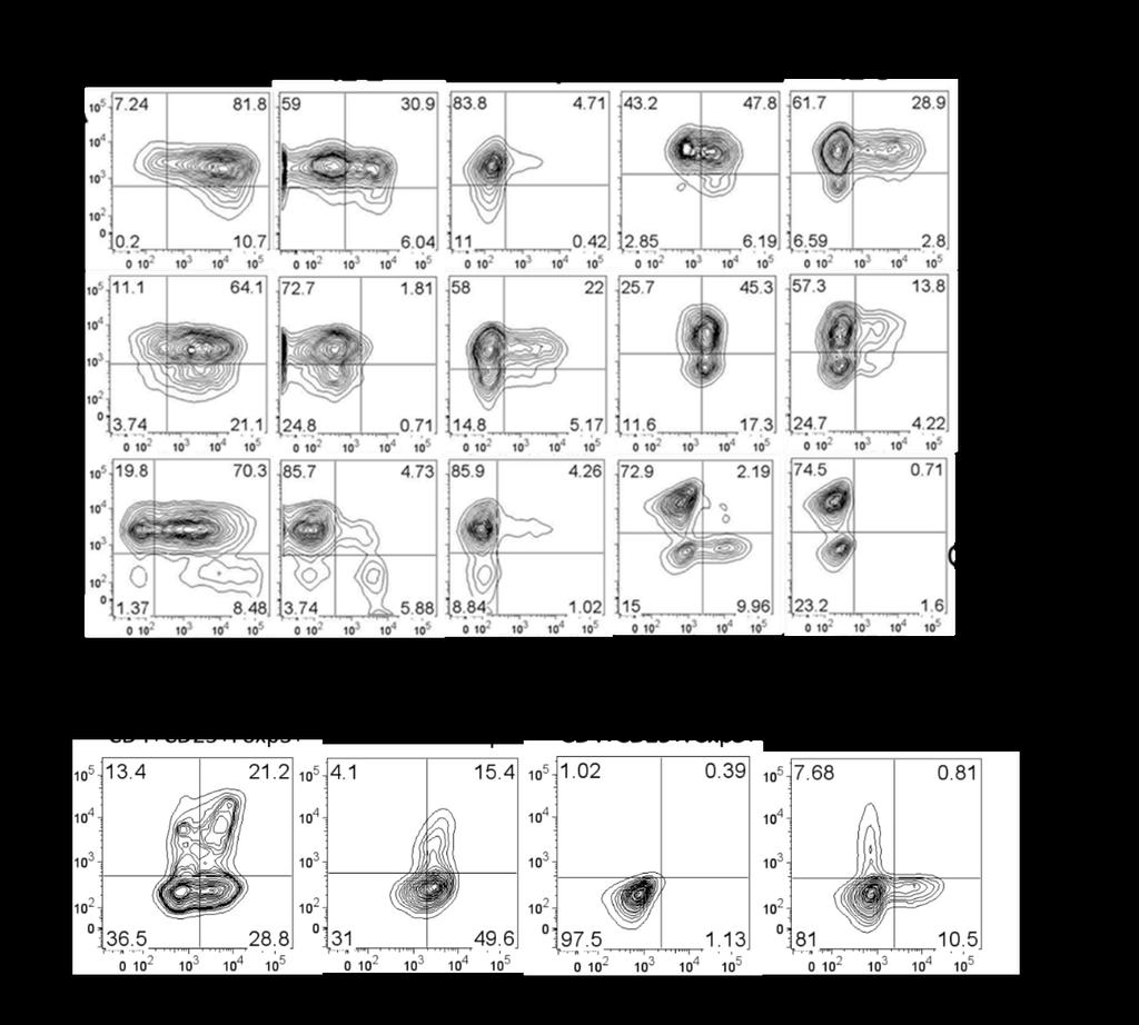
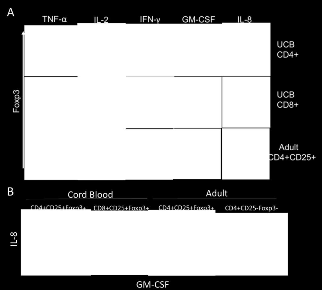
39 32
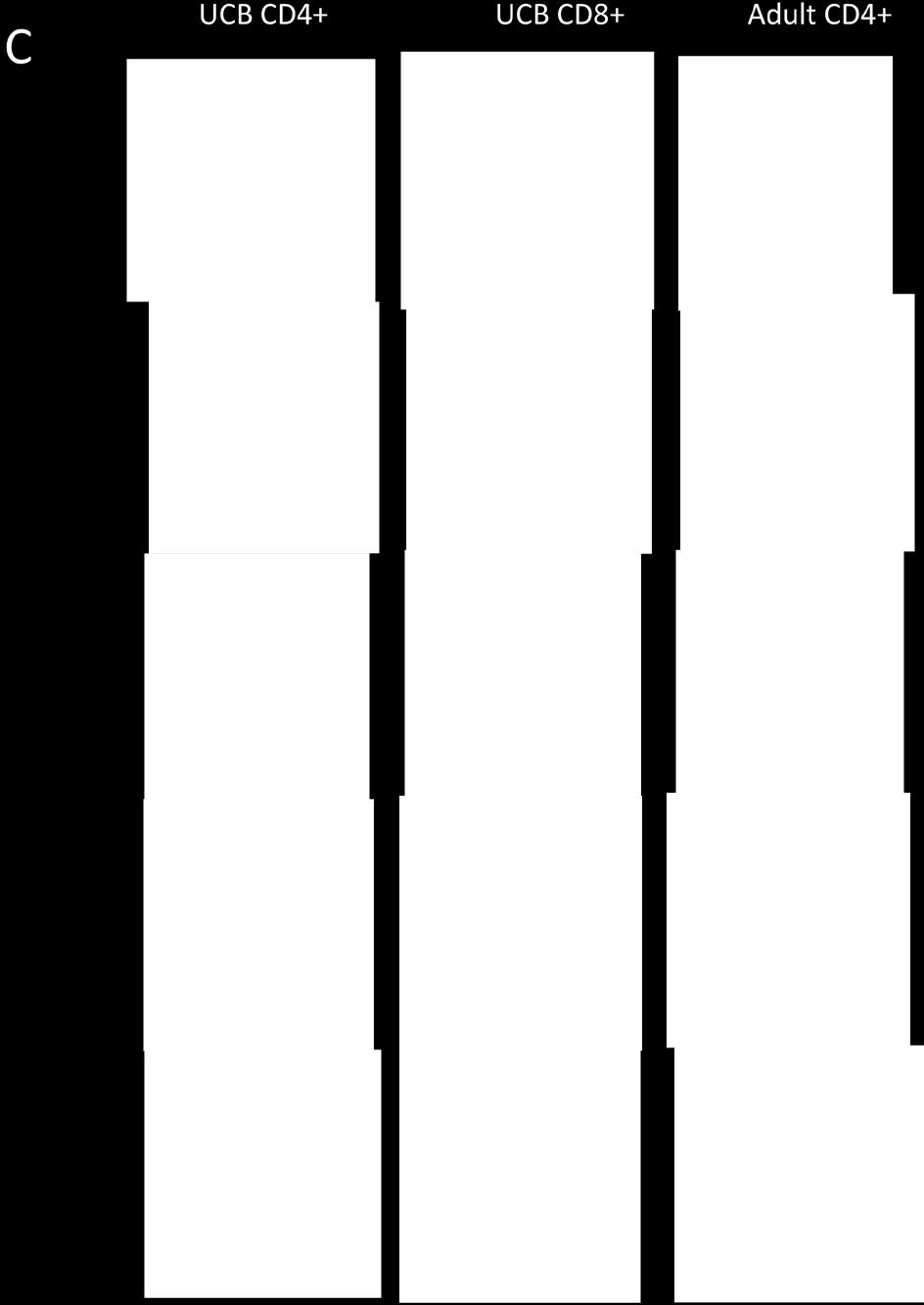
40 33
41 Figure 6. Distinct cytokine profile of cord blood Tregs. Cord blood and adult Tregs stimulated with PMA and ionomycin for 4 hours in the presence of monensin. (A) Cytokine profile of cord blood and adult Tregs (B) Co-expression of IL-8 and GM-CSF by cord blood Tregs in contrast to adult Tregs (CD4+CD25+Foxp3+) and conventional T cells (CD4+CD25- Foxp3-). Representative data from 6 donors for cord blood Tregs, and 3 donors for adult samples. (C) Frequency of cells staining positive for cytokines. Cells gated on CD4 or CD8 as indicated, followed by gating on Foxp3+ and Foxp3- populations prior to cytokine analysis. For adult samples, Foxp3+ were also gated on CD25+ and Foxp3- are gated on CD25-. Bars represent means of each sample. 34 Discussion Chemokine receptor expression. Cord blood Tregs express a distinct phenotype of chemokine receptor expression in comparison to adult Tregs, suggesting differential roles in tissue localization and trafficking ability. Expression of CXCR3 is limited predominantly to Th1 cells and mediates migration to sites of inflammation in response to CXCR3 ligands CXCL9, CXCL10, and CXCL Recently, a subset of Th1-like CXCR3 + Foxp3 + Tregs have been identified in adult peripheral blood with the ability to express IFN-γ and suppress Th1-mediated immune responses 57. In support of this, we observe a small population of CXCR3 + Tregs in adult peripheral blood. In contrast, however, the majority of CD4 + and CD8 + Tregs in cord blood express CXCR3, suggesting a role for these cells in suppressing Th1 immune responses and localizing to sites of Th1-mediated inflammation. During fetal development and early life, Th1 responses are mostly absent with the exception of responses to specific pathogens 146. In utero, Th1 responses are toxic to the placenta and may result in termination of pregnancy 152. Therefore, expression of CXCR3 by Tregs during fetal development may be important to control Th1 responses by trafficking to sites of Th1 inflammation and suppressing T cell proliferation and cytokine production.
42 35 Elevated CCR7 expression distinguishes cord blood Tregs from adult. CCR7 is required for T cell trafficking from peripheral tissues to lymphoid tissue in response to CCL19, the ligand for CCR CCL19 is expressed in T-cell zones in lymphoid tissue, suggesting a role for CCR7 + Tregs in modulation of T cell activation in germinal centers 154. While adult peripheral blood Tregs are known to express high levels of CCR4 and CCR6, expression of these receptors was reduced in cord blood Tregs 155. CCR4 + Tregs are associated with residence in epithelial tissue, and in mice the loss of CCR4 in Tregs results in lymphocyte infiltration and inflammation in skin and lungs 84. Expression of CCR6 mediates migration of both Th17 and Treg cells to areas of inflammation, suggesting a role of CCR6 + Tregs in suppressing Th17-mediated inflammation 156. Decreased expression of CCR4 and CCR6 on cord blood Tregs in comparison to adult suggests a diminished ability to migrate to epithelial tissue and sites of Th17-mediated inflammation, however this hypothesis has not been tested. Together, these data demonstrate a distinct phenotype of cord blood Tregs in comparison to adult and suggest that cord blood-derived Tregs would migrate differently than ttregs during immune responses and inflammation. Surface antigen phenotype. CD26, also known as dipeptidyl peptidase 4 (DPPIV), is a surface antigen associated with T cell activation and possesses enzymatic activity 157. CD26 can cleave a variety of hormones, growth factors, and chemokines which share proline or alanine residues at the N- terminus, thus modulating many physiologic processes including immune regulation and responses to infection 157. In particular, adenosine deaminase (ADA) is a known substrate for CD ADA is involved in purine metabolism and immune homeostasis, and genetic
43 36 deficiencies in ADA result in severe combined immune deficiency (SCID) and eventually death 159. As our data corroborate, CD26 is not expressed on adult Tregs, but in contrast we demonstrate high expression on cord blood Tregs, suggesting an important role for this enzyme in fetal or neonatal immune regulation or development 160. Future work will determine whether CD26 expression by cord blood Tregs modulates fetal or neonatal immune responses. Expression of TIM-3 is associated with Th1 cells in humans and regulates Th1 cytokine production as well as maintenance of tolerance in vivo 161. Specifically, interaction of TIM-3 with TIM-3 ligand is shown to inhibit Th1 responses in mice, thus maintaining peripheral tolerance 162. In humans, a paucity of TIM-3 expression is observed in multiple sclerosis (MS) patients compared with healthy controls and blockade of TIM-3 increases IFN-γ secretion by CD4 + T cells 163. Furthermore, expression of TIM-3 on cytotoxic CD8 + T cells is associated with anti-atherogenic cytokine production in human patients with atherosclerosis and blockade of TIM-3 in this context aggravates inflammation 164. Studies of tumor-bearing mice implicate tumor-resident TIM-3 + Tregs in tumor progression, identifying TIM-3 + Tregs as highly suppressive of effector T cell responses 165. Together, these studies demonstrate the importance of TIM-3 in the maintenance of tolerance in both human and murine disease. Expression of TIM-3 on cord blood Tregs, particularly CD8 + Tregs, suggests an important immunoregulatory role for this receptor. Future work will determine the function of TIM-3 in immune homeostasis in early life. TNF receptor superfamily members are highly conserved and control a wide variety of immune responses 166. One of these members, OX40, is important to regulation of CD4 and CD8 T cells responses as well as the differentiation and activity of regulatory T cells 167. OX40 is
44 37 expressed on activated T cells, where interaction with OX40 ligand (OX40L) on APCs induces NF-κB activation, enhancing T cell survival and proliferation 168. Ligation of OX40 with OX40L has been shown to reduce Foxp3 upregulation to block Treg differentiation and inhibit production of IL , 170. An interesting, but untested, possibility is that expression of OX40 on a subset of cord blood CD4 + Tregs may be a mechanism to limit Treg differentiation in neonates. CD31, or platelet endothelial cell adhesion molecule-1, is a transmembrane receptor expressed on a variety of immune cells, endothelial cells, and platelets 171. While the precise role of CD31 in Treg function has not been elucidated, some studies link CD31 expression to reduction in T cell activation and proliferation and others demonstrate a loss of Treg-mediated suppression following loss of CD31 in vivo 172, 173. In addition, CD31 signaling is suggested to limit clonal expansion by negatively regulating proliferation and inhibiting apoptosis 171. Expression of CD31 by cord blood Tregs, but not adult ttregs suggests that cord blood Tregs may be responsive to signaling which adult Tregs are not. However, the function of CD31 in neonatal immune homeostasis and Treg function remains unresolved. Cytokine profile. Cord blood Tregs exhibit potent suppressive activity in vitro but also produce inflammatory cytokines known to activate innate immune responses. Granulocyte macrophage colony-stimulating factor (GM-CSF) activates monocytes/macrophages and induces differentiation to modulate immune responses in peripheral tissues 174. Importantly, GM-CSF induces monocytes to differentiate into inflammatory M1 macrophages rather than antiinflammatory M2 macrophages 175. GM-CSF is produced by a variety of immune cells including B and T cells, macrophages, neutrophils, and eosinophils 176. Production of GM-CSF by T cells
45 38 is associated with pathology in several human diseases and their mouse models. In MS and the mouse model experimental autoimmune encephalomyelitis (EAE) CD4 + T cells drive inflammation and tissue destruction, and specifically Th17 cells have been tied to pathology of neuroinflammation 176. However, studies in mice demonstrate IL-17 is not required for development of EAE, but rather GM-CSF inhibition ameliorates progression of disease 177. In addition, production of GM-CSF by pathogenic T cells is associated with Th1 inflammation in rheumatoid arthritis and Th2-mediated allergic lung disease 176. Therefore, GM-CSF not only regulates immune homeostasis, but also exacerbates inflammation. GM-CSF production by T cells in cord blood may be important to differentiation of myeloid cells in responses to infection, although this possibility has not been tested. Expression of GM-CSF by Tregs suggests a multifunctional role for these cells, possibly negatively regulating adaptive immune responses while simultaneously driving innate immune responses and inflammation. Future work will test the ability of cord blood Tregs to induce M1 macrophage differentiation from monocytes. Furthermore, previous work demonstrates the requirement for monocytes in the differentiation of Tregs in cord blood (Lee et al., manuscript in preparation). Therefore, GM-CSF may induce differentiation of monocytes and reduce Treg differentiation in favor of effector T cell generation in a negative feedback loop to control Treg homeostasis in neonates. IL-8 also mediates innate immunity, resulting in recruitment and activation of neutrophils during immune responses 178. Neutrophils serve as the first line of defense against many pathogens, making IL-8 expression an essential aspect of initiation of immune responses during infection 179. In newborns, T cells readily produce IL-8 upon antigen encounter and infection, however production of IL-8 by T cells is rare in adults 105. Infants do not readily mount Th1
46 39 immune responses, so IL-8 production may be a compensatory mechanism to recruit neutrophils during bacterial infections. IL-8 production by Tregs has been identified in humans, and is hypothesized to modulate the recruitment of immune cells to inflamed tissues 180. Together, these data suggest IL-8 production by Tregs may be important for effective defense against bacterial infections in early life, recruiting neutrophils to peripheral tissues and activating innate immunity. Interferon gamma (IFN-γ) is produced by activated T helper and CD8 + cytotoxic T cells in response to viral and bacterial infections 181. Along with TNF, IFN-γ is considered a prototypic Th1 cytokine and contributes to pathogen recognition while initiating antiviral responses by inhibiting cell proliferation and inducing apoptosis 181. Regulatory T cells are generally considered unable to produce IFN-γ, however recently a subset of Th1-like Tregs have been described by expression of IFN-γ during ex vivo stimulation 57. We observe IFN-γ expression by CD8 + Foxp3 + Tregs from cord blood, suggesting these cells mediate immune homeostasis in response to intracellular pathogens. Viral infections are particularly dangerous during fetal development and in early life, with recent cases of Zika virus infections demonstrating a paucity of antiviral immune responses in the fetus 182. An interesting, but untested possibility is that CD8 + Tregs suppress adaptive immune responses to prevent damaging inflammation while modulating antiviral immunity by secreting IFN-γ during infection. Both cord blood and adult Tregs robustly express TNF in response to stimulation. The pro-inflammatory effects of TNF are well-described, however recent studies provide clear evidence of anti-inflammatory roles for this pleiotropic cytokine. In mice, TNF is important to development of inflammatory bowel disease. Paradoxically, however, TNF gene knockout
47 40 results in more severe intestinal inflammation rather than reducing pathology. Similarly, in humans inhibition of TNF in RA and Crohn s disease patients results in onset of autoimmune inflammation in some cases 124. These results suggest an important role for TNF in immune homeostasis that remains poorly understood. Expression of TNF by Tregs from both cord blood and adult peripheral blood raises the possibility of an important function for TNF in Treg homeostasis. Together, these results suggest a multifunctional role for Tregs in fetal and neonatal immunity. Expression of cytokines known to recruit and activate innate immune cells to sites of inflammation, along with distinct chemokine receptor expression may allow Tregs to migrate to peripheral tissues to control immune responses to pathogens. Tregs may be important to control adaptive immune responses during development while simultaneously protecting the newborn from infection by recruiting and activating macrophages and neutrophils.
48 CHAPTER FIVE FUNCTION OF TUMOR NECROSIS FACTOR SIGNALING IN TREGS Introduction The observation of robust TNF expression by both cord blood and adult Tregs suggests an important function for this cytokine. Recent reports disagree about the role of TNF-α signaling on Tregs; some reports suggest that TNF-α selectively activates and expands Tregs via tumor necrosis factor receptor 2 (TNFR2) while others indicate that TNF-α inhibits the suppressive function of human Tregs 127, 130. Furthermore, inhibition of TNF results in the onset or exacerbation of autoimmunity in some patients 124. However, expression of TNF by human Tregs has not been reported. For these reasons we decided to investigate the function of TNF produced by Tregs. Effect of TNF on Treg Differentiation TNF is reported to inhibit phosphorylation of Foxp3, therefore reducing suppressive function of Tregs in rheumatoid arthritis (RA) 127. However, other reports demonstrate a requirement for TNF signaling via TNFR2 on Tregs for maintenance of highly suppressive Tregs during inflammation 134. TNFR2 + Tregs reportedly upregulate Foxp3 and proliferate in response to TNF, suggesting a role of TNF in activation and expansion of Foxp3+ Tregs 135. Furthermore, interaction of transmembrane TNF (mtnf) with TNFR2 has been shown to block differentiation of Th17 cells in favor of Treg differentiation 131. To test the impact of TNF on 41

49 42 Figure 7. Effect of TNF inhibition on cord blood Treg generation. Cord blood Tregs were induced as previously described in the presence of anti-tnf, isotype control, or left untreated. At day 14, Treg phenotype was assess by flow cytometry. Representative data from 4 experiments. differentiation of Tregs, we used a neutralizing antibody against TNF to block TNF signaling during differentiation of cord blood Tregs, expecting to see a reduction of Foxp3 + CD25 + Tregs in cultures treated with anti-tnf if TNF is important to Treg generation. However, we observed no difference in differentiation of CD4 + or CD8 + Tregs from cord blood treated with anti-tnf, and isotype control antibody (migg1), or those left untreated (Fig. 7). Although only one concentration of anti-cd3 is shown above, concentrations as high as 25 µg/ml and as low as 40 ng/ml also had no effect on frequency of Tregs generated in vitro. Specifically, no change in
50 43 total Foxp3 frequency was observed in CD4 + or CD8 + Tregs between conditions. Furthermore, no change in Foxp3 + CD25 + phenotype was observed. These results suggest TNF does not affect cord blood Treg differentiation during induction in vitro. Effect of TNF on Suppressive Activity of Tregs TNF signaling has been linked to alterations in suppressive activity of Tregs, however discordant results prompted us to investigate the impact of TNF inhibition on in vitro suppressive activity of human Tregs 127, 129. We tested the effect of TNF neutralization on suppression of naïve T cell proliferation using a standard suppression assay. Induced Tregs from cord blood or adult Tregs isolated from adult peripheral blood were mixed (1:1) with naïve T cells (adult CD3 + CD45RA + FACS sorted after collection from peripheral blood) labeled with CFSE to track cell division. Cells were stimulated by anti-cd3 antibody and APCs (CD3 - FACS sorted after isolation from peripheral blood), and after 5 days proliferation of naïve T cells was determined by flow cytometric analysis of CFSE dilution. Naïve T cells and APCs were isolated from the same donor as Tregs in experiments with adult Tregs. To discern between Tregs and naïve T cells, Tregs were labeled with Cell Trace Violet (CTV) and removed from analysis by gating out CTV + cells. As a control for viability of naïve T cells, naïve T cells were stimulated with anti- CD3 and APCs in the absence of Tregs. Both cord blood derived Tregs and adult peripheral blood Tregs potently suppressed proliferation of naïve T cells, however no difference was observed between cells treated with anti-tnf antibody and those treated with the isotype control (Fig. 8). Neither cord blood nor adult Tregs exhibited a decrease in suppressive activity when TNF was inhibited, however a slight increase in suppression was observed. This small increase in suppression may be due to reverse signaling of mtnf when bound to anti-tnf, thus

51 activating Tregs and mildly increasing suppression. Together these results suggest that in vitro suppression of naïve T cell proliferation is not decreased by TNF inhibition. 44 Figure 8. Effect of TNF inhibition on suppressive activity of cord blood and adult Tregs. Whole cord blood derived Tregs or adult Tregs isolated from peripheral blood were tested for suppressive activity in the presence or absence of anti-tnf antibody. CFSE dilution in labeled naïve T cells was determined by flow cytometry after 5 days. Adult Foxp3 + Tregs Express Membrane TNF TNF is expressed as a type II transmembrane protein and must be cleaved by TACE to produce the soluble form 113. Both membrane and soluble forms of TNF are active and can signal via TNF receptors, however each form exhibits specific activity. Soluble TNF signals more readily via TNFR1, while mtnf has a greater affinity for TNFR TNFR1 contains an intracellular death domain and signals for cell cycle arrest and apoptosis, while TNFR2 lacks a death domain and activates NF-κB to promote growth and proliferation 116. Due to the different effects mediated by the two different forms of TNF, we investigated which cells in adult peripheral blood express mtnf.
52 45 Total adult PBMCs were collected and stimulated in vitro with PMA and ionomycin. Expression of mtnf was detected by surface staining followed by analysis by flow cytometry. We found that expression of mtnf was limited to CD4 + CD25 + cells (Fig. 9A). In contrast, CD4 + CD25 - cells did not express mtnf. Human T cells transiently express CD25 when activated, but Tregs constitutively express high amounts of CD To test whether Tregs express mtnf, Tregs were isolated from PBMCs by magnetic sorting and stimulated. Expression of mtnf on conventional T cells was compared by magnetic sorting for CD4 + CD25 - cells followed by stimulation. To ensure mtnf expression by Foxp3 + cells, both enriched populations were stained for TNF along with co-staining for CD4, CD25, and Foxp3 and analyzed by flow cytometry. We found that mtnf expression was indeed limited to a group of CD4 + CD25 + Foxp3 + cells, and found no mtnf expression on stimulated CD4 + CD25 - Foxp3 - cells (Fig. 9B). Although expression was low, greater than 50% of Foxp3 + T cells express detectable mtnf. These results demonstrate mtnf is preferentially expressed by Tregs in comparison to conventional T cells. TNF is traditionally considered a Th1 cytokine, so as a control we assessed production of the signature Th1 cytokine IFN-γ by Tregs. Total expression of TNF and IFN-γ were assessed by intracellular staining with Foxp3 costaining. We found that Foxp3 + Tregs expressed TNF when stimulated but did not express any IFN-γ (Fig. 9C). We also compared expression of TNF and IFN-γ between Foxp3 + Tregs and conventional T cells. Total CD4 + CD25 - Foxp3 - T cells expressed IFN-γ and TNF, however Foxp3 + T cells did not express IFN-γ (Fig. 9D).
53 Figure 9. Expression of TNF by human Foxp3 + Tregs Adult peripheral blood T cells were stimulated by PMA plus ionomycin for 4 hrs in the presence of monensin (details described in methods). The expression of cytokines was determined by intracellular staining or surface staining and analysis by flow cytometry. (A) Expression of the membrane form of TNF (mtnf) by CD4+CD25+ (right) and CD4+CD25- T cells (left). Representative data from 4 donors. Dashed lines indicate unstimulated samples and solid lines indicate stimulated samples (B) Expression of mtnf by CD4 + CD25 + Foxp3 + Tregs (left) or by CD4 + CD25 - Foxp3 - conventional T cells (right). A representative data set from three donors. (C) 46
54 Expression of total TNF (upper panels) and IFN-y (lower panels) by unstimulated (left panels) or stimulated (right panels) Foxp3 + Tregs by intracellular staining. A representative data set from three samples. (D) Frequencies of TNF (left) or IFN-y(right) expressing cells among CD4 + CD25 + Foxp3 + (Foxp3 + ) cells and CD4 + CD25 - Foxp3 - (Foxp3 - ) cells. TNF (n=6), IFN-y (n=3). * p<0.05 (student t test). (E) Expression of TNFR1 and TNFR2 on human Tregs by flow cytometry. (F) Expression of mtnf (n=3) and total (intracellular) TNF (n=5) by mouse CD4+ T cells. Representative data sets from 2 experiments. Foxp3+ Tregs (left) were gated first on live CD4+CD25+ cells, while Foxp3- T cells (right) were gated first on live CD4+CD25- cells. Dashed lines indicate unstimulated samples and solid lines represent stimulated samples. 47 The anti-inflammatory effect of TNF has been described in mice as well as humans, so we investigated whether TNF is expressed on mouse Tregs 126. We isolated total splenic CD4 + T cells from C57BL/6 mice and stimulated as previously described. Total TNF expression was assessed by intracellular staining after stimulation in the presence of monensin while mtnf expression was assessed by surface staining after stimulation without monensin. We found similar expression of total TNF by Foxp3 + and Foxp3 - T cells, however Foxp3 + T cells expressed a greater amount of mtnf (Fig. 9F). Some Foxp3 - T cells expressed detectable mtnf as well, however expression was lower than in Foxp3 + cells. These data demonstrate that Foxp3 + Tregs preferentially express the membrane form of TNF in both mice and humans, suggesting that antiinflammatory roles for TNF described in mouse and man may be related to mtnf expression by Tregs. Membrane TNF signals preferentially through TNFR2 and promotes survival and growth rather than death. To investigate the possible outcomes of TNF signaling by Tregs, we assessed the expression of TNF receptors by adult peripheral blood Tregs. In agreement with literature, we found that Foxp3+ T cells express TNFR2 but not TNFR1 (Fig. 9E). These data suggest that mtnf may signal Tregs for survival and proliferation via TNFR2 in an autocrine manner.
55 48 Function of Treg-derived TNF To test the hypothesis that mtnf signals Tregs for survival and proliferation in an autocrine manner, we developed an assay to test survival of adult Tregs in response to TNF inhibition. Tregs were isolated from peripheral blood, labeled with CFSE and stimulated ex vivo with plate-bound anti-cd3 and soluble CD28 in the presence of anti-tnf neutralizing antibody or isotype control. Importantly, IL-2 is considered a powerful growth factor for Tregs and is required for their survival in vitro 20. Therefore, to test the impact of TNF on Treg survival independent of IL-2, we did not add exogenous IL-2. Furthermore, we added a neutralizing antibody against IL-2 to block any endogenous IL-2 production. As a control for cell viability we included Tregs stimulated in the presence of IL-2. After 5 days, survival was assessed by counting live cells. In the presence of TNF inhibition, survival was significantly reduced compared to untreated cells (Fig. 10). Furthermore, increasing concentration of anti-tnf reduced survival of Tregs. Specifically, 5 µg/ml and 25 µg/ml anti-tnf significantly reduced survival in comparison to untreated control in a concentration-dependent manner. While reduced survival of Tregs treated with 1 µg/ml anti-tnf did not achieve significance, 2 donors exhibited a reduction in survival, suggesting that this concentration is the threshold for effect in this assay. Importantly, the isotype control did not affect cell survival in comparison to untreated cells. To confirm the role of cell proliferation and apoptosis in overall cell number after 5 days of stimulation, we analyzed CFSE dilution by flow cytometry and stained with annexin V to detect phosphatidyl inositol, a marker of apoptosis when exposed on outer leaflet of cell membranes. In the untreated and isotype controls, annexin V staining was positive in a small fraction of cells (~20%), indicating few cells undergoing apoptosis (Fig. 11, upper panel).
56 49 However, addition of 1 µg/ml anti-tnf increased annexin V + cells (~50%) and higher concentrations resulted in the majority of cells exhibiting an apoptotic phenotype (~80%). Cells treated with IL-2 did not bind annexin V and increased in size (data not shown). These data demonstrate that inhibition of TNF increases apoptosis, suggesting a role for TNF in survival of Tregs in the absence of IL-2. To test the role of TNF in Treg proliferation in vitro, CFSE dilution was measured by flow cytometry (Fig. 11, lower panel). About half of all untreated and isotype control-treated cells proliferated in response to stimulation (~50%) as indicated by dilution of CFSE. In contrast, fewer cells proliferated in anti-tnf-treated conditions, with almost total abrogation of cell division in the highest concentration of anti-tnf (~7% cells divided). Together, these data demonstrate that Tregs fail to proliferate and undergo apoptosis during ex vivo stimulation in the presence of TNF inhibition.
57 Figure 10. Treg survival in response to TNF inhibition. Tregs isolated from peripheral blood were stimulated by plate-bound anti-cd3 and soluble anti-cd28 in the presence of anti-tnf for isotype control antibody for 5 days. Cell survival was calculated by comparing cell number of treated samples to untreated control. Statistical analysis by paired T-test (*p<0.05, ***p<0.001) with bars representing mean and standard deviation. 50
 to detect apoptotic cells. Data representative of 3 independent experiments with 5 donors total.")
58 51 Figure 11. Effect of TNF neutralization on apoptosis and cell proliferation. Tregs were labeled with CFSE (bottom) prior to stimulation to track proliferation. After 5 days culture with anti-tnf, cells were stained with Annexin V (top) to detect apoptotic cells. Data representative of 3 independent experiments with 5 donors total. Discussion TNF drives inflammation but also contributes to anti-inflammatory immune responses. Studies in humans and mice demonstrate TNF mediates autoimmune inflammation, but inhibition of TNF also results in onset or exacerbation of autoimmunity 124. Multiple cells types can produce TNF and the pleiotropic effects of this cytokine are dependent upon the type of cell and receptor engaged. Discordant studies of TNF function on immune homeostasis, along with the identification of TNF expression by cord blood and adult Tregs prompted us to investigate the function of Treg-derived TNF. Several studies implicate TNF signaling in the differentiation of Tregs, however we did not observe any effect of TNF inhibition during cord blood Treg induction. The conditions used for ex vivo Treg induction in our lab may override any impact of TNF on Foxp3 expression. We provide exogenous IL-2 to drive Treg differentiation, raising the possibility that IL-2 potently
Lecture outline. Immunological tolerance and immune regulation. Central and peripheral tolerance. Inhibitory receptors of T cells. Regulatory T cells
 1 Immunological tolerance and immune regulation Abul K. Abbas UCSF 2 Lecture outline Central and peripheral tolerance Inhibitory receptors of T cells Regulatory T cells 1 The immunological equilibrium:
Central tolerance. Mechanisms of Immune Tolerance. Regulation of the T cell response
 Immunoregulation: A balance between activation and suppression that achieves an efficient immune response without damaging the host. Mechanisms of Immune Tolerance ACTIVATION (immunity) SUPPRESSION (tolerance)
Mechanisms of Immune Tolerance
 Immunoregulation: A balance between activation and suppression that achieves an efficient immune response without damaging the host. ACTIVATION (immunity) SUPPRESSION (tolerance) Autoimmunity Immunodeficiency
Effector mechanisms of cell-mediated immunity: Properties of effector, memory and regulatory T cells
 ICI Basic Immunology course Effector mechanisms of cell-mediated immunity: Properties of effector, memory and regulatory T cells Abul K. Abbas, MD UCSF Stages in the development of T cell responses: induction
Immunological Tolerance
 Immunological Tolerance Introduction Definition: Unresponsiveness to an antigen that is induced by exposure to that antigen Tolerogen = tolerogenic antigen = antigen that induces tolerance Important for
Effector T Cells and
 1 Effector T Cells and Cytokines Andrew Lichtman, MD PhD Brigham and Women's Hospital Harvard Medical School 2 Lecture outline Cytokines Subsets of CD4+ T cells: definitions, functions, development New
SEVENTH EDITION CHAPTER
 Judy Owen Jenni Punt Sharon Stranford Kuby Immunology SEVENTH EDITION CHAPTER 16 Tolerance, Autoimmunity, and Transplantation Copyright 2013 by W. H. Freeman and Company Immune tolerance: history * Some
T-cell activation T cells migrate to secondary lymphoid tissues where they interact with antigen, antigen-presenting cells, and other lymphocytes:
 Interactions between innate immunity & adaptive immunity What happens to T cells after they leave the thymus? Naïve T cells exit the thymus and enter the bloodstream. If they remain in the bloodstream,
Tolerance 2. Regulatory T cells; why tolerance fails. Abul K. Abbas UCSF. FOCiS
 1 Tolerance 2. Regulatory T cells; why tolerance fails Abul K. Abbas UCSF FOCiS 2 Lecture outline Regulatory T cells: functions and clinical relevance Pathogenesis of autoimmunity: why selftolerance fails
Tolerance 2. Regulatory T cells; why tolerance fails. FOCiS. Lecture outline. Regulatory T cells. Regulatory T cells: functions and clinical relevance
 1 Tolerance 2. Regulatory T cells; why tolerance fails Abul K. Abbas UCSF FOCiS 2 Lecture outline Regulatory T cells: functions and clinical relevance Pathogenesis of autoimmunity: why selftolerance fails
Cover Page. The handle holds various files of this Leiden University dissertation.
 Cover Page The handle http://hdl.handle.net/1887/23854 holds various files of this Leiden University dissertation. Author: Marel, Sander van der Title: Gene and cell therapy based treatment strategies
Supplemental Table I.
 Supplemental Table I Male / Mean ± SEM n Mean ± SEM n Body weight, g 29.2±0.4 17 29.7±0.5 17 Total cholesterol, mg/dl 534.0±30.8 17 561.6±26.1 17 HDL-cholesterol, mg/dl 9.6±0.8 17 10.1±0.7 17 Triglycerides,
Tolerance, autoimmunity and the pathogenesis of immunemediated inflammatory diseases. Abul K. Abbas UCSF
 Tolerance, autoimmunity and the pathogenesis of immunemediated inflammatory diseases Abul K. Abbas UCSF Balancing lymphocyte activation and control Activation Effector T cells Tolerance Regulatory T cells
Adaptive immune responses: T cell-mediated immunity
 MICR2209 Adaptive immune responses: T cell-mediated immunity Dr Allison Imrie allison.imrie@uwa.edu.au 1 Synopsis: In this lecture we will discuss the T-cell mediated immune response, how it is activated,
The Adaptive Immune Responses
 The Adaptive Immune Responses The two arms of the immune responses are; 1) the cell mediated, and 2) the humoral responses. In this chapter we will discuss the two responses in detail and we will start
T cell maturation. T-cell Maturation. What allows T cell maturation?
 T-cell Maturation What allows T cell maturation? Direct contact with thymic epithelial cells Influence of thymic hormones Growth factors (cytokines, CSF) T cell maturation T cell progenitor DN DP SP 2ry
The development of T cells in the thymus
 T cells rearrange their receptors in the thymus whereas B cells do so in the bone marrow. The development of T cells in the thymus The lobular/cellular organization of the thymus Immature cells are called
General Overview of Immunology. Kimberly S. Schluns, Ph.D. Associate Professor Department of Immunology UT MD Anderson Cancer Center
 General Overview of Immunology Kimberly S. Schluns, Ph.D. Associate Professor Department of Immunology UT MD Anderson Cancer Center Objectives Describe differences between innate and adaptive immune responses
Objectives. Abbas Chapter 11: Immunological Tolerance. Question 1. Question 2. Question 3. Definitions
 Objectives Abbas Chapter 11: Immunological Tolerance Christina Ciaccio, MD Children s Mercy Hospitals and Clinics February 1, 2010 To introduce the concept of immunologic tolerance To understand what factors
5/1/13. The proportion of thymus that produces T cells decreases with age. The cellular organization of the thymus
 T cell precursors migrate from the bone marrow via the blood to the thymus to mature 1 2 The cellular organization of the thymus The proportion of thymus that produces T cells decreases with age 3 4 1
Determinants of Immunogenicity and Tolerance. Abul K. Abbas, MD Department of Pathology University of California San Francisco
 Determinants of Immunogenicity and Tolerance Abul K. Abbas, MD Department of Pathology University of California San Francisco EIP Symposium Feb 2016 Why do some people respond to therapeutic proteins?
T Cell Activation, Costimulation and Regulation
 1 T Cell Activation, Costimulation and Regulation Abul K. Abbas, MD University of California San Francisco 2 Lecture outline T cell antigen recognition and activation Costimulation, the B7:CD28 family
Supplemental Figure 1. Signature gene expression in in vitro differentiated Th0, Th1, Th2, Th17 and Treg cells. (A) Naïve CD4 + T cells were cultured
 Naïve CD4 + T cells were cultured") Supplemental Figure 1. Signature gene expression in in vitro differentiated Th0, Th1, Th2, Th17 and Treg cells. (A) Naïve CD4 + T cells were cultured under Th0, Th1, Th2, Th17, and Treg conditions. mrna
Supplemental Figure 1. Signature gene expression in in vitro differentiated Th0, Th1, Th2, Th17 and Treg cells. (A) Naïve CD4 + T cells were cultured under Th0, Th1, Th2, Th17, and Treg conditions. mrna
ACTIVATION OF T LYMPHOCYTES AND CELL MEDIATED IMMUNITY
 ACTIVATION OF T LYMPHOCYTES AND CELL MEDIATED IMMUNITY The recognition of specific antigen by naïve T cell induces its own activation and effector phases. T helper cells recognize peptide antigens through
Immunology for the Rheumatologist
 Immunology for the Rheumatologist Rheumatologists frequently deal with the immune system gone awry, rarely studying normal immunology. This program is an overview and discussion of the function of the
Antigen Presentation and T Lymphocyte Activation. Abul K. Abbas UCSF. FOCiS
 1 Antigen Presentation and T Lymphocyte Activation Abul K. Abbas UCSF FOCiS 2 Lecture outline Dendritic cells and antigen presentation The role of the MHC T cell activation Costimulation, the B7:CD28 family
T Lymphocyte Activation and Costimulation. FOCiS. Lecture outline
 1 T Lymphocyte Activation and Costimulation Abul K. Abbas, MD UCSF FOCiS 2 Lecture outline T cell activation Costimulation, the B7:CD28 family Inhibitory receptors of T cells Targeting costimulators for
FOCiS. Lecture outline. The immunological equilibrium: balancing lymphocyte activation and control. Immunological tolerance and immune regulation -- 1
 1 Immunological tolerance and immune regulation -- 1 Abul K. Abbas UCSF FOCiS 2 Lecture outline Principles of immune regulation Self-tolerance; mechanisms of central and peripheral tolerance Inhibitory
Darwinian selection and Newtonian physics wrapped up in systems biology
 Darwinian selection and Newtonian physics wrapped up in systems biology Concept published in 1957* by Macfarland Burnet (1960 Nobel Laureate for the theory of induced immune tolerance, leading to solid
TCR, MHC and coreceptors
 Cooperation In Immune Responses Antigen processing how peptides get into MHC Antigen processing involves the intracellular proteolytic generation of MHC binding proteins Protein antigens may be processed
ACTIVATION AND EFFECTOR FUNCTIONS OF CELL-MEDIATED IMMUNITY AND NK CELLS. Choompone Sakonwasun, MD (Hons), FRCPT
, FRCPT") ACTIVATION AND EFFECTOR FUNCTIONS OF CELL-MEDIATED IMMUNITY AND NK CELLS Choompone Sakonwasun, MD (Hons), FRCPT Types of Adaptive Immunity Types of T Cell-mediated Immune Reactions CTLs = cytotoxic T lymphocytes
ACTIVATION AND EFFECTOR FUNCTIONS OF CELL-MEDIATED IMMUNITY AND NK CELLS Choompone Sakonwasun, MD (Hons), FRCPT Types of Adaptive Immunity Types of T Cell-mediated Immune Reactions CTLs = cytotoxic T lymphocytes
Optimizing Intracellular Flow Cytometry:
 Optimizing Intracellular Flow Cytometry: Simultaneous Detection of Cytokines and Transcription Factors An encore presentation by Jurg Rohrer, PhD, BD Biosciences 10.26.10 Outline Introduction Cytokines
T Cell Receptor & T Cell Development
 T Cell Receptor & T Cell Development Questions for the next 2 lectures: How do you generate a diverse T cell population with functional TCR rearrangements? How do you generate a T cell population that
Advances in Cancer Immunotherapy
 Advances in Cancer Immunotherapy Immunology 101 for the Non-Immunologist Arnold H. Zea, PhD azea@lsuhsc.edu Disclosures No relevant financial relationships to disclose This presentation does not contain
Allergy and Immunology Review Corner: Chapter 19 of Immunology IV: Clinical Applications in Health and Disease, by Joseph A. Bellanti, MD.
 Allergy and Immunology Review Corner: Chapter 19 of Immunology IV: Clinical Applications in Health and Disease, by Joseph A. Bellanti, MD. Chapter 19: Tolerance, Autoimmunity, and Autoinflammation Prepared
Topic (Final-02): Immunologic Tolerance
: Immunologic Tolerance") Topic (Final-02): Immunologic Tolerance Immunologic tolerance is defined as unresponsiveness to an antigen that is induced by previous exposure to that antigen. The term arose from the experimental observation
Topic (Final-02): Immunologic Tolerance Immunologic tolerance is defined as unresponsiveness to an antigen that is induced by previous exposure to that antigen. The term arose from the experimental observation
Immune Tolerance. Kyeong Cheon Jung. Department of Pathology Seoul National University College of Medicine
 Immune Tolerance Kyeong Cheon Jung Department of Pathology Seoul National University College of Medicine Immune tolerance Unresponsiveness to an antigen that is induced by previous exposure to that antigen
Immunology Lecture 4. Clinical Relevance of the Immune System
 Immunology Lecture 4 The Well Patient: How innate and adaptive immune responses maintain health - 13, pg 169-181, 191-195. Immune Deficiency - 15 Autoimmunity - 16 Transplantation - 17, pg 260-270 Tumor
Immune Regulation and Tolerance
 Immune Regulation and Tolerance Immunoregulation: A balance between activation and suppression of effector cells to achieve an efficient immune response without damaging the host. Activation (immunity)
Scott Abrams, Ph.D. Professor of Oncology, x4375 Kuby Immunology SEVENTH EDITION
 Scott Abrams, Ph.D. Professor of Oncology, x4375 scott.abrams@roswellpark.org Kuby Immunology SEVENTH EDITION CHAPTER 11 T-Cell Activation, Differentiation, and Memory Copyright 2013 by W. H. Freeman and
Blocking antibodies and peptides. Rat anti-mouse PD-1 (29F.1A12, rat IgG2a, k), PD-
, PD-") Supplementary Methods Blocking antibodies and peptides. Rat anti-mouse PD-1 (29F.1A12, rat IgG2a, k), PD- L1 (10F.9G2, rat IgG2b, k), and PD-L2 (3.2, mouse IgG1) have been described (24). Anti-CTLA-4 (clone
Supplementary Methods Blocking antibodies and peptides. Rat anti-mouse PD-1 (29F.1A12, rat IgG2a, k), PD- L1 (10F.9G2, rat IgG2b, k), and PD-L2 (3.2, mouse IgG1) have been described (24). Anti-CTLA-4 (clone
1. Overview of Adaptive Immunity
 Chapter 17A: Adaptive Immunity Part I 1. Overview of Adaptive Immunity 2. T and B Cell Production 3. Antigens & Antigen Presentation 4. Helper T cells 1. Overview of Adaptive Immunity The Nature of Adaptive
Medical Virology Immunology. Dr. Sameer Naji, MB, BCh, PhD (UK) Head of Basic Medical Sciences Dept. Faculty of Medicine The Hashemite University
 Head of Basic Medical Sciences Dept. Faculty of Medicine The Hashemite University") Medical Virology Immunology Dr. Sameer Naji, MB, BCh, PhD (UK) Head of Basic Medical Sciences Dept. Faculty of Medicine The Hashemite University Human blood cells Phases of immune responses Microbe Naïve
Medical Virology Immunology Dr. Sameer Naji, MB, BCh, PhD (UK) Head of Basic Medical Sciences Dept. Faculty of Medicine The Hashemite University Human blood cells Phases of immune responses Microbe Naïve
Immunity. Acquired immunity differs from innate immunity in specificity & memory from 1 st exposure
 Immunity (1) Non specific (innate) immunity (2) Specific (acquired) immunity Characters: (1) Non specific: does not need special recognition of the foreign cell. (2) Innate: does not need previous exposure.
Autoimmunity. Autoimmunity arises because of defects in central or peripheral tolerance of lymphocytes to selfantigens
 Autoimmunity Autoimmunity arises because of defects in central or peripheral tolerance of lymphocytes to selfantigens Autoimmune disease can be caused to primary defects in B cells, T cells and possibly
Supplemental Information. T Cells Enhance Autoimmunity by Restraining Regulatory T Cell Responses via an Interleukin-23-Dependent Mechanism
 Immunity, Volume 33 Supplemental Information T Cells Enhance Autoimmunity by Restraining Regulatory T Cell Responses via an Interleukin-23-Dependent Mechanism Franziska Petermann, Veit Rothhammer, Malte
I. Lines of Defense Pathogen: Table 1: Types of Immune Mechanisms. Table 2: Innate Immunity: First Lines of Defense
 I. Lines of Defense Pathogen: Table 1: Types of Immune Mechanisms Table 2: Innate Immunity: First Lines of Defense Innate Immunity involves nonspecific physical & chemical barriers that are adapted for
The Adaptive Immune Response: T lymphocytes and Their Functional Types *
 OpenStax-CNX module: m46560 1 The Adaptive Immune Response: T lymphocytes and Their Functional Types * OpenStax This work is produced by OpenStax-CNX and licensed under the Creative Commons Attribution
Innate immune regulation of T-helper (Th) cell homeostasis in the intestine
 cell homeostasis in the intestine") Innate immune regulation of T-helper (Th) cell homeostasis in the intestine Masayuki Fukata, MD, Ph.D. Research Scientist II Division of Gastroenterology, Department of Medicine, F. Widjaja Foundation,
Innate immune regulation of T-helper (Th) cell homeostasis in the intestine Masayuki Fukata, MD, Ph.D. Research Scientist II Division of Gastroenterology, Department of Medicine, F. Widjaja Foundation,
Question 1. Kupffer cells, microglial cells and osteoclasts are all examples of what type of immune system cell?
 Abbas Chapter 2: Sarah Spriet February 8, 2015 Question 1. Kupffer cells, microglial cells and osteoclasts are all examples of what type of immune system cell? a. Dendritic cells b. Macrophages c. Monocytes
Cytokines modulate the functional activities of individual cells and tissues both under normal and pathologic conditions Interleukins,
 Cytokines http://highered.mcgraw-hill.com/sites/0072507470/student_view0/chapter22/animation the_immune_response.html Cytokines modulate the functional activities of individual cells and tissues both under
Prof. Ibtesam Kamel Afifi Professor of Medical Microbiology & Immunology
 By Prof. Ibtesam Kamel Afifi Professor of Medical Microbiology & Immunology Lecture objectives: At the end of the lecture you should be able to: Enumerate features that characterize acquired immune response
Guarding the immune system: Suppression of autoimmunity by CD4 1 CD25 1 immunoregulatory T cells
 Immunology and Cell Biology (2006) 84, 487 501 doi:10.1111/j.1440-1711.2006.01471.x Review Article Guarding the immune system: Suppression of autoimmunity by CD4 1 CD25 1 immunoregulatory T cells TRICIA
HLA and antigen presentation. Department of Immunology Charles University, 2nd Medical School University Hospital Motol
 HLA and antigen presentation Department of Immunology Charles University, 2nd Medical School University Hospital Motol MHC in adaptive immunity Characteristics Specificity Innate For structures shared
Immune response. This overview figure summarizes simply how our body responds to foreign molecules that enter to it.
 Immune response This overview figure summarizes simply how our body responds to foreign molecules that enter to it. It s highly recommended to watch Dr Najeeb s lecture that s titled T Helper cells and
Primer on Tumor Immunology. International Society for Biological Therapy of Cancer. C. H. June, M.D. November 10, 2005
 Primer on Tumor Immunology International Society for Biological Therapy of Cancer C. H. June, M.D. November 10, 2005 Outline: Primer on Tumor Immunology T Cell Receptors T Cell Biology Tumor immunology
Defensive mechanisms include :
 Acquired Immunity Defensive mechanisms include : 1) Innate immunity (Natural or Non specific) 2) Acquired immunity (Adaptive or Specific) Cell-mediated immunity Humoral immunity Two mechanisms 1) Humoral
Examples of questions for Cellular Immunology/Cellular Biology and Immunology
 Examples of questions for Cellular Immunology/Cellular Biology and Immunology Each student gets a set of 6 questions, so that each set contains different types of questions and that the set of questions
Chapter 13: Cytokines
 Chapter 13: Cytokines Definition: secreted, low-molecular-weight proteins that regulate the nature, intensity and duration of the immune response by exerting a variety of effects on lymphocytes and/or
CYTOKINE RECEPTORS AND SIGNAL TRANSDUCTION
 CYTOKINE RECEPTORS AND SIGNAL TRANSDUCTION What is Cytokine? Secreted popypeptide (protein) involved in cell-to-cell signaling. Acts in paracrine or autocrine fashion through specific cellular receptors.
Adaptive Immunity: Specific Defenses of the Host
 17 Adaptive Immunity: Specific Defenses of the Host SLOs Differentiate between innate and adaptive immunity, and humoral and cellular immunity. Define antigen, epitope, and hapten. Explain the function
Cytokines (II) Dr. Aws Alshamsan Department of Pharmaceu5cs Office: AA87 Tel:
 Dr. Aws Alshamsan Department of Pharmaceu5cs Office: AA87 Tel:") Cytokines (II) Dr. Aws Alshamsan Department of Pharmaceu5cs Office: AA87 Tel: 4677363 aalshamsan@ksu.edu.sa Learning Objectives By the end of this lecture you will be able to: 1 Understand the physiological
Cytokines (II) Dr. Aws Alshamsan Department of Pharmaceu5cs Office: AA87 Tel: 4677363 aalshamsan@ksu.edu.sa Learning Objectives By the end of this lecture you will be able to: 1 Understand the physiological
Immunology MIMM-314 MID-TERM II EXAMINATION. 1 hour between 8:30 a.m. - 10:00 a.m. McIntyre Medical Rm 504 (Martin Amphitheatre)
") GROUP (Version) 1 Annotated version April 8, 2011, RGEP DEPARTMENT OF MICROBIOLOGY AND IMMUNOLOGY Immunology MIMM-314 MID-TERM II EXAMINATION Course Coordinator: Dr. Roger Palfree Date: Thursday, March
GROUP (Version) 1 Annotated version April 8, 2011, RGEP DEPARTMENT OF MICROBIOLOGY AND IMMUNOLOGY Immunology MIMM-314 MID-TERM II EXAMINATION Course Coordinator: Dr. Roger Palfree Date: Thursday, March
The encephalitogenicity of TH17 cells is dependent on IL-1- and IL-23- induced production of the cytokine GM-CSF
 CORRECTION NOTICE Nat.Immunol. 12, 568 575 (2011) The encephalitogenicity of TH17 cells is dependent on IL-1- and IL-23- induced production of the cytokine GM-CSF Mohamed El-Behi, Bogoljub Ciric, Hong
Chapter 22: The Lymphatic System and Immunity
 Bio40C schedule Lecture Immune system Lab Quiz 2 this week; bring a scantron! Study guide on my website (see lab assignments) Extra credit Critical thinking questions at end of chapters 5 pts/chapter Due
Adaptive Immunity. Jeffrey K. Actor, Ph.D. MSB 2.214,
 Adaptive Immunity Jeffrey K. Actor, Ph.D. MSB 2.214, 500-5344 Lecture Objectives: Understand role of various molecules including cytokines, chemokines, costimulatory and adhesion molecules in the development
Following T-cell activation and differentiation with HTRF reagents: IL-2, IFN-γ and IL-17
 Following T-cell activation and differentiation with HTRF reagents: IL-2, IFN-γ and IL-17 4 th HTRF Symposium for Drug Discovery Avignon, Sept. 24-26, 28 Introduction: T-cells have effector and helper
HLA and antigen presentation. Department of Immunology Charles University, 2nd Medical School University Hospital Motol
 HLA and antigen presentation Department of Immunology Charles University, 2nd Medical School University Hospital Motol MHC in adaptive immunity Characteristics Specificity Innate For structures shared
T Cell Effector Mechanisms I: B cell Help & DTH
 T Cell Effector Mechanisms I: B cell Help & DTH Ned Braunstein, MD The Major T Cell Subsets p56 lck + T cells γ δ ε ζ ζ p56 lck CD8+ T cells γ δ ε ζ ζ Cα Cβ Vα Vβ CD3 CD8 Cα Cβ Vα Vβ CD3 MHC II peptide
Development of B and T lymphocytes
 Development of B and T lymphocytes What will we discuss today? B-cell development T-cell development B- cell development overview Stem cell In periphery Pro-B cell Pre-B cell Immature B cell Mature B cell
LESSON 2: THE ADAPTIVE IMMUNITY
 Introduction to immunology. LESSON 2: THE ADAPTIVE IMMUNITY Today we will get to know: The adaptive immunity T- and B-cells Antigens and their recognition How T-cells work 1 The adaptive immunity Unlike
Introduction to Immunology Part 2 September 30, Dan Stetson
 Introduction to Immunology Part 2 September 30, 2016 Dan Stetson stetson@uw.edu 441 Lecture #2 Slide 1 of 26 CLASS ANNOUNCEMENT PLEASE NO TREE NUTS IN CLASS!!! (Peanuts, walnuts, almonds, cashews, etc)
Chapter 24 The Immune System
 Chapter 24 The Immune System The Immune System Layered defense system The skin and chemical barriers The innate and adaptive immune systems Immunity The body s ability to recognize and destroy specific
Immunology Basics Relevant to Cancer Immunotherapy: T Cell Activation, Costimulation, and Effector T Cells
 Immunology Basics Relevant to Cancer Immunotherapy: T Cell Activation, Costimulation, and Effector T Cells Andrew H. Lichtman, M.D. Ph.D. Department of Pathology Brigham and Women s Hospital and Harvard
The Adaptive Immune Response. B-cells
 The Adaptive Immune Response B-cells The innate immune system provides immediate protection. The adaptive response takes time to develop and is antigen specific. Activation of B and T lymphocytes Naive
DNA vaccine, peripheral T-cell tolerance modulation 185
 Subject Index Airway hyperresponsiveness (AHR) animal models 41 43 asthma inhibition 45 overview 41 mast cell modulation of T-cells 62 64 respiratory tolerance 40, 41 Tregs inhibition role 44 respiratory
Optimizing Intracellular Flow Cytometry:
 Optimizing Intracellular Flow Cytometry: Simultaneous Detection of Cytokines and Transcription Factors Presented by Jurg Rohrer, PhD, BD Biosciences 23-10780-00 Outline Introduction Cytokines Transcription
Phenotypical and Functional Analysis of Peripheral T Cells in Foxn1 Transgenic Mice: Effects of Aging
 Loyola University Chicago Loyola ecommons Master's Theses Theses and Dissertations 2010 Phenotypical and Functional Analysis of Peripheral T Cells in Foxn1 Transgenic Mice: Effects of Aging Paulette Krishack
Supplementary Figure 1. Characterization of basophils after reconstitution of SCID mice
 Supplementary figure legends Supplementary Figure 1. Characterization of after reconstitution of SCID mice with CD4 + CD62L + T cells. (A-C) SCID mice (n = 6 / group) were reconstituted with 2 x 1 6 CD4
Adaptive (acquired) immunity. Professor Peter Delves University College London
 immunity. Professor Peter Delves University College London") Adaptive (acquired) immunity Professor Peter Delves University College London p.delves@ucl.ac.uk Haematopoiesis Haematopoiesis Lymphocytes = adaptive response Recognition of pathogens by adaptive cells,
Adaptive (acquired) immunity Professor Peter Delves University College London p.delves@ucl.ac.uk Haematopoiesis Haematopoiesis Lymphocytes = adaptive response Recognition of pathogens by adaptive cells,
T Cell Activation. Patricia Fitzgerald-Bocarsly March 18, 2009
 T Cell Activation Patricia Fitzgerald-Bocarsly March 18, 2009 Phases of Adaptive Immune Responses Phases of T cell responses IL-2 acts as an autocrine growth factor Fig. 11-11 Clonal Expansion of T cells
Regulatory T Cells and Maintenance of Tolerance
 Regulatory Cells and Maintenance of olerance Stephen Canfield, MD, PhD Asst. Prof. Medicine smc12@columbia.edu What constitutes immune tolerance? Discrimination between: non-self and self e.g., virally
The Lymphatic System and Body Defenses
 PowerPoint Lecture Slide Presentation by Patty Bostwick-Taylor, Florence-Darlington Technical College The Lymphatic System and Body Defenses 12PART B Adaptive Defense System: Third Line of Defense Immune
SUPPLEMENTARY INFORMATION
 Supplemental Figure 1. Furin is efficiently deleted in CD4 + and CD8 + T cells. a, Western blot for furin and actin proteins in CD4cre-fur f/f and fur f/f Th1 cells. Wild-type and furin-deficient CD4 +
Supplementary Figure 1. IL-12 serum levels and frequency of subsets in FL patients. (A) IL-12
 IL-12") 1 Supplementary Data Figure legends Supplementary Figure 1. IL-12 serum levels and frequency of subsets in FL patients. (A) IL-12 serum levels measured by multiplex ELISA (Luminex) in FL patients before
1 Supplementary Data Figure legends Supplementary Figure 1. IL-12 serum levels and frequency of subsets in FL patients. (A) IL-12 serum levels measured by multiplex ELISA (Luminex) in FL patients before
Title: NATURAL KILLER CELL FUNCTIONS AND SURFACE RECEPTORS
 LECTURE: 14 Title: NATURAL KILLER CELL FUNCTIONS AND SURFACE RECEPTORS LEARNING OBJECTIVES: The student should be able to: Describe the general morphology of the NK-cells. Enumerate the different functions
Overview of the Lymphoid System
 Overview of the Lymphoid System The Lymphoid System Protects us against disease Lymphoid system cells respond to Environmental pathogens Toxins Abnormal body cells, such as cancers Overview of the Lymphoid
Cellular Immune response. Jianzhong Chen, Ph.D Institute of immunology, ZJU
 Cellular Immune response Jianzhong Chen, Ph.D Institute of immunology, ZJU Concept of adaptive immune response T cell-mediated adaptive immune response I. Concept of immune response A collective and coordinated
Follicular Lymphoma. ced3 APOPTOSIS. *In the nematode Caenorhabditis elegans 131 of the organism's 1031 cells die during development.
 Harvard-MIT Division of Health Sciences and Technology HST.176: Cellular and Molecular Immunology Course Director: Dr. Shiv Pillai Follicular Lymphoma 1. Characterized by t(14:18) translocation 2. Ig heavy
T cell and Cell-mediated immunity
 T cell and Cell-mediated immunity ( 第十章 第十二章第十二章 ) Lu Linrong ( 鲁林荣 ) PhD Laboratory of Immune Regulation Institute of Immunology Zhejiang University, School of Medicine Medical Research Building B815-819
This is a free sample of content from Immune Tolerance. Click here for more information or to buy the book.
 A ACPAs. See Antibodies to citrullinated peptide antigens Activation-induced cell death (AICD), 25 AICD. See Activation-induced cell death AIRE, 3, 18 19, 24, 88, 103 104 AKT, 35 Alefacept, 136 ALPS. See
Alessandra Franco MD PhD UCSD School of Medicine Department of Pediatrics Division of Allergy Immunology and Rheumatology
 Immunodominant peptides derived from the heavy constant region of IgG1 stimulate natural regulatory T cells: identification of pan- HLA binders for clinical translation Alessandra Franco MD PhD UCSD School
Attribution: University of Michigan Medical School, Department of Microbiology and Immunology
 Attribution: University of Michigan Medical School, Department of Microbiology and Immunology License: Unless otherwise noted, this material is made available under the terms of the Creative Commons Attribution
NK cell flow cytometric assay In vivo DC viability and migration assay
 NK cell flow cytometric assay 6 NK cells were purified, by negative selection with the NK Cell Isolation Kit (Miltenyi iotec), from spleen and lymph nodes of 6 RAG1KO mice, injected the day before with
Chapter 13 Lymphatic and Immune Systems
 The Chapter 13 Lymphatic and Immune Systems 1 The Lymphatic Vessels Lymphoid Organs Three functions contribute to homeostasis 1. Return excess tissue fluid to the bloodstream 2. Help defend the body against
The Immune Response in Time and Space
 The Immune Response in Time and Space Chapters 14 & 4 Sharon S. Evans, Ph.D. Department of Immunology 845-3421 sharon.evans@roswellpark.org September 18 & 23, 2014 Inflammation Inflammation Complex response
Properties & Overview of IRs Dr. Nasser M. Kaplan JUST, Jordan. 10-Jul-16 NM Kaplan 1
 Properties & Overview of IRs Dr. Nasser M. Kaplan JUST, Jordan 10-Jul-16 NM Kaplan 1 Major components of IS & their properties Definitions IS = cells & molecules responsible for: 1- Physiologic; protective
Part III Innate and Adaptive Immune Cells: General Introduction
 Innate and Adaptive Immune Cells: General Introduction Iván López-Expósito As an organ specialized in food digestion and nutrient absorption, the intestinal mucosa presents a huge surface area (almost
LYMPHOCYTES & IMMUNOGLOBULINS. Dr Mere Kende, Lecturer SMHS
 LYMPHOCYTES & IMMUNOGLOBULINS Dr Mere Kende, Lecturer SMHS Immunity Immune- protection against dangers of non-self/invader eg organism 3 components of immune system 1 st line: skin/mucosa/cilia/hair/saliva/fatty