epithelial cells that are seen following infection of infant mice with have firmly established its viral etiology. The virus is a
|
|
|
- Justin Harmon
- 6 years ago
- Views:
Transcription

1 ELECTRON-MICROSCOPIC STUDY OF THE INTESTINAL EPITHELIUM OF MICE INFECTED WITH THE AGENT OF EPIZOOTIC DIARRHEA OF INFANT MICE (EDIM VIRUS) W. ROBERT ADAMS, M.D., AND LISBETHI M. KRAFr, D.V.M.* From the Veterans Administration Hospital, West Haven, Conn., and the Department of Pathology, Yale University School of Medicine, New Haven, Conn. The highly contagious virus disease of infant mice that has recently come to be known as epizootic diarrhea of infant mice (EDIM) was first described in I947 by Cheever and Mueller.' These authors were persuaded that the disease was infectious in nature and probably had a viral etiology, but did not believe that their data furnished sufficient evidence to prove these points. The epidemiology and pathology of EDIM has now been reported in some detail,1-6 and transmission experiments7 have firmly established its viral etiology. The virus is a heat-resistant particle that can be serially transferred and is neutralized by specific hyperimmune antiserum from rabbits.7'8 Intracytoplasmic virus-like particles interpreted as EDIM virus have been recently demonstrated to occur in close association with the endoplasmic reticulum of intestinal epithelial cells of infected mice.9 The electron-microscopic studies described in this report were designed to characterize the ultrastructural changes in the intestinal epithelial cells that are seen following infection of infant mice with EDIM virus. MATERIALS AND METHODS Method of Infection Infant mice of the ICR strain, I-3 days old, were infected by oral administration of IO5-IOS ID50 of EDIM virus in the form of a purified infective intestinal filtrate prepared as previously described.7 The mice were from a colony shown to be free of and susceptible to EDIM virus infection.8 Fixation and Embedding Approximately i day after the appearance of overt symptoms of diarrhea, the mice were killed by decapitation, the small intestine immediately exposed, and portions of the jejunum fixed in i% osmium tetroxide buffered to ph 7.2 in o.o6 M veronal-acetate buffer for i-4 hr. The specimens were then dehydrated in graded Supported by Research Grants CA-o2738, from the National Cancer Institute, and AI-o6584 and AI-o4374 from the National Institute of Allergy and Infectious Diseases, U. S. Public Health Service. * Present address: Oak Ridge Institute for Nuclear Studies, Oak Ridge, Tenn. Accepted for publication Jan. 23, I

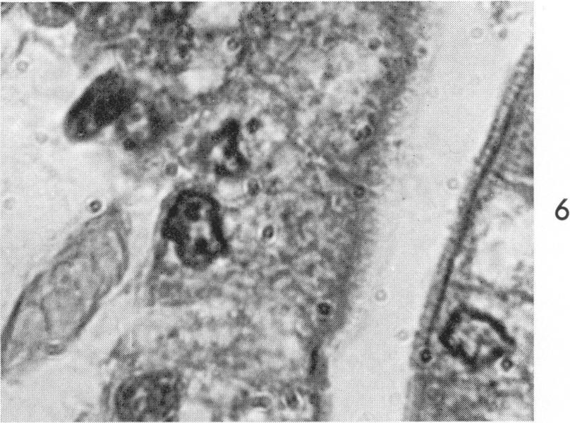
2 40 ADAMS AND KRAFT Vol. Sz, No. r alcohols and embedded in methacrylate. Sections were cut with a glass or diamond knife on a Sorvall Porter-Blum type microtome, stained with lead hydroxide10 or uranyl acetate-lead citrate1' and examined in an RCA EMU 3G or Siemens Elmiskop i electron microscope. Material for light microscopy was fixed in Bouin's fixative, or in Zenker's fixative with or without acetic acid, and embedded in paraffin; routine sections were stained with hematoxylin and eosin, Masson's trichrome stain, Laidlaw's inclusion-body stain,'2 alcian green stain for mucin, or Giemsa stain. RESULTS Light Microscopy One of the distinguishing features of this disease was the total lack of a cellular inflammatory exudate, even in severely affected portions of the bowel (Fig. i-8). The morphologic changes appeared to be limited exclusively to the extreme tips of the villi, which became bulbous as a result both of congestion of the small blood vessels and dilatation, in some cases extreme, of the lymphatics. Changes in the epithelial cells were limited to those covering the tips of the villi. The least severe changes consisted of a fine vacuolation affecting the cytoplasm of the cell between the nucleus and the striate border. This vacuolation had a fine vesicular appearance in the least affected cells, whereas in those most severely affected, large empty vesicles occupied the entire cytoplasm with concomitant disappearance of the usual complement of cytoplasmic organelles and matrix. The striate border remained intact in cells with mild and moderate stages of vacuolation, but when the latter was extreme, the border became greatly attenuated and finally disappeared. The nuclei of affected cells were relatively normal in appearance except where cytoplasmic vacuolation was extreme. Under this condition, the nuclei assumed a shrunken and pyknotic appearance. Large quantities of cellular debris could be seen within the lumen of the affected intestine, undoubtedly the result of massive sloughing of the epithelial cells. The extensive fluid losses accompanying the diarrhea that characterizes this disease would appear to result from the extensive epithelial sloughing at the dilated edematous tips of the villi. Cytoplasmic acidophilic inclusion bodies similar to those described by Pappenheimer 4 were seen (Fig. 6-8). These inclusions were usually circular or slightly ellipsoidal in cross section and measured from i to 4 1 in their greatest dimension. They were demonstrable with most of the stains employed but were most easily visualized in sections stained with Laidlaw's inclusion-body stain.'2 There appeared to be no preferential position for these cytoplasmic inclusions, approximately equal numbers being found in the juxtanuclear (Fig. 6), supranuclear (Fig. 7), and infranuclear (Fig. 8) positions. Intranuclear inclusions were never seen.

3 July I967 EDIM INFECTION 41 Electron Microscopy The least severe changes noted (Fig. 9) consisted of simple dilation of both smooth- and rough-surfaced endoplasmic reticulum. These dilated sacs of reticulum always contained varying quantities of a stippled, finely granular precipitate. Accompanying the dilatation were accumulations of lipid material within the sacs of reticulum (Fig. 9-I1). When the dilatation of endoplasmic reticulum and the accompanying deposition of lipid was rather severe, recognizable virus particles could be seen within the dilated sacs (Fig. 9-II and I3). The nucleus, mitochondria, and other cytoplasmic organelles of affected cells showed little if any morphologic change except when the cell had been converted to a virtual bag of lipid with virus particles sequestered in contiguous hugely dilated sacs of endoplasmic reticulum (Fig. i i). The microvilli remained unchanged in cells minimally or moderately involved (Fig. 9 and io) but were short, thick, and irregular in more severely affected cells (Fig. ii and I2). The Golgi apparatus was never significantly involved, the only notable change being minimal dilation in severely involved cells. Virus was never seen in Golgi vesicles. The material examined at a given time interval after infection contained cells with a spectrum of morphologic change varying from those indistinguishable from normal through a series of affected cells displaying severe morphologic changes. In general, the morphologic spectrum tended to correspond to the spatial distribution of the cells. There appeared to be a gradient of cellular susceptibility to infection, as judged by morphologic response, increasing in sensitivity from crypt to tip of villus (Fig.2 andi2). The majority of virus particles visualized in the infected cells were present within vesicles. These vesicles varied considerably in size, in some cases being as large as or larger than the nucleus of the cell (Fig. IO, ii, and I3), and by virtue of the ribosomes attached to their outer aspect (Fig. I3, I5, I7, and I8) could be identified as elements of the rough-surfaced reticulum. The virus particles were in many cases associated with the membrane of the endoplasmic reticulum in such a way as to suggest formation in the cytoplasm outside the endoplasmic reticulum (Fig. 21). In the electron micrographs particles appeared to protrude into the lumen of the endoplasmic reticulum, becoming invested in the process by an external coat apparently derived from the endoplasmic reticulum membrane (Fig. I4-2 i). A somewhat puzzling feature was the lack of such an external envelope on some of the intravesicular particles (Fig. 20 and 2I). This suggested that entrance into the vesicle of endoplasmic reticu-

4 42 ADAMS AND KRAFT Vol. SI, No. z lum could be gained without such investiture or that the membranederived outer envelope of the virus particle was variable or transient in nature. The distribution of virus-containing vesicles within the cytoplasm of infected cells was uniform throughout the cell. They were seen in the infranuclear, paranuclear, and supranuclear regions with approximately equal freuency. The acidophilic inclusion bodies seen in the light microscope could not be identified with certainty in the electron micrographs and hence could not be related to any ultrastructural component of infected cells. It is pertinent that the frequency of these acidophilic inclusions as judged from the paraffin sections was considerably less than the frequency of virus-containing cells as judged from the electron micrographs. This strongly suggests that, whatever their nature, the inclusions were not merely virus-filled vesicles. A number of electron micrographs, one of which is shown in Fig. I2, indicated that liberation of virus particles into the intestinal lumen occurred by way of rupture of afflicted cells with the disgorgement of both ruptured and intact virus-containing vesicles as well as large quantities of the virus-associated lipid into the intestinal lumen. A summary of the manifold morphologic variations, both of particle morphology and of the particles' relationships to the rough-surfaced reticulum, is presented in Fig. I3 through 22. The general particle form (Type i) averaged 8o mt in diameter and was circular in profile, suggesting that the particles were in most cases spheres; however, rare elongated or ellipsoidal forms were noted (Fig. i9). A typical round particle (Fig. I4) was composed of a circular, centrally located, dense core or nucleoid surrounded in turn by 2 sets of double membranes, the outer set of which could be seen to be in continuity with the vesicle membrane in the case of those particles that were protruding into the vesicle or were still attached to the membrane by a pedicle (Fig. I4-I 7, and I9). Most particles were present within vesicles of rough-surfaced reticulum, but occasional condensations of dense granular material occurred within the cytoplasm proper in which could be seen the faint outlines of round virus particles, possibly in early stages of formation (Fig. 2I). Other studies have indicated that similar cytoplasmic aggregates are nascent viroplasm, and the present material gave no indication that another interpretation is necessary here. In some cases virus particles were identified within dilated vesicles of endoplasmic reticulum that were in direct continuity with the nuclear envelope (Fig. i8); however, particles were never seen within the nucleus proper. A second class of particles (Type 2), averaging 65 mfa in diameter,

5 July I967 EDIM INFECTION 43 was occasionally seen (Fig. I7 and 20). These were similar to the first particle type except for their lack of the outer double membrane. Whether these particles had become divested of this outer coat achieved on passage through the vesicle membrane or whether they had simply failed to acquire such an outer vesicle-derived membrane was not suggested by any of the electron micrographs. A third possibility, of course, is that the latter smaller particles have become sequestered in a vesicle under unfavorable conditions and are either degenerating or have been altered in development in some way. The possibility that they are degenerating is suggested by the fact that in many cases not only do these smaller particles lack an outer membrane, but threads of material, interpreted as the virus nucleoprotein, extend from the core of the virus through apparent morphologic defects in the virus inner shell (Fig. 20 and 22). A third and rare type of elongated or ellipsoidal particle is shown in Fig. I9. It is possible that these are nonviable forms or that they represent developmental aberrations. Virus or virus-associated lipid was never seen in sections of ileum and jejunum from uninfected mice. Such control epithelium was always normal in appearance, resembling the cell at the lower left in Fig. 9. SUMMARY A light- and electron-microscopic study of intestinal epithelium of infant mice infected with epizootic diarrhea of infant mice (EDIM) virus has shown that (i) intracellular virus is seen exclusively in the cytoplasm of infected cells; (2) virus replication is probably initiated in the cytoplasmic matrix, after which the particle appears to enter cisternae of the granular endoplasmic reticulum by a process of "budding;" (3) the endoplasmic reticulum of infected cells appears dilated and contains virus particles and virus-associated lipid; (4) some infected cells (probably the most severely affected) rupture, liberating large numbers of virus particles into the intestinal lumen; and (5) cells exhibit a gradient of susceptibility (as judged by morphologic response) to infection, increasing in sensitivity from crypt to tip of villus. REFERENCES I. CHEEVER, F. S., and MUELLER, J. H. Epidemic diarrheal disease of suckling mice. I. Manifestations, epidemiology, and attempts to transmit the disease. J Exp Med 85: , I PAPPENHEIMER, A. M., and ENDERS, J. F. An epidemic diarrheal disease of suckling mice. II. Inclusions in the intestinal epithelial cells. J Exp Med 85: , 1947.

6 44 ADAMS AND KRAFT Vol. 51, No. z 3. CHEEVER, F. S., and MUELLER, J. H. Epidemic diarrheal disease of suckling mice. III. The effect of strain, litter, and season upon the incidence of the disease. J Exp Med 88:309-3I6, I PAPPENHEIMER, A. M., and CHEEVER, F. S. Epidemic diarrheal disease of suckling mice. IV. Cytoplasmic inclusion bodies in intestinal epithelium in relation to the disease. J Exp Med 88:3I7-324, I RUNNER, M. N., and PALM, J. Factors associated with the incidence of infantile diarrhea in mice. Proc Soc Exp Biol Med 82:I47-I50, I CHEEVER, F. S. Epidemic diarrheal disease of suckling mice. Ann NYAcad Sci 66:I96-203, I KRAFT, L. M. Studies on the etiology and transmission of epidemic diarrhea of infant mice. J Exp Med I06: , I KRAFT, L. M. Observations on the control and natural history of epidemic diarrhea of infant mice (EDIM). Yale J Biol Med 3I:I2I-I37, I ADAMS, W. R., and KRAFT, L. M. Epizootic diarrhea of infant mice: Identification of the etiologic agent. Science 141: , I963. 1O. MILLONIG, G. A modified procedure for lead staining of thin sections. J Biophy Biochem Cytol zz: , I96I. ii. REYNOLDS, E. S. The use of lead citrate at high ph as an electronopaque stain in electron microscopy. J Cell Biol z7:208-2i2, I963. I2. PAPPENHEIMER, A. M., and HAWTHORNE, J. J. Certain cytoplasmic inclusions of liver cells. Amer J Path 12: , I936. I3. MORGAN, C., ROSE, H. M., HOLDEN, M., and JONES, E. P. Electron microscopic observations on the development of herpes simplex virus. J Exp Med zio: , I959. I4. MORGAN, C., ELLISON, S. A., RoSE, H. M., and MooRE, D. H. Structure and development of viruses observed in the electron microscope. II. Vaccinia and fowl pox viruses. J Exp Med 100:30I-308, I954. I5. BERNHARD, W., OBERLING, C., and VIGIER, P. Ultrastructure de virus dans le sarcome de Rous leur rapport avec le cytoplasme des cellules tumorales. Bull Ass Franc Cancer 43: , I956. i6. JENSON, A. B., RABIN, E. R., PHILLIPS, C. A., and MELNICK, J. L. Reovirus encephalitis in newborn mice: An electron microscopic and virus assay study. Amer J Path 47: , I965. The skilled technical assistance of Miss Lillemor Wailmark is gratefully acknowledged.
7 July I967 EDIM INFECTION 45 [ Illustrations follow ]
8 ADAMS AND KRAFT 46 Vol. 5r, No. z LEGENDS FOR FIGURES Figures I to 8 show intestinal epithelium (jejunum) of control and infected mice fixed in Bouin's solution, embedded in paraffin, and stained with Laidlaw's inclusionbody stain. FIG. I. Normal control. Columnar cells are uniform in size and appearance and extend from base of villi to tips. Stroma of microvilli is normal in appearance and there is little if any tendency to club formation of tips. Vacuoles are rare and small. X I8o. FIG. 2. Epithelium from infected mouse showing enlargement and vacuolation of cells over tip of villi, increased cellularity of stroma, and dilatation of stromal lymphatics. Submucosal edema is also present. X i8o. FIG. 3. Tip of villus from normal control showing features mentioned in Fig. i and also demonstrating uniform layer of microvilli and general uniformity and polarity of cells and nuclei. X 360. FIG. 4. Tip of intestinal villus from infected mouse, showing coarse and fine vacuolation and enlargement of cells over tip of villus and dilatation of lymphatics in stromal core. Layer of microvilli is irregular over tip and has disappeared over most severely affected cells. X 360. FIG. S. Portion of cross section of villus from normal control showing uniformity of microvilli, cytoplasm, and nuclei. X I335. FIG. 6 to 8. Cross sections of portions of villi from infected mice showing cytoplasmic inclusions. These may be distinguished from small particles in embedding media by halos surrounding the latter. No intranuclear inclusions were seen. Dark structures suggestive of inclusions, present in some nuclei, are interpreted as normal nucleoli. X 1335.


9 July I967 EDIM INFECTION
10 48 ADAMS AND KRAFT Vol. 51, No. I Figures 9 to 22 are electron micrographs of jejunal epithelium from infected mice. The tissues were fixed in veronal acetate-buffered i% osmium tetroxide and embedded in methacrylate. Sections were poststained with uranyl acetate and lead citrate. FIG. 9. Cross section of villus of infected mouse showing supranuclear and infranuclear vesicles of granular reticulum containing virus particles and irregular clumps of lipid material. Nuclei, cell organelles, and microvilli appear uninvolved. Central cell has both infranuclear and supranuclear vesicles that contain virus particles and lipid. It is flanked by 2 less-involved cells that contain lipid but no visible virus. Cell at extreme lower left is normal in appearance. X 8050.

11 July I967 EDIM INFECTION 49 9 AWA.f it- _... :ie.
12 so ADAMS AND KRAFT Vol. 5r, No. r FIG. IO. Somewhat higher-power view of infected epithelial cell showing more extensive involvement. Huge vesicles of endoplasmic reticulum comprise majority of cell volume and are filled with clumps of virus particles and irregular masses of lipid material. No alterations are seen in those cytoplasmic organelles that remain visible. Microvilli appear to be uninvolved. X I3,800.

13 July I 967 EDIM INFECTION A4-5I qwln- I hmo- i..s.,..,{ 10.1 r.;"iatv, Er 1.0 f,..14 li: s. tr, 1- L~,4_1.:.4.1i'a PO,'. 'AM16, t' -Wkt. d. ii:... -
14 52 ADAMS AND KRAFT Vol. 5.r, No. I FIG. II. Cross section of jejunal villus from infected mouse showing more severely involved cell at periphery of villus. This cell consists essentially of mass of vesicles containing large quantities of lipid material and clumps of virus particles. Few indentifiable organelles are seen. Mitochondria appear somewhat swollen, but there is minimal loss of cristae. Microvilli have been greatly attenuated. Both adjacent cells, though less severely involved, contain clumps of virus particles and dilated vesicles. X 8i6o.

15 July I967 EDIM INFECTION 53 11
16 54 ADAMS AND KRAFT Vol. 5I, No. z FIG. I2. Cross section of jejunal villus from infected mouse showing formation of microbullae and liberation of cytoplasmic contents, including ruptured vesicles, free virus particles, virus-containing vesicles, and virus-associated lipid material, into lumen of intestine. X I6,900.

17 July' I967 EDIM INFECTION : 77T i &. 12 or, :14r :.1 l.. A.. 11 Fi W,U O..W i..7t. V 1. 0 riab :.JR.4.z* V. il, 0
18 56 ADAMS AND KRAFT Vol. 5r, No. r Figures I3 to 22 show portions of cytoplasm of intestinal epithelial cells from infected mice showing 3 different particle types and varied membrane relationships of virus particles. FIG. I3. Portion of jejunal epithelium cell from infected mouse showing particularly well-developed dilated vesicle of rough-surfaced reticulum (lower center) containing virus-associated lipid and large number of virus particles. Some smaller virus-containing vesicles are present. No changes in nucleus, mitochondria, or microvilli are seen. X 24,000.

19 July 1967 EDIM INFECTION 57 13

20 ADAMS AND KRAFT Vol. 5.r, No. I FIG. I4. Virus particles of first type, which have a generally circular profile and a diameter of 8o m,u. Most are probably spherical. Each has outer membrane composed of typical double layer measuring about 70 A in width. Particles all located within vesicle of dilated rough-surfaced reticulum. Portion of cytoplasm (upper right) contains virus particle that impinges on membrane at surface of vesicle with protrusion into vesicle lumen (extreme right). X 84,ooo. FIG. I5. Dilated vesicle of rough-surfaced reticulum with single virus particle still attached to vesicle membrane and protruding almost entirely into vesicle lumen. Present within vesicle is irregular dumbbell-shaped mass of virus-associated lipid and numerous fine electron-dense strands of material. x 6o,ooo. FIG. 16. Two vesicles of granular reticulum. Vesicle on left contains virus particle whose outer membrane is still continuous with vesicle membrane. Vesicle on right contains lipid material and usual irregular electron-dense strands of material. Mitochondrion at top is normal in appearance. X 6o,ooo. FIG. I7. Vesicle of rough-surfaced reticulum containing "emerging" virus particle (top) and 3 virus particles of lesser diameter (second particle type in Fig. 20). X 56,ooo. FIG. I8. Virus particles within dilated vesicle of endoplasmic reticulum that is in continuity with outer membrane of nucleus in upper right. X 52,000. FIG. I9. Third type of particle, occurring only sporadically, consisted of elongated and ellipsoidal forms (center and right). Rarity of these forms suggests that they may not be functional biological forms but may represent aberrations of development. X 84,000. FIG. 20. Dilated vesicle of endoplasmic reticulum occupies lower two-thirds of figure. Rim of cytoplasm can be seen along top border. In addition to Type I particles (Fig. I5), there are numerous smaller particles that lack outer double membrane (Type 2 particles), measure 65 mu in diameter, and have slightly more irregular structure than Type I particle, suggesting that they may be abnormal forms. X 6o,ooo. FIG. 2I. Possible formation of virus particles within cytoplasm immediately adjacent to dilated vesicle of endoplasmic reticulum (left and extreme top center). Central portion of figure shows faint circular particles measuring about 50 mu in diameter interspersed in granular deposits of dense material. Granules in latter material are smaller than ribosomes, 3 or 4 clumps of which are at extreme right. Virus particles imping upon membrane of vesicle and protrude into its lumen. X 6o,ooo.


21 July I967 EDIM INFECTION * *-m W_ ^ l ,, 0. ~t ' *tv K b-...i,, qr i '... l~.-j. Ir Mvi.,..4 f IA..., ': i: : w h 20 21


22 4- X. -.. il p- 1. ".. ;..... s ;..7..L. *ie,. I I Y...t., ",.0-22 s i t., 4.,.R R.5 l's...:.' 5 {. i:,'.. $...s.. 4.S *'' tv K *23. D.> FIG. 22. Type 2 particles within vesicle of rough-surfaced reticulum. A thin rim of cytoplasm extends along right-hand border, and edge of mitochondrion is also visible (lower right). Several particles contain morphologic defects in their outer coat and protruding threads of material about 30 A in width, interpreted as viral nucleic acid. Some ribosomes stud surface of vesicle membrane traversing figure vertically along right side, thus identifying it as part of granular reticulum. X I68,ooo.
ELECTRON MICROSCOPIC STUDY OF THE FORMATION OF BLUETONGUE VIRUS*
 Onderstepoort J. vet. Res. (1968), 35 (1), 139-150 Printed in the Repub. of S. Afr. by The Government Printer, Pretoria ELECTRON MICROSCOPIC STUDY OF THE FORMATION OF BLUETONGUE VIRUS* G. LECATSAS, Veterinary
(From The Rockefeller Institute) Materials and Methods. Observations with the Electron Microscope
 Materials and Methods. Observations with the Electron Microscope") ELECTRON MICROSCOPE STUDY OF THE DEVELOPMENT OF THE PAPILLOMA VIRUS IN THE SKIN OF THE RABBIT* BY ROBERT S. STONE,~ M.D., RICHARD E. SHOPE, M.D., DAN H. MOORE, P,~.D. (From The Rockefeller Institute) PLATES
ELECTRON MICROSCOPE STUDY OF THE DEVELOPMENT OF THE PAPILLOMA VIRUS IN THE SKIN OF THE RABBIT* BY ROBERT S. STONE,~ M.D., RICHARD E. SHOPE, M.D., DAN H. MOORE, P,~.D. (From The Rockefeller Institute) PLATES
The Fine Structure of the Epithelial Cells of the Mouse Prostate* II. Ventral Lobe Epithelium
 Published Online: 1 June, 1960 Supp Info: http://doi.org/10.1083/jcb.7.3.511 Downloaded from jcb.rupress.org on September 28, 2018 The Fine Structure of the Epithelial Cells of the Mouse Prostate* II.
Electron Microscope Studies of HeLa Cells Infected with Herpes Virus
 244 STOKER, M. G. P., SMITH, K. M. & Ross, R. W. (1958). J. gen. Microbiol. 19,244-249 Electron Microscope Studies of HeLa Cells Infected with Herpes Virus BY M: G. P. STOKER, K. M. SMITH AND R. W. ROSS
ELECTRON MICROSCOPIC STUDIES ON EQUINE ENCEPHALOSIS VIRUS
 Onderstepoort]. vet. Res. 40 (2), 53-58 (1973) ELECTRON MICROSCOPIC STUDIES ON EQUINE ENCEPHALOSIS VIRUS G. LECATSAS, B. J. ERASMUS and H. J. ELS, Veterinary Research Institute, Onderstepoort ABSTRACT
From the Bland-Sutton Institute of Pathology, Middlesex Hospital Medical School, London, England
 Published Online: 1 March, 1962 Supp Info: http://doi.org/10.1083/jcb.12.3.589 Downloaded from jcb.rupress.org on July 15, 2018 OBSERVATIONS ON THE MODE OF RELEASE OF HERPES VIRUS FROM INFECTED H~LA CELLS
New aspect of hepatic nuclear glycogenosis
 J. clin. Path. (1968), 21, 19 New aspect of hepatic nuclear glycogenosis in diabetes1 F. CARAMIA, F. G. GHERGO, C. BRANCIARI, AND G. MENGHINI From the Institute of General Pathology, University of Rome,
Some Observations on the Fine Structure of the Goblet Cells. Special Reference to the Well-Developed Agranular Endoplasmic Reticulum
 Okajimas Folia Anat. Jpn., 58(4-6) : 583-594, March 1982 Some Observations on the Fine Structure of the Goblet Cells in the Nasal Respiratory Epithelium of the Rat, with Special Reference to the Well-Developed
AN ELECTRON-MICROSCOPIC STUDY OF THE STARCH-CONTAINING PLASTIDS IN THE FERN TODEA BARBARA
 J. Cell Sci. 4, 211-221 (1969) 211 Printed in Great Britain AN ELECTRON-MICROSCOPIC STUDY OF THE STARCH-CONTAINING PLASTIDS IN THE FERN TODEA BARBARA H. M. SMITH* AND D. S. SMITHf Department of Biology,
Published Online: 25 November, 1956 Supp Info: on November 16, 2018 jcb.rupress.org Downloaded from
 Published Online: 25 November, 1956 Supp Info: http://doi.org/10.1083/jcb.2.6.799 Downloaded from jcb.rupress.org on November 16, 2018 B~IEF NOrmS 799 Permanganate--A New Fixative for Electron Microscopy.*
the structure of their ducts has been
 Tza JOURNAL 0? INVEa'riGATrVN DEBMATOLOOT Copyright t 1966 by The Williams & Wilkins Co. Vol. 46, No. I Printed in U.S.A. AN ELECTRON MICROSCOPIC STUDY OF THE ADULT HUMAN APOCRINE DUCT* KEN HASHIMOTO,
Ultrastructure of Connective Tissue Cells of Giant African Snails Achatina fulica (Bowdich)
") Kasetsart J. (Nat. Sci.) 36 : 285-290 (2002) Ultrastructure of Connective Tissue Cells of Giant African Snails Achatina fulica (Bowdich) Viyada Seehabutr ABSTRACT The connective tissue sheath of cerebral
Kasetsart J. (Nat. Sci.) 36 : 285-290 (2002) Ultrastructure of Connective Tissue Cells of Giant African Snails Achatina fulica (Bowdich) Viyada Seehabutr ABSTRACT The connective tissue sheath of cerebral
Initially, the patients did not receive extra vitamin E except for a very
 EFFECT OF VITAMIN E ON MEMBRANES OF THE INTESTINAL CELL BY I. MOLENAAR, F. A. HOMMES, W. G. BRAAMS, AND H. A. POLMAN CENTER FOR MEDICAL ELECTRON MICROSCOPY AND DEPARTMENT OF PEDIATRICS, UNIVERSITY OF GRONINGEN,
SBI3U7 Cell Structure & Organelles. 2.2 Prokaryotic Cells 2.3 Eukaryotic Cells
 SBI3U7 Cell Structure & Organelles 2.2 Prokaryotic Cells 2.3 Eukaryotic Cells No nucleus Prokaryotic Cells No membrane bound organelles Has a nucleus Eukaryotic Cells Membrane bound organelles Unicellular
10/13/11. Cell Theory. Cell Structure
 Cell Structure Grade 12 Biology Cell Theory All organisms are composed of one or more cells. Cells are the smallest living units of all living organisms. Cells arise only by division of a previously existing
IT has been shown (Chou, 1957 a, b) that there are three kinds of lipid
 that there are three kinds of lipid") 279 The Ultra-fine Structure of Lipid Globules in the Neurones of Helix aspersa By J. T. Y. CHOU and G. A. MEEK (From the Cytological Laboratory, Department of Zoology; and Department of Human Anatomy,
279 The Ultra-fine Structure of Lipid Globules in the Neurones of Helix aspersa By J. T. Y. CHOU and G. A. MEEK (From the Cytological Laboratory, Department of Zoology; and Department of Human Anatomy,
ON THE PRESENCE OF A CILIATED COLUMNAR EPITHELIAL CELL TYPE WITHIN THE BOVINE CERVICAL MUCOSA 1
 ON THE PRESENCE OF A CILIATED COLUMNAR EPITHELIAL CELL TYPE WITHIN THE BOVINE CERVICAL MUCOSA 1 R. I. Wordinger, 2 J. B. Ramsey, I. F. Dickey and I. R. Hill, Jr. Clemson University, Clemson, South Carolina
Cell Overview. Hanan Jafar BDS.MSc.PhD
 Cell Overview Hanan Jafar BDS.MSc.PhD THE CELL is made of: 1- Nucleus 2- Cell Membrane 3- Cytoplasm THE CELL Formed of: 1. Nuclear envelope 2. Chromatin 3. Nucleolus 4. Nucleoplasm (nuclear matrix) NUCLEUS
Yara shwabkeh. Osama Alkhader. Heba Kalbouneh
 2 Yara shwabkeh Osama Alkhader Heba Kalbouneh CELL OVERVIEW -Note ; the important thing is to know how the organelles appear under the microscope - the stains we usually use in Histology are composed of
COMPARATIVE DISTRIBUTION OF CARBOHYDRATES AND LIPID DROPLETS IN THE GOLGI APPARATUS OF INTESTINAL ABSORPTIVE CELLS
 COMPARATIVE DISTRIBUTION OF CARBOHYDRATES AND LIPID DROPLETS IN THE GOLGI APPARATUS OF INTESTINAL ABSORPTIVE CELLS JEAN A. SAGE and RALPH A. JERSILD, JR. Medical Center, Indianapolis, Indiana 46202 From
ELECTRON MICROSCOPY OF A SMALL PIGMENTED CUTANEOUS LESION*
 ELECTRON MICROSCOPY OF A SMALL PIGMENTED CUTANEOUS LESION* The description of the lesion in the title of this rcport is intentionally non-committal. Diagnosed clinically as a lentigo, it was removed as
The Cytoplasm Li Shulei Department of Histology & Embryology
 The Cytoplasm Li Shulei lishulei@tom.com Department of Histology & Embryology Cell components Cytoplasm Plasma membrane Organelles Cytoplasmic deposits Cytoskeleton Cytosol ( Matrix ) Nucleus Plasma membrane
Eukaryotic cells are essentially two envelope systems. Nuclear materials are separated from cytoplasm by nuclear membrane. Complex structure Also
 Dr. Gugale Pritesh Ramanlal M.Sc., Ph.D., B.Ed., D.M.L.T. Email id - pritesh.gugale09@gmail.com Contact numbernumber- 8446475310 Eukaryotic cells are essentially two envelope systems. Nuclear materials
ELECTRON MICROSCOPIC STUDIES ON REOVIRUS TYPE I IN BHK 21 CELLS
 Onderstepoort J. vet. Res. (1968), 35 (1), 151-158 Printed in the Republic of S. Afr. by the Government Printer, Pretoria ELECTRON MICROSCOPIC STUDIES ON REOVIRUS TYPE I IN BHK 21 CELLS G. LECATSAS, Veterinary
Cell Structure. Present in animal cell. Present in plant cell. Organelle. Function. strength, resist pressure created when water enters
 Cell Structure Though eukaryotic cells contain many organelles, it is important to know which are in plant cells, which are in animal cells and what their functions are. Organelle Present in plant cell
Acta Medica Okayama. Electron microscopic demonstration of a new virus isolated from a patient with SMON. Zensuke Ota DECEMBER 1970
 Acta Medica Okayama Volume 24, Issue 6 1970 Article 3 DECEMBER 1970 Electron microscopic demonstration of a new virus isolated from a patient with SMON Zensuke Ota Okayama University, Copyright c 1999
psittaci by Silver-Methenamine Staining and
 JOURNAL OF BACTERIOLOGY, July 1972, p. 267-271 Copyright 1972 American Society for Microbiology Vol. 111, No. 1 Printed in U.S.A. Location of Polysaccharide on Chlamydia psittaci by Silver-Methenamine
Silver-Impregnation of the Golgi Complex in Epididymal Epithelial Cells of Mice
 CELL STRUCTURE AND FUNCTION 8, 339-346 (1984) C by Japan Society for Cell Biology Silver-Impregnation of the Golgi Complex in Epididymal Epithelial Cells of Mice Ikuo Yamaoka, Sumie Katsuta and Yoshimi
QUESTIONSHEET 1. The diagram shows some of the cell structures involved in the secretion of an extracellular enzyme. C D
 QUESTIONSHEET 1 The diagram shows some of the cell structures involved in the secretion of an extracellular enzyme. C D A (a) Identify A,, C, and D. A:... :... C:... D:... [4] (b) Outline the role of each
The ultrastructure of the egg and
 The ultrastructure of the egg and central cell of Petunia J.L. van Went Botanisch Laboratorium, Universiteit, Nijmegen SUMMARY The egg and central cell of Petunia hybrida undergo a number of changes and
AN ELECTRON MICROSCOPE STUDY OF THE MORPHOLOGY AND DISTRIBUTION OF THE INTRACYTOPLASMIC "VIRUS-LIKE" PARTICLES OF EHRLICH ASCITES TUMOR CELLS*
 AN ELECTRON MICROSCOPE STUDY OF THE MORPHOLOGY AND DISTRIBUTION OF THE INTRACYTOPLASMIC "VIRUS-LIKE" PARTICLES OF EHRLICH ASCITES TUMOR CELLS* BY W. ROBERT ADAMS,~ M.D., AND ALFRED M. PRINCE, M.D. (From
Chapter 7. (7-1 and 7-2) A Tour of the Cell
 A Tour of the Cell") Chapter 7 (7-1 and 7-2) A Tour of the Cell Microscopes as Windows to the World of Cells Cells were first described in 1665 by Robert Hooke. By the mid-1800s, the accumulation of scientific evidence led
Chapter 7 (7-1 and 7-2) A Tour of the Cell Microscopes as Windows to the World of Cells Cells were first described in 1665 by Robert Hooke. By the mid-1800s, the accumulation of scientific evidence led
Structures in Cells. Cytoplasm. Lecture 5, EH1008: Biology for Public Health, Biomolecules
 Structures in Cells Lecture 5, EH1008: Biology for Public Health, Biomolecules Limian.zheng@ucc.ie 1 Cytoplasm Nucleus Centrioles Cytoskeleton Cilia Microvilli 2 Cytoplasm Cellular material outside nucleus
Cell Structure and Function. Biology 12 Unit 1 Cell Structure and Function Inquiry into Life pages and 68-69
 Cell Structure and Function Biology 12 Unit 1 Cell Structure and Function Inquiry into Life pages 45 59 and 68-69 Assignments for this Unit Pick up the notes/worksheet for this unit and the project There
Epstein-Barr Virus: Stimulation By 5 '-Iododeoxy uridine or 5 '-Brom odeoxy uridine in Human Lymphoblastoid Cells F ro m a Rhabdom yosarcom a*
 A n n a ls o f C l i n i c a l L a b o r a t o r y S c i e n c e, Vol. 3, No. 6 Copyright 1973, Institute for Clinical Science Epstein-Barr Virus: Stimulation By 5 '-Iododeoxy uridine or 5 '-Brom odeoxy
STUDIES OF THE HUMAN UNFERTILIZED TUBAL OVUM*t
 FERTILITY AND STERILITY Copyright @ 1973 by The Williams & Wilkins Co. Vol. 24, No.8, August 1973 Printed in U.S.A. STUDIES OF THE HUMAN UNFERTILIZED TUBAL OVUM*t C. NORIEGA, M.D., AND C. OBERTI, M.D.
Structures in Cells. Lecture 5, EH1008: Biology for Public Health, Biomolecules.
 Structures in Cells Lecture 5, EH1008: Biology for Public Health, Biomolecules Limian.zheng@ucc.ie 1 Cytoplasm Nucleus Centrioles Cytoskeleton Cilia Microvilli 2 Cytoplasm Cellular material outside nucleus
(Plates LXVIII-LXXI)
") [GANN, 54, 481-486; December, 1963] UDC 616.155.392-076.4:578.69 VIRUS-LIKE PARTICLES IN HUMAN CHLOROLEUKEMIA CELLS (Plates LXVIII-LXXI) Zensuke OTA, Shin-ya SUZUKI, and Satoru HIGASHI (Department of Internal
[GANN, 54, 481-486; December, 1963] UDC 616.155.392-076.4:578.69 VIRUS-LIKE PARTICLES IN HUMAN CHLOROLEUKEMIA CELLS (Plates LXVIII-LXXI) Zensuke OTA, Shin-ya SUZUKI, and Satoru HIGASHI (Department of Internal
Basophilic. Basophilic structures are stained by basic dyes: Mnemonic: Basophilic = Blue
 Cell Overview Basophilic Basophilic structures are stained by basic dyes: Basic dyes are positive Basophilic structures are negative (ex. DNA, RNA, ribosomes, RER) Mnemonic: Basophilic = Blue Acidophilic
Mansoura university Faculty of medicine Histology and cell Biology Department Curriculum Content And Logbook
 Mansoura university Faculty of medicine Histology and cell Biology Department Curriculum Content And Logbook For the 1 st year Medical Student s In Histology and cell Biology Mansoura university Faculty
TRANSFER OF PREMELANOSOMES INTO THE KERATINIZING CELLS OF ALBINO HAIR FOLLICLE
 TRANSFER OF PREMELANOSOMES INTO THE KERATINIZING CELLS OF ALBINO HAIR FOLLICLE PAUL F. PARAKKAL. From the Department of Dermatology, Boston University School of Medicine, Boston, Massachusetts 02118 INTRODUCTION
Gastroenteritis and viral infections
 Gastroenteritis and viral infections A Large number of viruses are found in the human gut; these include some that are associated with gastroenteritis Rotaviruses Adenoviruses 40/41 Caliciviruses Norwalk-like
Relationship of Ehrlichia canis-infected Mononuclear Cells to Blood Vessels of Lungs1
 INFECTION AND IMMUNITY, Sept. 1974, p. 590-596 Copyright 0 1974 American Society for Microbiology Vol. 10, No. 3 Printed in U.S.A. Relationship of Ehrlichia canis-infected Mononuclear Cells to Blood Vessels
Cell Cell
 Go to cellsalive.com. Select Interactive Cell Models: Plant and Animal. Fill in the information on Plant and Animal Organelles, then Click on Start the Animation Select Plant or Animal Cell below the box.
Eukaryotic cells contain organelles that allow the specializations and the separation of functions within the cell.
 Section 3: Eukaryotic cells contain organelles that allow the specializations and the separation of functions within the cell. K What I Know W What I Want to Find Out L What I Learned Essential Questions
Eukaryotic Cell Structures
 Comparing the Cell to a Factory Eukaryotic Cell Structures Structures within a eukaryotic cell that perform important cellular functions are known as organelles. Cell biologists divide the eukaryotic cell
ARANEUS SERICATUS CHANGES IN FINE STRUCTURE DURING SILK PROTEIN PRODUCTION IN THE AMPULLATE GLAND OF THE SPIDER. ALLEN L. BELL and DAVID B.
 Published Online: 1 July, 1969 Supp Info: http://doi.org/10.1083/jcb.42.1.284 Downloaded from jcb.rupress.org on September 18, 2018 CHANGES IN FINE STRUCTURE DURING SILK PROTEIN PRODUCTION IN THE AMPULLATE
The Microscopic World of Cells. The Microscopic World of Cells. The Microscopic World of Cells 9/21/2012
 Organisms are either: Single-celled, such as most prokaryotes and protists or Multicelled, such as plants, animals, and most fungi How do we study cells? Light microscopes can be used to explore the structures
Small intestine. Small intestine
 General features Tubular organ longest part; 5-6 m most of chemical digestion absorption of nutrients reabsorption of H2O occurs. Two structural features; maximize the lumenal surface area villi microvilli
R,;habdomyosarcoma, the most common
 Fine-structural classification of orbital rhabdomyosarcoma Arnold J. Kroll Six cases of orbital rhabdomyosarcoma were studied with the electron microscope. Tumor cells (rhabdomyoblasts) could be classified
CELL PART OF THE DAY. Chapter 7: Cell Structure and Function
 CELL PART OF THE DAY Chapter 7: Cell Structure and Function Cell Membrane Cell membranes are composed of two phospholipid layers. Cell membrane is flexible, not rigid The cell membrane has two major functions.
A Tour of the Cell. Chapter 6. Biology Eighth Edition Neil Campbell and Jane Reece. PowerPoint Lecture Presentations for
 Chapter 6 A Tour of the Cell PowerPoint Lecture Presentations for Biology Eighth Edition Neil Campbell and Jane Reece Lectures by Chris Romero, updated by Erin Barley with contributions from Joan Sharp
Ultrastructural Study of Human Natural Killer CNK) Cell*)
 Cell*)") Hiroshima Journal of Medical Sciences Vol. 31, No. 1, March, 1982 HJIM 31-6 31 Ultrastructural Study of Human Natural Killer CNK) Cell*) Yoshinori KAWAGUCHI, Eishi KITTAKA, Yoshito TANAKA, Takeo TANAKA
Hiroshima Journal of Medical Sciences Vol. 31, No. 1, March, 1982 HJIM 31-6 31 Ultrastructural Study of Human Natural Killer CNK) Cell*) Yoshinori KAWAGUCHI, Eishi KITTAKA, Yoshito TANAKA, Takeo TANAKA
Coronaviruses cause acute, mild upper respiratory infection (common cold).
.") Coronaviruses David A. J. Tyrrell Steven H. Myint GENERAL CONCEPTS Clinical Presentation Coronaviruses cause acute, mild upper respiratory infection (common cold). Structure Spherical or pleomorphic enveloped
Coronaviruses David A. J. Tyrrell Steven H. Myint GENERAL CONCEPTS Clinical Presentation Coronaviruses cause acute, mild upper respiratory infection (common cold). Structure Spherical or pleomorphic enveloped
Human height. Length of some nerve and muscle cells. Chicken egg. Frog egg. Most plant and animal cells Nucleus Most bacteria Mitochondrion
 10 m 1 m 0.1 m 1 cm Human height Length of some nerve and muscle cells Chicken egg Unaided eye 1 mm Frog egg 100 µm 10 µm 1 µm 100 nm 10 nm Most plant and animal cells Nucleus Most bacteria Mitochondrion
First to View Cells. copyright cmassengale
 CELL THEORY All living things are made of cells Cells are the basic unit of structure and function in an organism (basic unit of life) Cells come from the reproduction of existing cells (cell division)
By: Brooke Sheppard
 By: Brooke Sheppard What is a Cell? Cells are the basic structure of life for all organisms. Cells are microscopic, which means we can only view cells under a microscope. There are animal cells and plant
Cells. Variation and Function of Cells
 Cells Variation and Function of Cells Cell Theory states that: 1. All living things are made of cells 2. Cells are the basic unit of structure and function in living things 3. New cells are produced from
Yara Saddam. Amr Alkhatib. Ihsan
 1 Yara Saddam Amr Alkhatib Ihsan NOTE: Yellow highlighting=correction/addition to the previous version of the sheet. Histology (micro anatomy) :- the study of tissues and how they are arranged into organs.
Endomembrane system, *Chloroplasts, *Mitochondria. *Learn these from text/connect1. Fertilization of a human cell
 Key Concepts: - Cells are the Basic Unit of Life Cell Theory, Surface to Volume - 2 Cell Types Prokaryotic, Eukaryotic - Cell Membrane Membrane Structure - Cell Organelles Endomembrane system, *Chloroplasts,
:1c.c :& Preliminary and Short Report GRANULE FORMATION IN THE LANGERHANS CELL* structure with rounded ends and a striated lamella
 THE JOURNAL OF INVESTIGATIVE DERMATOLOGY Copyright 1566 by The Williams & Wilkins Co. Vol. 7, No. 5 Printed in U.S.A. Preliminary and Short Report GRANULE FORMATION IN THE LANGERHANS CELL* ALVIN S. ZELICKSON,
Cell Structure & Function. Source:
 Cell Structure & Function Source: http://koning.ecsu.ctstateu.edu/cell/cell.html Definition of Cell A cell is the smallest unit that is capable of performing life functions. http://web.jjay.cuny.edu/~acarpi/nsc/images/cell.gif
1) All organisms are made up of one or more cells and the products of those cells.
 All organisms are made up of one or more cells and the products of those cells.") CELL ORGANELLES - NOTES CELL THEORY Cells are the basic unit of life. The Cell Theory states that: 1) All organisms are made up of one or more cells and the products of those cells. 2) All cells carry
CELL ORGANELLES - NOTES CELL THEORY Cells are the basic unit of life. The Cell Theory states that: 1) All organisms are made up of one or more cells and the products of those cells. 2) All cells carry
Cells & Cell Transport. Cells
 Cells & Cell Transport Cells Cell Membrane Cell membrane (plasma membrane): a phospholipid bilayer surrounding the cell Each phospholipid has a polar phosphate head and lipid tails Selectively permeable:
A Tour of the Cell. Chapter 6. Biology. Edited by Shawn Lester. Inner Life of Cell. Eighth Edition Neil Campbell and Jane Reece
 Chapter 6 A Tour of the Cell Inner Life of Cell Edited by Shawn Lester PowerPoint Lecture Presentations for Biology Eighth Edition Neil Campbell and Jane Reece Lectures by Chris Romero, updated by Erin
4 A Tour of the Cell CAMPBELL BIOLOGY IN FOCUS. Urry Cain Wasserman Minorsky Jackson Reece
 CAMPBELL BIOLOGY IN FOCUS Urry Cain Wasserman Minorsky Jackson Reece 4 A Tour of the Cell Lecture Presentations by Kathleen Fitzpatrick and Nicole Tunbridge Overview: The Fundamental Units of Life All
Objectives. To determine the differences between plant and animal cells To discover the structure and function of cellular organelles.
 Cell Organelles 3.2 Objectives To determine the differences between plant and animal cells To discover the structure and function of cellular organelles. Basic Cellular Structures Cell membrane (cytoplasmic
African Trypanosomes
 African Trypanosomes Giemsa-stained blood smear of African trypanosomes viewed under the 100X objective lens. The block arrows denote trypomastigote forms of the African trypanosomes found within the blood
Chapter 2 Cell. Zhou Li Prof. Dept. of Histology and Embryology
 Chapter 2 Cell Zhou Li Prof. Dept. of Histology and Embryology The inner life of the cell Ⅰ. Plasma membrane (Plasmalemma) 1.1 The structure Unit membrane: inner layer 3-layered structure outer layer mediat
Electron Microscopy of Small Cells: Mycoplasma hominis
 JOURNAL of BAcTRiowOY, Dc. 1969, p. 1402-1408 Copyright 0 1969 American Society for Microbiology Vol. 100, No. 3 Printed In U.S.A. NOTES Electron Microscopy of Small Cells: Mycoplasma hominis JACK MANILOFF
Basic Structure of a Cell
 Basic Structure of a Cell 1 Introduction to Cells Cells are the basic units of organisms Cells can only be observed under microscope Basic types of cells: Animal Cell Plant Cell Bacterial Cell 2 Number
An Electron-Microscope Study of Germination of Conidia of Botrytis cinerea
 J. gen. Microbiol. (1963), 33, 43-46 With 2 plates Printed in Great Britain 43 An Electron-Microscope Study of Germination of Conidia of Botrytis cinerea BY LILIAN E. HAWKER AND R. J. HENDY Department
Cells. 1. Smallest living structures. 2. Basic structural and functional units of the body. 3. Derived from pre-existing cells. 4. Homeostasis.
 Cells The Cell The human body has about 75 trillion cells All tissues and organs are made up of cells Smallest functional unit of life Cytology Histology Cytology Epithelial cells Fibroblasts Erythrocytes
7-2 : Plasma Membrane and Cell Structures
 7-2 : Plasma Membrane and Cell Structures Plasma Membrane of aveolar sac But first... Let s Review What is cell theory? Light microscopes vs. electron microscopes Prokaryotic vs. eukaryotic Basic Cell
Acid phosphatase activity in the neutral red granules of mouse exocrine pancreas cells
 343 Acid phosphatase activity in the neutral red granules of mouse exocrine pancreas cells By JENNIFER M. BYRNE (From the Cytological Laboratory, Department of Zoology, University Museum, Oxford) With
Viruse associated gastrointestinal infection
 Viruse associated gastrointestinal infection Dr. Hala Al Daghistani Rotaviruses Rotaviruses are a major cause of diarrheal illness in human (infants), and young animals, including calves and piglets. Infections
Cell Structure and Function
 Cell Structure and Function Many Scientists Contributed to the Cell Theory! Hooke discovered cells while looking at cork under the microscope! Leewenhoek was the first to observe bacteria! Schleiden discovered
A. Major parts 1. Nucleus 2. Cytoplasm a. Contain organelles (see below) 3. Plasma membrane (To be discussed in Cellular Transport Lecture)
 3. Plasma membrane (To be discussed in Cellular Transport Lecture)") Lecture 5: Cellular Biology I. Cell Theory Concepts: 1. Cells are the functional and structural units of living organisms 2. The activity of an organism is dependent on both the individual and collective
Lecture 5: Cellular Biology I. Cell Theory Concepts: 1. Cells are the functional and structural units of living organisms 2. The activity of an organism is dependent on both the individual and collective
8/7/18. UNIT 2: Cells Chapter 3: Cell Structure and Function. I. Cell Theory (3.1) A. Early studies led to the development of the cell theory
 A. Early studies led to the development of the cell theory") 8/7/18 UNIT 2: Cells Chapter 3: Cell Structure and Function I. Cell Theory (3.1) A. Early studies led to the development of the cell theory 1. Discovery of Cells a. Robert Hooke (1665)-Used compound microscope
8/7/18 UNIT 2: Cells Chapter 3: Cell Structure and Function I. Cell Theory (3.1) A. Early studies led to the development of the cell theory 1. Discovery of Cells a. Robert Hooke (1665)-Used compound microscope
Detection of Viruslike Particles in Germ-Free Mice
 JOURNAL OF BACTERIOLOGY, Nov, 1965 Copyright 1965 American Society for Microbiology Vol 90, No 5 Printed in USA Detection of Viruslike Particles in Germ-Free Mice MASAHIRO KAJIMA AND MORRIS POLLARD Lobund
UNIT 2: Cells Chapter 3: Cell Structure and Function
 UNIT 2: Cells Chapter 3: Cell Structure and Function I. Cell Theory (3.1) A. Early studies led to the development of the cell theory 1. Discovery of Cells a. Robert Hooke (1665)-Used compound microscope
1. (a) (i) Ability to distinguish points (close together); 1 (ii) Electrons have a shorter wavelength; 1
 (i) Ability to distinguish points (close together); 1 (ii) Electrons have a shorter wavelength; 1") 1. (a) (i) Ability to distinguish points (close together); 1 Electrons have a shorter wavelength; 1 (b) (i) Golgi / nucleus / mitochondrion / endoplasmic reticulum / chromosome / larger ribosomes; R Membrane
1. (a) (i) Ability to distinguish points (close together); 1 Electrons have a shorter wavelength; 1 (b) (i) Golgi / nucleus / mitochondrion / endoplasmic reticulum / chromosome / larger ribosomes; R Membrane
The Cell Organelles. Eukaryotic cell. The plasma membrane separates the cell from the environment. Plasma membrane: a cell s boundary
 Eukaryotic cell The Cell Organelles Enclosed by plasma membrane Subdivided into membrane bound compartments - organelles One of the organelles is membrane bound nucleus Cytoplasm contains supporting matrix
PLANT CELL. Department of Biology, College of Science, Polytechnic University of the Philippines 2
 PLANT CELL Jhia Anjela D. Rivera 1,2 1 Department of Biology, College of Science, Polytechnic University of the Philippines 2 Department of Biological Sciences, School of Science and Technology, Centro
A Scanning and Transmission Electron Microscopic Study of Rotavirus-Induced Intestinal Lesions in Neonatal Gnotobiotic Dogs
 Vet. Pathol. 23:443-453 (1986) A Scanning and Transmission Electron Microscopic Study of Rotavirus-Induced Intestinal Lesions in Neonatal Gnotobiotic Dogs C. A. JOHNSON, T. G. SNIDER, 111, W. G. HENK,
Eucaryotic Cell Structure and Function
 Chapter 4 Eucaryotic Cell Structure and Function Eucaryotic and Procaryotic cells differ in the use of their cell membranes.! EC have membrane delimited nuclei! Play a role in the structure of many other
The Extracellular Nature of Enamel in the Rat*
 Published Online: 1 June, 1960 Supp Info: http://doi.org/10.1083/jcb.7.3.489 Downloaded from jcb.rupress.org on September 1, 2018 The Extracellular Nature of Enamel in the Rat* By MICHAEL L. WATSON, Ph.D.
Chapter 4: Cell Structure and Function
 Chapter 4: Cell Structure and Function Robert Hooke Fig. 4-2, p.51 The Cell Smallest unit of life Can survive on its own or has potential to do so Is highly organized for metabolism Senses and responds
7-2 : Plasma Membrane and Cell Structures
 7-2 : Plasma Membrane and Cell Structures Plasma Membrane of aveolar sac But first... Let s Review What is cell theory? Light microscopes vs. electron microscopes Prokaryotic vs. eukaryotic Basic Cell
BIOLOGY 111. CHAPTER 3: The Cell: The Fundamental Unit of Life
 BIOLOGY 111 CHAPTER 3: The Cell: The Fundamental Unit of Life The Cell: The Fundamental Unit of Life Learning Outcomes 3.1 Explain the similarities and differences between prokaryotic and eukaryotic cells
Different Particle Types in Tissue Culture and Intestinal Epithelium Infected with Rotavirus
 J. gen. ViroL 0977), 37, 443-45I Printed in Great Britain 443 Different Particle Types in Tissue Culture and Intestinal Epithelium Infected with Rotavirus By DAVID CHASEY Central Veterinary Laboratory,
Human Epithelial Cells
 The Cell Human Epithelial Cells Plant Cells Cells have an internal structure Eukaryotic cells are organized Protective membrane around them that communicates with other cells Organelles have specific jobs
Ch. 6 A Tour of the Cell BIOL 222
 Ch. 6 A Tour of the Cell BIOL 222 Overview: The Fundamental Units of Life All organisms are made of cells The cell is the simplest collec=on of ma>er that can live Cell structure is correlated to cellular
Section 7 2 Eukaryotic Cell Structure
 Section 7 2 Eukaryotic Cell Structure (pages 174 181) Key Concept What are the functions of the major cell structures? Comparing a Cell to a Factory (page 174) 1. What is an organelle? 2. Label the structures
MULTIPLE CHOICE. Choose the one alternative that best completes the statement or answers the question.
 Exam Name MULTIPLE CHOICE. Choose the one alternative that best completes the statement or answers the question. 1) All of the following are synthesized along various sites of the endoplasmic reticulum
Unit 2:The Cell. Section 3: Organelle Structure and Function Mrs. McNamara Biology
 Unit 2:The Cell Section 3: Organelle Structure and Function Mrs. McNamara Biology Organelle-cell part that performs a specific function for the cell Most are surrounded by a membrane Each helps to maintain
Cell Structure and and Function Chapter 4
 Cell Structure and Function Chapter 4 Robert Hooke (1635-1703) 1703) Discovered cells by studying the cork layer of bark from an oak tree. Found cells when studied tree stems, roots, and leaves. Antony
Plant Cells. Chapter 3
 Plant Cells Chapter 3 Major Learning Objectives Contrast prokaryotic and eukaryotic cells Describe the functions of 10 parts of a plant cell Summarize the similarities and differences between plant cells
Biology 12 Cell Structure and Function. Typical Animal Cell
 Biology 12 Cell Structure and Function Typical Animal Cell Vacuoles: storage of materials and water Golgi body: a series of stacked disk shaped sacs. Repackaging centre stores, modifies, and packages proteins
The Endocrine System Pituitary
 The Endocrine System Pituitary Look at your slide of the human pituitary with your naked eye. You should see a cellular region and a more fibrous region. Then view each region with your microscope under