African Trypanosomes
|
|
|
- Matilda Taylor
- 5 years ago
- Views:
Transcription

1 African Trypanosomes Giemsa-stained blood smear of African trypanosomes viewed under the 100X objective lens. The block arrows denote trypomastigote forms of the African trypanosomes found within the blood and tissue of the vertebrate host. The filled block arrows are replicating forms which are in the process of cell division. (Note the two nuclei, two kinetoplasts, and two undulating membranes.) Also indicated are reticulocytes (immature erythrocytes) and platelets. The inset in the lower left shows an enlarged trypomastigote with the kinetoplast (Kt), nucleus (Nu), and undulating membrane (um) indicated.

, and free flagellum (fl) are denoted. Comparison of T. gambiense and T.")


2 Trypanosoma cruzi Trypomastigotes and Amastigotes Blood-stage trypomastigotes of T. cruzi (left). Two examples of T. cruzi trypomastigotes from Giemsa-stained blood smears in which the kinetoplast (kt), nucleus (nu), undulating membrane (um), and free flagellum (fl) are denoted. Comparison of T. gambiense and T. cruzi blood-stage trypomastigotes. Be able to distinguish these two species and know why by the end of the lab sessions. Muscle Liver Spleen Amastigotes of T. cruzi taken from three different tissue sections (as indicated). The arrowheads in the muscle section denote particularly distinctive amastigotes. The arrowheads in the liver section denote particularly distinctive kinetoplasts. Depending on orientation the kinetoplasts will appear as either dark bars or dots. The nucleus (hn) of the infected host hepatocyte is also obvious in this section. The amastigote labels as 'a' in the spleen section has the nucleus and kinetoplast in the same focal plane. The nucleus and kinetoplast in the 'b' amastigote are in different focal planes and the kinetoplast appears to be on top of the nucleus.

3 Trypanosoma cruzi Epimastigotes Epimastigotes of T. cruzi. Shown is a Giemsa-stained smear from in vitro cultures of T. cruzi. The culture form is the same form found in the gut of the triatomine vector. T. cruzi epimastigotes have a prominent bar-shaped kinetoplast just anterior to the nucleus. Extending from the kinetoplast to the anterior end is an undulating membrane. The undulating membrane is sometimes difficult to see in epimastigotes and it often appears that the anterior end of the parasite is twisted. The open block arrows denote epimastigotes in which the features (i.e., nucleus, kinetoplast, undulating membrane) are relatively apparent. The filled block arrows denote dividing forms.
 appear as a collection of small dots using the 10X objective as indicated by the arrowheads. The infected host cells are sometimes called pseudocysts.")
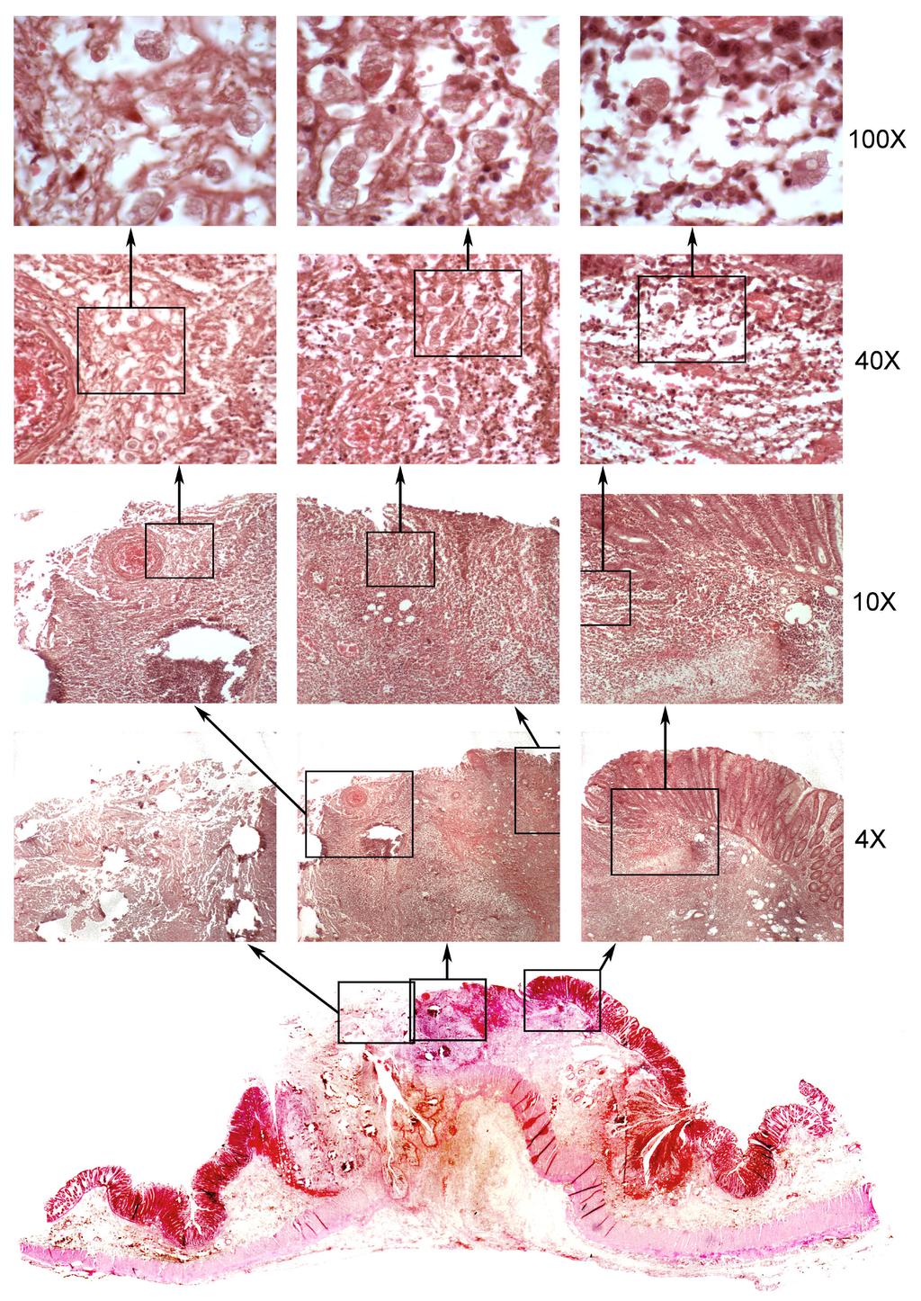
4 T. cruzi in heart muscle. The successive enlargements of the boxed areas are shown at the indicated magnifications. Infected myocytes (ie, muscle cells) appear as a collection of small dots using the 10X objective as indicated by the arrowheads. The infected host cells are sometimes called pseudocysts. These dots become more evident using the 40X objective. To clearly see the kinetoplasts it is necessary to use the 100X objective lens (ie, oil immersion). It is often difficult to see the individual amastigotes in the larger pseudocysts which are most evident at the lower magnifications. Distinct amastigotes (indicated by arrowheads) are usually easier to see in the pseudocysts contain less amastigotes. The inset at the lower left is a further enlargement of amastigotes in which the nuclei and kinetoplasts are more evident.


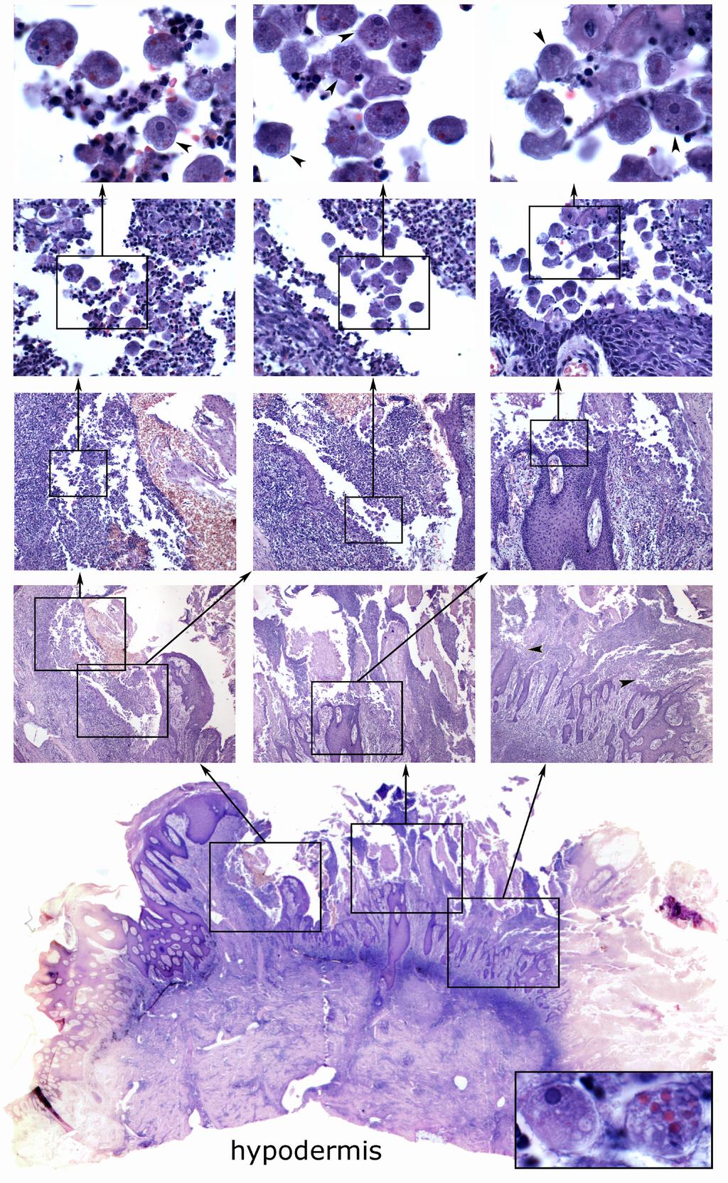
5 Leishmania Amastigotes Giemsa-stained impression smear of Leishmania amastigotes. (Left) Shown are two infected macrophages. The macrophage nuclei are marked with *. The inset shows and enlargement of some of the amastigotes. The kinetoplasts are denoted with arrowheads and the arrows denote nuclei. The morphology of amastigotes is generally better in impression smears that in tissue sections. (See plates on T. cruzi tissue sections.) (Right) Bone marrow smear of patient with visceral leishmaniasis. Free amastigotes (arrowheads) and an infected macrophage (block arrow) can be seen. Many of the amastigotes are out of focus.

6 Leishmania Promastigotes Promastigotes of Leishmania derived from in vitro culture. Promastigotes are found within the gut of the sandfly vector. Distinct kinetoplasts and nuclei are evident in many of the these promastigotes. Dividing forms are also evident. Note the variety of morphologies. Different morphological forms representing different developmental stages are also present within the gut of the vector.

 which consists of a circular muscle layer (inner) and a longitudinal muscle layer (outer).")
7 Entamoeba histolytica, colonic ulcer Gross morphology of the human colon and E. histolytica ulcers (above). The various features of the anatomical features of the colon and lesions are highlighted. The inset in the upper left shows a schematic representation of the human colon in cross-section with the boxed area denoting the relative position of the section. The mucosal layer, defined in part a layer of intestinal epithelial cells (1), faces the lumen. The lamina propria (2), also called the submucosal layer, lies below the intestinal epithelium. Below this is a muscle layer (3) which consists of a circular muscle layer (inner) and a longitudinal muscle layer (outer). A thin serosa (4), which is not always evident, makes up the outer most layer. Distinct areas within the large lesion are evident. An area with a high level of necrosis (5a) represents the site of the original lesion. This part of the lesion is characterized by extensive tissue damage and very few ameba. Adjacent to this area are areas exhibiting less damage and extensive inflammation (5b). Numerous ameba can be in these areas and represent the progression of the ameba from the original site. Note that the intestinal epithelium has sloughed off above the lesion. Note also that the muscle layer and serosa have been breached (5c). The smaller more recent lesion (5d) demonstrates the classic 'flask shape' of the E. histolytica ulcer. Microscopic characterization of the ulcer (next page). Progressively higher magnifications (as denoted with boxes and arrows and objective lens on the right) of different regions of the ulcer are shown. Very few ameba are detected in the highly necrotic area (4X magnification on the left). The ameba are most numerous along the top edge of the large lesion where the intestinal epithelium has sloughed off. Fewer ameba are detected in the area just below the intact epithelium (column on the right) and the smaller lesion, but the damage and inflammation are obvious. Nuclei and ingested erythrocytes will be evident in some of the trophozoites with the 40X and 100X objectives (see below). (Left) Trophozoites exhibiting nucleus (n) and ingested erythrocyte (e). It is sometimes difficult to distinguish nuclei from ingested erythrocytes because of the section plane or amount of digestion of the erythrocyte. The peripheral chromatin and occasionally the central karyosome will be evident in nuclei from a good section plane. The ingested erythrocytes tend to not have as distinct edges as the nuclei.
8
9
10

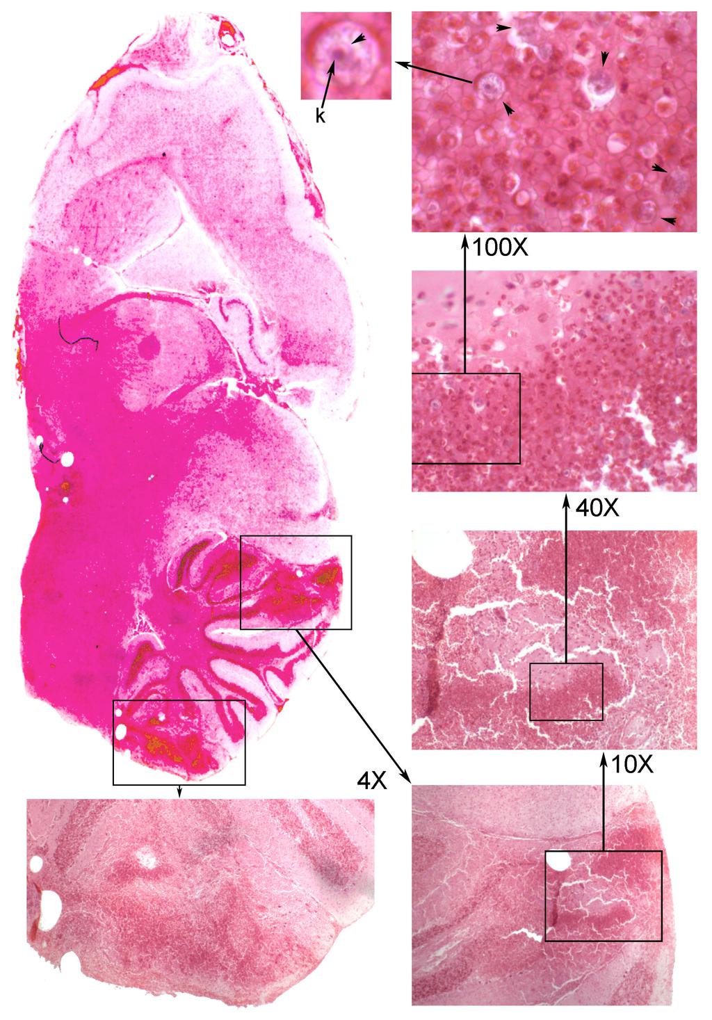
11 Naegleria fowleri in Brain Legend. Trophozoites of Naegleria can be found by scanning the foci of inflammation (see legend below) with the 40X objective. The trophozoites are a lavender color and appear to be foamy. Nuclei will be seen in some of the trophozoites depending on the section plane. The halo like appearance around some of the trophozoites is due to a higher level of shrinkage of the trophozoites as compared to the surrounding tissue during the fixation process. Five particularly prominent trophozoites are denoted with the arrowheads. The box indicates the region shown under oil immersion (100X objective). The vacuolated cytoplasm and nuclear (depending on section plane) is more obvious at this magnification. The nucleus is characterized by a large central karyosome and a thin line of peripheral chromatin (enlarged inset). The nuclei will not always be in the section plane. Legend to figure on other page. The figure depicts a progression going from the view with the naked eye through increasing magnification to the 100X objective (oil immersion). The areas enlarged by subsequent magnification are indicated with boxes and arrows pointing to the view with the subsequent objective lens (i.e., 4X, 10X, 40X, 100X). Two regions of inflammation can be seen when looking at this section with the naked eye (see boxed areas). These foci of inflammation are easily recognized as the darker areas (due to cellular infiltration) using the 4X and 10X objectives. Despite their small size the trophozoites of Naegleria can be detected using the 40X (high dry) objective. (See also the figure above.) The arrowheads in the 100X objective view denote trophozoites that are readily recognized. One of these trophozoites is further enlarged to highlight the nuclear morphology and vacuolated cytoplasm. Note the large karyosome (k) and the thin boundary of the nucleus (arrowhead).
 Oil immersion view of infected epithelial cells showing various morphological forms. The columnar epithelial cells are quite evident in the middle panel.")


12 Toxoplasma gondii Toxoplasma in Cat Intestine. (Left) Low power magnification showing the villi and intestinal epithelial cell layer. (Right) Oil immersion view of infected epithelial cells showing various morphological forms. The columnar epithelial cells are quite evident in the middle panel. Note that the parasite lies between the host cell nucleus (n) and the luminal surface of the epithelial cell. Cell #1 is infected with 2 trophozoites. Cell #2 is infected with a meront in which the individual crescent-shaped merozoites are evident. The merozoites can also appear round or oval depending on the angle of the section. Cell #3 contains a macrogamont (i.e., macrogametocyte). Cell #4 is infected with an immature microgamont (i.e., microgametocyte) as evidence by the multiple nuclei. The out-of-focus parasite to the left is probably also an immature microgamont. The nuclei of the mature microgamonts tend to be found around the periphery of the parasite. The far right panel shows three thick-walled refractive immature oocysts which have not yet been released into the intestinal lumen. The arrowheads denote a probable fungus. Toxoplasma in brain. Shown are successive magnifications (as indicated) of a tissue cyst. Under low power the tissue cysts will appear as round or oval objects with a dot-like appearance (arrowhead in left panel). Small blood vessels (bv) can have a similar size and appearance but are readily distinguished from the tissue cysts at higher magnifications. Individual bradyzoites are apparent at the higher magnifications. A thick wall surrounding the tissue cyst (arrowhead right panel) will sometimes be evident.
 denote foci of necrosis. These are active areas of infection.")
13 Toxoplasma in liver. Shown are successive enlargements of a liver section using the 10X objective (upper left), 40X objective (right side), and 100X objective (insets) as denoted by boxes and arrows. The arrowheads (upper left panel) denote foci of necrosis. These are active areas of infection. Most of the tachyzoites within these necrotic areas are extracellular and are sometimes difficult to detect because of the cellular debris within these lesions. The tachyzoites look like small round or oval bodies and some particularly evident tachyzoites are denoted with arrows in the lower left panel. Host cells infected with tachyzoites can usually be found by looking adjacent to the areas of necrosis as demonstrated by the insets on the upper and lower right corners.
Protozoa from tissues. Leishmania spp. Naegleria fowleri Toxoplasma gondii Trichomonas vaginalis Trypanosoma spp.
 Protozoa from tissues Leishmania spp. Naegleria fowleri Toxoplasma gondii Trichomonas vaginalis Trypanosoma spp. Leishmaniasis Leishmania infantum, Leishmania donovani, in macrophages of man. Female sandflies:
Blood Smears Only 07 February Sample Preparation and Quality Control 12B A
 NEW YORK STATE Parasitology Proficiency Testing Program Blood Smears Only 07 February 2012 The purpose of the New York State Proficiency Testing Program in the category of Parasitology Blood Smears Only
Laboratory diagnosis of Blood and tissue flagellates
 Laboratory diagnosis of Blood and tissue flagellates (Leishmania and trypanosma) Sarah Alharbi Clinical Laboratory department Collage of Applied Medical Sciences King Saud University Leishmania and trypanosma:
Blood Smears Only 6 October Sample Preparation and Quality Control 15B-K
 NEW YORK STATE Parasitology Proficiency Testing Program Blood Smears Only 6 October 5 The purpose of the New York State Proficiency Testing Program in the category of Parasitology - Blood Smears Only is
Morphological forms of hemoflagellates
 Parasitology Lecture: 1 Hemoflagellates (blood and tissue flagellates) *Classification: - Sub-kingdom: Protozoa -Phylum: Sarcomastigophora -Sub-phylum: Mastigiphora -Class: Zoomastigophora د. رائد *Flagellates
Laboratory diagnosis of parasitic diseases. (Amoebiasis)
") Laboratory diagnosis of parasitic diseases (Amoebiasis) Sarah Alharbi Clinical Laboratory department Collage of Applied Medical Sciences King Saud University This document contains materials modified or
Laboratory diagnosis of parasitic diseases (Amoebiasis) Sarah Alharbi Clinical Laboratory department Collage of Applied Medical Sciences King Saud University This document contains materials modified or
Blood Smears Only 20 May Sample Preparation and Quality Control
 NEW YORK STATE Parasitology Proficiency Testing Program Blood Smears Only 20 May 2014 The purpose of the New York State Proficiency Testing Program in the category of Parasitology - Blood Smears Only is
2.Trichomonas vaginalis
 2.Trichomonas vaginalis 1. Pathogenic to human &causes vaginitis (trichomoniasis). 2. troph. Is round or pear like in shape, contains 4-6 flagella, all originating from anterior end & only one extend posteriorly.
EDUCATIONAL COMMENTARY DISTINGUISHING MORPHOLOGIC LOOK-ALIKES
 EDUCATIONAL COMMENTARY DISTINGUISHING MORPHOLOGIC LOOK-ALIKES Educational commentary is provided through our affiliation with the American Society for Clinical Pathology (ASCP). To obtain FREE CME/CMLE
1 Trophozoite stage : The typical characteristics of this stage are :
 Lecturer : Nerran K.F.AL- Rubaey Practical parasites Lab - 2 - Genus : Entamoeba coli It is considered to be a nonpathogenic with world wide distribution. That frequently exists as a commensal parasite
HAEMOFLAGELLATES. Dr. Anuluck Junkum Department of Parasitology Faculty of Medicine
 HAEMOFLAGELLATES Dr. Anuluck Junkum Department of Parasitology Faculty of Medicine Objective Can describe the morphology, life cycle, pathology, diagnosis and prevention of Leishmania spp. and Trypanosoma
Non_ pathogenic Amoeba of humans:
 The parasite protozoa Phylum: Sarcomastigophora Sarcodina االميبات Amoebae Non_ pathogenic Amoeba of humans: 1. Entamoeba hartmanii, contain trophozoite and cyst 2. Entamoeba coli, cyst and trophozoite
Blood Smears Only 19 May Sample Preparation and Quality Control
 NEW YORK STATE Parasitology Proficiency Testing Program Blood Smears Only 9 May 205 The purpose of the New York State Proficiency Testing Program in the category of Parasitology - Blood Smears Only is
WSC , Conference 9. Case 1. Tissue from a rhesus macaque.
 Case 1. Tissue from a rhesus macaque. MICROSCOPIC DESCRIPTION: Esophagus: There is multifocal loss of the mucosal lining (1 pt). In these areas, the denuded subepithelial fibrous connective tissue is infiltrated
American Association of Bioanalysts 5615 Kirby Drive, Suite 870 Houston, TX
 Q3 2018 Parasitology American Association of Bioanalysts 5615 Kirby Drive, Suite 870 Houston, TX 77005 800-234-5315 281-436-5357 Specimen 1 Referees Extent 1 Extent 2 Total Few to 534 Giardia lamblia Many
This is the second learning component (Learning Component 2) in our first learning module (Learning Module 1). In this component we review a very
 in our first learning module (Learning Module 1). In this component we review a very") This is the second learning component (Learning Component 2) in our first learning module (Learning Module 1). In this component we review a very basic response to injury inflammation. We ll look at examples
This is the second learning component (Learning Component 2) in our first learning module (Learning Module 1). In this component we review a very basic response to injury inflammation. We ll look at examples
EEB 4274 Lecture Exam #1 Protozoa September 2011
 1 Name EEB 4274 Lecture Exam #1 Protozoa September 2011 Read through the exam once before you begin. Read the questions CAREFULLY; be certain to provide all of the information requested. In instances in
EDUCATIONAL COMMENTARY MORPHOLOGIC ABNORMALITIES IN LEUKOCYTES
 EDUCATIONAL COMMENTARY MORPHOLOGIC ABNORMALITIES IN LEUKOCYTES Educational commentary is provided through our affiliation with the American Society for Clinical Pathology (ASCP). To obtain FREE CME/CMLE
DIGESTIVE TRACT ESOPHAGUS
 DIGESTIVE TRACT From the lower esophagus to the lower rectum four fundamental layers comprise the wall of the digestive tube: mucosa, submucosa, muscularis propria (externa), and adventitia or serosa (see
Prelab #4 BLOOD; BONE MARROW; RESPIRATORY; INTEGUEMENT Page 1
 Prelab #4 BLOOD; BONE MARROW; RESPIRATORY; INTEGUEMENT Page 1 Blood Slide 101 This a classic slide of blood cells using a Wright stain. Inspect red blood cells and their appearance. Note the approximate
Parasitic Protozoa, Helminths, and Arthropod Vectors
 PowerPoint Lecture Slides for MICROBIOLOGY ROBERT W. BAUMAN Chapter 23 Parasitic Protozoa, Helminths, and Arthropod Vectors Parasitic Diseases Protozoan and helminthic parasites are emerging as serious
Histopathology: chronic inflammation
 Histopathology: chronic inflammation These presentations are to help you identify, and to test yourself on identifying, basic histopathological features. They do not contain the additional factual information
Common Clinical Presentations of Parasitic Infections
 Common Clinical Presentations of Parasitic Infections Hepatosplenomegaly Enlarged lymph nodes Anaemia Dysentery Parasites causing this clinical presentation How did the parasite produce this presentation
PARASITOLOGY CASE HISTORY 15 (HISTOLOGY) (Lynne S. Garcia)
 (Lynne S. Garcia)") PARASITOLOGY CASE HISTORY 15 (HISTOLOGY) (Lynne S. Garcia) A biopsy was performed on a 27-year-old man with no known travel history, presenting with a perianal ulcer. The specimen was preserved in formalin
PARASITOLOGY CASE HISTORY 15 (HISTOLOGY) (Lynne S. Garcia) A biopsy was performed on a 27-year-old man with no known travel history, presenting with a perianal ulcer. The specimen was preserved in formalin
VETERINARY HEMATOLOGY ATLAS OF COMMON DOMESTIC AND NON-DOMESTIC SPECIES COPYRIGHTED MATERIAL SECOND EDITION
 VETERINARY HEMATOLOGY ATLAS OF COMMON DOMESTIC AND NON-DOMESTIC SPECIES SECOND EDITION COPYRIGHTED MATERIAL CHAPTER ONE HEMATOPOIESIS GENERAL FEATURES All blood cells have a finite life span, but in normal
Kinetoplastids Handout
 Kinetoplastids Handout 1 Kinetoplastids widespread group of flagellated protozoa parasitize virtually all animal groups as well as plants and insects 3 distinct kinetoplastid species cause human disease
Flagellates I Genito-urinary & Intestinal flagellates
 Flagellates I Genito-urinary & Intestinal flagellates Dr. Anuluck Junkum PARA 317221 Objective Can describe the morphology, life cycle, pathology, diagnosis and prevention of pathogenic flagellate Classification
Asian Journal of Research in Pharmaceutical Sciences and Biotechnology
 Research Article ISSN: 2349 7114 Asian Journal of Research in Pharmaceutical Sciences and Biotechnology Journal home page: www.ajrpsb.com ENTAMO ENTAMOEBA COLI AS STRONG PHAGOCYTIC PARASITIC ORGANISM Mosab
Histopathology: gastritis and peptic ulceration
 Histopathology: gastritis and peptic ulceration These presentations are to help you identify, and to test yourself on identifying, basic histopathological features. They do not contain the additional factual
1. Toxoplasma gondii:
 Parasites affecting the central nervous system: 1. Toxoplasma gondii: It s a protozoa family member, more specifically a member of the apicomplexa just like plasmodium malaria. Causes toxoplasmosis Has
2015 Descriptive Vet Path Course. Histo Exam #3 KEY
 2015 Descriptive Vet Path Course Histo Exam #3 KEY Test 3, Slide 1 Tissue from a guinea pig. MORPHOLOGIC DIAGNOSIS: Heart: Multifocally and randomly (1 pt), within the left and right ventricular myocardium
WSC , Conference 9, Case 1. Tissue from a nyala.
 WSC 2009-2010, Conference 9, Case 1. Tissue from a nyala. MICROSCOPIC DESCRIPTION: Heart, atrium (1 pt.): Approximately 40% of the atrial myocardium is replaced by areas of fibrous connective tissue (1
Supplemental Figure 1. Quantification of proliferation in thyroid of WT, Ctns -/- and grafted
 Supplemental Figure 1. Quantification of proliferation in thyroid of WT, Ctns -/- and grafted Ctns -/- mice. Cells immunolabeled for the proliferation marker (Ki-67) were counted in sections (n=3 WT, n=4
Schistosome life cycle.
 Schistosomiasis infects approximately 200 million persons and kills approximately 280,000 annually. Most of the mortality comes from hepatic granulomas and fibrosis Schistosoma japonicum and Schistosoma
A adipose cells. B capillary. C epithelium
 EPITHELIA Objective The objective of this class is to observe how different epithelia vary in terms of cell shape, size and number of cell layers enabling them to be well adapted for functions in different
News and Notes. Parasitology Comprehensive 2 October Sample Preparation and Quality Control. 12 K (All Parasites)
") NEW YORK STATE Parasitology Proficiency Testing Program News and Notes Recent reports in the literature have indicated a high rate of Cryptosporidium sp. false positive associated with Rapid Cartridge
NEW YORK STATE Parasitology Proficiency Testing Program News and Notes Recent reports in the literature have indicated a high rate of Cryptosporidium sp. false positive associated with Rapid Cartridge
MORPHOLOGIC DIAGNOSIS: Liver: Hepatitis, necrotizing, multifocal to coalescing, severe, with numerous trichomonads. (3 pt)
") Case 1. Tissue from a pelican. MICROSCOPIC DESCRIPTION: Liver: Approximately 80% (1 pt) of the liver is replaced by multifocal to coalescing areas of coagulative and lytic necrosis. Centrally, within these
Case 1. Tissue from a pelican. MICROSCOPIC DESCRIPTION: Liver: Approximately 80% (1 pt) of the liver is replaced by multifocal to coalescing areas of coagulative and lytic necrosis. Centrally, within these
News and Notes. Parasitology Comprehensive 5 November 2013
 NEW YORK STATE Parasitology Proficiency Testing Program News and Notes Beginning with the May 2013 event a separate set of 3 samples were supplied for laboratories performing antigen detection. For the
EDUCATIONAL COMMENTARY DIFFERENTIATING IMMATURE PERIPHERAL BLOOD CELLS
 Educational commentary is provided through our affiliation with the American Society for Clinical Pathology (ASCP). To obtain FREE CME/CMLE credits click on Continuing Education on the left side of the
Epithelia will be discussed according to the following scheme: Type Number of layers Shape Line drawing. Squamous Cuboidal Columnar
 Epithelia Epithelia will be discussed according to the following scheme: Type Number of layers Shape Line drawing Simple Squamous Cuboidal Columnar Covering and Lining epithelium Pseudostratified Stratified
Intestinal Protozoa 1- Entamoeba Histolytica
 Check NOTES BOXES in sheet 27, just in case 1- Trophozoite Form (Vegetative form / tissue form). 2- Cyst Form (Luminal form), small and circular, can be: a- Immature cyst, which can be either Uninucleate
اعداد رغداحمد رغد جمال الدين
 اعداد رغداحمد رغد جمال الدين Trypanosoma Causes Trypanosomiasis West African Trypanosomiasis T.brucei gambiense Sleeping sickness East African Trypanosomiasis T.brucei rhodesiense American Trypanosomiasis
Blood Smears Only 3 February Sample Preparation and Quality Control
 NEW YORK STATE Parasitology Proficiency Testing Program Blood Smears Only 3 February 2015 The purpose of the New York State Proficiency Testing Program in the category of Parasitology - Blood Smears Only
Small intestine. Small intestine
 General features Tubular organ longest part; 5-6 m most of chemical digestion absorption of nutrients reabsorption of H2O occurs. Two structural features; maximize the lumenal surface area villi microvilli
SESSION 1: GENERAL (BASIC) PATHOLOGY CONCEPTS Thursday, October 16, :30am - 11:30am FACULTY COPY
 PATHOLOGY CONCEPTS Thursday, October 16, :30am - 11:30am FACULTY COPY") SESSION 1: GENERAL (BASIC) PATHOLOGY CONCEPTS Thursday, October 16, 2008 9:30am - 11:30am FACULTY COPY GOAL: Describe the basic morphologic (structural) changes which occur in various pathologic conditions.
SESSION 1: GENERAL (BASIC) PATHOLOGY CONCEPTS Thursday, October 16, 2008 9:30am - 11:30am FACULTY COPY GOAL: Describe the basic morphologic (structural) changes which occur in various pathologic conditions.
FACT OR ARTIFACT LEARN THE KEYS TO DIFFERENTIATE PARASITES FROM ARTIFACTS
 FACT OR ARTIFACT LEARN THE KEYS TO DIFFERENTIATE PARASITES FROM ARTIFACTS ACMLT CONFERENCE OCTOBER 1, 2005 PRESENTED BY PAULINE TOMLIN Public Health Microbiology 2 FACT OR ARTIFACT Many body sites and
Histopathology: healing
 Histopathology: healing These presentations are to help you identify, and to test yourself on identifying, basic histopathological features. They do not contain the additional factual information that
Entamoeba histolytica
 Entamoeba histolytica Occurence: -cosmopolitan amoeba lives in the form of trophozoite and cyst in the human colon, but in multifocal invasions may locate various organs including the lungs, -common in
Classification - Protozoa. Parasitology Intestinal Amoeba. Stools for O&P Examination. Stool Specimen. What to look for.
 Classification - Protozoa Parasitology Intestinal Amoeba Phylum Phylum Sarcomastigophora Sarcomastigophora Subkingdom Subkingdom Protozoa Protozoa Phylum Phylum Ciliophora Ciliophora Phylum Phylum Apicomplexa
HISTOLOGY VIRTUAL LABORATORY BLOOD AND LYMPHATICS SYSTEM
 HISTOLOGY VIRTUAL LABORATORY BLOOD AND LYMPHATICS SYSTEM Login: http://histopath.westernu.edu Histology Atlas AND Virtual Histology links. I. HEMATOLOGY - PERIPHERAL BLOOD Purpose: To be able to identify
Slide 154: Pancreas, H&E
 Slide 154: Pancreas, H&E the pancreas, located adjacent to the duodenum, is a mixed exocrine and endocrine gland; it is usually readily identifiable by the presence of the interspersed endocrine pancreatic
Genus : Chilomastix mesnili Considered to be a non-pathogen, it ' s resides in cecum and colon. The life cycle of this parasite have two stages :
 Lecturer : Nerran K.F.AL- Rubaey Practical parasites Lab - 4 - Genus : Chilomastix mesnili Considered to be a non-pathogen, it ' s resides in cecum and colon. The life cycle of this parasite have two stages
Protozoal Infections. By: Nader Alaridah MD, PhD
 Protozoal Infections By: Nader Alaridah MD, PhD Classification of Phylum Protozoa 1- According to the organ of locomotion 1- Class Rhizopoda (Amoebae) 2- Class Ciliata (Ciliates) 3- Class Zoomastigophora
Learning Objectives. 3. Epidemiology distribution; endemic; 4. Basic Morphology 5. Name of diesease 6. Prevention and Control
 Learning Objectives At the end of the class student will able to state 1. Life cycle Environment, Human, Animals 2. Name of parasite: (Genus), (Species) 3. Epidemiology distribution; endemic; 4. Basic
LYMPH GLAND. By : Group 1
 LYMPH GLAND By : Group 1 ANATOMY LYMPH NODE Lymphatic Organs Red bone marrow Thymus gland Lymph nodes Lymph nodules Spleen Primary organs Secondary organs Lymph Nodes Firm, smooth-surfaced, bean-shaped
Mitral valve repair for functional regurgitation caused by Chagas disease
 Mitral valve repair for functional regurgitation caused by Chagas disease Stevan K Martins, Alberto T Kiyose, Frederico C Mendonça, Veridiana S Andrade, Jairo Pinheiro Jr, Jeffer Moraes, Adib D Jatene
General Structure of Digestive Tract
 Dr. Nabil Khouri General Structure of Digestive Tract Common Characteristics: Hollow tube composed of a lumen whose diameter varies. Surrounded by a wall made up of 4 principal layers: Mucosa Epithelial
(b) Stomach s function 1. Dilution of food materials 2. Acidification of food (absorption of dietary Fe in small intestine) 3. Partial chemical digest
 Stomach s function 1. Dilution of food materials 2. Acidification of food (absorption of dietary Fe in small intestine) 3. Partial chemical digest") (1) General features a) Stomach is widened portion of gut-tube: between tubular and spherical; Note arranged of smooth muscle tissue in muscularis externa. 1 (b) Stomach s function 1. Dilution of food
(1) General features a) Stomach is widened portion of gut-tube: between tubular and spherical; Note arranged of smooth muscle tissue in muscularis externa. 1 (b) Stomach s function 1. Dilution of food
Cytoplasmic changes Nuclear changes
 The presence of infection in the female genital tract may procure certain cellular changes in the epithelium. Such changes are seen in nucleus and cytoplasm surrounding the nucleus. Cytoplasmic changes
CINtec p16 INK4a Staining Atlas
 CINtec p16 INK4a Staining Atlas Rating Rating Positive The rating positive will be assigned if the p16 INK4a -stained slide shows a continuous staining of cells of the basal and parabasal cell layers of
Lab Animal Tissue. LEARNING OBJECTIVES: To understand the relationship between the structure and function of different animal tissues
 Name: Bio A.P. PURPOSE: HYPOTHESIS: NONE Lab Animal Tissue BACKGROUND: In animals, groups of closely related cells specialized to perform the same function are called tissues. There are four general classes
Alimentary Canal (I)
") Alimentary Canal (I) Esophagus and Stomach (Objectives) By the end of this lecture, the student should be able to discuss the microscopic structure in correlation with the function of the following organs:
Alimentary Canal (I) Esophagus and Stomach (Objectives) By the end of this lecture, the student should be able to discuss the microscopic structure in correlation with the function of the following organs:
Anopheles freeborni. Courtesy
 Anopheles freeborni Courtesy Plasmodia seen with the microscope M. Lontie, MCH, Leuven, 2012 Diagnosis of malaria Thin film (better for species identification). Thick film (more sensitive). QBC (quantitative
HDF Case CRYPTOSPORIDIOSE
 HDF Case 986949 CRYPTOSPORIDIOSE 45 yo male with severe diarrhea. Known HIV positive. Endoscopic biopsy of duodenum, the colon and ileum. EXUDATIVE CHANGES GRANULAR BASOPHILIC BODIES Colonic biopsy shows
The Endocrine System Pituitary
 The Endocrine System Pituitary Look at your slide of the human pituitary with your naked eye. You should see a cellular region and a more fibrous region. Then view each region with your microscope under
Coccidia. Eucoccidioside
 Coccidia Kingdom Sub-Kingdom Phylum Class Order Family Genus Species Protista Protozoa Apicomplexa Sporozoasida Eucoccidioside Sarcocystidae Toxoplasma gondii 1 Toxoplasma gondii (life cycle) Sexual cycle
HISTOLOGY VIRTUAL LABORATORY GASTROINTESTINAL SYSTEM
 HISTOLOGY VIRTUAL LABORATORY GASTROINTESTINAL SYSTEM LIP (Slides GI 1, 2) Identify the outer portion lined by stratified squamous (keratinized) epithelium. Note the hair follicles and sebaceous glands
Effect of hydroxyurea on the intracellular multiplication of. gondii, Leishmania amazonensis and Trypanosoma
 Brazilian Journal of Medical and Biological Research (2003) 36: 65-69 ISSN 0100-879X Short Communication 65 Effect of hydroxyurea on the intracellular multiplication of Toxoplasma gondii, Leishmania amazonensis
Digestive system L 2. Lecturer Dr. Firdous M. Jaafar Department of Anatomy/Histology section
 Digestive system L 2 Lecturer Dr. Firdous M. Jaafar Department of Anatomy/Histology section objectives 1-Describe the general structure of digestive tract: a-mucosa. b-submucosa. c-muscularis externa d-adventitia
Modified mitochondrion near base of flagellum
 XV. The Flagellates (Chapters 5 & 6) 2011 A. Hemoflagellates 1. Classification of trypanosomes a. Phylum Euglenozoa b. Subphylum Kinetoplasta * c. Class Trypanosomatida d. Important genera (1) Trypanosoma
EEB 4274 Lecture Exam #1 Protozoa September 2014
 1 Name EEB 4274 Lecture Exam #1 Protozoa September 2014 Read through the exam once before you begin. Read the questions CAREFULLY; be certain to provide all of the information requested. In instances in
KINETOPLASTIDS. Kinetoplast. Nucleus
 KINETOPLASTIDS Kinetoplast Nucleus widespread parasites animals (fish humans) insects plants monophyletic group related to euglenoids unifying feature = kinetoplast Giemsa staining structure KINETOPLAST
Tongue In the buccal cavity of the digestive system
 Tongue In the buccal cavity of the digestive system same layers as those of tubular organs Mucosa, submucosa, and muscularis muscularis = the muscularis externa no muscularis mucosa 1 Tongue ling = tongue
HISTOLOGICAL PARAMETERS
 HISTOLOGICAL PARAMETERS ADDITIONAL FILE FOR THE MANUSCRIPT: Peritoneal Negative Pressure Therapy Prevents Multiple Organ Injury in a Chronic Porcine Sepsis and Ischemia/Reperfusion model Brian D. Kubiak
Giardia lamblia (flagellates)
") Giardia lamblia (flagellates) Dr. Hala Al Daghistani Giardia lamblia (Giardia duodenalis or Giardia intestinalis) is the causative agent of giardiasis and is the only common pathogenic protozoan found
Giardia lamblia (flagellates) Dr. Hala Al Daghistani Giardia lamblia (Giardia duodenalis or Giardia intestinalis) is the causative agent of giardiasis and is the only common pathogenic protozoan found
GUIDE TO: Diagnosing Coccidiosis & Necrotic Enteritis
 GUIDE TO: Diagnosing Coccidiosis & Necrotic Enteritis Site of Infection Species E. acervulina E. brunetti E. maxima E. mivati E. tenella E. necatrix Oocyst Size 2µ{ 18.3 x 14.6 24.6 x 18.8 30.5 x 20.7
Color Atlas of Clinical Laboratory Medicine. First Version
 Color Atlas of Clinical Laboratory Medicine First Version Addis Ababa, February 2015 Page 1 of 44 Table Contents 1. Hematology... 5 1.1. Cell Maturation... 5 1.2 Red Blood Cells Morphology... 6 1.3 Abnormal
Flagellates. Dr. Anuluck Junkum PARA
 Flagellates Dr. Anuluck Junkum PARA 317242 Objective Can describe the morphology, life cycle, pathology, diagnosis and prevention of pathogenic flagellates Classification of Protozoa Based on locomotive
Lab activity manual - Histology of the digestive system. Lab activity 1: esophagus stomach - small intestines
 Lab activity manual - Histology of the digestive system Jeanne Adiwinata Pawitan Prerequisite: Histology of the 4 basic tissues In this module we learn about the histology of the digestive system, from
HUMAN PARASITOLOGY. lumbricoides, Trichuris trichiura, hookworm. Human Parasitology (Code: ) Guideline
 Guideline") HUMAN PARASITOLOGY Human Parasitology (Code:1001021) Guideline I. Course Introduction 128 classes comprise 72 for lecturing, 54 for lab practice, and 2 for self-study. 6.0 credits; 5 th semester II. Lecturing
HUMAN PARASITOLOGY Human Parasitology (Code:1001021) Guideline I. Course Introduction 128 classes comprise 72 for lecturing, 54 for lab practice, and 2 for self-study. 6.0 credits; 5 th semester II. Lecturing
EDUCATIONAL COMMENTARY BLOOD CELL IDENTIFICATION
 EDUCATIONAL COMMENTARY BLOOD CELL IDENTIFICATION Educational commentary is provided through our affiliation with the American Society for Clinical Pathology (ASCP). To obtain FREE CME/CMLE credits click
Blood Smears Only 5 February Sample Preparation and Quality Control 13B A
 NEW YORK STATE Parasitology Proficiency Testing Program Blood Smears Only 5 February 2013 The purpose of the New York State Proficiency Testing Program in the category of Parasitology Blood Smears Only
川北医学院讲稿. Under low power note the testis is enclosed by a strong fibrous. layer of serous epithelium. These fibrous tissue
 川北医学院讲稿 Experiment 5: Male and Female Reproductive System Hello, everybody, class is begin,keep quiet, please. And this is the last experimental class. Today we will learn 5 slices and review all structures
PARASITOLOGY CASE HISTORY #14 (BLOOD PARASITES) (Lynne S. Garcia)
 (Lynne S. Garcia)") PARASITOLOGY CASE HISTORY #14 (BLOOD PARASITES) (Lynne S. Garcia) A 37-year-old woman, who had traveled to New Guinea for several weeks, presented to the medical clinic with fever, chills, and rigors within
PARASITOLOGY CASE HISTORY #14 (BLOOD PARASITES) (Lynne S. Garcia) A 37-year-old woman, who had traveled to New Guinea for several weeks, presented to the medical clinic with fever, chills, and rigors within
Parasitology. Lab. Amoeba
 Parasitology. Lab. Kingdom : Protista Subkingdom : Protozoa Phylum : Sacromastigophora Subphylum : Sarcodina Superclass : Rhizopoda Class : Lobosea Order : Amoebida Amoeba Protozoa Amoebae geneus Entamoeba
Blood Cell Identification Graded
 Blood Cell Identification Graded Case History The patient was a five-day-old girl with an elevated unconjugated bilirubin and a weakly positive direct antiglobulin test (DAT). Her CBC showed: WBC = 11.0
Disorders of Cell Growth & Neoplasia. Histopathology Lab
 Disorders of Cell Growth & Neoplasia Histopathology Lab Paul Hanna April 2010 Case #84 Clinical History: 5 yr-old, West Highland White terrier. skin mass from axillary region. has been present for the
Entamoeba histolytica
 Entamoeba histolytica cosmopolitan distribution no animal reservoirs facultative pathogen most clear the infection spontaneous in 6-12 months with mild or no symptoms can cause a serious invasive disease
Mass Histology Service
 Mass Histology Service A complete anatomical pathology laboratory www.masshistology.com Telephone: (877) 286-6004 Report on Pathology A Time Course Study of the Local Effects of Intramuscular XXXXXXX Injection
PBS Class #2 Introduction to the Immune System part II Suggested reading: Abbas, pgs , 27-30
 PBS 803 - Class #2 Introduction to the Immune System part II Suggested reading: Abbas, pgs. 15-25, 27-30 Learning Objectives Compare and contrast the maturation of B and T lymphocytes Compare and contrast
CELL AND TISSUE INJURY COURSE-II PATHOLOGY LABORATORY
 CELL AND TISSUE INJURY COURSE-II PATHOLOGY LABORATORY PATHOLOGY of INFECTIOUS DISEASES MICROSCOPY Rengin Ahıskalı Macroscopy samples are shown in the macroscopy presentations of the first two courses.
Cellular Pathology. Histopathology Lab #2 (web) Paul Hanna Jan 2018
 Paul Hanna Jan 2018") Cellular Pathology Histopathology Lab #2 (web) Paul Hanna Jan 2018 Slide #91 Clinical History: a necropsy was performed on an aged cat the gross pathological changes included: widespread subcutaneous edema
Cellular Pathology Histopathology Lab #2 (web) Paul Hanna Jan 2018 Slide #91 Clinical History: a necropsy was performed on an aged cat the gross pathological changes included: widespread subcutaneous edema
BLOOD PARASITES. Danka Petrovic
 BLOOD PARASITES Danka Petrovic Parasites Found in Blood Plasmodium species (Malaria) Babesia Trypanosoma spp Microfilariae Leishmania (Visceral) Plasmodium (Malaria) Four species are considered true parasites
Blood Cell Identification Graded
 BCP-21 Blood Cell Identification Graded Case History The patient is a 37-year-old female with a history of multiple sickle cell crises. She now presents with avascular necrosis of the left hip. Laboratory
CYTOMORPHOLOGY MODULE 28.1 INTRODUCTION OBJECTIVES 28.2 GENERAL GUIDELINES. Notes
 28 CYTOMORPHOLOGY 28.1 INTRODUCTION Light microscopic examination of stained cells in smears is the method of choice of diagnostic cytology. It allows classification of most normal cells as to type and
Pathogenic amoebae and ciliate. Dr. Narissara Jariyapan Department of Parasitology Faculty of Medicine Chiang Mai University
 Pathogenic amoebae and ciliate Dr. Narissara Jariyapan Department of Parasitology Faculty of Medicine Chiang Mai University Objectives After the lecture, students must know 1. General morphology of pathogenic
ccess safe drinking wa r is everyone s right Protozoans that cause diarrheal disease
 ccess safe drinking wa r is everyone s right Protozoa: Protozoans that cause diarrheal disease 1. Giardia lamblia 2. Entameba histolytica 3. Cryptosporidium parvum 4. Cyclospora cayetanensis 1 Giardia
Methodology Prevalence:
 Context of Research Free ranging domestic fowl are very susceptible to parasitic infections owing to their exposure to parasites during their roaming in the backyards in search of food. Heterakis gallinarum
Observations on the Pathology of Lesions Associated with Stephanofilaria dinniki Round, 1964 from the Black Rhinoceros (Diceros bicornis)
") Journal of Helminthology, ~ol. XXXVIII, Nos. 1/2, 1964, pp. 171-174. Observations on the Pathology of Lesions Associated with Stephanofilaria dinniki Round, 1964 from the Black Rhinoceros (Diceros bicornis)
Journal of Helminthology, ~ol. XXXVIII, Nos. 1/2, 1964, pp. 171-174. Observations on the Pathology of Lesions Associated with Stephanofilaria dinniki Round, 1964 from the Black Rhinoceros (Diceros bicornis)