The Most Common Parasitic Infections In Yemen. Medical Parasitology
|
|
|
- Merilyn Thomas
- 5 years ago
- Views:
Transcription


1 The Most Common Parasitic Infections In Yemen Medical Parasitology ﻓﺎﯾز اﻟﺧوﻻﻧﻲ / د
2 2
3 : is a vector-borne disease that transmitted by sandflies and caused by obligate intracellular protozoa of the genus Leishmania. About 21 of 30 species cause human infection. The different species are morphologically indistinguishable, but they can be differentiated by isoenzyme analysis, molecular methods, or monoclonal antibodies. The following table summarizes the clinical diseases caused by the most important Leishmania species. Leishmania species Disease Visceral L. donovani L. infantum L. chagasi L. tropica L. major L. aethiopica L. mexicana L. mazonensis leishmaniasis Cutaneous L. mexicana leishmaniasis Diffuse-cutaneous L. aethiopica leishmaniasis (DCL) Muco-cutaneous L. braziliensis L. panamensis leishmaniasis (MCL 3
4 Epidemiology and Distribution is endemic in 88 countries with an estimated 350 million people at risk of infection. The overall prevalence of the leishmaniasis estimated to be at 12 million cases with 0.5 million new visceral leishmaniasis cases per year and million new cutaneous leishmaniasis cases per year. More than 90% of visceral leishmaniasis occurs in Sudan, Bangladesh, Nepal, Brazil and India, and more than 90% of cutaneous leishmaniasis in Brazil, Peru, Afghanistan, Syria, Iran, Yemen and Saudi Arabia. In recent years, there have been major epidemics of visceral leishmaniasis in southern Sudan, eastern India, Bangladesh, and Brazil. Increased infections and the spread of leishmaniasis is related to environmental and behavioral changes and development, conflict and war, bringing non-immune people into closer contact with vectors and reservoir hosts. Transmission and life cycle Leishmania is mostly zoonotic (transmitted to humans from animals), and humans become infected only when accidentally exposed to the natural transmission cycle. However, humans are the sole reservoir hosts when the transmission occurs from human to human through the sand fly vector. 4
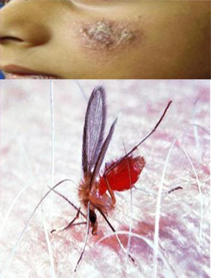
5 Leishmania species are transmitted by the bite of an infected female sandfly, belonging to the genus Phlebotomus in Africa, Asia and Europe, and the genus Lutzomyia in the Americas. About 30 species of sandflies act as vectors, infecting humans and animal reservoir hosts. Pattern of transmission Human-to-human transmission: man is the only source and reservoir of infection. Dog-to-human transmission: The infection source is domestic dogs and some rodents, which acts as a reservoir host. Classification of leishmaniasis according to the location NEW WORLD OLD WORLD (South America and Central (Africa, Asia, Europe) America) Leishmania species Leishmania species L. donovani L. chagasi Visceral leishmaniasis L. infantum Cutaneous leishmaniasis L. tropica L. major L. aethiopica L. guyanensis L. amazonensis L. Mexicana Diffuse cutaneous leishmaniasis L. aethiopica L. Mexicana L. amazonensis Mucocutaneous leishmaniasis L. braziliensis L. panamensis 5
6 Sandfly vectors The feeding, breeding and flight habits of sandflies are species specific. Most sandflies feed mainly on plant juices, but female flies also require blood meals for egg development. Most species feed at night, dusk or dawn. Morphology The parasite exists in two forms: 1. Amastigote 2. Promastigote Amastigote Amastigotes are round in man and other vertebrate hosts. Amastigotes live inside monocytes, polymorphonuclear leucocytes. They are small, round to oval bodies measuring μm in length (Fig below). They are stained well with Giemsa or Wright stain. In the stained preparation, the cytoplasm appears pale-blue and surrounded by a limiting membrane. The nucleus relatively is large and stained red. The kinetoplast lies at right angle to the nucleus. It is slender, rod-shaped and is stained deep red. Axoneme arises from the kinetoplast and extends to margin of the body. Vacuole, which is a clear unstained space, lies alongside the axoneme. 6

7 Leishmania. Amastigote Promastigote Promastigotes are excited in the digestive tract of sand fly (vector) and in the culture media. The fully developed promastigotes are long, slender and spindle-shaped. They measure 14.3 to 20 μm in length and 1.5to 1.8 μm in breadth. A single nucleus lies at the center. The kinetoplast lies transversely near the anterior end. The flagellum is single, delicate and measures15-28 um. With Leishman stain, cytoplasm appears blue, the nucleus pink and the kinetoplast blight red (Figure below). 7

8 Fig. ( ) Leishmania species Promastigote Life cycle in man The life cycle of Leishmania species is summarized in Fig below. It consists of two forms: amastigote, which presents in the human macrophages and promastigote, which presents in the sandfly and culture media. Life cycle of Leishmania species involves two hosts: human host (vertebrate host) and insect host (vector host, invertebrate host). Because it is not identified the sexual stages of the parasite, the definitive host is not recognized yet. The life cycle begins with injection the infective stage metacyclic promastigote into the human host at the time of taking blood meal by the female sandfly. The skin macrophages phagocytize the promastigotes by a process of phagocytosis then transform into intracellular forms called amastigotes. 8

9 Amstigotes multiply within skin macrophages (in case of cutaneous leishmaniasis), liberate from the macrophages, and infect new cells. In visceral leishmaniasis, the amastigotes multiply in the macrophages of the spleen, liver, bone marrow and lymph glands of the reticuloendothelial system. Blood monocytes are also infected. Life cycle in sandfly When intracellular and free amastigotes are ingested by a female sandfly the life cycle is continued After about 72 hours, the amastigotes become flagellated promastigotes in the midgut of the sandfly. They multiply and fill the lumen of the gut. After days (depending on species), the promastigotes move forward to the head and mouth-parts of the sandfly. Sandfly the leishmaniasis vector 9


10 Injection of metacyclic promastigote the infective stage into the human skin Life cycle of Leishmania 10
11 Symptoms of Visceral leishmaniasis (VL) This is the most severe form of leishmaniasis. It is caused by L. donovani and L. infantum in the old world and L. chagasi in the new world. In the endemic areas, the disease is more chronic with young adults and children being more commonly infected. In epidemics, all age groups are susceptible (except those with acquired immunity), and the disease is often acute. Without treatment, VL is usually fatal. Symptoms in acute VL, there is splenomegaly, high undulating fever with two peaks in the day, chills, profuse sweating, weight loss, fatigue, anaemia, and leucopenia. Symptoms in chronic VL include irregular fever, massive splenomegaly, hepatomegaly, and/or lymphadenopathy, marked loss of weight with wasting, diarrhea, low white cell and platelet counts, and anaemia. The local Indian name for VL, kala-azar (meaning black sickness or black fever) is a reference to the darken color of the infected patients. Malnutrition and other infections increase the risk of developing symptomatic VL. 11


12 Massive splenomegaly in VL Post kala-azar dermal leishmaniasis (PKDL) In India and occasionally in Africa, a cutaneous form of leishmaniasis can occur about 2 years after treatment and recovery from visceral leishmaniasis. This is referred to as post kala-azar dermal leishmaniasis and affects about 20% of patients in India. Hypopigmented and raised erythematous patches appear on the face, trunk of the body, and limbs. These may develop into nodules and resemble those of lepromatous leprosy, fungal infections or other skin disorders. Occasionally there is ulceration of the lips and tongue. Amastigotes are present in the papules and nodules. 12


")

13 Figure ( ) PKDL in sudan Figure ( Figure ( Papular and nodular PKDL ) PKDL affecting the earlobe ) PKDL: macular lesions, some are confluent. 13
14 Immunity Absence of Gamma Interferon IFN ᵧ and Interleukin 2 during Active Visceral Inhibition of parasitic Ag presentation by the antigen presenting cells (APCs)-macrophage because lysis of intracellular amastigote is blocked. Leishmania proamastigote has surface inhibiter molecules called lipophosphoglycan (LPG) that inhibits the toxic effect of macrophage nitric oxide. Nitric oxide or nitrogen mediators are a potent toxic oxidant that destroys intracellular pathogens however, promastigote of leishmania parasite has the ability of inhibition the nitric oxide-dependent killing mechanism of the macrophage. Because the acidic ph is an environment suitable for living and multiplication of the amastigote, thus lysis of intra-phagosome amstigote does not achieved by the macrophage lysosomes. As a result, rupture of parasitized phagocyte occurs and releasing of amastigotes that in turn infects other macrophages. T lymphocytes are not activated unless recognizes the pathogen Ag which must be expressed on the surface macrophage. 14

15 Macrophages have the key role in an initiating the cell-mediated immune response, and one of some immune cells that act as an antigen presenting cells. In normal phagocytosis, the phagocyte lysosomes destructs the intracellular pathogen into small peptides, these peptides are then expressed on the macrophage surface via the Major Histocompatibility molecules class 1(MHC molecules). After an antigen presenting, macrophage release a cytokines molecules called Il-2 and INF ᵧ, which activate T lymphocytes. The consequences of T lymphocytes activation is the direct killing of infected cells and controlling the disease prognosis. 15
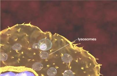
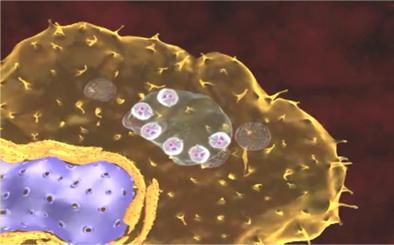
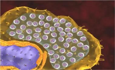
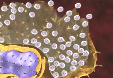
16 Illustration of leishmania parasite during invasion of macrophage 16

17 Development of the immune response to protozoan and helminth infection 17

18 Cutaneous leishmaniasis (CL) Cutaneous leishmaniasis is a potentially severe and disfiguring disease in some people. The clinical forms of CL vary according to the species of parasite, region, and immune response of the patient. People with cutaneous leishmaniasis have one or several long-lasting lesions on the skin, usually without fever or general symptoms. New cases are emerging in areas previously free of the disease. Over new cases of cutaneous leishmaniasis are reported annually to WHO by countries in the Region, but the actual incidence is estimated to be three to five times higher since many patients never 18

19 seek medical attention and not all patients with a diagnosis of cutaneous leishmaniasis are reported to health authorities. Old world cutaneous leishmaniasis (CL) Cutaneous leishmaniasis caused by L. tropica Infection is often referred to as dry urban oriental sore. Dry painless ulcers mm in diameter are produced which are self-healing usually after 1 2 years but often leave disfiguring scars. The patient acquires immune to reinfection. Rarely, multiple unhealed lesions may develop, often on the face. It can last many years and is difficult to treat; this condition is known as leishmaniasis recidivans (LR). Untreated LR may leads to destruction and disfiguration of the infected parts. Initial brownish nodule of dry, urban type of cutaneous leishmaniasis 19


20 Plaque lesion of dry, urban type of cutaneous leishmaniasis Dry, urban and anthroponotic type of cutaneous leishmaniasis recidivans due to (L. tropica) from Morocco.Note the healed scar from which new lesions develop. 20

21 Chronic cutaneous leishmanaisis of face with areas of scarring and reactivation of disease ( recidivans) recidivans due to (L. tropica) from Afghanistan. Note the healed scars from which new lesions develop. Cutaneous leishmaniasis caused by L. major Infection is often referred to as wet oriental sore. The early papule is often inflamed and resembles a boil of 5 10 mm in diameter which rapidly develops into a large uneven ulcer which is self-healing in as little as 3 6 months. Multiple lesions may occur in non-immune persons. L. major infections show permanent immunity against reinfection. 21


22 Wet ulcer in CL Cutaneous leishmaniasis caused by L. aethiopica A cutaneous lesion that is similar to typical oriental sore with healing in 1 3 years. Localized cutaneous lesions may spread to involve large cutaneous area, forming a nodules often associated with scaling. This form is known as diffuse cutaneous leishmaniasis. The patients who have diffuse cutaneous leishmaniasis are more likely of little or no cell -mediated immunity against the parasite. Chronic localized cutaneous leishmanisis of face Non-healing chronic cutaneous leishmaniasis 22

23 Diffuse cutaneous leishmaniasis (DCL) Both L. aethiopica (Old World) and L. amazonensis (New World) are the causes of diffuse cutaneous leishmaniasis. Skin lesions develop over a large area of the body. The lesions on the eyebrows, nose and ears resemble those of lepromatous leprosy. At first, the lesions are smooth, and firm. Later they become scaly and rough. The nodules contain large numbers of amastigotes. The lesions do not heal spontaneously and this is an incurable condition characterized by the formation of disfiguring nodules over the surface of the body. DCL caused by L. amazonensis is resistant to treatment. DCL caused by L. aethiopica, relapses occur after treatment. Diffuse cutaneous leishmaniasis- Venezuela 23
 In New World, both L. braziliensis and L. panamensis can cause Mucocutaneous leishmaniasis (MCL).")
24 Diffuse Cutaneous (DCL) from Ethiopia. The patient originating from the Highlands where CL and not VL is endemic; there is no previous history of VL. Leishmania parasites were found in a skin scraping. Mucocutaneous leishmaniasis (MCL) In New World, both L. braziliensis and L. panamensis can cause Mucocutaneous leishmaniasis (MCL). In south America Mucocutaneous leishmaniasis (MCL) known as, espundia. Rarely MCL is caused by L. tropica and L. aethiopica in the old world. MCL is the most severe and destructive form of cutaneous leishmaniasis in South America. Lesions are similar in development to those of oriental sore and the resulting ulcers may become very large and long lasting. 24

25 Disfiguration is extreme with complete destruction of the infected part, such as nasal septum if the nose is the primary lesion and damage to the tissues of the lips and ear cartilage. Mucosal lesions do not heal spontaneously and severe secondary bacterial infections can occur. A Sudanese form of MCL is referred to as oro-nasal leishmaniasis. Mucocutaneous leishmaniasis (MCL) Treatment of Most sores will heal spontaneously within one year. Treatment of cutaneous and muco-cutaneous leishmaniasis is the same while the latter needs more intensive treatment due to the more severe and destructive complications. 25
26 Pentavalent antimony: Pentostam Unfortunately, some cases of leishmaniasis may treated by topical steroid preparation. This changes the clinical picture, deteriorates the lesion, which becomes later more chronic and decreases its response to the specific medications. For adults, we give 6 cc of Pentostam I.M. daily for 10 days. This usually gives very good results, causing rapid healing of the ulcers. The dose is adjusted according to the age (20 mg/kg of body weight). Neostibosan Neostibosan (Bayer): is also an effective medication. The daily dose is 5mg/kg body weight. A dose of mg. can be given for older children and adults daily for 16 days is proved to be effective. Patients with diffuse cutaneous leishmaniasis require treatment for a longer time. Liposomal amphotericin-b (AmBisome ) Is the drug of choice for VL. It is given in a dose of 3 mg/kg per day on days 1-5, day 14 and day
27 Laboratory Diagnosis Diagnosis of visceral leishmaniasis The laboratory diagnosis of visceral leishmaniasis (VL) is by: Finding amastigotes in: o material aspirated from the spleen, bone marrow or an enlarged lymph node, nasal secretion. o peripheral blood monocytes and less commonly in neutrophils (buffy coat preparations). Culturing aspirates and peripheral blood and examining cultures for promastigotes. Other tests Formol gel (aldehyde) test. This is a non-specific screening test which detects marked increases in IgG. Large amounts of polyclonal nonspecific immunoglobulin are produced by patients with active VL. Haematological investigations including: o measurement of haemoglobin, o total and differential white cell (leukocyte) count, o platelet (thrombocyte) count. Detection of anti-leishmanial antibody In visceral leishmaniasis, specific antibody as well as non-specific polyclonal Ig G and Ig M are produced. 27
28 Several techniques have been developed to detect and measure specific anti-leishmanial antibodies in patients sera. Those being used in district laboratories and field surveys include: Direct agglutination test (DAT) or rk39 dipstick to detect anti-rk39 antibody. Diagnosis of cutaneous and mucocutaneous The laboratory diagnosis of CL and MCL is by: Detecting amastigotes in smears taken from infected ulcers or nodules. In MCL, the parasites are scanty and difficult to find in smears. Culturing ulcer material and examining cultures for promastigotes. Serological diagnosis of CL and MCL Because of the poor antibody response in CL, serological tests are of little value in diagnosis. Leishmanin skin test (Montenegro test) It is a delayed hypersensitivity skin test. In this test, 0.2ml of Leishmania antigen (containing 100,000,000 promastigotes of L. donovani in l ml of 0.5% phenol saline) is injected intradermally. The test is read after 48 to 72 hours. A positive test shows an area of erythema and induration of 5 mm in diameter or larger, which heals in days. 28
29 Positive reaction indicates prior exposure to leishmanial parasites. In kala-azar, the skin test becomes positive usually only 6 to 8 weeks after cure from the disease, it is negative in active cases. Culture of ulcer material Culture is of value when cutaneous leishmaniasis is suspected and parasites cannot be found in smears. Measures to prevent and control leishmaniasis Early detection and treatment of infected persons, especially in areas where humans are the only or important reservoirs of infection. Personal protection from sandfly bites by: Using insect repellants, although in hot and humid conditions they are of limited use due to profuse sweating. Avoiding endemic areas especially at times when sandflies are most active. Use of insecticide impregnated bed nets and curtains. Vector control by the use of light traps, sticky paper traps, or residual insecticide spraying of houses and farm buildings where this is practical, or alternatively using insecticide paints in a slow-release emulsifiable solution. Destruction of stray dogs and infected domestic dogs in areas where dogs are the main reservoir hosts. 29


30 Elimination and control of rodents in areas where these are sources of human infections. Leishmania amastigotes in Giemsa stained skin slit smear. Leishmania amastigotes in monocyte in a Giemsa stained blood film. Giemsa stained amastigotes of L.donovani. Right: As seen in bone marrow. Left: As seen in splenic aspirate. 30
 and negative ( - )")
31 Formol gel test showing positive (+ ) and negative ( - ) reactions. 31
HAEMOFLAGELLATES. Dr. Anuluck Junkum Department of Parasitology Faculty of Medicine
 HAEMOFLAGELLATES Dr. Anuluck Junkum Department of Parasitology Faculty of Medicine Objective Can describe the morphology, life cycle, pathology, diagnosis and prevention of Leishmania spp. and Trypanosoma
Laboratory diagnosis of Blood and tissue flagellates
 Laboratory diagnosis of Blood and tissue flagellates (Leishmania and trypanosma) Sarah Alharbi Clinical Laboratory department Collage of Applied Medical Sciences King Saud University Leishmania and trypanosma:
Leishmaniasis. CDR R.L. Gutierrez Oct 2014
 Leishmaniasis CDR R.L. Gutierrez Oct 2014 Overview Protozoan parasite(s) of tissue and WBCs Many species / Many Syndromes (Cutaneous / Visceral) Pathogen: Location - Old World vs. New World Host: Immune
Leishmaniasis, Kala Azar(The Black Fever)
") Leishmaniasis, Kala Azar(The Black Fever) By Lawrence Hall Etiologic agent Protist obligate intracellular parasite, Transmission Vectors Phylum: Euglenozoa (genus Leishmania) Over 21 species that infect
Leishmaniasis, Kala Azar(The Black Fever) By Lawrence Hall Etiologic agent Protist obligate intracellular parasite, Transmission Vectors Phylum: Euglenozoa (genus Leishmania) Over 21 species that infect
Morphological forms of hemoflagellates
 Parasitology Lecture: 1 Hemoflagellates (blood and tissue flagellates) *Classification: - Sub-kingdom: Protozoa -Phylum: Sarcomastigophora -Sub-phylum: Mastigiphora -Class: Zoomastigophora د. رائد *Flagellates
What is Kala-azar? What are Signs & Symptoms of Kala-Azar?
 What is Kala-azar? Kala-azar is a slow progressing indigenous disease caused by a protozoan parasite of genus Leishmania In India Leishmania donovani is the only parasite causing this disease The parasite
Leishmaniasis. MAJ Kris Paolino September 2014
 Leishmaniasis MAJ Kris Paolino September 2014 Thanks to COL (Ret) Kent Kester MAJ Leyi Lin http://www.niaid.nih.gov/topics/leishmaniasis History Sir William Boog Leishman (1865-1926) Matriculated at the
~Trichinella Spiralis:
 Musculoskeletal System **Today we are going to talk about the parasites that affect the musculoskeletal system ~Trichinella Spiralis: It s a small nematode that measures to about 2-3mm in length. In general
VISERAL LEISHMANIASI S (KALA-AZAR)
") VISERAL LEISHMANIASI S (KALA-AZAR) :OUTLINES DEFINITION. EPIDEMIOLOGY. PARASITE & VECTOR. PATHOLOGY CLINICAL & LIFE CYCLE. PICTURE. COMPLICATIONS. DIAGNOSIS. INVESTIGATIONS. MANAGEMENT TREATMENT S CONTROL.
VISERAL LEISHMANIASI S (KALA-AZAR) :OUTLINES DEFINITION. EPIDEMIOLOGY. PARASITE & VECTOR. PATHOLOGY CLINICAL & LIFE CYCLE. PICTURE. COMPLICATIONS. DIAGNOSIS. INVESTIGATIONS. MANAGEMENT TREATMENT S CONTROL.
2.Trichomonas vaginalis
 2.Trichomonas vaginalis 1. Pathogenic to human &causes vaginitis (trichomoniasis). 2. troph. Is round or pear like in shape, contains 4-6 flagella, all originating from anterior end & only one extend posteriorly.
World Health Organization Department of Communicable Disease Surveillance and Response
 WHO Report on Global Surveillance of Epidemic-prone Infectious Diseases World Health Organization Department of Communicable Disease Surveillance and Response This document has been downloaded from the
BIO Parasitology Spring 2009
 BIO 475 - Parasitology Spring 2009 Stephen M. Shuster Northern Arizona University http://www4.nau.edu/isopod Lecture 5 Discovery of the Disease In 1924 the Kala-Azar Commission noted that the distribution
Leishmaniaand Leishmaniasis
 Leishmaniaand Leishmaniasis Methodenseminar SS2014 IRMA SCHABUSSOVA, PhD Institute of Specific Prophylaxis and Tropical Medicine; Medical University Vienna Kinderspitalgasse 15, A-1090 Vienna, Austria
Welcome to the Jungle! Dr Aileen Oon, 2017 Microbiology Registrar
 Welcome to the Jungle! Dr Aileen Oon, 2017 Microbiology Registrar AA 55M presented with sores on left olecranon and umbilical area Umbilical sores present for 3 weeks Left olecranon lesions for 1 week
SUMMARY. Cutaneous leishmaniasis with only skin involvement: single to multiple skin ulcers, satellite lesions and nodular lymphangitis.
 SUMMARY Leishmaniasis is a disease affecting predominantly people in the developing countries; 350 million people worldwide are at risk and yearly more than 2 million new cases occur. Leishmaniasis is
Protozoa from tissues. Leishmania spp. Naegleria fowleri Toxoplasma gondii Trichomonas vaginalis Trypanosoma spp.
 Protozoa from tissues Leishmania spp. Naegleria fowleri Toxoplasma gondii Trichomonas vaginalis Trypanosoma spp. Leishmaniasis Leishmania infantum, Leishmania donovani, in macrophages of man. Female sandflies:
GENUS: LEISHMANIA. Under the genus Leishmania, there are 2 subgenus: SPECIES PARASITIC IN MEN. Under subgenus Leishmania, there are following species:
 GENUS: LEISHMANIA Species parasitic in man: Under the genus Leishmania, there are 2 subgenus: 1. Leishmania 2. Viannia SPECIES PARASITIC IN MEN Under subgenus Leishmania, there are following species: LEISHMANIA
Sodium Stibogluconate treatment for cutaneous leishmaniasis: A clinical study of 43 cases from the north of Jordan
 Sodium Stibogluconate treatment for cutaneous leishmaniasis: A clinical study of 43 cases from the north of Jordan Mamoun Mohammad Al-Athamneh Hiathem Qasem Abu Al-haija Ra ed Smadi Ayman S. Qaqaa Heba
Blood Smears Only 6 October Sample Preparation and Quality Control 15B-K
 NEW YORK STATE Parasitology Proficiency Testing Program Blood Smears Only 6 October 5 The purpose of the New York State Proficiency Testing Program in the category of Parasitology - Blood Smears Only is
Control of leishmaniasis
 SIXTIETH WORLD HEALTH ASSEMBLY A60/10 Provisional agenda item 12.3 22 March 2007 Control of leishmaniasis Report by the Secretariat BACKGROUND 1. Leishmaniasis is endemic in 88 countries in the world and
Cutaneous Leishmaniasis : Global overview
 Cutaneous Leishmaniasis : Global overview Meeting of stakeholders for selected Health R&D Demostration Projects, 7-10 May 2014, WHO, Geneva Dr. Daniel Argaw Dagne, NTD/WHO CSR - DDC AFRO Leishmaniasis
Frequently Asked Questions on Visceral Leishmaniasis (Kala-azar)
") SEA-CD-274 Frequently Asked Questions on Visceral Leishmaniasis (Kala-azar) World Health Organization 2013 All rights reserved. Requests for publications, or for permission to reproduce or translate WHO
SEA-CD-274 Frequently Asked Questions on Visceral Leishmaniasis (Kala-azar) World Health Organization 2013 All rights reserved. Requests for publications, or for permission to reproduce or translate WHO
Kinetoplastids Handout
 Kinetoplastids Handout 1 Kinetoplastids widespread group of flagellated protozoa parasitize virtually all animal groups as well as plants and insects 3 distinct kinetoplastid species cause human disease
اعداد رغداحمد رغد جمال الدين
 اعداد رغداحمد رغد جمال الدين Trypanosoma Causes Trypanosomiasis West African Trypanosomiasis T.brucei gambiense Sleeping sickness East African Trypanosomiasis T.brucei rhodesiense American Trypanosomiasis
Leishmaniasis WRAIR- GEIS 'Operational Clinical Infectious Disease' Course
 Leishmaniasis WRAIR- GEIS 'Operational Clinical Infectious Disease' Course UNCLASSIFIED Acknowledgments LTC James E. Moon, MD Chief, Sleep Trials Branch Walter Reed Army Institute of Research CDR Ramiro
Leishmaniasis. caused by Leishmania spp. 利什曼原蟲 transmitted by Phlebotomine flies (Sandflies 白蛉 )
") Leishmaniasis caused by Leishmania spp. 利什曼原蟲 transmitted by Phlebotomine flies (Sandflies 白蛉 ) Cutaneous Leishmaniasis Mucocutaneous Leishmaniasis Visceral Leishmaniasis About 12 million people are currently
Leishmaniasis caused by Leishmania spp. 利什曼原蟲 transmitted by Phlebotomine flies (Sandflies 白蛉 ) Cutaneous Leishmaniasis Mucocutaneous Leishmaniasis Visceral Leishmaniasis About 12 million people are currently
Leishmaniasis. caused by Leishmania spp. 利什曼原蟲 transmitted by Phlebotomine flies (Sandflies 白蛉 ) 皮膚型黏膜型內臟型. Cutaneous Leishmaniasis
 皮膚型黏膜型內臟型. Cutaneous Leishmaniasis") Leishmaniasis caused by Leishmania spp. 利什曼原蟲 transmitted by Phlebotomine flies (Sandflies 白蛉 ) 皮膚型黏膜型內臟型 Cutaneous Leishmaniasis Mucocutaneous Leishmaniasis Visceral Leishmaniasis About 12 million people
Leishmaniasis caused by Leishmania spp. 利什曼原蟲 transmitted by Phlebotomine flies (Sandflies 白蛉 ) 皮膚型黏膜型內臟型 Cutaneous Leishmaniasis Mucocutaneous Leishmaniasis Visceral Leishmaniasis About 12 million people
Prevalence of Cutaneous Leishmaniasis among HIV and Non-HIV Patients attending some Selected Hospitals in Jos Plateau State
 International Journal of Current Microbiology and Applied Sciences ISSN: 2319-7706 Volume 7 Number 06 (2018) Journal homepage: http://www.ijcmas.com Original Research Article https://doi.org/10.20546/ijcmas.2018.706.307
New insights on leishmaniasis in immunosuppressive conditions
 New insights on leishmaniasis in immunosuppressive conditions Javier Moreno Immunoparasitology Unit WHO Collaborative Center for Leishmaniasis Centro Nacional de Microbiología INSTITUTO DE SALUD CARLOS
Post Kala-azar Dermal Leishmaniasis (PKDL) from the field to the cellular and the subcellular levels
 from the field to the cellular and the subcellular levels") Post Kala-azar Dermal Leishmaniasis (PKDL) from the field to the cellular and the subcellular levels A M EL Hassan Institute of Endemic Diseases University of Khartoum Introduction PKDL is a VL related
Post Kala-azar Dermal Leishmaniasis (PKDL) from the field to the cellular and the subcellular levels A M EL Hassan Institute of Endemic Diseases University of Khartoum Introduction PKDL is a VL related
History of Leishmaniasis (from Wikipedia)
") History of Leishmaniasis (from Wikipedia) Descriptions of conspicuous lesions similar to cutaneous leishmaniasis (CL) has been discovered on tablets from King Ashurbanipal from the 7th century BC, some
History of Leishmaniasis (from Wikipedia) Descriptions of conspicuous lesions similar to cutaneous leishmaniasis (CL) has been discovered on tablets from King Ashurbanipal from the 7th century BC, some
Elimination of VL in the Indian subcontinent is it achievable?
 Elimination of VL in the Indian subcontinent is it achievable? P Das Rajendra Memorial Research Institute, Patna Jorge Alvar DNDi, Geneva Bhawna Sharma DNDi, India Leishmaniasis 350 million at risk worldwide
Review Cutaneous Manifestations of Human and Murine Leishmaniasis
 Review Cutaneous Manifestations of Human and Murine Leishmaniasis Breanna M. Scorza 1, Edgar M. Carvalho 2,3 and Mary E. Wilson 1,4,5,6, * 1 Interdisciplinary Graduate Program in Immunology, University
Flagellates. Dr. Anuluck Junkum PARA
 Flagellates Dr. Anuluck Junkum PARA 317242 Objective Can describe the morphology, life cycle, pathology, diagnosis and prevention of pathogenic flagellates Classification of Protozoa Based on locomotive
Summary of Cases & Epidemiology Aspects of Leishmaniasis in Thailand
 Summary of Cases & Epidemiology Aspects of Leishmaniasis in Thailand Sukmee T. 1, Mungthin M. 2, Apiwathanasorn C. 3, Leelayoova S. 2 1 Department of Microbiology, Phramongkutklao College of Medicine 2
Leishmaniasis. By Joseph Knight, PA-C. 2. Explain the differences in the reasons leishmaniasis is spreading in Afghanistan and India.
 Leishmaniasis By Joseph Knight, PA-C Objectives 1. Identify the two types of leishmaniasis. 2. Explain the differences in the reasons leishmaniasis is spreading in Afghanistan and India. 3. Discuss how
(From the Department of Medicine, the Peiping Union Medical College, Peiping, China)
") Published Online: 1 April, 1934 Supp Info: http://doi.org/10.1084/jem.59.4.491 Downloaded from jem.rupress.org on October 9, 2018 VIABLE LEISHMANIA DONOVANI IN NASAL AND ORAL SECRETIONS OF PATIENTS WITH
Published Online: 1 April, 1934 Supp Info: http://doi.org/10.1084/jem.59.4.491 Downloaded from jem.rupress.org on October 9, 2018 VIABLE LEISHMANIA DONOVANI IN NASAL AND ORAL SECRETIONS OF PATIENTS WITH
Epidemiological Study of Cutaneous Leishmaniasis in Tuz
 International Journal of Current Microbiology and Applied Sciences ISSN: 2319-7706 Volume 6 Number 1 (2017) pp. 477-483 Journal homepage: http://www.ijcmas.com Original Research Article http://dx.doi.org/10.20546/ijcmas.2017.601.056
Leishmaniasis: A forgotten disease among neglected people
 ISPUB.COM The Internet Journal of Health Volume 11 Number 2 Leishmaniasis: A forgotten disease among neglected people Y Homsi, G Makdisi Citation Y Homsi, G Makdisi. Leishmaniasis: A forgotten disease
Proceedings of the World Small Animal Veterinary Association Sydney, Australia 2007
 Proceedings of the World Small Animal Veterinary Association Sydney, Australia 2007 Hosted by: Australian Small Animal Veterinary Association (ASAVA) Australian Small Animal Veterinary Association (ASAVA)
The Immune System. These are classified as the Innate and Adaptive Immune Responses. Innate Immunity
 The Immune System Biological mechanisms that defend an organism must be 1. triggered by a stimulus upon injury or pathogen attack 2. able to counteract the injury or invasion 3. able to recognise foreign
Jarmila Kliescikova, MD. Leishmaniasis
 Jarmila Kliescikova, MD Leishmaniasis Leishmania spp. Kinetoplastida More than 20 pathogenic species Disease Cutaneous Transmission: inoculative Amastigotes multiply intracellulary Mucocutaneous Systemic
DENDRITIC CELLS ACTIVATED BY CpG MOTIFS ARE POTENT INDUCERS OF A TH1 IMMUNE RESPONSE THAT PROTECTS MICE AGAINST LEISHMANIASIS
 DENDRITIC CELLS ACTIVATED BY CpG MOTIFS ARE POTENT INDUCERS OF A TH1 IMMUNE RESPONSE THAT PROTECTS MICE AGAINST LEISHMANIASIS Dissertation zur Erlangung des naturwissenschaftlichen Doktorgrades der Bayerischen
Blood Smears Only 07 February Sample Preparation and Quality Control 12B A
 NEW YORK STATE Parasitology Proficiency Testing Program Blood Smears Only 07 February 2012 The purpose of the New York State Proficiency Testing Program in the category of Parasitology Blood Smears Only
WMLN Case Study. Necrotizing Palate Biopsy Specimen. Youngmi Kim Sr. Microbiologist TB Lab & Parasitology Lab MS, M(ASCP)
") WMLN Case Study Necrotizing Palate Biopsy Specimen Youngmi Kim Sr. Microbiologist TB Lab & Parasitology Lab MS, M(ASCP) 44 year old male from Honduras Immigrated to US 12 years ago to WI 4 years ago Seen
WMLN Case Study Necrotizing Palate Biopsy Specimen Youngmi Kim Sr. Microbiologist TB Lab & Parasitology Lab MS, M(ASCP) 44 year old male from Honduras Immigrated to US 12 years ago to WI 4 years ago Seen
Laboratory investigation of Cutaneous Leishmaniasis in Karachi
 Laboratory investigation of Cutaneous Leishmaniasis in Karachi Pages with reference to book, From 248 To 250 G.M. Rajpar, M.A. Khan, A. Hafiz ( Department of Microbiology, Jinnah Postgraduate Medical Centre,
Studying the Dynamics of Visceral Leishmaniasis Epidemic in India - A System Dynamic Approach for Policy Development
 Studying the Dynamics of Visceral Leishmaniasis Epidemic in India - A System Dynamic Approach for Policy Development Hiba Ahmed Supervised by: Pål Davidsen Submitted in Partial Fulfillment Of the Requirement
Leishmaniasis in the WhO european region
 Leishmaniasis in the WhO european region This information leaflet contains six sections and is intended for a generic and public health audience: 1.Leishmaniasis is present in europe. What are the risks
African Trypanosomes
 African Trypanosomes Giemsa-stained blood smear of African trypanosomes viewed under the 100X objective lens. The block arrows denote trypomastigote forms of the African trypanosomes found within the blood
Control of leishmaniasis
 EXECUTIVE BOARD EB118/4 118th Session 11 May 2006 Provisional agenda item 5.1 Control of leishmaniasis Report by the Secretariat BACKGROUND 1. Leishmaniasis is endemic in 88 countries in the world and
ESCMID Online Lecture Library ESCMID PGTW PARASITIC INFECTIONS OF THE ARABIAN PENINSULA AL AIN, UAE MARCH by author
 ESCMID PGTW PARASITIC INFECTIONS OF THE ARABIAN PENINSULA AL AIN, UAE 17-19 MARCH 2016 Leishmania Míriam J. Álvarez-Martínez M.D., Ph.D. Microbiology Department Hospital Clinic, Barcelona (Spain) ISGlobal
CUTANEOUS LEISHMANIASIS
 CUTANEOUS LEISHMANIASIS Why are you neglecting me? A WHO initiative to control Cutaneous Leishmaniasis in selected Old World areas This document has been produced as the result of a WHO Informal Consultative
Cutaneous Leishmaniasis in an Immigrant Saudi Worker: A Case Report
 J HEALTH POPUL NUTR 2014 Jun;32(2):372-376 ISSN 1606-0997 $ 5.00+0.20 INTERNATIONAL CENTRE FOR DIARRHOEAL DISEASE RESEARCH, BANGLADESH CASE STUDY Cutaneous Leishmaniasis in an Immigrant Saudi Worker: A
Animal Models of Leishmaniasis Relevant to Vaccine Development. David Sacks Laboratory of Parasitic Diseases NIAID, NIH
 Animal Models of Leishmaniasis Relevant to Vaccine Development David Sacks Laboratory of Parasitic Diseases NIAID, NIH Leishmaniasis: A spectrum of diseases associated with a diversity of parasite species,
Visceral leishmaniasis: an endemic disease with global impact
 Visceral leishmaniasis: an endemic disease with global impact Professor Olivier Lortholary, MD, PhD Department of Infectious and Tropical diseases Hôpital Necker-Enfants Malades Université Paris Descartes
KINETOPLASTIDS. Kinetoplast. Nucleus
 KINETOPLASTIDS Kinetoplast Nucleus widespread parasites animals (fish humans) insects plants monophyletic group related to euglenoids unifying feature = kinetoplast Giemsa staining structure KINETOPLAST
Lymphatic System. Where s your immunity idol?
 Lymphatic System Where s your immunity idol? Functions of the Lymphatic System Fluid Balance Drains excess fluid from tissues Lymph contains solutes from plasma Fat Absorption Lymphatic system absorbs
Innate Immunity. Bởi: OpenStaxCollege
 Innate Immunity Bởi: OpenStaxCollege The vertebrate, including human, immune system is a complex multilayered system for defending against external and internal threats to the integrity of the body. The
Cutaneous leishmaniasis in an overseas Filipino worker who responded favorably to oral itraconazole
 CSE REPORT Cutaneous leishmaniasis in an overseas Filipino worker who responded favorably to oral itraconazole udi, MD 1, ilza ellamy R. Limcangco, MD, FPDS 2, Johannes F, Dayrit, MD, FPDS 2,3 Introduction:
Acta Dermatovenerol Croat 2008;16(2):60-64 CLINICAL ARTICLE
:60-64 CLINICAL ARTICLE") 2008;16(2):60-64 CLINICAL ARTICLE Clinical Efficacy of Intramuscular Meglumine Antimoniate Alone and in Combination with Intralesional Meglumine Antimoniate in the Treatment of Old World Cutaneous Leishmaniasis
2008;16(2):60-64 CLINICAL ARTICLE Clinical Efficacy of Intramuscular Meglumine Antimoniate Alone and in Combination with Intralesional Meglumine Antimoniate in the Treatment of Old World Cutaneous Leishmaniasis
Lecture 5: Dr. Jabar Etaby
 Lecture 5: Dr. Jabar Etaby 1 2 Onchocerca volvulus (Blinding filariasis; river blindness) Microfilaria of Onchocerca volvulus, from skin snip from a patient seen in Guatemala. Wet preparation 3 Some important
1 INTRODUCTION. 1.1 Historical background
 1 Parasites are living organisms that use other living creatures like our body for food and a place to live. We can get them from contaminated food or water, a bug bite, or sexual contact. Parasitic diseases
Common Clinical Presentations of Parasitic Infections
 Common Clinical Presentations of Parasitic Infections Hepatosplenomegaly Enlarged lymph nodes Anaemia Dysentery Parasites causing this clinical presentation How did the parasite produce this presentation
Neglected Diseases (NDs) Landscape in Brazil and South America
 Landscape in Brazil and South America") Frontiers in Science on Neglected Diseases 13 th November 2014 Neglected Diseases (NDs) Landscape in Brazil and South America Jeffrey Shaw São Paulo University Biomedical Sciences Institute jeffreyj@usp.br
Frontiers in Science on Neglected Diseases 13 th November 2014 Neglected Diseases (NDs) Landscape in Brazil and South America Jeffrey Shaw São Paulo University Biomedical Sciences Institute jeffreyj@usp.br
18 : 1. Shyam Sundar, Anup Singh, Arun Shah, Varanasi EPIDEMIOLOGY: DIAGNOSIS OF VL:
 18 : 1 Visceral leishmaniasis-current scenario Visceral leishmaniasis (VL) is a systemic disease that is fatal if left untreated and is caused by an obligate intracellular protozoan parasite of genus leishmania.
WRAIR- GEIS 'OPERATIONAL CLINICAL INFECTIOUS DISEASE' COURSE
 Leishmaniasis WRAIR- GEIS 'OPERATIONAL CLINICAL INFECTIOUS DISEASE' COURSE The opinions or assertions contained herein are the private views of the author, and are not to be construed as official, or as
CONTROLE DAS LEISHMANIOSES O QUE FALTA FAZER? Centro de Convenções de Reboças Red Room 17: 00h
 CONTROLE DAS LEISHMANIOSES O QUE FALTA FAZER? Centro de Convenções de Reboças 08.04.2014 Red Room 17: 00h Leishmaniasis - a Global Problem Visceral 2012 300 000 cases 20,000 deaths (6.7%) 310 million at
Malaria. Population at Risk. Infectious Disease epidemiology BMTRY 713 (Lecture 23) Epidemiology of Malaria. April 6, Selassie AW (DPHS) 1
 Epidemiology of Malaria. April 6, Selassie AW (DPHS) 1") Infectious Disease Epidemiology BMTRY 713 (A. Selassie, DrPH) Lecture 23 Vector-Borne Disease (Part II) Epidemiology of Malaria Learning Objectives 1. Overview of malaria Global perspectives 2. Identify
Infectious Disease Epidemiology BMTRY 713 (A. Selassie, DrPH) Lecture 23 Vector-Borne Disease (Part II) Epidemiology of Malaria Learning Objectives 1. Overview of malaria Global perspectives 2. Identify
Parasitic Protozoa, Helminths, and Arthropod Vectors
 PowerPoint Lecture Slides for MICROBIOLOGY ROBERT W. BAUMAN Chapter 23 Parasitic Protozoa, Helminths, and Arthropod Vectors Parasitic Diseases Protozoan and helminthic parasites are emerging as serious
Children with Visceral Leishmaniasis Presented to Omdurman Emergency Hospital for Children
 Original Article Children with Visceral Leishmaniasis Presented to Omdurman Emergency Hospital for Children Elfakey Walyeldinl, Ahmed Muawial, Mohamed Abdurrahman2 and Suwar M 03 Department of Paediatrics
Cutaneous Leishmania
 DISCLOSURE OF RELEVANT RELATIONSHIPS WITH INDUSTRY Cutaneous Leishmania Ibrahim Khalifeh, MD I do not have any relevant relationships with industry. Cutaneous Leishmania: Is it a Legend? Ibrahim Khalifeh,
Invest in the future, defeat malaria
 Invest in the future, defeat malaria Malaria is caused by parasites from the genus Plasmodium, which are spread to people by infected mosquitoes. There are five species of Plasmodium that can infect humans.
Recommendations for Coping with Leishmaniasis: A Review of Control Strategies. Centro de Convenções de Reboças Red Room 17: 00h
 Recommendations for Coping with Leishmaniasis: A Review of Control Strategies Centro de Convenções de Reboças 08.04.2014 Red Room 17: 00h Leishmaniasis - a Global Problem Visceral 2012 300 000 cases 20,000
Visceral Leishmaniasis in the Indian Subcontinent: Modelling Epidemiology and Control
 Visceral Leishmaniasis in the Indian Subcontinent: Modelling Epidemiology and Control Anette Stauch 1., Ram Rup Sarkar 2., Albert Picado 3, Bart Ostyn 3, Shyam Sundar 4, Suman Rijal 5, Marleen Boelaert
Therapeutic Options for Visceral Leishmaniasis
 Drugs (2013) 73:1863 1888 DOI 10.1007/s40265-013-0133-0 REVIEW ARTICLE Therapeutic Options for Visceral Leishmaniasis Begoña Monge-Maillo Rogelio López-Vélez Published online: 30 October 2013 Ó The Author(s)
Unit 5 The Human Immune Response to Infection
 Unit 5 The Human Immune Response to Infection Unit 5-page 1 FOM Chapter 21 Resistance and the Immune System: Innate Immunity Preview: In Chapter 21, we will learn about the branch of the immune system
Medical Virology Immunology. Dr. Sameer Naji, MB, BCh, PhD (UK) Head of Basic Medical Sciences Dept. Faculty of Medicine The Hashemite University
 Head of Basic Medical Sciences Dept. Faculty of Medicine The Hashemite University") Medical Virology Immunology Dr. Sameer Naji, MB, BCh, PhD (UK) Head of Basic Medical Sciences Dept. Faculty of Medicine The Hashemite University Human blood cells Phases of immune responses Microbe Naïve
Medical Virology Immunology Dr. Sameer Naji, MB, BCh, PhD (UK) Head of Basic Medical Sciences Dept. Faculty of Medicine The Hashemite University Human blood cells Phases of immune responses Microbe Naïve
Physiology Unit 3. ADAPTIVE IMMUNITY The Specific Immune Response
 Physiology Unit 3 ADAPTIVE IMMUNITY The Specific Immune Response In Physiology Today The Adaptive Arm of the Immune System Specific Immune Response Internal defense against a specific pathogen Acquired
CASE OF CUTANEOUS LEISHMANIASIS OF THE LID*
 Brit. J. Ophthal. (1965) 49, 542 CASE OF CUTANEOUS LEISHMANIASIS OF THE LID* BY GWYN MORGAN Department ofpathology, Institute of Ophthalmology, University of London THE lid is involved only in about 2
PARASITOLOGY CASE HISTORY #4 (BLOOD PARASITES) (Lynne S. Garcia)
 (Lynne S. Garcia)") PARASITOLOGY CASE HISTORY #4 (BLOOD PARASITES) (Lynne S. Garcia) A 52-year old male presented after complaining of a week of fever and headaches. Other symptoms included malaise and lack of appetite, and
PARASITOLOGY CASE HISTORY #4 (BLOOD PARASITES) (Lynne S. Garcia) A 52-year old male presented after complaining of a week of fever and headaches. Other symptoms included malaise and lack of appetite, and
The status of Visceral Leishmaniasis in India
 Available online www.jocpr.com Journal of Chemical and Pharmaceutical Research, 2016, 8(8):195-201 Review Article ISSN : 0975-7384 CODEN(USA) : JCPRC5 The status of Visceral Leishmaniasis in India Naveen
Immunology. Prof. Nagwa Mohamed Aref (Molecular Virologist & Immunology)
") Host Defenses Overview and Nonspecific Defenses I Immunology Prof. Nagwa Mohamed Aref (Molecular Virologist & Immunology) The Nature of Host Defenses 2 3 4 1st line of defense - intact skin mucous membranes
Host Defenses Overview and Nonspecific Defenses I Immunology Prof. Nagwa Mohamed Aref (Molecular Virologist & Immunology) The Nature of Host Defenses 2 3 4 1st line of defense - intact skin mucous membranes
40% (90% (500 BC)
") MALARIA causative agent = Plasmodium species 40% of world s population lives in endemic areas 3-500 million clinical cases per year 1.5-2.7 million deaths (90% Africa) known since antiquity early medical
MALARIA causative agent = Plasmodium species 40% of world s population lives in endemic areas 3-500 million clinical cases per year 1.5-2.7 million deaths (90% Africa) known since antiquity early medical
Immunity and Infection. Chapter 17
 Immunity and Infection Chapter 17 The Chain of Infection Transmitted through a chain of infection (six links) Pathogen: Disease causing microorganism Reservoir: Natural environment of the pathogen Portal
Report of the. Fifth Consultative Meeting on Leishmania/HIV Coinfection. Addis Ababa, Ethiopia, March 2007
 Report of the Fifth Consultative Meeting on Leishmania/HIV Coinfection Addis Ababa, Ethiopia, 20 22 March 2007 WHO/CDS/NTD/IDM/2007.5 Report of the Fifth Consultative Meeting on Leishmania/HIV Coinfection
Lymphatic System. Chapter 14. Introduction. Main Channels of Lymphatics. Lymphatics. Lymph Tissue. Major Lymphatic Vessels of the Trunk
 Lymphatic System Chapter 14 Components Lymph is the fluid Vessels lymphatics Structures & organs Functions Return tissue fluid to the bloodstream Transport fats from the digestive tract to the bloodstream
TOWARD AN APPROACH FOR CUTANEOUS LEISHMANIA TREATMENT. Mohammed Wael Daboul
 Original Articles DOI: 10.7241/ourd.20131.09 Source of Support: Nil Competing Interests: None TOWARD AN APPROACH FOR CUTANEOUS LEISHMANIA TREATMENT Mohammed Wael Daboul Laboratory Medicine Specialist,
The Immune System is the Third Line of Defense Against Infection. Components of Human Immune System
 Chapter 17: Specific Host Defenses: The Immune Response The Immune Response Immunity: Free from burden. Ability of an organism to recognize and defend itself against specific pathogens or antigens. Immune
Leishmania/HIV co-infections in the second decade
 Review Article Indian J Med Res 123, March 2006, pp 357-388 Leishmania/HIV co-infections in the second decade Israel Cruz, Javier Nieto, Javier Moreno, Carmen Cañavate, Philippe Desjeux* & Jorge Alvar**
Series Editors Samuel J. Black, University of Massachusetts, Amherst, MA, US.A. J. Richard Seed, University of North Carolina, Chapel Hill, NC, US.A.
 LEISHMANIA World Class Parasites VOLUME 4 Volumes in the World Class Parasites book series are written for researchers, students and scholars who enjoy reading about excellent research on problems of global
Blood and Immune system Acquired Immunity
 Blood and Immune system Acquired Immunity Immunity Acquired (Adaptive) Immunity Defensive mechanisms include : 1) Innate immunity (Natural or Non specific) 2) Acquired immunity (Adaptive or Specific) Cell-mediated
immunity produced by an encounter with an antigen; provides immunologic memory. active immunity clumping of (foreign) cells; induced by crosslinking
 cells; induced by crosslinking") active immunity agglutination allografts immunity produced by an encounter with an antigen; provides immunologic memory. clumping of (foreign) cells; induced by crosslinking of antigenantibody complexes.
active immunity agglutination allografts immunity produced by an encounter with an antigen; provides immunologic memory. clumping of (foreign) cells; induced by crosslinking of antigenantibody complexes.
Immune system. Aims. Immune system. Lymphatic organs. Inflammation. Natural immune system. Adaptive immune system
 Aims Immune system Lymphatic organs Inflammation Natural immune system Adaptive immune system Major histocompatibility complex (MHC) Disorders of the immune system 1 2 Immune system Lymphoid organs Immune
 Malaria parasites Malaria parasites are micro-organisms that belong to the genus Plasmodium. There are more than 100 species of Plasmodium, which can infect many animal species such as reptiles, birds,
Research Article Efficacy and Safety of Paromomycin in Treatment of Post-Kala-Azar Dermal Leishmaniasis
 ISRN Parasitology, Article ID 548010, 4 pages http://dx.doi.org/10.1155/2014/548010 Research Article Efficacy and Safety of Paromomycin in Treatment of Post-Kala-Azar Dermal Leishmaniasis Shyam Sundar,
Immune responses in kala-azar
 Review Article Indian J Med Res 123, March 2006, pp 245-266 Immune responses in kala-azar Samiran Saha, Smriti Mondal, Antara Banerjee, Jayeeta Ghose, Sudipta Bhowmick & Nahid Ali Infectious Diseases Group,
Leishmania and Human Immunodeficiency Virus Coinfection: the First 10 Years
 CLINICAL MICROBIOLOGY REVIEWS, Apr. 1997, p. 298 319 Vol. 10, No. 2 0893-8512/97/$04.00 0 Copyright 1997, American Society for Microbiology Leishmania and Human Immunodeficiency Virus Coinfection: the
Necrotizing and suppurative lymphadenitis in Leishmania
 Tropical Medicine and International Health VOLUME I NO. 2 PP 243-250 APRIL 1996 Necrotizing and suppurative lymphadenitis in Leishmania major infections A. Gaafar r, A. Ismai13, A. Y. El Kadaro4, Elsir
Antimonial Resistance & Combination therapy in Indian Visceral Leishmaniasis. Shyam Sundar Banaras Hindu University Varanasi, India
 Antimonial Resistance & Combination therapy in Indian Visceral Leishmaniasis Shyam Sundar Banaras Hindu University Varanasi, India History of the Current Epidemic in India & Antimony Resistance 6 Official