Morning Report. copyright The University of Colorado. 11/25/09 Emily McCourt MD
|
|
|
- Gerald Lang
- 6 years ago
- Views:
Transcription
1 Morning Report 11/25/09 Emily McCourt MD
2 HPI 46 year old presents to denver health eye clinic for urgent eval. Complains of 3 days of red eye on right No previous episodes mild pain, no fbs Has spent all weekend writing a chapter, thinks his eyes are dry. 8/27/2015 2
3 HPI Vision unchanged, but not great. He states that he has had somewhat decreased vision in the right eye for 6-12 months. Last saw ophtho (?optom) in India while on vacation who was having trouble getting his glasses prescription right. Patient attributes this to possible unequal presbyopia 8/27/2015 3
4 History PMH: none PSH: None Meds: None Allergies: NKDA ROS: None FH: no significant eye history Social: no bad habits. Married. Adolescent medicine physician at TCH, DG, Univ. 8/27/2015 4
5 Ocular History Refractive error Abnormally shaped right pupil x about 6 years. Oval in shape. Unknown etiology. No eye trauma. No previous surgery. 8/27/2015 5
6 Exam Vacc Tp Pupils 20/50 PH 20/40 20/ Irreg, 4 to 3; no APD 3 to 2; no APD EOM 8/27/ Full Alignment Ortho

7 8/27/2015 7
8

9
10

11 8/27/

12 8/27/
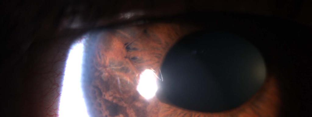
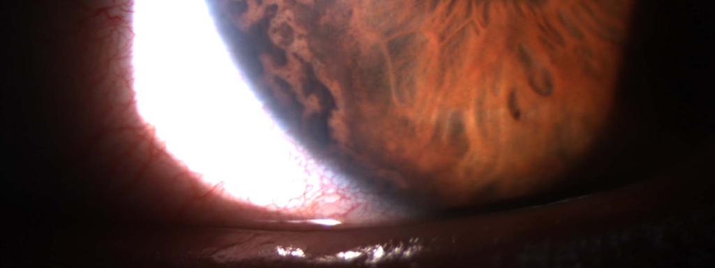
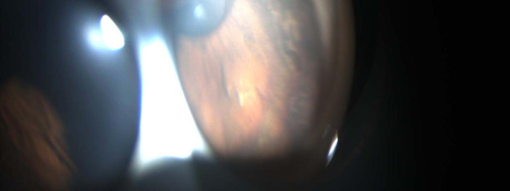
13 Exam L/L: C/S: Cornea: Normal both 2-3+ injection right Clear left 2+ edema right Clear left Endothelium with beaten bronze appearance AC: Iris: Lens: Gonio: Deep and quiet both Right: see photo Left: normal Clear both Right with diffuse PAS, areas of hyperpigmented iris peripherally 8/27/
14 r v s r e t h ig r y p o 8/27/2015 c e h T i n U ity o lo o fc 14 o d a
15
16 Differential Diagnosis 8/27/
17 Differential Diagnosis Iridocorneal Endothelial Sydnrome Uveitic Glaucoma Iridodialysis Posterior polymorphous dystrophy Iris nevus, cyst or tumor (iris melanoma) Siderosis Neovascular glaucoma Epithelial down growth 8/27/
18 Differential Diagnosis Iridocorneal Endothelial Sydnrome Uveitic Glaucoma Iridodialysis Posterior polymorphous dystrophy Iris nevus, cyst or tumor (iris melanoma) Siderosis Neovascular glaucoma Epithelial down growth
19 ICE Iridocorneal Endothelial Sydnrome Unilateral Not heritable Increased risk in middle aged women Patients years old Abnormal corneal endothelium proliferation Migration across tm and onto iris Endothelium has features resembling epithelial cells Possible association with HSV or EBV
20 ICE Iridocorneal Endothelial Sydnrome Unilateral Not heritable Increased risk in middle aged women Patients years old Abnormal corneal endothelium proliferation Migration across tm and onto iris Endothelium has features resembling epithelial cells Possible association with HSV or EBV 8/27/
21 ICE Iridocorneal Endothelial Sydnrome Often asymptomatic Decreased vision Occ monocular diplopia due to iris or pupil abnormalities Corneal changes with beaten bronze appearance to the endothelium in all three forms Glaucoma occurs in 50% of patients with ICE Three forms: Iris Nevus Syndrome (Cogan-Reese), Chandler Syndrome, Essential Iris Atrophy 8/27/


22 Iris Nevus Syndrome Corectopia Pigmented iris nodules from the contraction of proliferating endothelial cells pigmented, pedunculated nodules are composed of iris stroma pinched off by abnormal cellular membrane

23 Chandler Syndrome Corneal changes with beaten bronze appearance to the endothelium Can have corneal edema without increased IOP Most common form, about 50% Yanoff & Ducker, Ophthalmology, 3 rd Ed

24 Essential Iris Atrophy Iris stromal loss with corectopia and ectropion uvea Iris hole formation may be associated with ischemia of the iris Yanoff & Ducker, Ophthalmology, 3 rd Ed

25 Histopathology Essential Iris Atrophy P: peripheral synechia IP: total loss of the central iris pigment epithelium C: cornea CB: ciliary body; IR: iris root L: lens 8/27/ Yanoff & Ducker, Ophthalmology, 3 rd Ed
26 Pathology Corneal endothelium: fine, hammered silver material, similar to the guttae seen in Fuchs corneal endothelial dystrophy This appearance comes from the abnormal endothelial cells located posterior to the normal Descemet s membrane and varies in thickness (normal endothelial cells are a monolayer) The abnormal endothelial cells may have the potential to move Endothelial dysfunction causes the K edema High PAS is caused by the contraction of the endothelial cell layer and surrounding tissues These membranes can cause progressive angle closure. 8/27/
27 TREATMENT IOP and corneal edema are treated directly. If the IOP level remains uncontrolled despite medical treatment, filtration surgery - tube Late surgical failures are due to endothelialization of the fistular opening - may be reopened with a YAG. 8/27/
28 Our patient Diamox 375 bid All IOP lowering gtts maximized IOP remained in the 20s with decreased K edema, Va 20/30 Express placed yesterday 8/27/
Clinical and Specular Microscopic Manifestations of Iridocorneal Endothelial Syndrome
 Clinical and Specular Microscopic Manifestations of Iridocorneal Endothelial Syndrome Yuan-Kuei Liu, I-Jong Wang, Fung-Rong Hu, Por-Tying Hung and Huai-Wen Chang Department of Ophthalmology, National Taiwan
Gonioscopy and Slit Lamp Exam for the Glaucoma Suspect. Disclosure GONIOSCOPY: Gonioscopy Why?? What should I look for? GONIOSCOPY
 Gonioscopy and Slit Lamp Exam for the Glaucoma Suspect Disclosure Michael Chaglasian has the following disclosures:» 1. Advisory Board: Alcon, Allergan, Bausch+Lomb, Carl Zeiss Meditec, Merck, Sucampo»
Fleck. Pre-Descemet Dystrophies (generally good vision and comfort) Primary Pre-Descemet Dystrophy
 Primary Pre-Descemet Dystrophy") Fleck Etiology: bilateral, sometimes asymmetric, autosomal dominant opacities located in all levels of stroma as early as 1 st decade Slit lamp: well demarcated, small round gray-white doughnut-like, wreath-like
Fleck Etiology: bilateral, sometimes asymmetric, autosomal dominant opacities located in all levels of stroma as early as 1 st decade Slit lamp: well demarcated, small round gray-white doughnut-like, wreath-like
Journal of Ophthalmic Medical Technology. Fuchs Dystrophy Amy Hischier
 Journal of Ophthalmic Medical Technology Volume 8, Number 1 October 2013 www.jomtonline.com Fuchs Dystrophy Amy Hischier Patient History: A 55 year old female complained that both of her eyes were red,
Meet Libby. Corneal Dysgenesis, Degeneration, and Dystrophies Definitions. Dr. Victor Malinovsky
 Meet Libby Corneal Dysgenesis, Degeneration, and Dystrophies 2006 Dr. Victor Malinovsky Definitions Dysgenesis: (congenital anomalies) A development disorder that results in a congenital malformation of
Aging & Ophthalmology
 Aging & Ophthalmology Pr Jean-Marie Rakic Dr Denis Malaise January 2018 Major ocular diseases 1. Cataract 2. Age-related macular degeneration 3. Ischemic optic neuropathy 4. Horton arteritis 5. Glaucoma
Around The Globe in 60 Minutes
 Around The Globe in 60 Minutes Around the GLOBE in Sixty Minutes Basic Ocular Anatomy, Examination, and Diagnostic Techniques Introduction Focusing on canine and feline ocular anatomy and basic examination
Morning Report. copyright The University of Colorado. Ashley Laing 09/01/10 Preceptor- Dr. Pantcheva
 Morning Report Ashley Laing 09/01/10 Preceptor- Dr. Pantcheva Case Presentation CC: Right eye pain and headache HPI: 52 yo Caucasian male Presents to ED with OD pain/redness, decreased vision, headache
I ridocorneal endothelial (ICE) syndrome is a rare disease
 syndrome is a rare disease") 64 EXTENDED REPORT Ultrasound biomicroscopy of Chinese eyes with iridocorneal endothelial syndrome M Zhang, J Chen, L Liang, A M Laties, Z Liu... See end of article for authors affiliations... Correspondence
64 EXTENDED REPORT Ultrasound biomicroscopy of Chinese eyes with iridocorneal endothelial syndrome M Zhang, J Chen, L Liang, A M Laties, Z Liu... See end of article for authors affiliations... Correspondence
Cases CFEH. CFEH Facebook Case #4
 CFEH Cases CFEH Facebook Case #4 A 42 year old female has noticed a floater in her left eye for many years but no flashes. She also reports hazy vision in this eye that has been present all her life. She
Differential diagnosis of the red eye. Carol Slight Nurse Practitioner Ophthalmology
 Differential diagnosis of the red eye Carol Slight Nurse Practitioner Ophthalmology The red eye Conjunctivitis HSV Keratitis Acute angle closure glaucoma Anterior Uveitis Red eye Scleritis Subconjunctival
Glaucoma & Inflammation. Jorge L. Fernandez Bahamonde, MD.
 Glaucoma & Inflammation. Jorge L. Fernandez Bahamonde, MD. Definition. Inflammatory ocular conditions compromise outflow of aqueous humor. Keratitis Episcleritis. Scleritis. Uveitis Glaucoma & Keratitis.
Secondary open-angle glaucoma
 Secondary open-angle glaucoma Kathy Hondeghem ZNA Middelheim MaNaMa 12/10/13 Definition Open anterior chamber angle (at least 270 ) Trabecular meshwork (and thus aqueous humor outflow) is occluded by a
The iris naevus (Cogan-Reese) syndrome:
 syndrome:") British Journal of Ophthalmology, 1980, 64, 446-452 The iris naevus (Cogan-Reese) syndrome: light and electron microscopic observations RALPH C. EAGLE, JR., RAMON L. FONT,' MYRON YANOFF,2 AND BEN S. FINE'
British Journal of Ophthalmology, 1980, 64, 446-452 The iris naevus (Cogan-Reese) syndrome: light and electron microscopic observations RALPH C. EAGLE, JR., RAMON L. FONT,' MYRON YANOFF,2 AND BEN S. FINE'
OCULAR DISORDERS REPORT BOSTON TERRIER
 OCULAR DISORDERS REPORT BOSTON TERRIER 1991-1999 2000-2009 2010-2012 TOTAL DOGS EXAMINED 2723 6803 2004 Diagnostic Name # % # % # % GLOBE 0.110 microphthalmia 1 0.0% 1 0.0% 0 EYELIDS 20.140 ectopic cilia
5/2/2016 EYE EMERGENCIES. Nathaniel Pelsor, O.D., FAAO Talley Medical-Surgical Eye Care Associates. Anatomy. Tools
 EYE EMERGENCIES Nathaniel Pelsor, O.D., FAAO Talley Medical-Surgical Eye Care Associates Anatomy Tools 1 Contact dermatitis Blepharitis HSV Preseptal Cellulitis Anterior Chamber Subconjunctival hemorrhage
Morning Report. copyright The University of Colorado. Daniel Corbett, PGY-3. Preceptor: Drs. Singh and Gelston
 Morning Report Daniel Corbett, PGY-3 Preceptor: Drs. Singh and Gelston 3 day old male with report of poor red reflex and cloudy cornea in both eyes OB Hx: Born via SVD @ 39.5 weeks. No trauma/forceps during
dystrophy: value of endothelial specular microscopy
 212 Moorfields Eye Hospital and Institute of Ophthalmology, London H C Laganowski E S Sherrard MG K Muir R J Buckley Correspondence to: H C Laganowski, Moorfields Eye Hospital, City Road, London ECIV 2PD.
What are some common conditions that affect the cornea?
 What are some common conditions that affect the cornea? Injuries After minor injuries or scratches, the cornea usually heals on its own. Deeper injuries can cause corneal scarring, resulting in a haze
Revitalization of the Anterior Segment: Corneal Transplantation and Secondary Lens Repair
 Revitalization of the Anterior Segment: Corneal Transplantation and Secondary Lens Repair CATHERINE REPPA, MD CORNEA SPECIALIST, ASSISTANT PROFESSOR TTUHSC DEPARTMENT OF OPHTHALMOLOGY AND VISUAL SCIENCES
A Curious Case of Bilateral Optic Disc Edema Brittney Dautremont, DO, MPH
 A Curious Case of Bilateral Optic Disc Edema Brittney Dautremont, DO, MPH PGY2 Ophthalmology Resident Grandview Medical Center Dayton, OH CASE PRESENTATION 51 year old white female presenting with blurred
How Strongly Do You Feel That This Patient Has Glaucoma? % % % % %
 My Favorite Cases Anthony B. Litwak, OD, FAAO VA Medical Center Baltimore, Maryland Dr. Litwak is a speaker and on advisory boards for Alcon and Zeiss Meditek CASE CR 35 yohf Neg PMH +FOH mother and grandmother
Eye Care for Animals Micki Armour VMD DACVO THE CORNEA
 Eye Care for Animals Micki Armour VMD DACVO THE CORNEA ANATOMY 0.5-0.6mm thick 4 primary layers Epithelium (5-7 cell layers) Stroma (90% total thickness) Descemet s membrane Endothelium (1 layer) ANATOMY-
3/16/2018. Ultrasound Biomicroscopy in Glaucoma By Ahmed Salah Abdel Rehim. Prof. of Ophthalmology Al-Azhar University
 Ultrasound Biomicroscopy in Glaucoma By Ahmed Salah Abdel Rehim Prof. of Ophthalmology Al-Azhar University 1 Ultrasound biomicroscopy (UBM) is a recent technique to visualize anterior segment with the
My Favourite Cases Anthony B. Litwak, OD, FAAO VA Medical Center Baltimore, MD
 My Favourite Cases Anthony B. Litwak, OD, FAAO VA Medical Center Baltimore, MD Dr. Litwak is a speaker and on advisory boards for Alcon and Zeiss Meditek CASE CR! 35 YOHF! Neg PMH! +FOH mother and grandmother
Chronicity. Narrow Minded. Course Outline. Acute angle closure. Subacute angle closure. Classification of Angle Closure 5/19/2014
 Chronicity Narrow Minded The management of narrow angles in the optometric practice Acute Subacute Chronic Aaron McNulty, OD, FAAO Course Outline Classification of Angle Closure Evaluation of narrow angles
Breaking the Cycle. Yijie (Brittany) Lin, MD, MBA, Reena Garg, MD New York Eye and Ear Infirmary of Mount Sinai
 Lin, MD, MBA, Reena Garg, MD New York Eye and Ear Infirmary of Mount Sinai") Lin, Garg Ophthalmology Times 1 Breaking the Cycle Yijie (Brittany) Lin, MD, MBA, Reena Garg, MD New York Eye and Ear Infirmary of Mount Sinai Abstract A 32 year-old female with a history of LASIK surgery
Lin, Garg Ophthalmology Times 1 Breaking the Cycle Yijie (Brittany) Lin, MD, MBA, Reena Garg, MD New York Eye and Ear Infirmary of Mount Sinai Abstract A 32 year-old female with a history of LASIK surgery
3/20/2018. Top Ten Pathology Pitfalls DEMOGRAPHICS DICTATE DEMOGRAPHICS DICTATE DILATE PATIENTS LOOK AT BOTH EYES. Disclosures Jill Autry, OD, R.Ph.
 Jill Autry, OD, RPh Eye Center of Texas Houston, Sugar Land, Pasadena, Katy, Woodlands, Clear Lake Speaker s Bureau/Consultant/KOL Boards Allergan Alcon B&L Owner/Partner Tropical CE Topicalce.com Eye
Glaucoma Glaucoma is a complication which has only recently been confirmed as a feature of
 1.2.4 OPHTHALMOLOGICAL ABNORMALITIES Ocular abnormalities are well documented in patients with NPS 6 62 81 95. 1.2.4.1 Glaucoma Glaucoma is a complication which has only recently been confirmed as a feature
Corporate Medical Policy
 Corporate Medical Policy Optical Coherence Tomography (OCT) Anterior Segment of the Eye File Name: Origination: Last CAP Review: Next CAP Review: Last Review: optical_coherence_tomography_(oct)_anterior_segment_of_the_eye
ACTIVATED OR NOT? RETINAL CASE PRESENTATION Shorye Payne, MD Medical Retinal Specialist Robley Rex VA Eye Clinic
 ACTIVATED OR NOT? RETINAL CASE PRESENTATION Shorye Payne, MD Medical Retinal Specialist Robley Rex VA Eye Clinic C We anticipate that the future management of posterior uveal melanoma (PUM) will focus
Dr Jo-Anne Pon. Dr Sean Every. 8:30-9:25 WS #70: Eye Essentials for GPs 9:35-10:30 WS #80: Eye Essentials for GPs (Repeated)
") Dr Sean Every Ophthalmologist Southern Eye Specialists Christchurch Dr Jo-Anne Pon Ophthalmologist Southern Eye Specialists, Christchurch Hospital, Christchurch 8:30-9:25 WS #70: Eye Essentials for GPs
Dr Sean Every Ophthalmologist Southern Eye Specialists Christchurch Dr Jo-Anne Pon Ophthalmologist Southern Eye Specialists, Christchurch Hospital, Christchurch 8:30-9:25 WS #70: Eye Essentials for GPs
10 EYE EMERGENCIES. Who goes, who you better not send! Brant Slomovic, MD, FRCPC University Health Network
 10 EYE EMERGENCIES Who goes, who you better not send! Brant Slomovic, MD, FRCPC University Health Network DISCLOSURES I have none PVD CASE 1 WHAT IS A PVD? a process of aging (45-55) liquefaction of vitreous
Management of Angle Closure Glaucoma Hospital Authority Convention 18 May 2015
 Management of Angle Closure Glaucoma Hospital Authority Convention 18 May 2015 Jimmy Lai Clinical Professor Department of Ophthalmology The University of Hong Kong 1 Primary Angle Closure Glaucoma PACG
JOURNAL OF OPHTHALMOLOGY AND RELATED SCIENCES
 JOURNAL OF OPHTHALMOLOGY AND RELATED SCIENCES BILATERAL ACUTE TRANSILLUMINATION OF THE IRIS Kavitha Avadhani 1, MD, MS, Jay Kalliath 1, MS, FRCS 1 Department of Ophthalmology, NMC Speciality Hospital,
_ Assessment of the anterior chamber. Review of anatomy of the angle
 Assessment of the anterior chamber Dr Simon Barnard PhD BSc FCOptom FAAO DCLP Department of Optometry & Visual Science City University London, UK Review of anatomy of the angle Figure 1. Anatomical section
Descemet s membrane endothelial keratoplasty (DMEK) surgery
 surgery") Patient information Descemet s membrane endothelial keratoplasty (DMEK) surgery This information leaflet tells you what to expect if you have DMEK surgery an operation on the cornea of the eye along with
Patient information Descemet s membrane endothelial keratoplasty (DMEK) surgery This information leaflet tells you what to expect if you have DMEK surgery an operation on the cornea of the eye along with
Role of Initial Preoperative Medical Management in Controlling Post-Operative Anterior Uveitis in Patients of Phacomorphic Glaucoma
 Original Article Role of Initial Preoperative Medical Management in Controlling Post-Operative Anterior Uveitis in Patients of Phacomorphic Glaucoma Irfan Qayyum Malik, M. Moin, A. Rehman, Mumtaz Hussain
Ocular Pathology. I. Congenital and/or developmental. A. Trisomy 21. Hypertelorism (widely spaced eyes) Keratoconus (cone shaped cornea)
 Keratoconus (cone shaped cornea)") I. Congenital and/or developmental Robbins Pathologic Basis of Disease, 6 th Ed. A. Trisomy 21 Hypertelorism (widely spaced eyes) Keratoconus (cone shaped cornea) Focal hypoplasia of iris Cataracts frequently
I. Congenital and/or developmental Robbins Pathologic Basis of Disease, 6 th Ed. A. Trisomy 21 Hypertelorism (widely spaced eyes) Keratoconus (cone shaped cornea) Focal hypoplasia of iris Cataracts frequently
OCCLUSIVE VASCULAR DISORDERS OF THE RETINA
 OCCLUSIVE VASCULAR DISORDERS OF THE RETINA Learning outcomes By the end of this lecture the students would be able to Classify occlusive vascular disorders (OVD) of the retina. Correlate the clinical features
Are traditional assessments a waste of time? NZAO 2015
 Are traditional assessments a waste of time? NZAO 2015 Disclosures No financial interests other than Optometry Practice owner Full time optometrist Not a glaucoma prescriber ODOB Board Chair Previously
Ophthalmology Unit Referral Guidelines
 Ophthalmology Unit Referral Guidelines Austin Health Ophthalmology Unit holds sub-specialty sessions to discuss and plan the treatment of patients with specific ocular conditions. General including cataract
Dr. D. Y. Patil Medical College, Pimpri, Pune
 Dr. D. Y. Patil Medical College, Pimpri, Pune - 411 018 Period : 04/July/16 to 22/September/16 Semester : 7 th Semester Department : Ophthalmology Lecture Lesson Plan Sr No Date Topic Learning objectives
Degenerations. Conditions with cloudy cornea at birth or in infancy
 Dermoids The lesions are choristomas, which are congenital masses of tissue that have been dislocated from their normal position Limbal dermoids--overlapping the cornea and sclera, often inferotemporally
PATIENT INFORMATION ON CORNEAL GRAFT
 PATIENT INFORMATION ON CORNEAL GRAFT (TRANSPLANT) SURGERY M ANANDAN What is the cornea? The clear window of the eye approximately 0.5mm thick and 12mm across. It lies in front of the fluid filled anterior
30 Years of Clinical Challenges
 Case RM 30 Years of Clinical Challenges Anthony B. Litwak, OD, FAAO VA Medical Center Baltimore, Maryland 62 yowm PMH: HTN POH unremarkable -FOH c/o eyes are scratchy, uses OTC zaditor BVA 20/20 OD 20/30
5/18/2014. Fundamentals of Gonioscopy Workshop Aaron McNulty, OD, FAAO Walt Whitley, OD, MBA, FAAO
 1 Fundamentals of Gonioscopy Workshop Aaron McNulty, OD, FAAO Walt Whitley, OD, MBA, FAAO 2 3 4 5 6 Optometry s Meeting 2014 The Most Valuable Glaucoma Tool Glaucoma Diagnosis Gonioscopy Central corneal
CATARACT SURGERY IN UVEITIS. Professor Harminder Singh Dua
 Research Institute of Ophthalmology, Cairo 11 th International Conference, 3-4 February, 2017 CATARACT SURGERY IN UVEITIS Professor Harminder Singh Dua MBBS, DO, DO(Lond), MS, MNAMS, FRCS, FRCOphth., FEBO,
02/03/2014. Average Length: 23mm (Infant ~16mm) Approximately the size of a quarter Volume: ~5mL
 Approximately the size of a quarter Volume: ~5mL") Identify the anatomy of the eye. Explain the basic physiology of the parts of the eye. Briefly discuss various surgeries related to different parts of the anatomy. Average Length: 23mm (Infant ~16mm) Approximately
Identify the anatomy of the eye. Explain the basic physiology of the parts of the eye. Briefly discuss various surgeries related to different parts of the anatomy. Average Length: 23mm (Infant ~16mm) Approximately
Systems for Anterior Chamber Angle Evaluation 長庚紀念醫院青光眼科吳秀琛
 Systems for Anterior Chamber Angle Evaluation 長庚紀念醫院青光眼科吳秀琛 Clinical Techniques for Assessing Angle Width A light from the side showing physiological iris bombe Slit lamp-grading of peripheral AC depth
Department of Ophthalmology
 Period : 03/July/17 to 07/September/17 Semester : 7 th Semester Department of Ophthalmology Lecture Lesson Plan Sr 1 03.07.17 Uvea-Anatomy, Uvea-Anatomy, Classification of Uveitis Dr R Paranjpe Classification
Patient AB. Born in 1961 PED
 Clinical Atlas Patient AB Born in 1961 PED Autofluorescence Dilated 45 EasyScan Zero-dilation IR 45 Fundus Dilated 45 In the fundus photos (Canon CX1) the PED is not able to be seen. However, the extent
WHAT IS YOUR DIAGNOSIS? By ADREA R. BENKOFF M.D.
 WHAT IS YOUR DIAGNOSIS? By ADREA R. BENKOFF M.D. Anterior Chamber Inflammation and Iris Depigmentation Noted 25 Years After Cataract Extraction Decreasing Vision Over a 5- Year Period 64 year old white
Department of Ophthalmology
 Department of Ophthalmology Period : 02/July/18 to 30/August/18 Semester : 7 th Semester Lecture Lesson Plan Sr. Date Topic Lesson plan Name of Faculty No. 1 02.07.18 Lens- Lens-Anatomy, Classification
Entropion uveae: Early sphincter atrophy, signposting primary angle closure glaucoma?
 European Journal of Ophthalmology / Vol. 14 no. 4, 2004 / pp. 290-297 Entropion uveae: Early sphincter atrophy, signposting primary angle closure glaucoma? R. SIHOTA, R. SAXENA, H.C. AGARWAL Glaucoma Service,
the raised IOP is associated with a primary ocular or systemic disease.
 Ù Glaucoma - Group of disorders characterized by a progressive op6c neuropathy resul6ng in characteris6c appearance of op6c disc and a specific pa:ern of irreversible visual field defects associated with
SHARED CARE CATARACT SCHEME LUTON & DUNSTABLE HOSPITAL
 Guidelines and Pathway for Optometrists This scheme allows accredited optometrists to assess patients with cataract in the community, and refer those who are visually disabled by cataract directly to the
Secondary Glaucomas. Mr Nick Strouthidis MBBS MD PhD FRCS FRCOphth FRANZCO Consultant Ophthalmologist, Glaucoma Service, Moorfields Eye Hospital
 Secondary Glaucomas Mr Nick Strouthidis MBBS MD PhD FRCS FRCOphth FRANZCO Consultant Ophthalmologist, Glaucoma Service, Moorfields Eye Hospital Introduction: What is glaucoma? Glaucoma is the name given
Ophthalmology. Juliette Stenz, MD
 Ophthalmology Juliette Stenz, MD Required Slide Disclosures NO SIGNIFICANT FINANCIAL, GENERAL, OR OBLIGATION INTERESTS TO REPORT Required Slide At the end of this session, students will be able to: 1.
Understanding Angle Closure
 Case Understanding Angle Closure Dominick L. Opitz, OD, FAAO Associate Professor Illinois College of Optometry 56 year old Caucasian Male Primary Eye Exam BCVA: 20/25 OD with+1.25 DS 20/25 OS with +1.75
Goals. Glaucoma PARA PEARL TO DO. Vision Loss with Glaucoma
 Glaucoma Janet R. Fett, OD Drs. Kincaid, Fett and Tharp So Sioux City, NE eyewear21@hotmail.com Goals Understand Glaucoma Disease process Understand how your data (objective and subjective) assists in
Grand Rounds. CYSTINOSIS Denis Jusufbegovic, M.D. University of Louisville Department of Ophthalmology and Visual Sciences 10/05/12
 Grand Rounds CYSTINOSIS Denis Jusufbegovic, M.D. University of Louisville Department of Ophthalmology and Visual Sciences 10/05/12 Subjective CC: bilateral ocular opacities x yrs HPI: : 11 yo WF referred
Routine OCT and UBM of the anterior segment
 Reprinted from No. 160 december 2012 Volume 17 Routine OCT and UBM of the anterior segment Michel Puech ISSN : 1274-5243 R e p r i n t e d w i t h t h e s u p p o r t o f t h e L a b o r a t o r y Q u
Glaucoma Grand Rounds: What was done wrong? COPE #45911-GL
 Glaucoma Grand Rounds: What was done wrong? COPE #45911-GL Robert E. Prouty, O.D., FAAO Specialty Eye Care Parker, Co RProuty@DrMyii.com Case I: 60 Y/O white female CC: Presents for glaucoma update and
Senile: flattening of vertical meridian, thinning of periphery, lack of luster
 Pterygia Etiology: triangular, fibrovascular, connective tissue overgrowths of bulbar conjunctiva onto cornea; distribution of ultraviolet energy- heat, wind, dust, dry atmosphere,higher prevalence nearer
Preliminary ASCRS CyPass Withdrawal Consensus Statement 1
 Preliminary ASCRS CyPass Withdrawal Consensus Statement 1 ASCRS CyPass Withdrawal Task Force Leads: Douglas Rhee, MD; Nathan Radcliffe, MD; and Francis Mah, MD Glaucoma: Leon Herndon, MD; Marlene Moster,
PRESENTED By DR. FAISAL ALMOBARAK, MD
 PRESENTED By DR. FAISAL ALMOBARAK, MD Early FAC associated with hypotony is an important complication after glaucoma filtering procedures, especially trabeculectomy. The reported incidence after trabeculectomy
ALTERNATIVES TO PHAKIC IMPLANT SURGERY
 Visian ICL Consent INTRODUCTION This information is being provided to you so that you can make an informed decision about having eye surgery to reduce or eliminate your nearsightedness. Only you and your
Grand Rounds. Eddie Apenbrinck M.D. University of Louisville School of Medicine Department of Ophthalmology & Visual Sciences 6/20/2014
 Grand Rounds Eddie Apenbrinck M.D. University of Louisville School of Medicine Department of Ophthalmology & Visual Sciences 6/20/2014 Subjective CC: sudden painless loss of vision OD HPI: 75 year old
Corporate Medical Policy
 Corporate Medical Policy File Name: Origination: Last CAP Review: Next CAP Review: Last Review: endothelial_keratoplasty 9/2009 6/2018 6/2019 6/2018 Description of Procedure or Service Endothelial keratoplasty
A LITTLE ANATOMY. three layers of eye: 1. outer: corneosclera. 2. middle - uvea. anterior - iris,ciliary body. posterior - choroid
 GLAUCOMA A LITTLE ANATOMY three layers of eye: 1. outer: corneosclera 2. middle - uvea anterior - iris,ciliary body posterior - choroid connection at the pars plana between post and ant uvea 3. retina
Subject Index. Atopic keratoconjunctivitis (AKC) management 16 overview 15
 management 16 overview 15") Subject Index Acanthamoeba keratitis, see Infective keratitis Acute allergic conjunctivitis AKC, see Atopic keratoconjunctivitis Allergy acute allergic conjunctivitis 15 atopic keratoconjunctivitis 15
Subject Index Acanthamoeba keratitis, see Infective keratitis Acute allergic conjunctivitis AKC, see Atopic keratoconjunctivitis Allergy acute allergic conjunctivitis 15 atopic keratoconjunctivitis 15
MEDICAL POLICY SUBJECT: CORNEAL ULTRASOUND PACHYMETRY. POLICY NUMBER: CATEGORY: Technology Assessment
 MEDICAL POLICY SUBJECT: CORNEAL ULTRASOUND,, PAGE: 1 OF: 5 If a product excludes coverage for a service, it is not covered, and medical policy criteria do not apply. If a commercial product, including
Why DMEK? EK: Complications and Controversies
 Why DMEK? EK: Complications and Controversies Quicker visual recovery Lower rejection rate Michael J. Taravella, MD Director: Cornea and Refractive Surgery University of Colorado 1 Less stroma? Potential
History. Examination. Diagnosis/Course
 History A 51 year-old female with a history of chronic dry eyes and photosensitivity was referred for evaluation. She reported a five year history of symptoms of frequent irritation and photophobia in
The Anterior Segment & Glaucoma Visual Recognition & Interpretation of Clinical Signs
 The Anterior Segment & Glaucoma Visual Recognition & Interpretation of Clinical Signs Quiz created by Jane Macnaughton MCOptom & Peter Chapman BSc MCOptom FBDO CET Accreditation C19095 2 CET Points (General)
Dystrophies. Molecular Causes. Anterior Membrane Dystrophies (epithelium, basement membrane and Bowman s layer)
") Dystrophies Characteristics of corneal dystrophies About half the members of appropriate age to have the dystrophy( usually autosomal dominant): inherited Usually seen in the first or second decade of
Dystrophies Characteristics of corneal dystrophies About half the members of appropriate age to have the dystrophy( usually autosomal dominant): inherited Usually seen in the first or second decade of
Nate Lighthizer, O.D., F.A.A.O. Assistant Professor, NSUOCO Assistant Dean, Clinical Care Services Director of CE Chief of Specialty Care Clinics
 Nate Lighthizer, O.D., F.A.A.O. Assistant Professor, NSUOCO Assistant Dean, Clinical Care Services Director of CE Chief of Specialty Care Clinics Chief of Electrodiagnostics Clinic lighthiz@nsuok.edu YAG
UC SF. g h. Eye Trauma. Martha Neighbor, MD Emergency Services San Francisco General Hospital University of California
 UC SF Eye Trauma sf g h Martha Neighbor, MD Emergency Services San Francisco General Hospital University of California Goals Recognize vision threatening eye emergencies Treat them when we can Know when
2/6/2018. Andrew Siedlecki, M.D.
 Andrew Siedlecki, M.D. Siedlecki Cataract and Vision Care Optimization- Improved Uncorrected VA Reduced Spectacle Dependency Minimize Complications Fast Rehabilitation Provide Options for Patients Optometrists
FACING YOUR FUNDIC FEARS: EXAMINATION OF THE OCULAR FUNDUS J. Seth Eaton, VMD, DACVO Cornell University Veterinary Specialists
 FACING YOUR FUNDIC FEARS: EXAMINATION OF THE OCULAR FUNDUS J. Seth Eaton, VMD, DACVO Cornell University Veterinary Specialists The goal of a thorough fundus examination is to clinically evaluate the structures
IMAGE OF THE MOMENT PRACTICAL NEUROLOGY
 178 PRACTICAL NEUROLOGY IMAGE OF THE MOMENT Gawn G. McIlwaine*, James H. Vallance* and Christian J. Lueck *Princess Alexandra Eye Pavilion, Chalmers Street, Edinburgh UK; The Canberra Hospital, P.O. Box
FUCH S DYSTROPHY & CATARACT SURGERY TREATMENT ALGORITHM
 FUCH S DYSTROPHY & CATARACT SURGERY TREATMENT ALGORITHM ΙΟΑΝΝΙS Α. MALLIAS, MD, PHD Director of the Dept. of Ophthalmology, Mediterraneo Hospital, Glyfada, Athens, Greece Clinical Fellow in Cornea and
SAFE, PERMANENT EYE-COLOR CHANGE
 SAFE, PERMANENT EYE-COLOR CHANGE Prepared by Gregg Homer JSD (PhD) February 1, 2012 THE PIGMENTARY GLAUCOMA ISSUE Glaucoma Defined Glaucoma is currently defined as a disturbance of the structural or functional
Acute Eyes for ED. Enis Kocak. The Alfred Ophthalmology
 Acute Eyes for ED Enis Kocak The Alfred Ophthalmology The problem with eyes Things to cover Ocular anatomy Basic assessment Common presentations Eye first aid and procedures Ophthalmic emergencies What
Ocular Urgencies and Emergencies
 Ocular Urgencies and Emergencies Pam Boyce, O.D., F.A.A.O. Boyce Family Eye Care, Ltd. 528 Devon Ave. Park Ridge, IL 60068 847-518-0303 Somebody s going to lose an eye Epidemiology 2.4 million ocular and
DNB QUESTIONS 2014 PAPER 1. b) What are the Clinical Conditions in Which Nystagmus is Seen? c) Management of Nystagmus.
 What are the Clinical Conditions in Which Nystagmus is Seen? c) Management of Nystagmus.") DNB QUESTIONS 2014 PAPER 1 1. a) How Will you investigate a case of Nystagmus? b) What are the Clinical Conditions in Which Nystagmus is Seen? c) Management of Nystagmus. 2. a) What is the Principle of
DNB QUESTIONS 2014 PAPER 1 1. a) How Will you investigate a case of Nystagmus? b) What are the Clinical Conditions in Which Nystagmus is Seen? c) Management of Nystagmus. 2. a) What is the Principle of
Corneal Ulceration. Client Information Sheet Copyright Bilton Veterinary Centre All rights Reserved. What is the cornea?
 What is the cornea? Corneal Ulceration The cornea is the central clear part of the eye that is surrounded by the white of the eye called the Sclera. Looking through the cornea, you can see the coloured
Anterior segment imaging
 Article Date: 11/1/2016 Anterior segment imaging AS OCT vs. UBM vs. endoscope; case based approaches BY BENJAMIN BERT, MD, FACS AND BRIAN FRANCIS, MD, MS Currently, numerous imaging modalities are available
GLAUCOMA SUMMARY BENCHMARKS FOR PREFERRED PRACTICE PATTERN GUIDELINES
 SUMMARY BENCHMARKS FOR PREFERRED PRACTICE PATTERN GUIDELINES Introduction These are summary benchmarks for the Academy s Preferred Practice Pattern (PPP) guidelines. The Preferred Practice Pattern series
Infra-red transillumination stereophotography of the iris in Fuchs's heterochromic cyclitis
 British Journal of Ophthalmology, 1978, 62, 110-115 Infra-red transillumination stereophotography of the iris in Fuchs's heterochromic cyclitis M. SAARI, I. VUORRE, AND H. NIEMINEN From the University
Downloaded from:
 Philippin, H; Shah, P; Burton, M (2012) The next step: Detailed assessment of an adult glaucoma patient. Community eye health / International Centre for Eye Health, 25 (79-80). pp. 50-53. ISSN 0953-6833
THE EYE: RETINA AND GLOBE
 Neuroanatomy Suzanne Stensaas February 24, 2011, 10:00-12:00 p.m. Reading: Waxman Ch. 15. Your histology and gross anatomy books should be useful. Reading: Histology of the Eye from any histology book
Ultrasound Biomicroscopy & Glaucoma Care
 S P E C IA L S E C T I O N S P O N S O R E D B Y Q UA N T E L M E D I C A L Ultrasound Biomicroscopy & Glaucoma Care Visualizing angle closure and its mechanisms from screening to post-surgical assessment
THE CHRONIC GLAUCOMAS
 THE CHRONIC GLAUCOMAS WHAT IS GLAUCOMA People with glaucoma have lost some of their field of all round vision. It is often the edge or periphery that is lost. That is why the condition can be missed until
JINNAH SINDH MEDICAL UNIVERSITY STUDY GUIDE- OPHTHALMOLOGY YEAR 4,
 INTRODUCTION Pakistan, the 7th most populous country in the world, has an urban population of 38.8% and rural dwellers of 61.2%. The country has faced challenges with vision impairment and blindness as
ANTERIOR SEGMENT EXAMINATION TECHNIQUES
 ANTERIOR SEGMENT EXAMINATION TECHNIQUES GERS October 2017 Amanda Harding - Principal Optometrist, MREH MSc., MCOptom, Dip Glauc., Dip TP (IP). Anterior Segment Examination Depth of anterior chamber Central
Anterior segment imaging
 CET CONTINUING Sponsored by 1 CET POINT Anterior segment imaging Sundeep Vaswani, BSc (Hons), MCOptom 39 The anterior segment of the eye encompasses all structures from the front surface of the cornea
Vascular changes in the iris in chronic
 Vascular changes in the iris in chronic anterior uveitis LEILA LAATIKAINEN From the Department of Ophthalmology, University of Helsinki, Finland British Journal of Ophthalmology, 1979, 63, 145-149 SUMMARY
OPHTHALMOLOGY DEPARTMENT Primary care referral guidelines
 OPHTHALMOLOGY DEPARTMENT Primary care referral guidelines Contents REFERRAL CATEGIES... 2 Emergency... 2 Urgent... 2 Semi urgent/routine... 2 Not accepted... 2 OPHTHALMOLOGY CONDITIONS NOT ACCEPTED...