OCCLUSIVE VASCULAR DISORDERS OF THE RETINA
|
|
|
- Benedict Chapman
- 5 years ago
- Views:
Transcription
1 OCCLUSIVE VASCULAR DISORDERS OF THE RETINA
2 Learning outcomes By the end of this lecture the students would be able to Classify occlusive vascular disorders (OVD) of the retina. Correlate the clinical features of OVD of the retinal with the underlying pathophysiological changes. List treatment modalities for OVD List the complications of OVDs
3 Let us look at a case a 54-year-old school teahcer presented with acute visual loss in the left eye. He had history of uncontrolled hypertension and open angle glaucoma. Laboratory tests including complete hypercoagulability and thrombotic workup were completed. Ophthalmologic exam showed best corrected visual acuity (BCVA)of 6/9 OD, 6/60 OS. Pupil exam showed sluggish right pupil with relative afferent papillary defect, and a reactive pupil on the left. Intraocular pressures were 19 mmhg in the right, and 11 mmhg in the left. Slit lamp exam showed normal anterior segments with open angles bilaterally.
4
5 Retinal vein occlusion Central retinal vein occlusion (CRVO) Branch retinal vein occlusion (BRVO) Hemiretinal vein occlusion (HRVO)

6 BRVO CRVO HRVO
7 Epidemiology Retinal vein occlusion is the second most common cause of visual loss due to retinal vascular disease BRVO is the most common type It is a significant cause of severe visual loss in people over the age of 40 yrs


8 Pathophysiology Can you think of some anatomical factors that could predispose retinal veins to occlusion?
9 Pathophysiology Various local and systemic factors play a role in the pathological closure of the retinal veins The central retinal artery and vein share a common adventitial sheath as they exit the optic nerve head and pass through a narrow opening in the lamina cribrosa. Retinal arterioles and their corresponding vein share the adventitia. Thickening & hardening of the arteries can compress the veins. This anatomical position predisposes to thrombus formation in the central retinal vein by various factors, including slowing of the blood stream, changes in the vessel wall, and changes in the viscosity of the blood. Arteriosclerotic changes in the central retinal artery transform the artery into a rigid structure and impinge upon the pliable central retinal vein, causing hemodynamic disturbances, endothelial damage, and thrombus formation.


10 With occlusion of the central retinal vein (CRVO) increased venous & capillary pressure stagnation of the blood in the retinal venous system and increased resistance to venous blood flow ischemic damage to the retina increased production of vascular endothelial growth factor (VEGF) neovascularization of the posterior and anterior segment Capillary leakage Neovascular Glaucoma, NVD, NVE,RD Complications of RVO Macular oedema
11 CRVO With occlusion of the vein there is increased venous & capillary pressure and stagnation of the blood in the retinal venous system and increased resistance to venous blood flow. ischemic damage to the retina increased production of vascular endothelial growth factor (VEGF) in the vitreous cavity. Increased levels of VEGF stimulate neovascularization of the posterior and anterior segment responsible for secondary complications due to RVO. VEGF causes capillary leakage leading to macular edema. The prognosis depends upon the reestablishment of patency of the venous system by recanalization, dissolution of clot, or formation of optociliary shunt vessels.

12 Risk factors Blood Systemic Vessel wall
13 Risk factors Age: 50% of these cases occur in patient over 65 years of age Systemic diseases like diabetes mellitus, hypertension, hyperlipidemics states, smoking & obesity Inflammatory diseases like sarcoidosis, Bechet disease Hyperviscosity syndromes like polycythemia, paraproteinemias Clotting disorders - Activated protein C resistance, lupus anticoagulant, anticardiolipin antibodies, protein C, protein S, antithrombin III Raised Intraocular pressure (IOP) Oral contraceptive use
14 CRVO Clinical presentation Clinical entities Non ischemic CRVO (about 75% of cases) Ischemic CRVO (worse prognosis) How would you diagnose retinal ischemia?
15 CRVO Clinical presentation Asymptomatic Decreased vision Visual loss sudden or gradual, over a period of days to weeks. ranges from mild to severe. Patients can present with transient obscurations of vision initially, later progressing to constant visual loss. Photophobia Redness of eyes Painful blind eye
16 Clinical examination Patients should undergo a complete eye examination, including visual acuity, pupillary reactions, slit lamp examination of the anterior and posterior segments, undilated examination of the iris, gonioscopy, Dilated fundus examination
17 Signs Visual acuity: (Best-corrected vision acuity) It is one of the important indicators of the final visual prognosis. Pupillary reactions: normal/ relative afferent pupillary reflex. If the iris has abnormal blood vessels, the pupil may not react. Conjunctiva: Advanced stages may show congestion on conjunctival and ciliary vessels. Cornea: Advanced stages may show diffuse corneal edema obscuring the visibility of internal structures. Iris: normal/ neovascularization The anterior chamber angle: it may show neovascularization with open angles and later show total peripheral anterior synechia and closed angles.
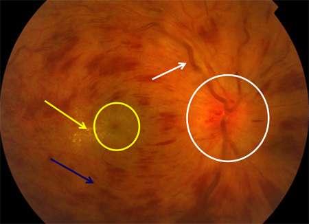
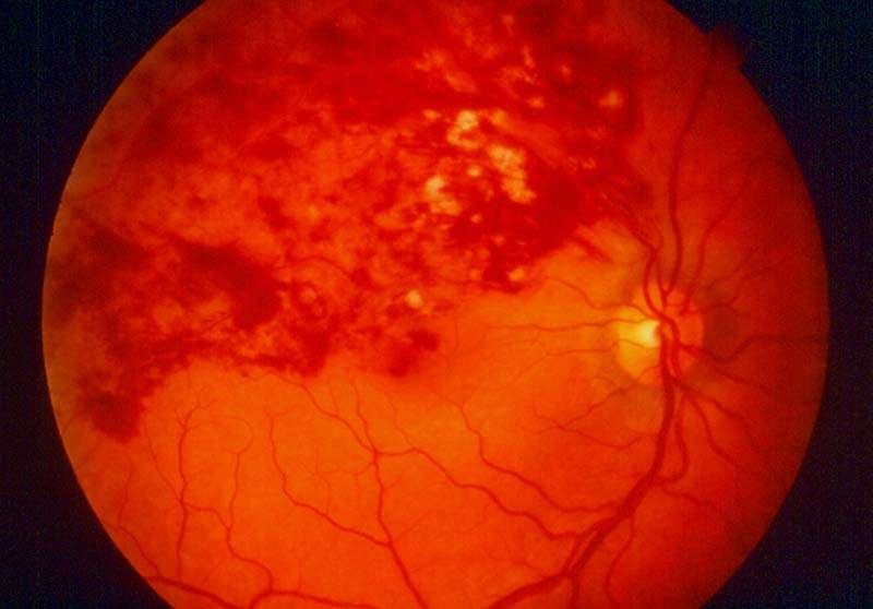
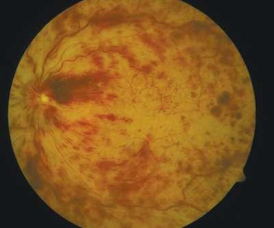
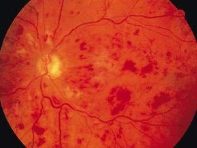
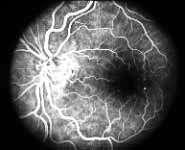
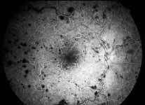
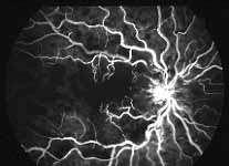
18 Signs Fundus examination: Retinal hemorrhages Dilated tortuous veins Optic disc edema Cotton-wool spots Macular oedema Late signs: Neovascularization (NVD, NVE), optic disce cupping, optociliary shunt vessels at the disc (a prognostic sign), pigmentary changes in the macula
19
20
21
22
23
24

25 Rubeosis 100 day glaucoma
26 Investigations Risk factors screening (Lab tests) FFA Electro-retinogram (ERG)- amplitude of the b-wave is decreased relative to the a-wave
27
28 TREATMENT Depends on the type & stage of CRVO Principles of treatment are Treat the underlying cause Monitor Treatment modalities Intravitreal corticosteroid and anti-vegf injections Dexamethasone intravitreal implant Laser photocoagulation Chorioretinal venous anastomosis
29 Follow -up Regular follow up babysit" 1 for these eyes during that period when they are at maximum risk of developing neovascular glaucoma, i.e. first 7-8 months
30 Prognosis For nonischemic CRVO, complete recovery with good visual recovery occurs only in about 10% of cases. Fifty percent of patients will have 6/60 or worse vision. About one third of patients convert to ischemic CRVO within 3 years; 15% within the first 4 months. For ischemic CRVO, more than 90% of patients will have 6/60 or worse vision. About 60% of patients develop ocular neovascularization About 10% of patients can develop CRVO or other type of vein occlusions within either the same eye or the contralateral eye within 2 years.
31 Conclusion? Lacrimal system Cornea Conjunctiva Cataract Refractive errors
Vascular Disease Ocular Manifestations of Systemic Hypertension
 Vascular Disease Ocular Manifestations of Systemic Hypertension Maynard L. Pohl, OD, FAAO Pacific Cataract & Laser Institute 10500 NE 8 th Street, Suite 1650 Bellevue, WA 98004 USA 425-462-7664 Cerebrovascular
Venous Occlusive Diseases
 Venous Occlusive Diseases Bruce R. Saran, MD Adjunct Assistant Clinical Professor of Medicine Scheie Eye Institute University of Pennsylvania School of Medicine Philadelphia, PA -a division of: RVO Demographics
Retinal Vein Occlusion (RVO) Treatment pathway- Northeast England. Retinal Vein Occlusion (RVO) with Macular oedema (MO)
 Treatment pathway- Northeast England. Retinal Vein Occlusion (RVO) with Macular oedema (MO)") Retinal Vein Occlusion (RVO) Treatment pathway- Northeast England (Royal Victoria Infirmary, Sunderland Eye Infirmary, James Cook University Hospital, Darlington Memorial Hospital, University Hospital
Retinal Vein Occlusion (RVO) Treatment pathway- Northeast England (Royal Victoria Infirmary, Sunderland Eye Infirmary, James Cook University Hospital, Darlington Memorial Hospital, University Hospital
Neovascular Glaucoma Associated with Cilioretinal Artery Occlusion Combined with Perfused Central Retinal Vein Occlusion
 Neovascular Glaucoma Associated with Cilioretinal Artery Occlusion Combined with Perfused Central Retinal Vein Occlusion Man-Seong Seo,* Jae-Moon Woo* and Jeong-Jin Seo *Department of Ophthalmology, Chonnam
Diabetic Retinopathy. Barry Emara MD FRCS(C) Giovanni Caboto Club October 3, 2012
 Giovanni Caboto Club October 3, 2012") Diabetic Retinopathy Barry Emara MD FRCS(C) Giovanni Caboto Club October 3, 2012 Outline Statistics Anatomy Categories Assessment Management Risk factors What do you need to do? Objectives Summarize the
Diabetic Retinopathy Barry Emara MD FRCS(C) Giovanni Caboto Club October 3, 2012 Outline Statistics Anatomy Categories Assessment Management Risk factors What do you need to do? Objectives Summarize the
 measure of your overall performance. An isolated glucose test is helpful to let you know what your sugar level is at one moment, but it doesn t tell you whether or not your diabetes is under adequate control
4/27/2010 INTRODUCTION TO RETINAL VASCULAR DISEASE VENOUS/VENULAR CENTRAL RETINAL VEIN OBSTRUCTION / CRVO ADDITIONAL FEATURES /COMPLICATIONS
 INTRODUCTION TO RETINAL VASCULAR DISEASE VENOUS/VENULAR Leo Semes, OD Professor, UAB Optometry 2 CENTRAL RETINAL VEIN OBSTRUCTION CENTRAL RETINAL VEIN OBSTRUCTION / OCCLUSION (CRVO) obstruction of the
Recurrent intraocular hemorrhage secondary to cataract wound neovascularization (Swan Syndrome)
") Recurrent intraocular hemorrhage secondary to cataract wound neovascularization (Swan Syndrome) John J. Chen MD, PhD; Young H. Kwon MD, PhD August 6, 2012 Chief complaint: Recurrent vitreous hemorrhage,
Recurrent intraocular hemorrhage secondary to cataract wound neovascularization (Swan Syndrome) John J. Chen MD, PhD; Young H. Kwon MD, PhD August 6, 2012 Chief complaint: Recurrent vitreous hemorrhage,
Mild NPDR. Moderate NPDR. Severe NPDR
 Diabetic retinopathy Diabetic retinopathy is the most common cause of blindness in adults aged 35-65 years-old. Hyperglycaemia is thought to cause increased retinal blood flow and abnormal metabolism in
Preliminary report on effect of retinal panphotocoagulation on rubeosis iridis and
 British Journal of Ophthalmology, 1977, 61, 278-284 Preliminary report on effect of retinal panphotocoagulation on rubeosis iridis and neovascular glaucoma LEILA LAATIKAINEN From Moorfields Eye Hospital,
FRANZCO, MD, MBBS. Royal Darwin Hospital
 Diabetes and Eye By Dr. Nishantha Wijesinghe FRANZCO, MD, MBBS Consultant Ophthalmologist Royal Darwin Hospital 98% of Diabetics do not need to suffer from severe visual loss Yet Diabetic eye disease is
Case Follow Up. Sepi Jooniani PGY-1
 Case Follow Up Sepi Jooniani PGY-1 Triage 54 year old M Pt presents to prelim states noticed today he had reddness to eyes, states worse in R eye. Pt denies any pain or itching. No further complaints.
RVO RETINAL VEIN OCCLUSION
 RVO RETINAL VEIN OCCLUSION A guide to understanding RVO Take some time to learn about RVO - it may help you hold on to your vision Retinal vein occlusion is a common disorder of the retina and a leading
PART 1: GENERAL RETINAL ANATOMY
 PART 1: GENERAL RETINAL ANATOMY General Anatomy At Ora Serrata At Optic Nerve Head Fundoscopic View Of Normal Retina What Is So Special About Diabetic Retinopathy? The WHO definition of blindness is
NEOVASCULAR GLAUCOMA IN A NIGERIAN AFRICAN POPULATION
 October 2006 E AST AFRICAN MEDICAL JOURNAL 559 East African Medical Journal Vol. 83 No. 10 October 2006 NEOVASCULAR GLAUCOMA IN A NIGERIAN AFRICAN POPULATION: A.O. Ashaye, FWACS, MSc, Department of Ophthalmology,
The Common Clinical Competency Framework for Non-medical Ophthalmic Healthcare Professionals in Secondary Care
 The Common Clinical Competency Framework for Non-medical Ophthalmic Healthcare Professionals in Secondary Care Medical Retina November 2016 Association of Health Professions in Ophthalmology General basic
RETINAL VEIN OCCLUSIONS (RVO) PREFERRED PRACTICE PATTERNS (PPP) Philippines: 2016
 PREFERRED PRACTICE PATTERNS (PPP) Philippines: 2016") RETINAL VEIN OCCLUSIONS (RVO) PREFERRED PRACTICE PATTERNS (PPP) Philippines: 2016 The Retinal Vein Occlusions (RVO) Preferred Practice Patterns (PPP) Philippines: 2016 was prepared by the VitreoRetina
RETINAL VEIN OCCLUSIONS (RVO) PREFERRED PRACTICE PATTERNS (PPP) Philippines: 2016 The Retinal Vein Occlusions (RVO) Preferred Practice Patterns (PPP) Philippines: 2016 was prepared by the VitreoRetina
The Human Eye. Cornea Iris. Pupil. Lens. Retina
 The Retina Thin layer of light-sensitive tissue at the back of the eye (the film of the camera). Light rays are focused on the retina then transmitted to the brain. The macula is the very small area in
FA Conference. Lara Rosenwasser Newman, M.D. 10/2/14 University of Louisville Department of Ophthalmology and Visual Sciences
 FA Conference Lara Rosenwasser Newman, M.D. 10/2/14 University of Louisville Department of Ophthalmology and Visual Sciences Patient Presentation CC: (sent by optometrist) Blurry/foggy vision HPI: 62 yo
Diagnosis and treatment of diabetic retinopathy. Blake Cooper MD Ophthalmologist Vitreoretinal Surgeon Retina Associates Kansas City
 Diagnosis and treatment of diabetic retinopathy Blake Cooper MD Ophthalmologist Vitreoretinal Surgeon Retina Associates Kansas City Disclosures Consulted for Novo Nordisk 2017,2018. Will be discussing
GLAUCOMA SUMMARY BENCHMARKS FOR PREFERRED PRACTICE PATTERN GUIDELINES
 SUMMARY BENCHMARKS FOR PREFERRED PRACTICE PATTERN GUIDELINES Introduction These are summary benchmarks for the Academy s Preferred Practice Pattern (PPP) guidelines. The Preferred Practice Pattern series
ZEISS AngioPlex OCT Angiography. Clinical Case Reports
 Clinical Case Reports Proliferative Diabetic Retinopathy (PDR) Case Report 969 PROLIFERATIVE DIABETIC RETINOPATHY 1 1-year-old diabetic female presents for follow-up of proliferative diabetic retinopathy
Treatment of Retinal Vein Occlusion (RVO)
") Manchester Royal Eye Hospital Medical Retina Services Information for Patients Treatment of Retinal Vein Occlusion (RVO) What is a Retinal Vein Occlusion (RVO)? The retina is the light sensitive layer
Manchester Royal Eye Hospital Medical Retina Services Information for Patients Treatment of Retinal Vein Occlusion (RVO) What is a Retinal Vein Occlusion (RVO)? The retina is the light sensitive layer
LECTURE # 7 EYECARE REVIEW: PART III
 LECTURE # 7 EYECARE REVIEW: PART III HOW TO TRIAGE EYE EMERGENCIES STEVE BUTZON, O.D. EYECARE REVIEW: HOW TO TRIAGE EYE EMERGENCIES FOR PRIMARY CARE PHYSICIANS Steve Butzon, O.D. Member Director IDOC President
The Foundation WHAT IS THE RETINA?
 The Foundation American Society of Retina Specialists Committed to improving the quality of life of all people with retinal disease. Branch Retinal Vein Occlusion Retinal vein occlusions occur when there
ROLE OF LASER PHOTOCOAGULATION VERSUS INTRAVITREAL TRIAMCINOLONE ACETONIDE IN ANGIOGRAPHIC MACULAR EDEMA IN DIABETES MELLITUS
 ORIGINAL ARTICLE ROLE OF LASER PHOTOCOAGULATION VERSUS INTRAVITREAL TRIAMCINOLONE ACETONIDE IN ANGIOGRAPHIC MACULAR EDEMA IN DIABETES MELLITUS Aggarwal Somesh VP 1, Shah Sonali N 2, Bharwada Rekha M 3,
OCULAR MANIFESTATIONS OF SYSTEMIC DISEASES THUCANH MULTERER, MD
 OCULAR MANIFESTATIONS OF SYSTEMIC DISEASES THUCANH MULTERER, MD UNDERGRADUATE: Philadelphia College of Pharmacy and Science 1996 MEDICAL SCHOOL: MCP Hahnemann School of Medicine, Philadelphia PA 2000 RESIDENCY:
Dr/ Marwa Abdellah EOS /16/2018. Dr/ Marwa Abdellah EOS When do you ask Fluorescein angiography for optic disc diseases???
 When do you ask Fluorescein angiography for optic disc diseases??? 1 NORMAL OPTIC DISC The normal optic disc on fluorescein angiography is fluorescent due to filling of vessels arising from the posterior
2/26/2017. Sameh Galal. M.D, FRCS Glasgow. Lecturer of Ophthalmology Research Institute of Ophthalmology
 Sameh Galal M.D, FRCS Glasgow Lecturer of Ophthalmology Research Institute of Ophthalmology No financial interest in the subject presented 1 Managing cataracts in children remains a challenge. Treatment
Central Retinal Vein Occlusion
 Central Retinal Vein Occlusion Sohan Singh Hayreh, MD, MS, PhD, DSc, FRCS, FRCOphth Professor Emeritus of Ophthalmology Ocular Vascular Clinic Department of Ophthalmology and Visual Sciences The University
PRECISION PROGRAM. Injection Technique Quick-Reference Guide. Companion booklet for the Video Guide to Injection Technique
 Injection Technique Quick-Reference Guide PRECISION PROGRAM Companion booklet for the Video Guide to Injection Technique Available at www.ozurdexprecisionprogram.com Provides step-by-step directions with
Case Report: Indocyanine Green Dye Leakage from Retinal Artery in Branch Retinal Vein Occlusion
 Case Report: Indocyanine Green Dye Leakage from Retinal Artery in Branch Retinal Vein Occlusion Hiroki Fujita, Kyoko Ohno-Matsui, Soh Futagami and Takashi Tokoro Department of Visual Science, Tokyo Medical
Central retinal vein occlusion in a young adult Case report
 Romanian Journal of Ophthalmology, Volume 60, Issue 2, April-June 2016. pp:120-124 CASE REPORT Central retinal vein occlusion in a young adult Case report Călugăru Dan*, Călugăru Mihai** *Department of
The Anterior Segment & Glaucoma Visual Recognition & Interpretation of Clinical Signs
 The Anterior Segment & Glaucoma Visual Recognition & Interpretation of Clinical Signs Quiz created by Jane Macnaughton MCOptom & Peter Chapman BSc MCOptom FBDO CET Accreditation C19095 2 CET Points (General)
Chris Brown, M.D. Eye Specialty Group, PLC Continuing Education Series
 Chris Brown, M.D. Eye Specialty Group, PLC 2018 Continuing Education Series Disclaimer I have no financial interests in this lecture or any information discussed therein Objectives Fluorescein Angiogram
Speaker Disclosure Statement. " Dr. Tim Maillet and Dr. Vladimir Kozousek have no conflicts of interest to disclose.
 Speaker Disclosure Statement Dr. Tim Maillet and Dr. Vladimir Kozousek have no conflicts of interest to disclose. Diabetes Morbidity Diabetes doubles the risk of stroke. Diabetes quadruples the risk of
What you can expect with OZURDEX
 Important Information About Macular Edema Following Branch or Central Retinal Vein Occlusion (RVO) and Treatment For patients with RVO What you can expect with OZURDEX Approved Use OZURDEX (dexamethasone
Perspectives on Screening for Diabetic Retinopathy. Dr. Dan Samaha, Optometrist, MSc Clinical Lecturer School of Optometry, Université de Montréal
 Perspectives on Screening for Diabetic Retinopathy 1 Dr. Dan Samaha, Optometrist, MSc Clinical Lecturer School of Optometry, Université de Montréal Current standards 2 According to the Canadian Diabetes
Moncef Khairallah, MD
 Moncef Khairallah, MD Department of Ophthalmology, Fattouma Bourguiba University Hospital Faculty of Medicine, University of Monastir Monastir, Tunisia INTRODUCTION IU: anatomic form of uveitis involving
The Common Clinical Competency Framework for Non-medical Ophthalmic Healthcare Professionals in Secondary Care
 The Common Clinical Competency Framework for Non-medical Ophthalmic Healthcare Professionals in Secondary Care Cataract November 2016 Association of Health Professions in Ophthalmology General basic competences
Prognosis for rubeosis iridis following central
 British Journal of Ophthalmology, 1979, 63, 735-743 Prognosis for rubeosis iridis following central retinal vein occlusion STEPHEN H. SINCLAIR AND EVANGELOS S. GRAGOUDAS From the Eye Research Institute
Diabetic Retinopathy
 Diabetic Retinopathy Diabetes can be classified into type 1 diabetes mellitus and type 2 diabetes mellitus, formerly known as insulin-dependent diabetes mellitus, and non-insulin diabetes mellitus, respectively.
NATIONAL INSTITUTE FOR HEALTH AND CARE EXCELLENCE. Health Technology Appraisal. Aflibercept for treating diabetic macular oedema.
 NATIONAL INSTITUTE FOR HEALTH AND CARE EXCELLENCE Health Technology Appraisal Aflibercept for treating diabetic macular oedema Final scope Final remit/appraisal objective To appraise the clinical and cost
Interferon-Associated Retinopathy: Communicating with Internal Medicine Ari Wes, Esther S. Hong, MD, and Thomas A. Oetting, MS, MD
 Interferon-Associated Retinopathy: Communicating with Internal Medicine Ari Wes, Esther S. Hong, MD, and Thomas A. Oetting, MS, MD July 26, 2010 Chief Complaint: New floaters in both eyes. History of Present
Optimal Treatment of Retinal Vein Occlusion: Canadian Expert Consensus
 Original Paper Received: December 9, 2014 Accepted: February 25, 2015 Published online: June 12, 2015 Optimal Treatment of Retinal Vein Occlusion: Canadian Expert Consensus Alan R. Berger a Alan F. Cruess
EyePACS Grading System (Part 3): Detecting Proliferative (Neovascular) Diabetic Retinopathy. George Bresnick MD MPA Jorge Cuadros OD PhD
: Detecting Proliferative (Neovascular) Diabetic Retinopathy. George Bresnick MD MPA Jorge Cuadros OD PhD") EyePACS Grading System (Part 3): Detecting Proliferative (Neovascular) Diabetic Retinopathy George Bresnick MD MPA Jorge Cuadros OD PhD Anatomy of the eye: 3 Normal Retina Retinal Arcades Macula Optic
EyePACS Grading System (Part 3): Detecting Proliferative (Neovascular) Diabetic Retinopathy George Bresnick MD MPA Jorge Cuadros OD PhD Anatomy of the eye: 3 Normal Retina Retinal Arcades Macula Optic
3/16/2018. Optic Nerve Examination. Hassan Eisa Swify FRCS Ed (Ophthalmology) Air Force Hospital
 Air Force Hospital") Optic Nerve Examination Hassan Eisa Swify FRCS Ed (Ophthalmology) Air Force Hospital 1 Examination Structure ( optic disc) Function Examination of the optic disc The only cranial nerve (brain tract) which
Optic Nerve Examination Hassan Eisa Swify FRCS Ed (Ophthalmology) Air Force Hospital 1 Examination Structure ( optic disc) Function Examination of the optic disc The only cranial nerve (brain tract) which
Year 2 MBChB Clinical Skills Session Ophthalmoscopy. Reviewed & ratified by: Mr M Batterbury Consultant Ophthalmologist
 Year 2 MBChB Clinical Skills Session Ophthalmoscopy Reviewed & ratified by: o Mr M Batterbury Consultant Ophthalmologist Learning objectives o To understand the anatomy and physiology of the external and
Clinically Significant Macular Edema (CSME)
") Clinically Significant Macular Edema (CSME) 1 Clinically Significant Macular Edema (CSME) Sadrina T. Shaw OMT I Student July 26, 2014 Advisor: Dr. Uwaydat Clinically Significant Macular Edema (CSME) 2
Clinically Significant Macular Edema (CSME) 1 Clinically Significant Macular Edema (CSME) Sadrina T. Shaw OMT I Student July 26, 2014 Advisor: Dr. Uwaydat Clinically Significant Macular Edema (CSME) 2
Macular edema (ME) is the most common
 is the most common") MANAGEMENT OF RETINAL VEIN OCCLUSIONS * Peter A. Campochiaro, MD ABSTRACT Macular edema (ME) is the most common cause of reduced vision in patients with retinal vein occlusions (RVOs). The primary cause
MANAGEMENT OF RETINAL VEIN OCCLUSIONS * Peter A. Campochiaro, MD ABSTRACT Macular edema (ME) is the most common cause of reduced vision in patients with retinal vein occlusions (RVOs). The primary cause
Update and review of central retinal vein occlusion Nikolas J.S. London and Gary Brown
 Update and review of central retinal vein occlusion Nikolas J.S. London and Gary Brown Retina Service, Wills Eye Institute, Thomas Jefferson University, Philadelphia, Pennsylvania, USA Correspondence to
Diabetic Retinopathy Screening Program in the Cree Region of James Bay of Quebec
 RUIS McGILL VIRTUAL HEALTH AND SOCIAL SERVICES CENTRE (CvSSS) SIMPLIFYING TELEHEALTH! Diabetic Retinopathy Screening Program in the Cree Region of James Bay of Quebec Nurse and Imager Training Prepared
The Common Clinical Competency Framework for Non-medical Ophthalmic Healthcare Professionals in Secondary Care
 The Common Clinical Competency Framework for Non-medical Ophthalmic Healthcare Professionals in Secondary Care Glaucoma November 2016 Association of Health Professions in Ophthalmology General basic competences
A Case of Carotid-Cavernous Fistula
 A Case of Carotid-Cavernous Fistula By : Mohamed Elkhawaga 2 nd Year Resident of Ophthalmology Alexandria University A 19 year old male patient came to our outpatient clinic, complaining of : -Severe conjunctival
Marcus Gonzales, OD, FAAO Cedar Springs Eye Clinic
 Marcus Gonzales, OD, FAAO Cedar Springs Eye Clinic 25.6 million adults 11.3% of the adult population 10.9 million adults 65 years and older 26.9% of this age population 79 million people are Pre-diabetic!!
Management of Angle Closure Glaucoma Hospital Authority Convention 18 May 2015
 Management of Angle Closure Glaucoma Hospital Authority Convention 18 May 2015 Jimmy Lai Clinical Professor Department of Ophthalmology The University of Hong Kong 1 Primary Angle Closure Glaucoma PACG
Diabetic and the Eye: An Introduction
 Diabetic and the Eye: An Introduction Lawrence Iu FRCSEd (Ophth), FCOphthHK, FHKAM (Ophthalmology) Department of Ophthalmology, Grantham Hospital & Queen Mary Hospital Background Diabetes mellitus (DM)
Clinical Case Presentation. Branch Retinal Vein Occlusion. Sarita M. Registered Nurse Whangarei Base Hospital
 Clinical Case Presentation on Branch Retinal Vein Occlusion Sarita M. Registered Nurse Whangarei Base Hospital Introduction Case Study Pathogenesis Clinical Features Investigations Treatment Follow-up
Neuropathy (NAION) and Avastin. Clinical Assembly of the AOCOO-HNS Foundation May 9, 2013
 and Avastin. Clinical Assembly of the AOCOO-HNS Foundation May 9, 2013") Non Arteritic Ischemic Optic Neuropathy (NAION) and Avastin Shalom Kelman, MD Clinical Assembly of the AOCOO-HNS Foundation May 9, 2013 Anterior Ischemic Optic Neuropathy Acute, painless, visual loss,
Non Arteritic Ischemic Optic Neuropathy (NAION) and Avastin Shalom Kelman, MD Clinical Assembly of the AOCOO-HNS Foundation May 9, 2013 Anterior Ischemic Optic Neuropathy Acute, painless, visual loss,
 Research Article http://www.alliedacademies.org/clinical-ophthalmology-and-vision-science/ A 2-year retrospective study of the treatment of retinal vein occlusion with dexamethasone 0.7 mg intravitreal
The use of a high-intensity laser to create an anastomotic
 Case Report 866 Laser Chorioretinal Venous Anastomosis for Progressive Nonischemic Central Retinal Vein Occlusion Chih-Hsin Chen, MD; Chien-Hsiung Lai 1, MD; Hsi-Kung Kuo, MD The use of high or medium-intensity
Diabetic Retinopathy A Presentation for the Public
 Diabetic Retinopathy A Presentation for the Public Ray M. Balyeat, MD The Eye Institute Tulsa, Oklahoma The Healthy Eye Light rays enter the eye through the cornea, pupil and lens. These light rays are
COMPARISON OF INTRAVITREAL TRIAMCINOLONE INJECTION VS LASER PHOTOCOAGULATION IN ANGIOGRAPHIC MACULAR EDEMA IN DIABETIC RETINOPATHY
 Original Article COMPARISON OF INTRAVITREAL TRIAMCINOLONE INJECTION VS LASER PHOTOCOAGULATION IN ANGIOGRAPHIC MACULAR EDEMA IN DIABETIC RETINOPATHY Aggarwal Somesh V 1, Shah Sonali N 2, Bharwada Rekha
MANAGEMENT OF NEOVASCULAR GLAUCOMA
 MSO EXPRESS: ISSUE 3 MANAGEMENT OF NEOVASCULAR GLAUCOMA Associate Professor Dr. Norlina Mohd Ramli, Dr. Ng Ker Hsin Associate Professor Dr. Norlina Mohd Ramli MBBS (UK) MRCOphth (UK) MS Ophthal (Mal) Fellowship
Glaucoma Glaucoma is a complication which has only recently been confirmed as a feature of
 1.2.4 OPHTHALMOLOGICAL ABNORMALITIES Ocular abnormalities are well documented in patients with NPS 6 62 81 95. 1.2.4.1 Glaucoma Glaucoma is a complication which has only recently been confirmed as a feature
Goals. Glaucoma PARA PEARL TO DO. Vision Loss with Glaucoma
 Glaucoma Janet R. Fett, OD Drs. Kincaid, Fett and Tharp So Sioux City, NE eyewear21@hotmail.com Goals Understand Glaucoma Disease process Understand how your data (objective and subjective) assists in
Themes for conferences No 42
 Themes for conferences No 42 Systemic Arterial Hypertension Venous Occlusive Disease Arterial Occlusive Disease Ocular Ischemic Syndrome Med. pract. Anton R. Xavier Dr. med. Claudia Zawinka Dr. med. Stephan
Ophthalmology. Juliette Stenz, MD
 Ophthalmology Juliette Stenz, MD Required Slide Disclosures NO SIGNIFICANT FINANCIAL, GENERAL, OR OBLIGATION INTERESTS TO REPORT Required Slide At the end of this session, students will be able to: 1.
Retinal Vein Occlusion
 Retinal Update 2018 Retinal Vein Occlusion Case Presentations to Myself Branch Vein Occlusion What medical evaluation do you recommend for this 72 year old patient? Is there anything you ask of your medical
Ophthalmology Unit Referral Guidelines
 Ophthalmology Unit Referral Guidelines Austin Health Ophthalmology Unit holds sub-specialty sessions to discuss and plan the treatment of patients with specific ocular conditions. General including cataract
aflibercept 40mg/mL solution for injection (Eylea ) SMC No. (1074/15) Bayer
 SMC No. (1074/15) Bayer") aflibercept 40mg/mL solution for injection (Eylea ) SMC No. (1074/15) Bayer 07 August 2015 The Scottish Medicines Consortium (SMC) has completed its assessment of the above product and advises NHS Boards
aflibercept 40mg/mL solution for injection (Eylea ) SMC No. (1074/15) Bayer 07 August 2015 The Scottish Medicines Consortium (SMC) has completed its assessment of the above product and advises NHS Boards
DIABETIC RETINOPATHY
 DIABETIC RETINOPATHY C. L. B. Canny, MD FRCSC Diabetic retinopathy is the most serious eye manifestation of diabetes and is responsible for most of the blindness caused by diabetes. Diabetic retinopathy
EyePACS Grading System (Part 2): Detecting Presence and Severity of Background (Non-Proliferative) Diabetic Retinopathy Lesion
: Detecting Presence and Severity of Background (Non-Proliferative) Diabetic Retinopathy Lesion") EyePACS Grading System (Part 2): Detecting Presence and Severity of Background (Non-Proliferative) Diabetic Retinopathy Lesion George Bresnick MD MPA Jorge Cuadros OD PhD Anatomy of the eye: 3 Normal Retina
EyePACS Grading System (Part 2): Detecting Presence and Severity of Background (Non-Proliferative) Diabetic Retinopathy Lesion George Bresnick MD MPA Jorge Cuadros OD PhD Anatomy of the eye: 3 Normal Retina
INTRODUCTION AND SYMPTOMS
 CHAPTER 1 INTRODUCTION AND SYMPTOMS Introduction of Diabetic Retinopathy Diabetic retinopathy (DR) is a potentially blinding complication of diabetes. It is defined as presence of one or more definite
Jay M. Haynie, O.D.; F.A.A.O. Olympia Tacoma Renton Kennewick Washington
 Jay M. Haynie, O.D.; F.A.A.O. Olympia Tacoma Renton Kennewick Washington I Jay M. Haynie, OD, FAAO have received honoraria from the following companies: Reichert Technologies Notal Vision Carl Zeiss Meditec
IMAGE OF THE MOMENT PRACTICAL NEUROLOGY
 178 PRACTICAL NEUROLOGY IMAGE OF THE MOMENT Gawn G. McIlwaine*, James H. Vallance* and Christian J. Lueck *Princess Alexandra Eye Pavilion, Chalmers Street, Edinburgh UK; The Canberra Hospital, P.O. Box
Dr. D. Y. Patil Medical College, Pimpri, Pune
 Dr. D. Y. Patil Medical College, Pimpri, Pune - 411 018 Period : 04/July/16 to 22/September/16 Semester : 7 th Semester Department : Ophthalmology Lecture Lesson Plan Sr No Date Topic Learning objectives
NEPTUNE RED BANK BRICK
 NEPTUNE RED BANK BRICK Diabetes & The Eye Diabetics are more likely to develop Cataracts at a younger age. Diabetics are twice as likely to develop Glaucoma when compared to non-diabetics. The primary
INTRAVITREOUS BEVACIZUMAB IN THE TREATMENT OF MACUAR EDEMA FROM BRANCH RETINAL VEIN OCCLUSION AND HEMISPHERE RETINAL VEIN OCCLUSION (AN AOS THESIS)
") INTRAVITREOUS BEVACIZUMAB IN THE TREATMENT OF MACUAR EDEMA FROM BRANCH RETINAL VEIN OCCLUSION AND HEMISPHERE RETINAL VEIN OCCLUSION (AN AOS THESIS) BY Gary Edd Fish MD ABSTRACT Purpose: To compare intravitreous
INTRAVITREOUS BEVACIZUMAB IN THE TREATMENT OF MACUAR EDEMA FROM BRANCH RETINAL VEIN OCCLUSION AND HEMISPHERE RETINAL VEIN OCCLUSION (AN AOS THESIS) BY Gary Edd Fish MD ABSTRACT Purpose: To compare intravitreous
Common Causes of Vision Loss
 Common Causes of Vision Loss Learning Objectives To identify the most common causes of vision loss in the United States To differentiate the most common forms of agerelated macular degeneration and diabetic
A Patient s Guide to Diabetic Retinopathy
 Diabetic Retinopathy A Patient s Guide to Diabetic Retinopathy 840 Walnut Street, Philadelphia PA 19107 www.willseye.org Diabetic Retinopathy 1. Definition Diabetic retinopathy is a complication of diabetes
SCIENTIFIC PROGRAM. Pediatric ophthalmology- Optometry challenges (max 20 attendees)
") FRIDAY, 22 SEPTEMBER 2017 WORKSHOPS (OPHTHALMICA Eye Institute) 08:30-09:00 Registration-Welcome 09:00-11:30 Workshops SCIENTIFIC PROGRAM I. Cornea & Refractive surgery (max 30 attendees) - Clinical examination
FRIDAY, 22 SEPTEMBER 2017 WORKSHOPS (OPHTHALMICA Eye Institute) 08:30-09:00 Registration-Welcome 09:00-11:30 Workshops SCIENTIFIC PROGRAM I. Cornea & Refractive surgery (max 30 attendees) - Clinical examination
Glaucoma Clinical Update. Barry Emara MD FRCS(C) Giovanni Caboto Club October 3, 2012
 Giovanni Caboto Club October 3, 2012") Glaucoma Clinical Update Barry Emara MD FRCS(C) Giovanni Caboto Club October 3, 2012 Objectives Understand the different categories of glaucoma Recognize the symptoms and signs of open angle and angle-closure
Glaucoma Clinical Update Barry Emara MD FRCS(C) Giovanni Caboto Club October 3, 2012 Objectives Understand the different categories of glaucoma Recognize the symptoms and signs of open angle and angle-closure
Determining the Diagnosis Complete guide to ICD-10-CM coding conventions and guidelines
 ICD-10 ESSENTIALS 2019 Determining the Diagnosis Complete guide to ICD-10-CM coding conventions and guidelines Power up your coding optum360coding.com Contents List of Case Studies... iii List of Figures...
Understanding Angle Closure
 Case Understanding Angle Closure Dominick L. Opitz, OD, FAAO Associate Professor Illinois College of Optometry 56 year old Caucasian Male Primary Eye Exam BCVA: 20/25 OD with+1.25 DS 20/25 OS with +1.75
Glaucoma. Glaucoma. Optic Disc Cupping
 Glaucoma What is Glaucoma? Bruce James A group of diseases in which damage to the optic nerve occurs as a result of intraocualar pressure being above the physiological norm for that eye Stoke Mandeville
Diabesity A Public Health Crisis: AOA Evidence Based Translation to Care Series
 Diabesity A Public Health Crisis: AOA Evidence Based Translation to Care Series Joseph J. Pizzimenti, OD, FAAO Associate Professor Nova Southeastern University The Eye Care Institute pizzimen@nova.edu
Diabetic Retinopathy Screening in Hong Kong. Dr. Rita Gangwani M.S, FRCS (Ophth), FCOphth(HK), FHKAM Eye Institute, The University of Hong Kong
, FCOphth(HK), FHKAM Eye Institute, The University of Hong Kong") Diabetic Retinopathy Screening in Hong Kong Dr. Rita Gangwani M.S, FRCS (Ophth), FCOphth(HK), FHKAM Eye Institute, The University of Hong Kong Co-Investigators Prof. David Wong Prof. Sarah McGhee Dr. Wico
Diabetic Retinopathy Screening in Hong Kong Dr. Rita Gangwani M.S, FRCS (Ophth), FCOphth(HK), FHKAM Eye Institute, The University of Hong Kong Co-Investigators Prof. David Wong Prof. Sarah McGhee Dr. Wico
Comparison of BRVO and CRVO management
 Comparison of BRVO and CRVO management Francesco Bandello, MD, FEBO Department of Ophthalmology University Vita-Salute Scientific Institute San Raffaele Milan, Italy 1 Financial Disclosure Advisory Board
What is Age-Related Macular Degeneration?
 Intravitreal Injections Eylea / Lucentis Patient Information What is Age-Related Macular Degeneration? Age-related macular degeneration (AMD) is an eye condition found in older people, which may lead to
Department of Ophthalmology
 Period : 03/July/17 to 07/September/17 Semester : 7 th Semester Department of Ophthalmology Lecture Lesson Plan Sr 1 03.07.17 Uvea-Anatomy, Uvea-Anatomy, Classification of Uveitis Dr R Paranjpe Classification
Eyes on Diabetics: How to Avoid Blindness in Diabetic Patient
 Eyes on Diabetics: How to Avoid Blindness in Diabetic Patient Rova Virgana FK Unpad Pusat Mata Nasional RS Mata Cicendo Bandung Eye Center (Hospital and Clinic) PIT IDI Jabar 2018 Keys Facts from WHO
The Prevalence of diabetic optic neuropathy in type 2 diabetes mellitus
 The Prevalence of diabetic optic neuropathy in type 2 diabetes mellitus Received: 25/4/2016 Accepted: 8/12/2016 Introduction Diabetic papillopathy is an atypical form of non-arteritic anterior ischemic
Neuro-Ocular Grand Rounds Anthony B. Litwak,OD, FAAO VA Medical Center Baltimore, Maryland
 Neuro-Ocular Grand Rounds Anthony B. Litwak,OD, FAAO VA Medical Center Baltimore, Maryland Dr. Litwak is on the speaker and advisory boards for Alcon and Zeiss Meditek COMMON OPTIC NEUROPATHIES THAT CAN
For details on measurement and recording of visual acuity, refer to Annex 1. VISION INTERPRETING RESULTS ABSTRACT
 management update on functional decline in older adults 2012 Unit No. 5 VISION Dr Au Eong Kah Guan, Ms Yulianti, Ms Fifiana ABSTRACT Among Singaporean adults of Chinese origin aged 40 to 79 years old,
Grand Rounds: Interesting and Exemplary Cases From Guanajuato and Djibouti
 Learning Community: January 25, 2015 Grand Rounds: Interesting and Exemplary Cases From Guanajuato and Djibouti JORGE CUADROS, OD, PHD EyePACS In Guanajuato Program started in 2007 Cameras go from clinic
Intro to Glaucoma/2006
 Intro to Glaucoma/2006 Managing Patients with Glaucoma is Exciting Interesting Challenging But can often be frustrating! Clinical Challenges To identify patients with risk factors for possible glaucoma.
A Curious Case of Bilateral Optic Disc Edema Brittney Dautremont, DO, MPH
 A Curious Case of Bilateral Optic Disc Edema Brittney Dautremont, DO, MPH PGY2 Ophthalmology Resident Grandview Medical Center Dayton, OH CASE PRESENTATION 51 year old white female presenting with blurred
Ocular Pathology. I. Congenital and/or developmental. A. Trisomy 21. Hypertelorism (widely spaced eyes) Keratoconus (cone shaped cornea)
 Keratoconus (cone shaped cornea)") I. Congenital and/or developmental Robbins Pathologic Basis of Disease, 6 th Ed. A. Trisomy 21 Hypertelorism (widely spaced eyes) Keratoconus (cone shaped cornea) Focal hypoplasia of iris Cataracts frequently
I. Congenital and/or developmental Robbins Pathologic Basis of Disease, 6 th Ed. A. Trisomy 21 Hypertelorism (widely spaced eyes) Keratoconus (cone shaped cornea) Focal hypoplasia of iris Cataracts frequently
Implementing New & Revised ICD-10 Codes John A. McGreal Jr., O.D. Missouri Eye Associates McGreal Educational Institute
 Implementing New & Revised ICD-10 Codes John A. McGreal Jr., O.D. Missouri Eye Associates McGreal Educational Institute Excellence in Optometric Education John A. McGreal Jr., O.D. Missouri Eye Associates