The uvea. Zita Makra DVM Equine Department and Clinic Veterinary Faculty, SZIU.
|
|
|
- Toby Carson
- 6 years ago
- Views:
Transcription


1 The uvea Zita Makra DVM Equine Department and Clinic Veterinary Faculty, SZIU
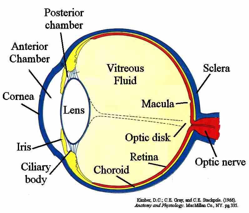
2 Terminology Uvea=vascular layer (tunica vasculosa) ANTERIOR UVEA: Iris & ciliary body POSTERIOR UVEA: Choroid Immunologicaly active (Ly-s can form lymphoid follicles)
3
 Layers: -")
 - posterior pigment epithel (pars")
4 Iris Separates the anterior and posterior chambers Regulates the amount of light (pupil) Layers: - anterior epithel - stroma (muscles, vessels, pigment-cells) - posterior pigment epithel (pars iridica retinae)


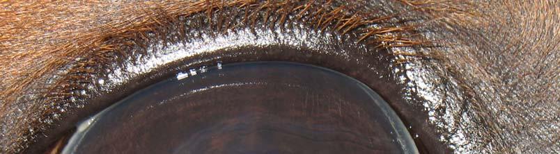
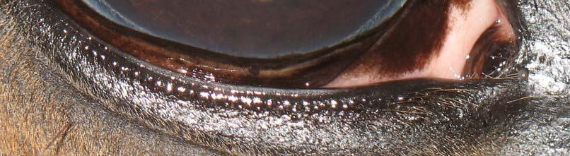
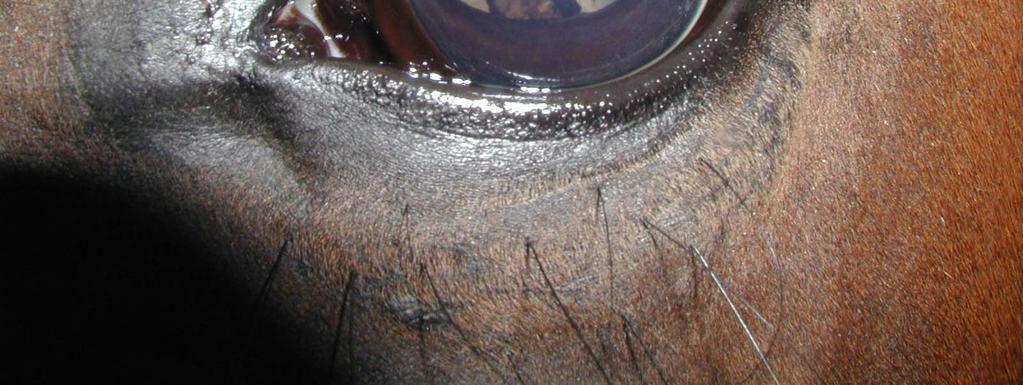
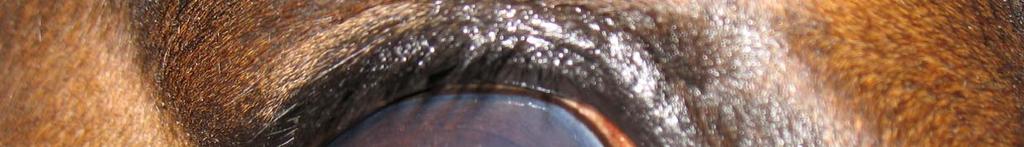
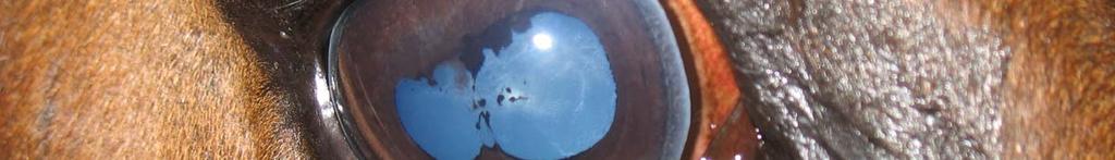
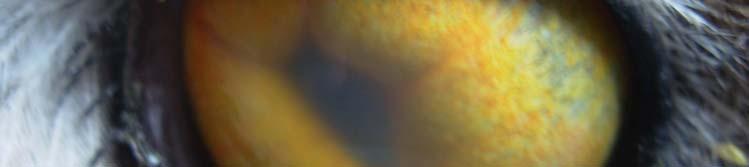
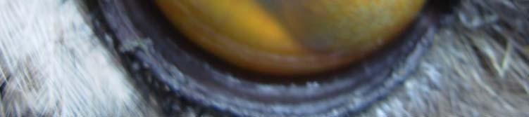
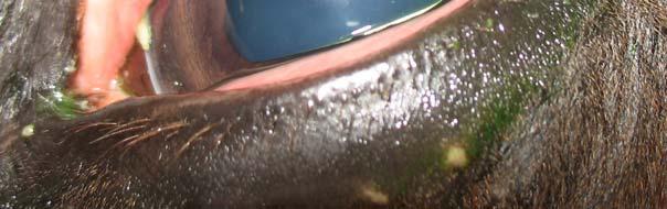
5 Iris Iris surface has many folds and furrows Most irises are brown Others are blue, gold, white Heterochromia -husky, paint horse, appaloosa, pinto Granula iridica (Eq, Ru)
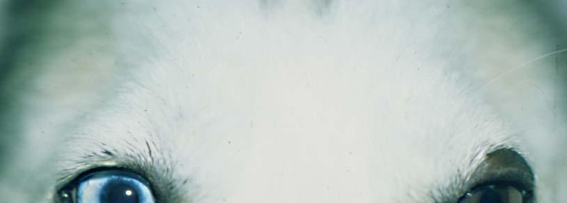
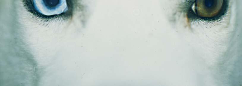
6 Heterochromia
7 Iris musculature Iris dilator muscle -sympathetic innervation -better developed in vertical meridians Iris sphincter muscle -parasympathetic innervation -circumferencially near pupil Slatter, 2001.
8 Ciliary body triangular outline Ciliary muscle (parasympathetic innervation) Pars plicata: - ciliary body processes produce aqueous humor - lens zonules hold the lens in place (+accomodation) Parsplana: -joins the retina: ora ciliaris retinae


9 Ciliary body Slatter, 2001.

 At the dorsal")

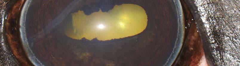
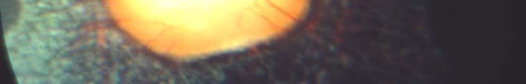
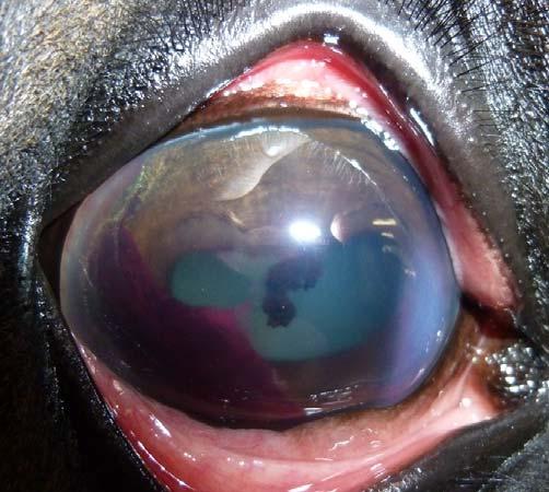
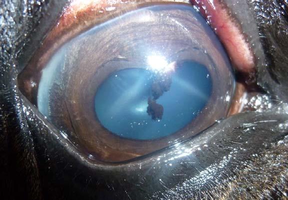
10 Choroid Between the retina and sclera, rich in vessels Histologically has 4 layers Supplies the outer layer of the retina(eq:whole retina) At the dorsal fundus, between the retina and chorioid tapetum (reflective layer, except the pig), stars of Winslow

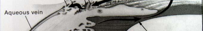
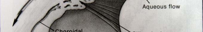
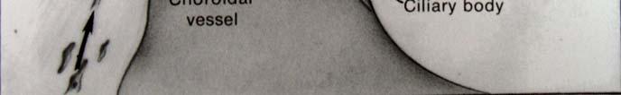
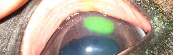
11 Aqueous humor Irido-corneal outflow conventional Slatter,2001. Gilger,2005. Uveo-scleral outflow horse
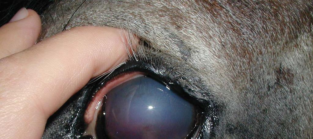
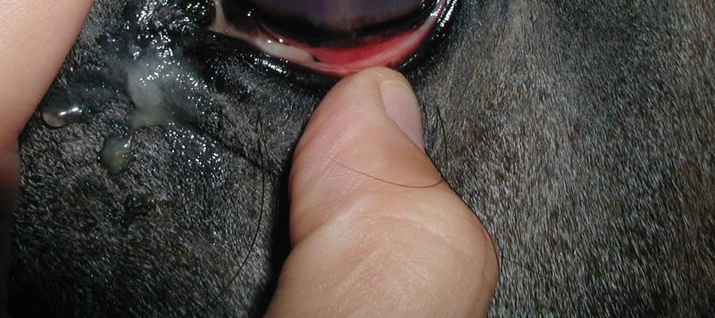
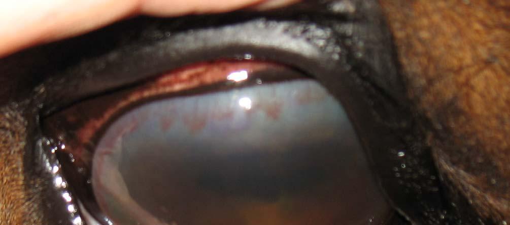
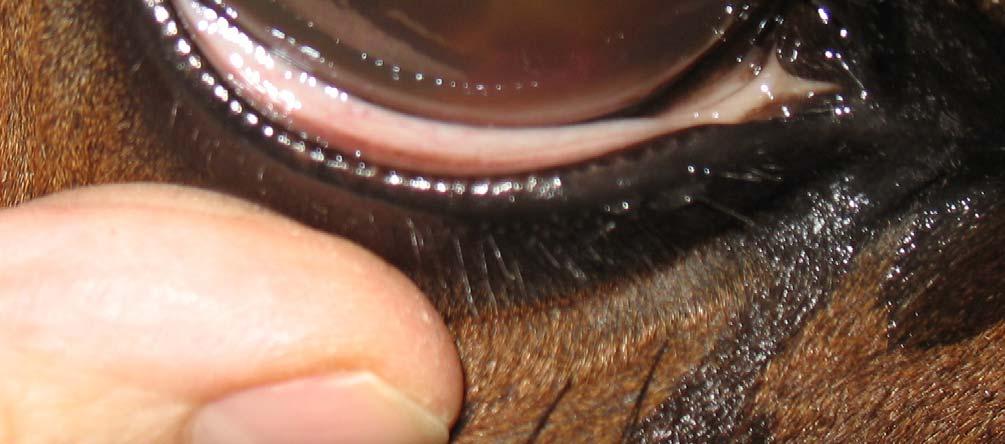
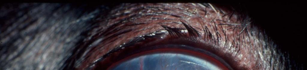
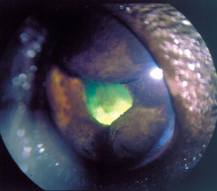
12 Abnormal aqueous humor Breakdown of the blood-aqueous barrier -Aqueousflare: increased protein in AC -Fibrinin AC -Hypopyon=white blood cells in AC -Hyphema=blood in AC These changes of the aqueous humor are usually associated with anterior UVEITIS!

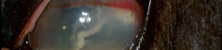
13 fibrin hypopyon hyphema
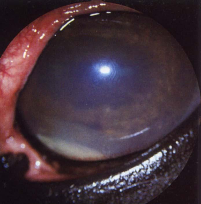
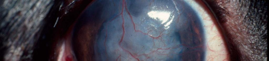
 -intraocular neoplasia Potentially life-threatening condition!")
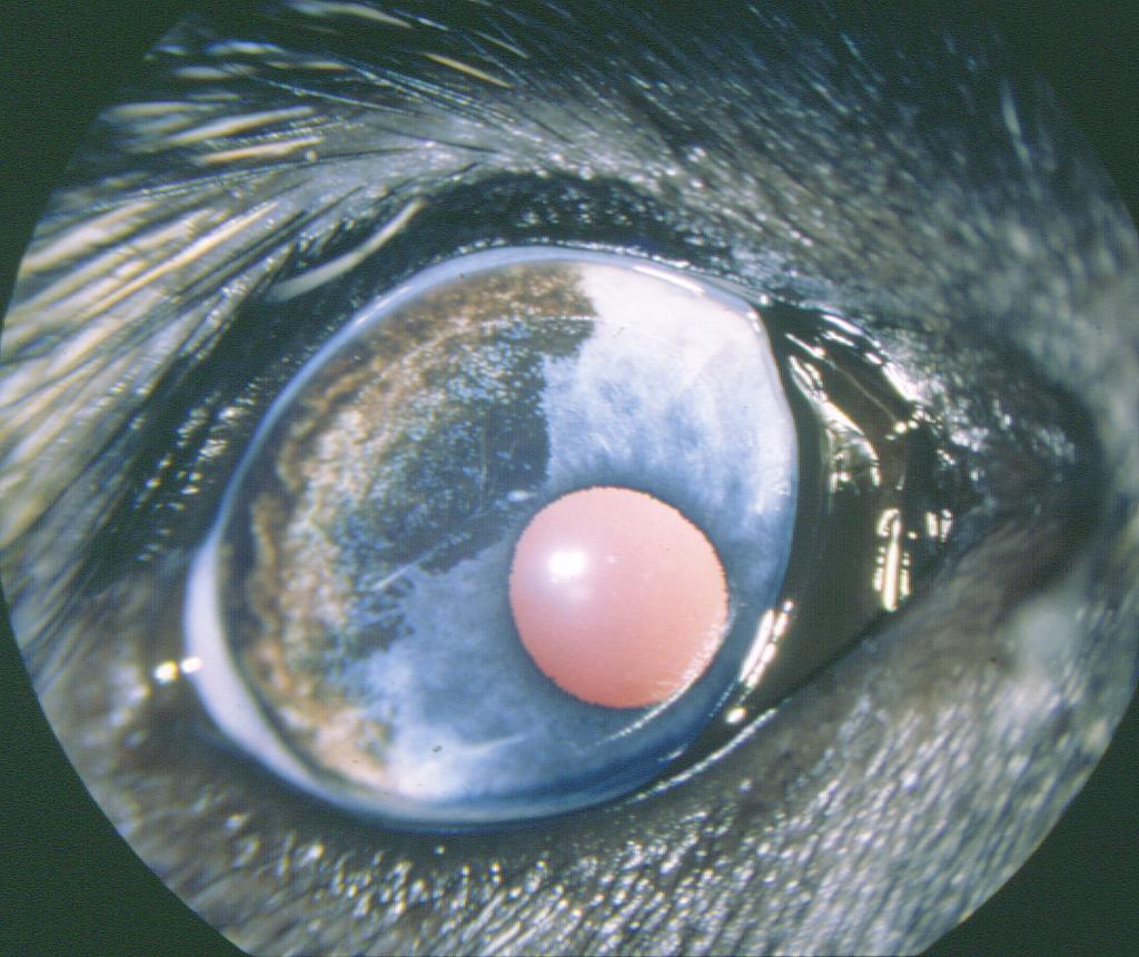
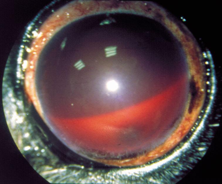
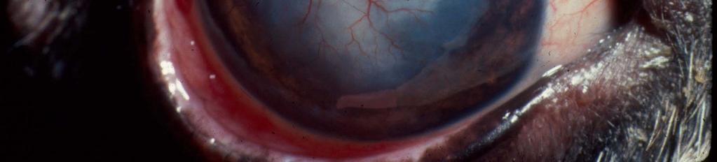
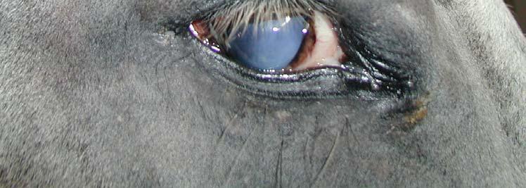
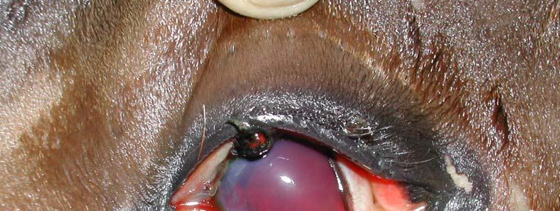
14 Hyphema Blood in AC Possible causes: -trauma -anterior uveitis -bleeding disorder (thrombocytopenia) -intraocular neoplasia Potentially life-threatening condition! Consider ocular ultrasonography!
15 Rule #1. With unilateral or bilateral breakdown of the blood-aqueous barrier consider possible underlying systemic disease, unless there is an obvious explanation such as corneal disease or ocular trauma!

16 Keratic precipitates (KPs) - Accumulation of inflammatory cells on the inner surface of cornea - Indication of ANTERIOR UVEITIS
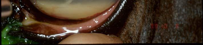
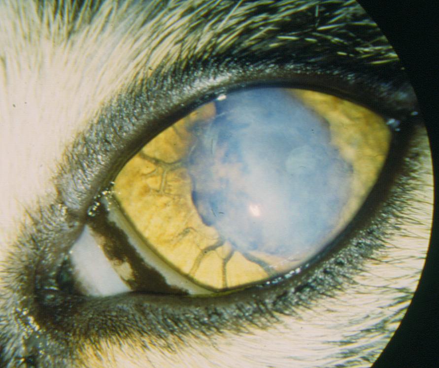
17 Congenital disorders Persistent pupillary membrane -remnants of the anterior tunica vasculosa lentis -usually regress over first 6-12 months of life Barnettetal, 1995.

18 PPM With corneal adhesion
19 Congenital disorders Iris coloboma/iris hypoplasia -congen. abscence of tissue, color dilute breeds -inferior position(6 o clock) is typical, often with other ocular abnormalities


20 Congenital disorders Iris cysts -usually attached to granula iridica:transilluminated tumor (ultrasound) -may be free floating -cause no problems (burst with laser)
21 Congenital disorders Anterior segment dysgenesis/aniridia (rocky mountain horses) Policoria, acoria, excentric pupil
22 Uveitis, terminology Anterior uveitis= iridocyclitis (inflammation of the iris and ciliary body) Posterior uveitis= chorioiditis ( inflammation of the chorioid, retina often affected as well=chorioretinitis) Panuveitis= anterior+posterior uveitis
23 Uveitis Causes idiopathic autoimmun with infect. syst. disease with noninfect. syst. disease trauma 2.reflex uveitis(corneal ulcer) toxic lens induced uveodermatological (immun.med.) ERU Type fibrinous suppurati ve haemorr hagic granulomatous Existence acute chronic recurrent
24 Uveitis Clinical findings Acute: blepharospasm, epiphora, photophobia conjunctival hyperemia aqueous flare inflammatory deposits:hypopion, keratic prec. iridal petechia, hyphema miosis decreased intraocular pressure (IOP ) corneal edema, ciliary injection swollen, dark infiltrated iris hyalitis, chorioiditis

25 Acute uveitis
26 Uveitis Clinical findings II. Acute chronic/complications: Corneal endothelial degeneration/dystrophy corneal vascularization/precipitates lens luxation/subluxation vitreal opacities (hyalitis) focal chorioretinitis, retinal detachment


27
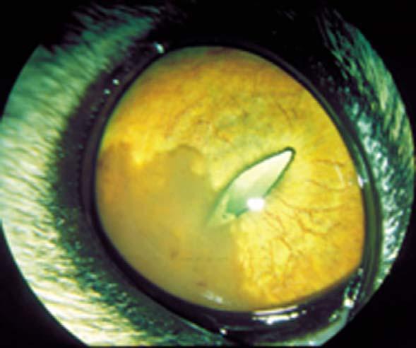
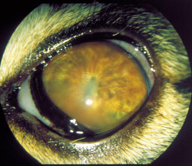
28 Uveitis Clinical findings III. Chronic: organised exsudates in the AC posterior synechia fibropupillary membrane, dyscoria occlusion of pupil, iris bombe keratic precipitates iris hyperpigmentation/neovascularization catract glaucoma endstage: phthisis bulbi

29 Chronic uveitis

30 Cat uveitis+cataract Uveitis-FIP Uveitis+iris bombae

31 Iris bombe

32 Phthisis bulbi
33 Rule # 2. If the IOP is normal or in an eye with clinical signs of anterior uveitis, you have to suspect the presence of glaucoma!


34 Rule # 3. Every red eye (with or without uveitis) needs to be stained with FLUORESCEIN!
35 Treatment of uveitis (1.) Aims: Elimination of the cause, if possible (treat syst. disease) Preserve vision Control discomfort and active inflammation Minimize permanent changes Clienteducation: -clinical signs to look for -re-initiation of treatment

36 TREATMENT OF UVEITIS. I. LOOK FOR SYSTEMIC CAUSE: History Systemic examination Bloodwork Urinalysis, imaging Aqueous paracentesis
37 Treatment of uveitis (2.) Topical antiinflammatories: -Corticosteroids 1-6x daily, depending on severity -Make sure that cornea is fluorescein negativ! -Prednisolone acetate 1% -Dexametasone 0,1% -Triamcinolon In subconjunctival injection!


38 Rule # 4. It is contraindicated to apply topical corticosteroids in an eye with corneal ulcer!
39 Ijured cell- cellmembrane phospholipid +mediators of mast cells Corticosteroids Phospholipase A 2 enzime Cyclooxygenase inhibitors (flunixin,phenilbutazone, ketoprofen,aspirin) Arachidonic acid Lipoxygenase inhibitors (ketoprofen) Prosztanoids - Prosztaglandins (miosis, IOP ) - Prosztacyclin (blood-aqueous barrier breakdown) Leukotrienes (cellular infiltration of uvea, miosis)
40 Treatment of uveitis (3.) Topical antiinflammatories - non-steroidals 1-6x daily - can be used with fluorescein stain uptake, but be careful! - Diclofenac 0,1% -Flurbiprofen0,3% Systemicanti-inflammatories -non-steroidals (NSAIDs): -flunixin meglumine 1,1mg/kg 2x daily -phenylbutazone 1-4 g/horse/day -aspirin/ketoprofen po. -Monitor for GI ulcers!
41 Treatment of uveitis (4.) Mydritics and cycloplegics -Atropin 1-2% (dilate pupil) -In severe cases up to q 4hours, in mild cases 1x daily -Potential complication: Colic -Monitor gut motility!
42 Effects of atropine Mydriatic=dilates pupil -minimize adhesions (synechiae) -may not be able to break down synechiae in chronic cases Cycloplegic=relaxes ciliary muscle -relieve ciliary muscle spasm -pain relief Stabilizes blood-aqueous barrier
43 Rule # 5. The effectiveness of atropine to keep the pupil dilated gives us information about the severity of uveitis. The longer and better the pupil stays dilated, the milder the uveitis.

.")
44 Rule # 5. In a normal eye, one dose of atropine can keep the pupil dilated for up to 1-4 weeks (only for therapeutic purpose). Eyes with a brown iris stay longer dilated than eyes with blue iris. atropine poisoning
45 Treatment of uveitis (5.) As the clinical signs improve, the frequency of drug application can slowly be decreased. If the clinical signs worsen as the medication is decreased, then the dosing should be temporarly increased again.
 Alternative methods: acupuncture")
46 Treatment of uveitis (6.) Alternative methods: acupuncture Eye hood+box rest


47 Endophthalmitis Severe uveitis with involvement of aqueous humor and vitreous, but not sclera Panophthalmitis: severe inflammation that also involves the sclera & orbital tissues Clinicalsigns:see uveitis, but more severe Treatment:see uveitis, +systemic antibiotics
48 Endophthalmitis (2.) Consider culturing aqueous humor and vitreous aspirate! Intravitreal injections (last chance before enucleation) µg gentamicin: Gr -2,2 µg cefazolin: Gr + - 0,1 mg miconazole/fluconazole: fungal - Should be done by specialist!
49 Trauma I. Penetrating/blunt Check for periorbital skull fractures Penetrating trauma: see corneal perforation -iris prolapse Careful clinical examination Consider ultrasonography if hyphema prevents examination Clinical signs: see uveitis (hyphema, miosis)


50 Trauma II. Prognosis is guarded with intraocular hemorrhage Treatment: -symptomatic treatment of uveitis -topical mydriatics -systemic and topical anti-inflammatories -surgery for penetrating injury

51 Traumatic uveitis


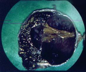
52 poor prognosis consider enucleation
53 Other diseases of the anterior uvea Iris neoplasia Rare Melanomamost common, esp. grey horses Clinicalsigns: - Dark mass in AC - Distorsion of pupil Treatment: - enucleation or sector iridectomy Other rare tumors: medulloepithelioma, multicentric lymphoma

54 melanoma Melanoma+ glaucoma
55 Iris atrophy Thinning of the iris (also with age) Older horses, esp. with heterochromia iridis Atrophy of granula iridica with chronic uveitis or glaucoma
56
Anterior Uveitis in Dogs
 Customer Name, Street Address, City, State, Zip code Phone number, Alt. phone number, Fax number, e-mail address, web site Anterior Uveitis in Dogs (Inflammation of the Front Part of the Eye, Including
Around The Globe in 60 Minutes
 Around The Globe in 60 Minutes Around the GLOBE in Sixty Minutes Basic Ocular Anatomy, Examination, and Diagnostic Techniques Introduction Focusing on canine and feline ocular anatomy and basic examination
UVEITIS. Dr. Yılmaz ÖZYAZGAN
 UVEITIS Dr. Yılmaz ÖZYAZGAN UVEITIS DEFINITION BY STRICT DEFINITION, UVEITIS IS AN INFLAMMATION OF UVEAL TRACT. BUT IN PRACTICAL, IT IS GENERALLY NOT RESTRICTED TO THE UVEA AND INVOLVES OTHER ADJACENT
Equine Recurrent Uvei2s/ ERU. Equine Recurrent Uvei2s. Uvei2s. Uvea. Blood- ocular barrier. Uvea
 Equine Recurrent Uvei2s/ ERU Equine Recurrent Uvei2s Moon blindness Iridocycli2s Periodic ophthalmia Tijn J.P. Spoormakers dipl. ECVS, cert ISELP Most common cause of blindness Uvei2s Inflamma2on of uvea
Update on management of Anterior Uveitis
 Update on management of Anterior Uveitis Parthopratim Dutta Majumder Senior Consultant, Department of Uvea & Intraocular Inflammation Medical Research Foundation, Sankara Nethralaya ABCD of Treating a
Table of Contents 1 Orbit 3 2 Eyelids 7
 Table of Contents Preface, x List of abbreviations xi Glossary xii Section I Atlas 1 1 Orbit 3 Clinical signs associated with orbital neoplasia 3 Clinical signs associated with orbital cellulitis 3 Enophthalmos
Case Study: Fuzz April 18th
 Case Study: Fuzz April 18th 33 year old Quarter Horse Had been battling corneal ulcer for several weeks before seeing us No foreign debris found Culture and cytology were taken. Started on topical antibiotics,
2) Uveal Tract & Uveitis - Dr. Bakhtyar
 Uveal Tract & Uveitis - Dr. Bakhtyar") Vascular Pigmented Layer Iris 2) Uveal Tract & Uveitis - Dr. Bakhtyar The uveal tract composed of the second (the vascular) coat of the globe which consists, from back to front, of the choroids, the ciliary
Vascular Pigmented Layer Iris 2) Uveal Tract & Uveitis - Dr. Bakhtyar The uveal tract composed of the second (the vascular) coat of the globe which consists, from back to front, of the choroids, the ciliary
Head prof. MUDr. E. Vlková, CSc.
 MUDr. Karkanová Michala, Oční klinika LF MU a FN Brno Head prof. MUDr. E. Vlková, CSc. 3 parts: iris (iris) ciliary body (corpus ciliare) choroid (choroidea) Function: regulating the entry of light into
Management of uveitis
 Management of uveitis DR. ANUPAMA KARANTH Anti-inflammatory agents -itis = inflammation Treatment : stop inflammation Use anti-inflammatory drugs Most potent of such agents : Corticosteroids Corticosteroids
Ocular Urgencies and Emergencies
 Ocular Urgencies and Emergencies Pam Boyce, O.D., F.A.A.O. Boyce Family Eye Care, Ltd. 528 Devon Ave. Park Ridge, IL 60068 847-518-0303 Somebody s going to lose an eye Epidemiology 2.4 million ocular and
Cases CFEH. CFEH Facebook Case #4
 CFEH Cases CFEH Facebook Case #4 A 42 year old female has noticed a floater in her left eye for many years but no flashes. She also reports hazy vision in this eye that has been present all her life. She
Department of Ophthalmology
 Department of Ophthalmology Period : 02/July/18 to 30/August/18 Semester : 7 th Semester Lecture Lesson Plan Sr. Date Topic Lesson plan Name of Faculty No. 1 02.07.18 Lens- Lens-Anatomy, Classification
EYE INJURIES OBJECTIVES COMMON EYE EMERGENCIES 7/19/2017 IMPROVE ASSESSMENT OF EYE INJURIES
 EYE INJURIES BRITTA ANDERSON D.O. DMC PRIMARY CARE SPORTS MEDICINE ASSOCIATE TEAM PHYSICIAN DETROIT TIGERS OBJECTIVES IMPROVE ASSESSMENT OF EYE INJURIES UNDERSTAND WHAT IS CONSIDERED AN EMERGENCY DEVELOP
OCULAR DISORDERS REPORT BOSTON TERRIER
 OCULAR DISORDERS REPORT BOSTON TERRIER 1991-1999 2000-2009 2010-2012 TOTAL DOGS EXAMINED 2723 6803 2004 Diagnostic Name # % # % # % GLOBE 0.110 microphthalmia 1 0.0% 1 0.0% 0 EYELIDS 20.140 ectopic cilia
HLA-B27-related anterior Uveitis
 HLA-B27-related anterior Uveitis Nicholas Jones Manchester Uveitis Clinic The Royal Eye Hospital Manchester Anterior means anterior only IUSG classification: Anterior uveitis = Iris & pars plicata AU
Eye Examination Techniques in Horses
 Eye Examination Techniques in Horses Dennis E. Brooks DVM, PhD Dip ACVO University of Florida brooksd@mail.vetmed.ufl.edu Basic Instruments How to tell the potential of vision? PLRs (retina, CN 2, chiasm,
IS THE CAT S EYE BLIND, PAINFUL, OR CHANGING COLORS? Kerry L. Ketring, DVM, DAVCO
 IS THE CAT S EYE BLIND, PAINFUL, OR CHANGING COLORS? Kerry L. Ketring, DVM, DAVCO OPHTHALMOLOGY Anterior uveitis or iridocyclitis is inflammation of the iris and ciliary body. This is the second most common
FACING YOUR FUNDIC FEARS: EXAMINATION OF THE OCULAR FUNDUS J. Seth Eaton, VMD, DACVO Cornell University Veterinary Specialists
 FACING YOUR FUNDIC FEARS: EXAMINATION OF THE OCULAR FUNDUS J. Seth Eaton, VMD, DACVO Cornell University Veterinary Specialists The goal of a thorough fundus examination is to clinically evaluate the structures
Optometric Postoperative Cataract Surgery Management
 Financial Disclosures Optometric Postoperative Cataract Surgery Management David Dinh, OD Oak Cliff Eye Clinic Dallas Eye Consultants March 10, 2015 Comanagement Joint cooperation between two or more specialists
LENS INDUCED GLAUCOMA
 LENS INDUCED GLAUCOMA PRESENTER P SHILPA RAVI 2 ND YEAR PG DEPT OF OPHTHALMOLOGY LENS INDUCED GLAUCOMA It is a form of secondary glaucoma where intraocular pressure is raised due to disorder in crystalline
Moncef Khairallah, MD
 Moncef Khairallah, MD Department of Ophthalmology, Fattouma Bourguiba University Hospital Faculty of Medicine, University of Monastir Monastir, Tunisia INTRODUCTION IU: anatomic form of uveitis involving
Secondary open-angle glaucoma
 Secondary open-angle glaucoma Kathy Hondeghem ZNA Middelheim MaNaMa 12/10/13 Definition Open anterior chamber angle (at least 270 ) Trabecular meshwork (and thus aqueous humor outflow) is occluded by a
Anterior Segment Disease and the Systemic Link Mile Brujic, OD, FAAO
 Anterior Segment Disease and the Systemic Link Mile Brujic, OD, FAAO brujic@prodigy.net Summary As optometry s role in health care increases, so does our responsibility to appropriately diagnose and appropriately
Focus on Ophthalmology Inside the Eye of the Horse
 www.ivis.org Proceedings of the American Association of Equine Practitioners - Focus Meeting Focus on Ophthalmology Inside the Eye of the Horse Raleigh, NC, USA 2012 Next Focus Meetings: August 4-6, 2013
Wildlife Ophthalmology D R. H E A T H E R R E I D T O R O N T O W I L D L I F E C E N T R E T O R O N T O, O N C A N A D A
 Wildlife Ophthalmology D R. H E A T H E R R E I D T O R O N T O W I L D L I F E C E N T R E T O R O N T O, O N C A N A D A Why understand eyes? Wildlife need to have excellent vision to survive in the
Glaucoma & Inflammation. Jorge L. Fernandez Bahamonde, MD.
 Glaucoma & Inflammation. Jorge L. Fernandez Bahamonde, MD. Definition. Inflammatory ocular conditions compromise outflow of aqueous humor. Keratitis Episcleritis. Scleritis. Uveitis Glaucoma & Keratitis.
PAINFUL PAINLESS Contact lens user BOV
 Common Causes Allergies Infections Ocular Cornea, uveitis, endophthalmitis Orbital Orbital cellulitis Inflammation Uveitis Scleritis / episcleritis Glaucomas Trauma Foreign bodies Chemical injuries History
Ocular Pathology. I. Congenital and/or developmental. A. Trisomy 21. Hypertelorism (widely spaced eyes) Keratoconus (cone shaped cornea)
 Keratoconus (cone shaped cornea)") I. Congenital and/or developmental Robbins Pathologic Basis of Disease, 6 th Ed. A. Trisomy 21 Hypertelorism (widely spaced eyes) Keratoconus (cone shaped cornea) Focal hypoplasia of iris Cataracts frequently
I. Congenital and/or developmental Robbins Pathologic Basis of Disease, 6 th Ed. A. Trisomy 21 Hypertelorism (widely spaced eyes) Keratoconus (cone shaped cornea) Focal hypoplasia of iris Cataracts frequently
OOGZIEKTEN VOOR DE HUISARTS F. GOES, JR.
 OOGZIEKTEN VOOR DE HUISARTS F. GOES, JR. HET RODE OOG F. GOES, JR. Condition Signs Symptoms Causes Conjunctivitis Viral Normal vision, normal pupil size Mild to no pain, diffuse Adenovirus (most common),
Department of Ophthalmology
 Period : 03/July/17 to 07/September/17 Semester : 7 th Semester Department of Ophthalmology Lecture Lesson Plan Sr 1 03.07.17 Uvea-Anatomy, Uvea-Anatomy, Classification of Uveitis Dr R Paranjpe Classification
Dr. D. Y. Patil Medical College, Pimpri, Pune
 Dr. D. Y. Patil Medical College, Pimpri, Pune - 411 018 Period : 04/July/16 to 22/September/16 Semester : 7 th Semester Department : Ophthalmology Lecture Lesson Plan Sr No Date Topic Learning objectives
Understanding and Treating Equine Recurrent Uveitis. Jacquelin Boggs, DVM, MS, Diplomate ACVIM. Area Veterinarian, Pfizer Animal Health
 Understanding and Treating Equine Recurrent Uveitis Jacquelin Boggs, DVM, MS, Diplomate ACVIM Area Veterinarian, Pfizer Animal Health Objectives: Review of the etiology and pathophysiology of uveitis.
Glaucoma. Glaucoma. Glaucoma. Trevor Arnold, MS, DVM, DACVO
 Glaucoma Trevor Arnold, MS, DVM, DACVO Glaucoma Physiology of Aqueous Humor Produced in the ciliary body Flows out the iridocorneal angle and ciliary cleft High intraocular pressures are caused by a decreased
Ocular and periocular trauma
 Ocular and periocular trauma No financial disclosures. Tina Rutar M.D. Assistant Professor of Clinical Ophthalmology and Pediatrics Director, Visual Center for the Child University of California San Francisco
Acute Eyes for ED. Enis Kocak. The Alfred Ophthalmology
 Acute Eyes for ED Enis Kocak The Alfred Ophthalmology The problem with eyes Things to cover Ocular anatomy Basic assessment Common presentations Eye first aid and procedures Ophthalmic emergencies What
CATARACT SURGERY IN UVEITIS. Professor Harminder Singh Dua
 Research Institute of Ophthalmology, Cairo 11 th International Conference, 3-4 February, 2017 CATARACT SURGERY IN UVEITIS Professor Harminder Singh Dua MBBS, DO, DO(Lond), MS, MNAMS, FRCS, FRCOphth., FEBO,
Focus on Ophthalmology Inside the Eye of the Horse
 www.ivis.org Proceedings of the American Association of Equine Practitioners - Focus Meeting Focus on Ophthalmology Inside the Eye of the Horse Raleigh, NC, USA 2012 Next Focus Meetings: August 4-6, 2013
in Uveitis Euretina Hamburg 2013 Nicholas Jones Royal Eye Hospital Manchester, UK
 Cataract Surgery in Uveitis Euretina Hamburg 2013 Nicholas Jones Royal Eye Hospital Manchester, UK Cataract surgery in eyes with uveitis is not routine It requires much more pre-operative planning It may
2/19/14. Garret Pachtinger, VMD, DACVECC. COO, VetGirl. Justine A. Lee, DVM, DACVECC, DABT CEO, VetGirl. Shelby Reinstein DVM, MS, DACVO
 Shelby Reinstein, DVM, MS, DACVO Garret Pachtinger, VMD, DACVECC COO, VetGirl Justine A. Lee, DVM, DACVECC, DABT CEO, VetGirl Shelby Reinstein DVM, MS, DACVO! Emailed to you 48 hours after the webinar!
Glaucoma Basics OVERVIEW GENETICS SIGNALMENT/DESCRIPTION OF PET
 Glaucoma Basics OVERVIEW Glaucoma is a disease of the eye, in which the pressure within the eye is increased (pressure within the eye is known as intraocular pressure or IOP) High intraocular pressure
Uveitis. Pt Info Brochure. Q: What is Uvea?
 Pt Info Brochure Uveitis Q: What is Uvea? A: Uvea is the middle layer of the eye. It is the most vascular structure of the eye. It provides nutrition to the other parts of the eye. The uvea is made up
Dr Jo-Anne Pon. Dr Sean Every. 8:30-9:25 WS #70: Eye Essentials for GPs 9:35-10:30 WS #80: Eye Essentials for GPs (Repeated)
") Dr Sean Every Ophthalmologist Southern Eye Specialists Christchurch Dr Jo-Anne Pon Ophthalmologist Southern Eye Specialists, Christchurch Hospital, Christchurch 8:30-9:25 WS #70: Eye Essentials for GPs
Dr Sean Every Ophthalmologist Southern Eye Specialists Christchurch Dr Jo-Anne Pon Ophthalmologist Southern Eye Specialists, Christchurch Hospital, Christchurch 8:30-9:25 WS #70: Eye Essentials for GPs
EYE TRAUMA: INCIDENCE
 Introduction EYE TRAUMA: INCIDENCE 2.5 million eye injuries per year in U.S. 40,000 60,000 of eye injuries lead to visual loss Introduction Final visual outcome of many ocular emergencies depends on prompt,
Vitrectomy as a Diagnostic and Therapeutic Approach for Equine Recurrent Uveitis (ERU)
") Vitrectomy as a Diagnostic and Therapeutic Approach for Equine Recurrent Uveitis (ERU) Hartmut Gerhards, Dr.Med.Vet.; Bettina Wollanke, Dr.Med.Vet.; and Siegfried Brem, Dr.Med.Vet. Vitrectomy in horses
Vitrectomy as a Diagnostic and Therapeutic Approach for Equine Recurrent Uveitis (ERU) Hartmut Gerhards, Dr.Med.Vet.; Bettina Wollanke, Dr.Med.Vet.; and Siegfried Brem, Dr.Med.Vet. Vitrectomy in horses
Ulcerative Keratitis (Type of Inflammation of the Cornea) Basics
 Basics") Ulcerative Keratitis (Type of Inflammation of the Cornea) Basics OVERVIEW Keratitis is inflammation of the cornea; the cornea is the clear outer layer of the front of the eye The corneal epithelium is
Ulcerative Keratitis (Type of Inflammation of the Cornea) Basics OVERVIEW Keratitis is inflammation of the cornea; the cornea is the clear outer layer of the front of the eye The corneal epithelium is
An Injector s Guide to OZURDEX (dexamethasone intravitreal implant) 0.7 mg
 0.7 mg") An Injector s Guide to OZURDEX (dexamethasone intravitreal implant) 0.7 mg This guide is intended to provide injectors with information on the recommended injection technique and the important risks related
An Injector s Guide to OZURDEX (dexamethasone intravitreal implant) 0.7 mg This guide is intended to provide injectors with information on the recommended injection technique and the important risks related
Photodynamic therapy for IMMK in horses
 Photodynamic therapy for IMMK in horses Overview Immune mediated keratitis in horses Traditional treatment options Sustained release implant Photodynamic therapy Use in veterinary medicine Treatment for
Ophthalmology. Juliette Stenz, MD
 Ophthalmology Juliette Stenz, MD Required Slide Disclosures NO SIGNIFICANT FINANCIAL, GENERAL, OR OBLIGATION INTERESTS TO REPORT Required Slide At the end of this session, students will be able to: 1.
PRECISION PROGRAM. Injection Technique Quick-Reference Guide. Companion booklet for the Video Guide to Injection Technique
 Injection Technique Quick-Reference Guide PRECISION PROGRAM Companion booklet for the Video Guide to Injection Technique Available at www.ozurdexprecisionprogram.com Provides step-by-step directions with
2/26/2017. Sameh Galal. M.D, FRCS Glasgow. Lecturer of Ophthalmology Research Institute of Ophthalmology
 Sameh Galal M.D, FRCS Glasgow Lecturer of Ophthalmology Research Institute of Ophthalmology No financial interest in the subject presented 1 Managing cataracts in children remains a challenge. Treatment
The Anterior Segment & Glaucoma Visual Recognition & Interpretation of Clinical Signs
 The Anterior Segment & Glaucoma Visual Recognition & Interpretation of Clinical Signs Quiz created by Jane Macnaughton MCOptom & Peter Chapman BSc MCOptom FBDO CET Accreditation C19095 2 CET Points (General)
WHAT IS YOUR DIAGNOSIS? By ADREA R. BENKOFF M.D.
 WHAT IS YOUR DIAGNOSIS? By ADREA R. BENKOFF M.D. Anterior Chamber Inflammation and Iris Depigmentation Noted 25 Years After Cataract Extraction Decreasing Vision Over a 5- Year Period 64 year old white
OPHTHALMOLOGY AND ULTRASOUND
 Vet Times The website for the veterinary profession https://www.vettimes.co.uk OPHTHALMOLOGY AND ULTRASOUND Author : JAMES OLIVER Categories : Vets Date : April 28, 2008 JAMES OLIVER discusses why ultrasound
Differential diagnosis of the red eye. Carol Slight Nurse Practitioner Ophthalmology
 Differential diagnosis of the red eye Carol Slight Nurse Practitioner Ophthalmology The red eye Conjunctivitis HSV Keratitis Acute angle closure glaucoma Anterior Uveitis Red eye Scleritis Subconjunctival
Test Bank for Medical Surgical Nursing An Integrated Approach 3rd Edition by White
 Test Bank for Medical Surgical Nursing An Integrated Approach 3rd Edition by White Link full download : http://testbankair.com/download/test-bank-for-medical-surgical-nursing-anintegrated-approach-3rd-edition-by-white/
The Pathology and Pathogenesis of Acute Glaucoma in Dogs. Richard R Dubielzig
 The Pathology and Pathogenesis of Acute Glaucoma in Dogs Richard R Dubielzig Overview of Glaucoma Intraocular Pressure too High to Support a Healthy Optic Nerve Terminology used in the classification of
UNDERSTAND MORE ABOUT UVEITIS UVEITIS
 UNDERSTAND MORE ABOUT UVEITIS UVEITIS Uveitis What is uveitis? Uveitis is inflammation of the uvea, the middle layer of your eye. The eye is shaped much like a tennis ball, with three different layers
The Orbit. The Orbit OCULAR ANATOMY AND DISSECTION 9/25/2014. The eye is a 23 mm organ...how difficult can this be? Openings in the orbit
 The eye is a 23 mm organ...how difficult can this be? OCULAR ANATOMY AND DISSECTION JEFFREY M. GAMBLE, OD COLUMBIA EYE CONSULTANTS OPTOMETRY & UNIVERSITY OF MISSOURI DEPARTMENT OF OPHTHALMOLOGY CLINICAL
Proceeding of the ACVP/ASVCP Concurrent Annual Meetings
 http://www.ivis.org Proceeding of the ACVP/ASVCP Concurrent Annual Meetings Dec.3-7, 2011 Nashville, Tennessee, USA Next Meeting : December 1-5, 2012 - Seattle, WA, USA Reprinted in the IVIS website with
Uveitis Update DISCLOSURE STATEMENT. Featured Speaker: Dr. Kyle Cheatham, FAAO, DIP ABO
 Uveitis Update Featured Speaker: Dr. Kyle Cheatham, FAAO, DIP ABO DISCLOSURE STATEMENT We have no direct financial or proprietary interest in any companies, products or services mentioned in this presentation.
Nausheen Khuddus, MD Melissa Elder, MD, PhD
 Nausheen Khuddus, MD Melissa Elder, MD, PhD Nausheen Khuddus, MD Pediatric Ophthalmologist and Strabismus Specialist Accent Physicians Gainesville, Florida What Is Uveitis? Uveitis is caused by inflammatory
MANAGEMENT OF NEOVASCULAR GLAUCOMA
 MSO EXPRESS: ISSUE 3 MANAGEMENT OF NEOVASCULAR GLAUCOMA Associate Professor Dr. Norlina Mohd Ramli, Dr. Ng Ker Hsin Associate Professor Dr. Norlina Mohd Ramli MBBS (UK) MRCOphth (UK) MS Ophthal (Mal) Fellowship
Choroidal Neovascularization in Sympathetic Ophthalmia
 Choroidal Neovascularization in Sympathetic Ophthalmia Lucia Sobrin, Miguel Cordero Coma, C. Stephen Foster Case Report A 49-year-old man presented after a ruptured globe repair of his left eye status
Retinal Detachment. Basics OVERVIEW GENETICS SIGNALMENT/DESCRIPTION OF PET
 Customer Name, Street Address, City, State, Zip code Phone number, Alt. phone number, Fax number, e-mail address, web site Retinal Detachment Basics OVERVIEW Retinal refers to the retina; the retina is
DNB Question Paper. December 1
 DNB Question Paper December 1 December,2013 DNB Examination 2013 (December) IMPORTANT INSTRUCTIONS: This question paper consists of 10 questions divided into Part A and Part B, each part containing 5 questions.
02/03/2014. Average Length: 23mm (Infant ~16mm) Approximately the size of a quarter Volume: ~5mL
 Approximately the size of a quarter Volume: ~5mL") Identify the anatomy of the eye. Explain the basic physiology of the parts of the eye. Briefly discuss various surgeries related to different parts of the anatomy. Average Length: 23mm (Infant ~16mm) Approximately
Identify the anatomy of the eye. Explain the basic physiology of the parts of the eye. Briefly discuss various surgeries related to different parts of the anatomy. Average Length: 23mm (Infant ~16mm) Approximately
The Ins and Outs of Ocular Trauma in the Horse. Noelle T. McNabb, DVM, DACVO. Peterson and Smith Equine Hospital. Ocala, Florida
 The Blown Up Eye The Ins and Outs of Ocular Trauma in the Horse Noelle T. McNabb, DVM, DACVO Peterson and Smith Equine Hospital Ocala, Florida The Prominent Equine Eye Vulnerable to trauma DESPITE protection
Vision I. Steven McLoon Department of Neuroscience University of Minnesota
 Vision I Steven McLoon Department of Neuroscience University of Minnesota 1 Eye Cornea Sclera Conjunctiva 2 Eye The conjunctiva lines the inner surface of the eyelids and outer surface of the sclera. 3
_ Assessment of the anterior chamber. Review of anatomy of the angle
 Assessment of the anterior chamber Dr Simon Barnard PhD BSc FCOptom FAAO DCLP Department of Optometry & Visual Science City University London, UK Review of anatomy of the angle Figure 1. Anatomical section
Measure #192: Cataracts: Complications within 30 Days Following Cataract Surgery Requiring Additional Surgical Procedures
 Measure #192: Cataracts: Complications within 30 Days Following Cataract Surgery Requiring Additional Surgical Procedures 2012 PHYSICIAN QUALITY REPORTING OPTIONS FOR INDIVIDUAL MEASURES: REGISTRY ONLY
Conjunctival Hemorrhage
 IN THE NAME OF GOD Lid Laceration Conjunctival Hemorrhage a) No therapy is necessary b) Usually resolve in 7-12 days. Subconjunctival Hemorrhage Corneal Abrasion Abrasions Many small abrasions can
THE RED EYE Cynthia McNamara, MD Week 25
 THE RED EYE Cynthia McNamara, MD Week 25 Educational Objectives: 1. Know the differential diagnosis and presentation of specific etiologies of the red eye 2. Be able to evaluate patients presenting with
Bilateral mydriasis in a senior neutered toy poodle
 Vet Times The website for the veterinary profession https://www.vettimes.co.uk Bilateral mydriasis in a senior neutered toy poodle Author : Negar Hamzianpour Categories : Companion animal, Vets Date :
Assessment and Management of Ocular Trauma. Disclosure I have no direct financial interests in today s subject matter. 3/25/2019. Normal Eye Anatomy
 Assessment and Management of Ocular Trauma Samiksha Fouzdar Jain, MD,FRCS Department of Ophthalmology & Visual Sciences Truhlsen Eye Institute Disclosure I have no direct financial interests in today s
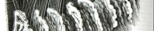
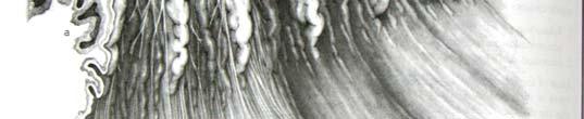
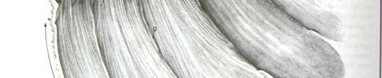
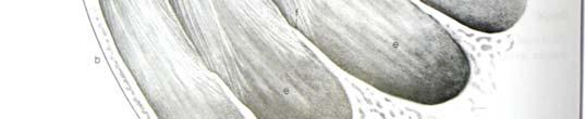
of Ciliary body, Iris and Irido-corneal angle in Adult Marwari Goat (Capra hircus) R. K. Barhaiyaa *, Malsawmkima, D. M. Bhayani and Y. L.
 R. K. Barhaiyaa *, Malsawmkima, D. M. Bhayani and Y. L.") DHR International Journal Of Biomedical and Life Sciences (DHR-IJBLS) ISSN 2278-8301, Vol.5(3), 2014 Available online http://www.doublehelixresearch.com/dhrijbls Double Helix Research Original Article
DHR International Journal Of Biomedical and Life Sciences (DHR-IJBLS) ISSN 2278-8301, Vol.5(3), 2014 Available online http://www.doublehelixresearch.com/dhrijbls Double Helix Research Original Article
Role of Intraocular Leptospira Infections in the Pathogenesis of Equine Recurrent Uveitis in the Southern United States
 Louisiana State University LSU Digital Commons LSU Master's Theses Graduate School 2012 Role of Intraocular Leptospira Infections in the Pathogenesis of Equine Recurrent Uveitis in the Southern United
Clinical Practice Guide for the Diagnosis, Treatment and Management of Anterior Eye Conditions. April 2018
 Clinical Practice Guide for the Diagnosis, Treatment and Management of Anterior Eye Conditions This Clinical Practice Guide provides evidence-based information about current best practice in the management
Diffuse infiltrating retinoblastoma
 Brit. 1. Ophthal. (I 971) 55, 6oo Diffuse infiltrating retinoblastoma GWYN MORGAN Department of Pathology, Institute of Ophthalmology, University of London The term "diffuse infiltrating retinoblastoma"
Pediatric Ocular Sonography
 Pediatric Ocular Sonography Cicero J Torres A Silva, MD Associate Professor of Radiology 2016 SPR Pediatric Ultrasound Course Yale University School of Medicine None Disclosures Objectives of Presentation
Ocular Anatomy for the Paraoptometric
 Ocular Anatomy for the Paraoptometric Minnesota Optometric Association Paraoptometric CE Friday September 30, 2016 Lindsay A. Sicks, OD, FAAO Assistant Professor, Illinois College of Optometry lsicks@ico.edu
10 EYE EMERGENCIES. Who goes, who you better not send! Brant Slomovic, MD, FRCPC University Health Network
 10 EYE EMERGENCIES Who goes, who you better not send! Brant Slomovic, MD, FRCPC University Health Network DISCLOSURES I have none PVD CASE 1 WHAT IS A PVD? a process of aging (45-55) liquefaction of vitreous
THE EYE: RETINA AND GLOBE
 Neuroanatomy Suzanne Stensaas February 24, 2011, 10:00-12:00 p.m. Reading: Waxman Ch. 15. Your histology and gross anatomy books should be useful. Reading: Histology of the Eye from any histology book
2008 Gross Ocular Pathology. Gross Pathology 2
 2008 Gross Ocular Pathology Gross Pathology 2 08rd1281 Feline T-Cell Lymphoma 08rd1300 Canine Iridociliary Adenoma Foam Cell Variant 08rd1331 Feline Feline Iridociliary Adenoma 08rd1340 Canine Retinal
Rare Presentation of Ocular Toxoplasmosis
 Case Report Rare Presentation of Ocular Toxoplasmosis Rakhshandeh Alipanahi MD From Department of Ophthalmology, Nikookari Eye Hospital, Tabriz University of Medical Sciences, Tabriz, Iran. Correspondence:
A LITTLE ANATOMY. three layers of eye: 1. outer: corneosclera. 2. middle - uvea. anterior - iris,ciliary body. posterior - choroid
 GLAUCOMA A LITTLE ANATOMY three layers of eye: 1. outer: corneosclera 2. middle - uvea anterior - iris,ciliary body posterior - choroid connection at the pars plana between post and ant uvea 3. retina
UVEITIS IN GENERAL. Information for patients UVEITIS CLINIC WHAT IS UVEITIS? MAIN CATEGORIES OF UVEITIS
 Information for patients UVEITIS CLINIC UVEITIS IN GENERAL WHAT IS UVEITIS? The uvea is a name given to the pigmented layer of tissue inside the eye. When all or part of the uvea becomes inflamed, the
DO YOU SEE WHAT I SEE? OPHTHALMIC EXAM BASICS
 DO YOU SEE WHAT I SEE? OPHTHALMIC EXAM BASICS Shelby Reinstein, DVM, MS, DACVO Veterinary Specialty and Emergency Center 301 Veterans Highway Levittown, PA 19057 Ophthalmic History Obtaining a thorough
JOURNAL OF OPHTHALMOLOGY AND RELATED SCIENCES
 JOURNAL OF OPHTHALMOLOGY AND RELATED SCIENCES BILATERAL ACUTE TRANSILLUMINATION OF THE IRIS Kavitha Avadhani 1, MD, MS, Jay Kalliath 1, MS, FRCS 1 Department of Ophthalmology, NMC Speciality Hospital,
TRAUMATIC CATARACT DR.KHUTEJA FATIMA IIND YEAR PG DEPT OF OPHTHALMOLOGY
 TRAUMATIC CATARACT DR.KHUTEJA FATIMA IIND YEAR PG DEPT OF OPHTHALMOLOGY Traumatic cataract :Traumatic lens damage caused by mechanical injury and by physical forces (Ionising radiation,ir radiation, electrical
Ocular warning signs in GP practice: Paediatric Eye Pointers
 Ocular warning signs in GP practice: Paediatric Eye Pointers Dr Benjamin Chang MB, BCh, BAO, MMedSci, FRCS(Irel), FRCS(Edin), FRCOphth(Lond) Senior Consultant Ophthalmology and Visual Sciences Khoo Teck
5/2/2016 EYE EMERGENCIES. Nathaniel Pelsor, O.D., FAAO Talley Medical-Surgical Eye Care Associates. Anatomy. Tools
 EYE EMERGENCIES Nathaniel Pelsor, O.D., FAAO Talley Medical-Surgical Eye Care Associates Anatomy Tools 1 Contact dermatitis Blepharitis HSV Preseptal Cellulitis Anterior Chamber Subconjunctival hemorrhage
Gross Ocular Pathology
 Gross Ocular Pathology 06rd0850 Feline Metastatic sarcoma 06rd0878 Feline Cryptococcosis 06rd0880 Canine Malignant Melanoma 06rd0929 Feline Conjunctivalization of the epithelium 06rd0931 Name the Species
Objectives. Tubes, Ties and Videotape: Financial Disclosure. Five Year TVT Results IOP Similar
 Tubes, Ties and Videotape: Surgical Video of Glaucoma Implants and Financial Disclosure I have no financial interests or relationships to disclose. Herbert P. Fechter MD, PE Eye Physicians and Surgeons
Eye Care for Animals Micki Armour VMD DACVO THE CORNEA
 Eye Care for Animals Micki Armour VMD DACVO THE CORNEA ANATOMY 0.5-0.6mm thick 4 primary layers Epithelium (5-7 cell layers) Stroma (90% total thickness) Descemet s membrane Endothelium (1 layer) ANATOMY-
 Traumatic Cataract Orbital Wall Fracture Vitreous Hemorrhage Optic Disc Hemorrhage a) Amblyopia b) Strabismus c) Trauma Playing with other children Sports Fire works BB gun Injecting needles .
The uvea and vitreous body
 The uvea and vitreous body Development of the eye Primary vitreous Mesenchymal invasion Secondary vitreous From the retina: tertiary vitreous Mesenchymal cells: uvea, cornea stroma, slera Vascular tunic
2/5/2018. Trauma. Subdivided into two main categories: Closed globe Open Globe
 1 2 3 4 5 Ocular Trauma Guide for Eye Care Office Staff Winter Thaw 2018 Aaron Yatskevich OD Definition A broad term used to describe a physical or chemical wound to the eye or eye socket. Ocular trauma