KERATITIS. Edited by Muthiah Srinivasan
|
|
|
- Jordan McKenzie
- 5 years ago
- Views:
Transcription

1 KERATITIS Edited by Muthiah Srinivasan
2 KERATITIS Edited by Muthiah Srinivasan
3 Keratitis Edited by Muthiah Srinivasan Published by InTech Janeza Trdine 9, Rijeka, Croatia Copyright 2012 InTech All chapters are Open Access distributed under the Creative Commons Attribution 3.0 license, which allows users to download, copy and build upon published articles even for commercial purposes, as long as the author and publisher are properly credited, which ensures maximum dissemination and a wider impact of our publications. After this work has been published by InTech, authors have the right to republish it, in whole or part, in any publication of which they are the author, and to make other personal use of the work. Any republication, referencing or personal use of the work must explicitly identify the original source. As for readers, this license allows users to download, copy and build upon published chapters even for commercial purposes, as long as the author and publisher are properly credited, which ensures maximum dissemination and a wider impact of our publications. Notice Statements and opinions expressed in the chapters are these of the individual contributors and not necessarily those of the editors or publisher. No responsibility is accepted for the accuracy of information contained in the published chapters. The publisher assumes no responsibility for any damage or injury to persons or property arising out of the use of any materials, instructions, methods or ideas contained in the book. Publishing Process Manager Jana Sertic Technical Editor Teodora Smiljanic Cover Designer InTech Design Team First published April, 2012 Printed in Croatia A free online edition of this book is available at Additional hard copies can be obtained from orders@intechopen.com Keratitis, Edited by Muthiah Srinivasan p. cm. ISBN
4
5 Contents Preface VII Chapter 1 An Overview of Fungal Keratitis and Case Report on Trichophyton Keratitis 1 Ivana Mravičić, Iva Dekaris, Nikica Gabrić, Ivana Romac, Vlade Glavota and Emilija Mlinarić- Missoni Chapter 2 Bacterial Keratitis Causes, Symptoms and Treatment 15 Hadassah Janumala, Praveen Kumar Sehgal and Asit Baran Mandal Chapter 3 Chapter 4 Keratitis Caused by Onchocerciasis: Wolbachia Bacteria Play a Key Role 31 G. Kluxen and A. Hoerauf Corneal Collagen Cross-Linking Using Riboflavin and Ultraviolet-A Irradiation in Keratitis Treatment 45 Vassilios Kozobolis, Maria Gkika and Georgios Labiris
6
7 Preface Readers may have access to several textbooks and peer reviewed journals about Keratitis, both in print and on-line versions. Recently due to advanced information technology made available even in developing nations; online publications have been viewed by several health professionals and public as a reliable source of information about health. This kind of publications works cheaper and could be shared by many users. Traditionally the title Keratitis covers non-infective and infective forms but the later one had been described elaborately in most of the languages. The contributing authors of this book have shifted their focus towards recent advances about Keratitis with their vast experience and expertise. Over the past few decades we have witnessed major expansion in the field of Keratitis and these chapters in this book will certainly improve the skill and knowledge of the readers involving patient management, clinical and basic research. Keeping up with the broad advance in technology and treatment strategy is challenging but is essential if we are to utilize this knowledge effectively for patient care. As you open this book I hope you as a reader will appreciate the efforts of dedicated authors, reviewer and publisher to further enhancing the health science for better outcome. Dr. Muthiah Srinivasan Chief Medical Officer, Aravind Eye Hospital and Postgraduate Institute of Ophthalmology, India
8
9 1 An Overview of Fungal Keratitis and Case Report on Trichophyton Keratitis Ivana Mravičić 1, Iva Dekaris 1, Nikica Gabrić 1, Ivana Romac 1, Vlade Glavota 1 and Emilija Mlinarić- Missoni 2 1 University Depertment of Ophthalmology, Eye hospital Svjetlost, Zagreb, 2 Croatian National Institute of Public Health, Zagreb, Croatia 1. Introduction 1.1 Keratitis Keratitis represents corneal inflammation from various causes and clinical manifestations. (Table 1.) It might affect populations of all ages, both males and females with variable incidence. The cornea has a few different defensive mechanisms because it is constantly exposed to external pathogens and enviromental influences: reflexive eye closing flushing effect of tearing epithelium diffusion barrier very quick regeneration ability Due to impaired defensive mechanisms caused by injuries or little epithelial defects, different types of pathogens or environmental influences might induce corneal inflammation (keratitis). The form of corneal inflammation called superficial keratitis involves just the corneal surface (epithelium). If the inflammation involves corneal stroma it is called stromal or interstitial keratitis. Keratitis might be mild, moderate or severe and may involve other parts of the eye. It might be acute or chronic, infectious or noninfectious. All conditions that lead to epithelial break are possible risk factors to induce keratitis. Microorganisms cannot invade an intact, healthy cornea and infection rarely occurs in the normal eye because the human cornea is naturally resistant to infection. Noninfectious Superficial punctate keratitis Exposure keratitis Neuroparalytic keratitis Keratitis Infectious Bacterial keratitis Viral keratitis Fungal keratitis Protozoal keratitis Table 1. Types of keratitis
10 2 Keratitis Noninfectious keratitis Noninfectious keratitis represents corneal inflammation with no known infectious cause. There is a wide spectrum of eye disorders that might be the very reason for this frequent corneal manifestation causing defects of corneal epithelium. This includes: tear film disturbances eyelid anomalies and inflammations physical or chemical trauma allergies problems with contact lens wearing facial neuropathy Persisting epithelial defects are typically accompanied by this form of keratitis. Pain, tearing, redness, photophobia and decreased visual acuity are the most common clinical symptoms. Alongside symptomatic therapy, treatment also depends on the specific cause and it is aimed at promoting corneal healing Infectious keratitis Infectious keratitis (microbial keratitis) as the most frequent cause of keratitis is a sight threatening process characterized by defects of corneal epithelium with inflammation of underlying corneal stroma. Bacteria, viruses, fungi and parasitic organisms are all possible causes of this medical emergency condition. The most common predisposing risk factors to develop infectious keratitis include overnight or extended contact lens wear, inadequate disinfection of contact lenses (contamination of the contact lens storage case or contact lens solution), trauma, previous ocular and eyelid surgery, especially corneal surgery (refractive surgery and keratoplasty), chronic ocular surface disease (tear film defficiencies, corneal exposure due to abnormalities of eyelid anatomy and function, systemic diseases (diabetes mellitus, immunocompromised status), extended use of topical corticosteroids. In about 10%of patients with infectious keratitis none of these risk factors was recognized. Clinical presentation varies and depends on the type of causative agent. Patients usually present with redness, tearing, rapid onset of pain and blurred vision. Varied physical findings can be revealed using slit lamp biomicroscopy and external examination will reveal : eyelid edema, conjunctival hyperemia, corneal ulceration, corneal infiltrates, stromal inflammation of the cornea, anterior chamber reaction with or without hypopion, Descemet folds, endothelial inflammatory plaques, posterior synechiae. Infectious keratitis as a serious and possible eye threatening medical emergency requires prompt and adequate treatment. Treatment options include local and systemic antibacterial, antifungal and antiviral medications that depend on the underlying cause and severity of disease. 1.2 Fungal keratitis Fungal keratitis or keratomycosis refers to an infective process of the cornea caused by any fungal species capable of invading the ocular surface. It is a result of fungal colonization or epithelial infiltration and/or invasion of the corneal stroma. It is most typically a slow, relentless disease that must be differentiated from other types of corneal conditions with similar presentation; especially its bacterial counterpart.
11 An Overview of Fungal Keratitis and Case Report on Trichophyton Keratitis 3 Fungal keratitis is a very serious, potentially sight-threatening corneal infection which most commonly develops in patients after trauma or those with a compromised corneal surface. It is relatively rare. However, with the increasing and extensive use of antibiotics and corticosteroids there has been an increase in the incidence of fungal keratitis over the past 20 years. Worldwide, the reported incidence of fungal keratitis is 17% to 36%. Despite advances in diagnosis and medical treatment of keratomycosis, 15% to 27% of patients require surgical intervention such as keratoplasty, enucleation, or evisceration because of either failed medical treatment or advanced disease at presentation Etiology Fungi are eukaryotic organisms that have rigid walls and multiple chromosomes containing both DNA and RNA. They are either saprophytic, free organisms living out of decaying organic matter or pathological, those that need a living host for perpetuation. At least 35 genera of fungi have been reported and are associated with corneal infection, the more common among which are Candida, Fusarium, Cephalosporium and Aspergillus. Keratomycosis can be caused by moulds (filamentous multicellular fungi that produce tubular projections known as hyphae): hyalohyphomycete (from any of the 30 genera with over 55 species) and pheohyphomycete (from 19 genera and over 30 species), or yeasts (unicellular fungi that reproduce by budding and may occasionally form hyphae or pseudohyphae) (from 7 genera, over 18 species).( Table 2). Hyalohyphomycetes Moulds Pheohyphomycetes Yeasts Fusarium spp. Alternaria spp. Candida spp. Aspergillus spp. Curvularia spp. Cryptococcus spp. Acremonium spp. Bipolaris spp. Geotrichum spp. Paecilomyces spp. Cladosporium spp. Malassezia spp. Penicillium spp. Lecytophora spp Rhodotorula spp. Pseudallescheria boydii Phialophora spp. Torulopsis spp. Verticillium spp. Phoma spp. Trichosporon spp. Rhizopus spp. Aureobasidium spp. Cryptococcus spp. Table 2. Most common agents of keratomycosis The most common type of keratomycosis is keratohyalohyphomycosis caused by moulds of the Fusarium and Aspergillus genera. Some studies into the etiology of keratohyalohyphomycosis in the USA proved that species of the Fusarium genus (F. solani and F. oxysporum, in particular) were the causative agents in 64% of the cases. Species of Aspergillus genus (e.g. A. fumigatus, A. flavus, A. niger) rank second most frequent agents of keratomycosis. Other less common agents of keratohyalohyphomycosis belong to the
12 4 Keratitis following genera: Acremonium, Cylindrocarpon, Paecilomyces, Penicillium, Pseudallescheria and Scopulariopsis. Zygomycete moulds (genera Absidia, Apophysomyces, Rhizopus) are also documented agents of keratomycosis. Keratopheohyphomycosis is a less common fungal infection of the cornea, more prevalent in tropical and subtropical regions, mostly caused by species of the genera Curvularia, Alternaria, Bipolaris, Drechslera, Exserohilum and Phialophora. Moulds are responsible for most cases of fungal keratitis in tropical climates. Yeasts rarely cause keramycosis, mostly species of the Candida genus. Risk factors for contracting keratocandidosis is a chronic disease of the cornea. Yeasts are responsible for most cases of fungal keratitis in temperate climates. Causative agents of keratomycosis documented over the last five years by the Ministry of Health and Social Welfare Referal Center for mycological diagnostics or systemic and disseminated infections at the Croatian National Institute of Public Health, Zagreb, are species from the Fusarium, Verticilium and Acremonium genera. Extremely rare causative agents are primary pathogenic species of dermatophytic moulds eg. Trichophyton spp Pathogenesis Fungal keratitis is more common in males than in females. Risk factors are previous history of ocular trauma (especially if organic matter is involved), agricultural occupations, age, pre-existing ocular disease, exposure keratopathy, chronic keratitis, hydrophillic contact lenses, chronic use of steroids, diabetes, systemic immunosuppressive disease. Corneal trauma is the most frequent and major risk factor for fungal keratitis. There should be a high level of suspicion if a patient presents with a history of corneal trauma, particularly with plant or soil matter.previous corneal trauma was documented in % of keratomycosis patients. Fungal keratitis frequently occurs in farmers and outdoor workers after ocular injuries (sometimes trivial) involving some type of vegetable matter. It would appear that the fungus is inoculated into the cornea by the injuring material rather than by subsequent contamination of the epithelial defect by environmental organisms in most of the cases reported. The trauma that accompanies contact lens wear is miniscule. Contact lenses are not a common risk factor of fungal keratitis. Candida is the principal cause of keratitis associated with therapeutic contact lense wear and filamentous fungi are the ones associated with refractive contact lens wear. Environmental conditions including temperature, annual rainfall, windy seasons and the harvest period have a significant role in increasing the incidence. The incidence of fungal infections is higher in tropical and semitropical areas and is much more frequent in developing countries. In some hot and humid regions it accounts for 50% of cases. Fungal keratitis is an important ophthalmic problem in all parts of the world, because it leads to corneal blindness and sometimes to loss of the eye.
13 An Overview of Fungal Keratitis and Case Report on Trichophyton Keratitis 5 Normal conjunctival microbiota usually does not consist of fungi. In some specific life or work circumstances, however, individuals are exposed to corneal trauma and thus become more prone to develop mycotic infection. Corneal epithelium is firmly built and resistant to prospective invasion of microorganisms. The infection probably starts when the epithelial integrity is broken either due to trauma or ocular surface disease and fungi gain access into the tissue, proliferate and elicit a severe inflammatory response that can cause stromal necrosis and melting. Proteases, collagenases, and phospholipases, extracellular enzymes of moulds and yeasts facilitate their penetration into corneal stroma. These enzymes, fungal antigens and toxins liberated into the cornea can result with necrosis and damage of its architecture thus compromising the eye integrity and function. Once in the anterior chamber, the infection is very difficult to eradicate and aggressive surgery is usually required. Wearing safety glasses while gardening will diminish the risk of ocular trauma, also general hygiene, proper contact lens care and avoidance of nonessential steroid use should diminish the probability of mycotic infection. Fungal keratitis was first described by Theodor Leber in 1879 in a farmer who had his cornea injured by wheat blades. The causative agent was Aspergillus glaucus species. Since then and until the middle of the last century, individual cases of keratomycosis caused by species of the Aspergillus genus, occurring through an injury of the cornea, were documented, mostly in the tropical regions. Whilst this entity is not a common cause of corneal infection, it certainly represents one of the major causes of infectious keratitis in tropical areas of the world. The incidence of fungal keratitis has increased over the past 30 years as a result of the frequent use of ocular corticosteroids; a rise in the number of patients who are immunocompromised, and the availability of laboratory diagnostic techniques that aid in its diagnosis. Fungal keratitis, if not diagnosed and treated with celerity, can be rapidly destructive to the integrity of the eye, resulting in devastating ocular damage. Unfortunately,delayed diagnosis is common, primarily because of the lack of suspicion and even if the diagnosis is made accurately,management remains a challenge. Poor corneal penetration and limited commercial availability of antifungal drugs further exacerbate the management problem Clinical manifestations Symptoms of fungal keratitis are similar to any corneal infection and include unilateral red eye, pain, foreign body sensation, photophobia, tearing, decreased vision and discharge. Pain and photophobia are initially mild, but become severe relative to the clinical signs. The clinical appearance of fungal keratitis varies greatly depending on the duration and severity of infection. Patients often have a history of trauma, chronic ocular surface disease or corticosteroid eye drop usage. Signs vary with the infectious agent. In early disease there tends to be less redness and lid swelling than with bacterial infection. The corneal surface typically appears grey with a dry rough texture.

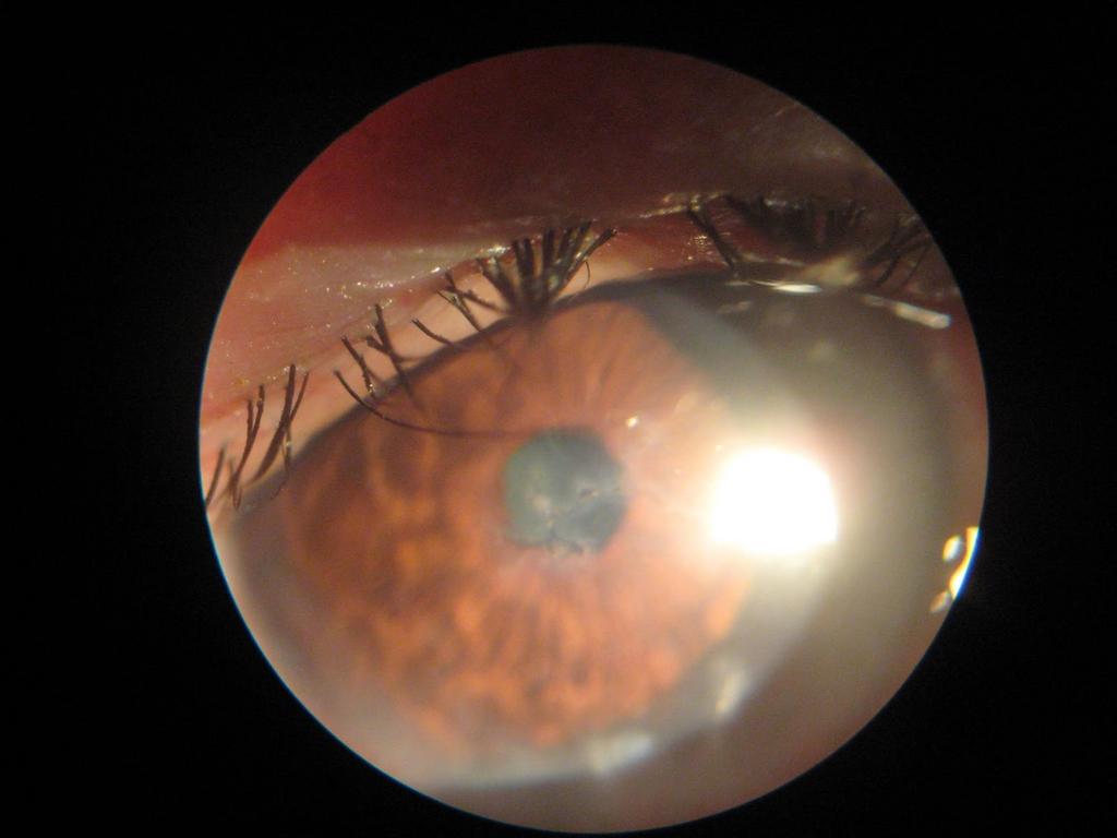
14 6 Keratitis Non specific signs of fungal keratitis include conjunctival injection; epithelial defect; suppuration;stromal infiltration; anterior chamber reaction; hypopyon; aqueous flare and corneal neovascularization. Specific signs of fungal keratitis include infiltrates with feathery margins, rough texture, raised borders, brown pigmentation, associated endothelial plaque, and satellite lesions; deep stromal infiltrates with an intact epithelium; dull grey appearance of the cornea with possible heaping of epithelium. Sclerotic scatter can be used to highlight the density and scalloped borders of the fungal lesion. Many fungal ulcers demonstrate no striking morphological pattern, and often it is not possible to differentiate clinically between fungal keratitis and bacterial keratitis. Filamentous keratitis is characterized by a grey-yellow stromal infiltrate with indistinct margins, a progressive infiltration, often surrounded by satellite lesions and hypopyon. Filamentous fungi classically grow in a feathery branching pattern, but may be very rapidly progressive and indistinguishable from bacterial keratitis. (Figure 1.) Fig. 1. Fungal corneal ulcer Candida keratitis is characterized by a yellow-white infiltrate associated with dense suppuration. Candida species produces a small ulcer with expanding infiltrate in a collarstud configuration, often superimposed on a debilitating corneal condition. There may be an endothelial plaque under the lesion and satellite lesions at the edges. Suppurative keratitis, fibrinoid uveitis, hypopyon and elevated IOP may occur. Several features of fungal keratitis are characteristic if not pathognomonic and may permit an immediate or early diagnosis. These features are: 1. The surface of the lesion is usually gray or dirty white with a dry rough texture. 2. Areas of the lesion may be raised above the plane of the uninvolved cornea. 3. The margins of the ulcer tend to be irregular. 4. There may be satellite lesions. 5. There may be a complete or partial white immune ring around the lesion. This is said to be formed by the fungal antigen and host antibody response.
15 An Overview of Fungal Keratitis and Case Report on Trichophyton Keratitis Diagnostics After establishing the patient's general condition the examiner should look for evidence of ocular surface disease. Determine the amount and type of secretions and lid swelling. The upper eyelid should be everted to exclude a retained foreign body. The examiner should measure the size and depth of the lesion as well as the presence of satellite lesions. Also the intraocular pressure should be ascertained. Anterior chamber reaction and evidence of hypopyon should be recorded. Vitreous reaction if present may suggest intraocular spread of the disease. Under the slit lamp, early in the evolution, the lesion might look like an unhealed corneal abrasion with scanty infiltrates and no secretions. With time the ulcer develops thicker infiltrates and fuzzy margins. The presence of satellite lesions strongly suggests a fungal infection. Redness and periocular edema are also common. This combined with a history of trauma, especially with vegetable matter, ocular surface disease or chronic use of topical steroids should alert about the possibility of a mycotic etiology. We should ask the patient about ocular or systemic disease: keratocandidosis is commonest in debilitated patients or those with preexisting corneal disease. Ocular trauma is associated with filamentous fungi, e.g. Aspergillus or Fusarium spp. A diagnosis of fungal keratitis is based on a matrix of the following: 1. Case history 2. Clinical signs 3. Confirmation from cytology and/or culture results. The most important step in the initial managment of suspected fungal keratitis is to obtain corneal material for direct smears and inoculation of media. It is important to scrape multiple sites in the ulcer crater, particularly at the margins, to enhance recovery of the organisms. Corneal scrapings are taken from deep into the lesion with a surgical blade or sterile spatula. To perform a corneal biopsy a dermatological 2 mm punch can be used. Laboratory diagnostics should be performed before starting antifungal therapy. Filamentous fungi tend to proliferate anterior to Descement membrane and a deep stromal biopsy may be required (similar in technique to performing a trabeculectomy-the excised deep tissue is sent for culture). Sometimes the diagnosis can only be confirmed following anterior chamber tap or excisional keratoplasty. Direct microscopy of corneal smears can be performed with special methods such as KOH, calcofluor white, Gram or/and Giemsa staining.. Gram stain may identify the yeast forms of Candida, and Giemsa stain is more likely to detect filamentous fungus.(fig. 2) Cultivation of causative agents can be done on Sabouraud s dextrose agar, although most fungi will also grow on blood agar or in enrichment media at 27 deg celcius or at room temperature within 3 days. PCR with pan fungal primers are used as an adjunct to culture. Antifungal susceptibility testing can be performed in reference laboratories but the relevance of these results to clinical effectiveness is uncertain. Histology involving periodic acid-schiff(pas) stain and Grocott silver stain of corneal tissue are the most sensitive.
We are IntechOpen, the world s leading publisher of Open Access books Built by scientists, for scientists. International authors and editors
 We are IntechOpen, the world s leading publisher of Open Access books Built by scientists, for scientists 4,000 116,000 120M Open access books available International authors and editors Downloads Our
Mycotic Keratitis in Patients Attending a Tertiary Care Hospital
 International Journal of Current Microbiology and Applied Sciences ISSN: 2319-7706 Volume 6 Number 10 (20) pp. 1665-1670 Journal homepage: http://www.ijcmas.com Original Research Article https://doi.org/10.20546/ijcmas.20.610.201
Identification of Fungal Species in Proved Cases of Fungal Corneal Ulcer
 www.jmscr.igmpublication.org Impact Factor 3.79 ISSN (e)-2347-176x Identification of Fungal Species in Proved Cases of Fungal Corneal Ulcer Authors Madhusudhan C.N 1, Tanushree V 2, H.T.Venkategowda 3,
Prevalence of Oculomycosis in a Tertiary Care Centre
 AJMS Al Ameen J Med Sci (2 011 )4 (4 ):3 3 4-3 3 8 (A US National Library of Medicine enlisted journal) I S S N 0 9 7 4-1 1 4 3 C O D E N : A A J M B G ORIGI NAL ARTICLE Prevalence of Oculomycosis in a
Introduction. Study of fungi called mycology.
 Fungi Introduction Study of fungi called mycology. Some fungi are beneficial: ex a) Important in production of some foods, ex: cheeses, bread. b) Important in production of some antibiotics, ex: penicillin
The Role of Ultrasonographic Biomicroscopy in the management of a patient with presumed Dematiaceous Mycotic Keratitis
 ISPUB.COM The Internet Journal of Ophthalmology and Visual Science Volume 6 Number 2 The Role of Ultrasonographic Biomicroscopy in the management of a patient with presumed Dematiaceous Mycotic Keratitis
PAINFUL PAINLESS Contact lens user BOV
 Common Causes Allergies Infections Ocular Cornea, uveitis, endophthalmitis Orbital Orbital cellulitis Inflammation Uveitis Scleritis / episcleritis Glaucomas Trauma Foreign bodies Chemical injuries History
Post-LASIK infections
 Post-LASIK infections By Mohamed El-moddather Assiss. Prof. and head of department of ophthalmology AL-Azhar unizersity Assuit LASIK has become a common refractive procedure and is generally considered
CORNEAL CONDITIONS CORNEAL TRANSPLANTATION
 GENERAL INFORMATION CORNEAL CONDITIONS CORNEAL TRANSPLANTATION WHAT ARE CORNEAL CONDITIONS? The cornea is the clear outer layer of the eye. Shaped like a dome, it helps to protect the eye from foreign
Clinical Decision making in Infectious Keratitis
 Clinical Decision making in Infectious Stephen D. McLeod, MD Theresa M. and Wayne M. Caygill, MD Distinguished Professor and Chair Department of Ophthalmology Francis I. Proctor Foundation University of
Lamellar Keratoplasty for the Treatment of Fungal Keratitis
 Cornea 21(1): 33 37, 2002. 2002 Lippincott Williams & Wilkins, Inc., Philadelphia Lamellar Keratoplasty for the Treatment of Fungal Keratitis Lixin Xie, M.D., Weiyun Shi, M.D., Zhaosheng Liu, M.D., and
ISHAM Symposium: S33: Ocular aspects of Fungal Infections Friday, 8 May 2015, ; MR101/102 Level 1
 ISHAM Symposium: S33: Ocular aspects of Fungal Infections Friday, 8 May 2015, 14.15 15.45; MR101/102 Level 1 Chairs: Ariya Chindamporn, TH and Phillip A Thomas, IN 14:15-14:35 AGENDA Clinical overview
INCIDENCE OF CURVULARIA ORGANISM IN MYCOTIC CORNEAL ULCER K. Anjaneyulu 1, Balla Vidya Sagar 2
 INCIDENCE OF CURVULARIA ORGANISM IN MYCOTIC CORNEAL ULCER K. Anjaneyulu 1, Balla Vidya Sagar 2 HOW TO CITE THIS ARTICLE: K. Anjaneyulu, Balla Vidya Sagar. Incidence of Curvularia Organism in Mycotic Corneal
SCHEDULING STATUS Schedule 4 PROPRIETARY NAME AND DOSAGE FORM
 Page 1 of 5 SCHEDULING STATUS Schedule 4 PROPRIETARY NAME AND DOSAGE FORM FML Liquifilm Sterile Eye Suspension COMPOSITION FML Liquifilm Sterile Eye Suspension contains: Fluorometholone 1,0 mg/ml Liquifilm
Acute Eyes for ED. Enis Kocak. The Alfred Ophthalmology
 Acute Eyes for ED Enis Kocak The Alfred Ophthalmology The problem with eyes Things to cover Ocular anatomy Basic assessment Common presentations Eye first aid and procedures Ophthalmic emergencies What
A Clinical Microbiological Study of Corneal Ulcer Patients at Western Gujarat, India
 A Clinical Microbiological Study of Corneal Ulcer Patients at Western Gujarat, India Rajesh Somabhai Katara 1, Nilesh Dhanjibhai Patel 2, and Mala Sinha 3 1 Department of Microbiology, B. J. Medical College,
Nasreen A. Syed, MD F.C. Blodi Eye Pathology Laboratory University of Iowa
 Nasreen A. Syed, MD F.C. Blodi Eye Pathology Laboratory University of Iowa No financial interest in any of the material discussed in this presentation There will be discussion of off label use of medications,
Clinical Practice Guide for the Diagnosis, Treatment and Management of Anterior Eye Conditions. April 2018
 Clinical Practice Guide for the Diagnosis, Treatment and Management of Anterior Eye Conditions This Clinical Practice Guide provides evidence-based information about current best practice in the management
PRECISION PROGRAM. Injection Technique Quick-Reference Guide. Companion booklet for the Video Guide to Injection Technique
 Injection Technique Quick-Reference Guide PRECISION PROGRAM Companion booklet for the Video Guide to Injection Technique Available at www.ozurdexprecisionprogram.com Provides step-by-step directions with
The Epidemiological Features and Laboratory Results of Fungal Keratitis
 Cornea 21(6): 555 559, 2002. 2002 Lippincott Williams & Wilkins, Inc., Philadelphia The Epidemiological Features and Laboratory Results of Fungal Keratitis A 10-Year Review at a Referral Eye Care Center
Dry Eye Assessment and Management Study ELIGIBILITY OCULAR EVALUATION FORM
 Page 1 of 13 BEFORE COMPLETING THE OCULAR EXAMINATION, YOU MUST BE ABLE TO ANSWER YES TO THE FOLLOWING QUESTIONS: Have you done MMP9? (SVonly) The Following are done at Baseline: Have you done Tear Osmolarity?
Sclerokeratoplasty David S. Chu, M.D. Cases
 Sclerokeratoplasty David S. Chu, M.D. Cases Case 1 40 year-old female from Peru presented to our Service with inflamed OS for 2 months duration. Her symptoms began as red painful OS, which progressively
EPIDEMIOLOGICAL AND MICROBIOLOGICAL PROFILE OF PATIENT S HAVING MICROBIAL KERATITIS
 Original Article EPIDEMIOLOGICAL AND MICROBIOLOGICAL PROFILE OF PATIENT S HAVING MICROBIAL KERATITIS Saurabh Patel 1, Akshay M Chaudhari 2, Trupti M Solu 3, Vaibhav Gharat 4 Financial Support: None declared
number Done by Corrected by Doctor د.حامد الزعبي
 number Fungi#1 Done by نرجس الس ماك Corrected by مهدي الشعراوي Doctor د.حامد الزعبي Introduction to Mycology -Terms: -Medical Mycology: The study of mycosis and their etiological agents -Mycosis: Disease
Dr Jo-Anne Pon. Dr Sean Every. 8:30-9:25 WS #70: Eye Essentials for GPs 9:35-10:30 WS #80: Eye Essentials for GPs (Repeated)
") Dr Sean Every Ophthalmologist Southern Eye Specialists Christchurch Dr Jo-Anne Pon Ophthalmologist Southern Eye Specialists, Christchurch Hospital, Christchurch 8:30-9:25 WS #70: Eye Essentials for GPs
Dr Sean Every Ophthalmologist Southern Eye Specialists Christchurch Dr Jo-Anne Pon Ophthalmologist Southern Eye Specialists, Christchurch Hospital, Christchurch 8:30-9:25 WS #70: Eye Essentials for GPs
EYE CARE PROTOCOL FOR PATIENTS IN ITU
 EYE CARE PROTOCOL FOR PATIENTS IN ITU Back to contents Developed by SUE LIGHTMAN PROFESSOR OF CLINICAL OPHTHALMOLOGY/CONSULTANT OPHTHALMOLOGIST MOORFIELDS EYE HOSPITAL Amended for UCLU ICU by Caroline
Subject Index. Atopic keratoconjunctivitis (AKC) management 16 overview 15
 management 16 overview 15") Subject Index Acanthamoeba keratitis, see Infective keratitis Acute allergic conjunctivitis AKC, see Atopic keratoconjunctivitis Allergy acute allergic conjunctivitis 15 atopic keratoconjunctivitis 15
Subject Index Acanthamoeba keratitis, see Infective keratitis Acute allergic conjunctivitis AKC, see Atopic keratoconjunctivitis Allergy acute allergic conjunctivitis 15 atopic keratoconjunctivitis 15
ISPUB.COM. Management of Mycotic Keratitis. V Sharma, M Purohit, S Vaidya INTRODUCTION
 ISPUB.COM The Internet Journal of Ophthalmology and Visual Science Volume 6 Number 2 V Sharma, M Purohit, S Vaidya Citation V Sharma, M Purohit, S Vaidya.. The Internet Journal of Ophthalmology and Visual
Subcutaneous Fungi 10/13/2009. General Characteristics. Pathogenesis. Epidemiology. Laboratory Diagnosis. Specimens. Growth rate: 1-4 weeks
 General Characteristics Growth rate: 1-4 weeks Subcutaneous Fungi Clinical Laboratory Science Program Carol Larson MSEd, MT(ASCP) Dematiaceous septate hyphae Hyaline septate hyphae Branching GPR Epidemiology
INVELTYS (loteprednol etabonate ophthalmic suspension) 1%, for topical ophthalmic use Initial U.S. Approval: 1998
 1%, for topical ophthalmic use Initial U.S. Approval: 1998") HIGHLIGHTS OF PRESCRIBING INFORMATION These highlights do not include all the information needed to use INVELTYS safely and effectively. See full prescribing information for INVELTYS. INVELTYS (loteprednol
HIGHLIGHTS OF PRESCRIBING INFORMATION These highlights do not include all the information needed to use INVELTYS safely and effectively. See full prescribing information for INVELTYS. INVELTYS (loteprednol
2016 Week 2. Corneal Ulcer Culture Collection & Foreign Body Removal
 2016 Week 2 Corneal Ulcer Culture Collection & Foreign Body Removal Collection of corneal ulcer specimen Entering acuities Thorough SLE; anesthetic likely necessary for SLE & definitely for corneal ulcer
Eye Care for Animals Micki Armour VMD DACVO THE CORNEA
 Eye Care for Animals Micki Armour VMD DACVO THE CORNEA ANATOMY 0.5-0.6mm thick 4 primary layers Epithelium (5-7 cell layers) Stroma (90% total thickness) Descemet s membrane Endothelium (1 layer) ANATOMY-
Fungal Keratitis CURRENT CONCEPTS. Aetiology. Epidemiology. Fungi causing human keratitis. Predisposing factors. Local
 June 2008 N. Bindu - Fungal Keratitis 169 CURRENT CONCEPTS Fungal Keratitis Dr. N. Bindu MS Fungal infections of the cornea constitute an important eye problem in outdoor workers in tropical & subtropical
Strategies for Anterior Segment Disease Management Mile Brujic, OD, FAAO 1409 Kensington Blvd Bowling Green, OH
 Strategies for Anterior Segment Disease Management Mile Brujic, OD, FAAO 1409 Kensington Blvd Bowling Green, OH 43402 brujic@prodigy.net 419-261-9161 Summary As optometry s scope of practice continues
FUNGAL CORNEAL ULCER. Arundhati Dvivedi final year p.g Dept.of Ophthalmology 2018/7/31
 FUNGAL CORNEAL ULCER Arundhati Dvivedi final year p.g Dept.of Ophthalmology Introduction: Fungal Keratitis is one of the most difficult forms of microbial keratitis to diagnose & to treat successfully
Dr.saifalshamarti. Objective. Where is cornea? Functions of the cornea
 Cornea Dr.saifalshamarti Objective Functions Anatomy: detailed description of the 5 layers: epithelium, Bowman s layer, stroma, Descement s membrane, endothelium. Diseases of the cornea: - infection: bacterial
ICD-10 Coding for Contact Lens Problems. The EyeCodingForum.com
 ICD-10 Coding for Contact Lens Problems The EyeCodingForum.com Jeffrey Restuccio, CPC, CPC-H, MBA Memphis TN (901) 517-1705 jeff@eyecodingforum.com www.eyecodingforum.com EyeCodingForum.com 1 Coding for
Acanthameba Keratitis
 Acanthameba Keratitis CHARALAMBOS S. SIGANOS, MD, PHD ASSOC. PROFESSOR OF OPHTHALMOLOGY UNIVERSITY OF CRETE DEPARTMENT OF OPHTHALMOLOGY HERAKLION UNIVERSITY HOSPITAL CRETE GREECE I declare no conflict
THE RED EYE Cynthia McNamara, MD Week 25
 THE RED EYE Cynthia McNamara, MD Week 25 Educational Objectives: 1. Know the differential diagnosis and presentation of specific etiologies of the red eye 2. Be able to evaluate patients presenting with
The Immune System & Non- Infectious Disease. Ch. 18: Sections 1, 2, & 4
 The Immune System & Non- Infectious Disease Ch. 18: Sections 1, 2, & 4 What is the Immune System? The purpose of the immune system is to: keep infectious microorganisms, such as certain bacteria, viruses,
founder of McDonald s Restaurants
 Press On Nothing in the world can take the place of persistence. Talent will not; nothing is more common than unsuccessful men with talent. Genius will not; unrewarded genius is almost a proverb. Education
Meet Libby. Corneal Dysgenesis, Degeneration, and Dystrophies Definitions. Dr. Victor Malinovsky
 Meet Libby Corneal Dysgenesis, Degeneration, and Dystrophies 2006 Dr. Victor Malinovsky Definitions Dysgenesis: (congenital anomalies) A development disorder that results in a congenital malformation of
Dermatophytes Dr. Hala Al Daghistani
 Dermatophytes Dr. Hala Al Daghistani Dermatophytoses are superficial infections of the skin and its appendages, commonly known as ringworm, athlete s foot, and jock itch. They are caused by species of
Spectrum of Mycotic corneal ulcers in Mid Western peripheral region of Terrain belt of Nepal and Indo-Nepal Border
 Nepal Journal of Medical Sciences Original Article Spectrum of Mycotic corneal ulcers in Mid Western peripheral region of Terrain belt of Nepal and Indo-Nepal Border Bastola P, 1* Mishra A, 2 Chaudhary
Fungi. Eucaryotic Rigid cell wall(chitin, glucan) Cell membrane ergosterol Unicellular, multicellular Classic fungus taxonomy:
 Cell membrane ergosterol Unicellular, multicellular Classic fungus taxonomy:") MYCOLOGY Mycology I Fungi Eucaryotic Rigid cell wall(chitin, glucan) Cell membrane ergosterol Unicellular, multicellular Classic fungus taxonomy: Morphology Spore formation FFungi Yeast Mold Yeastlike
MYCOLOGY Mycology I Fungi Eucaryotic Rigid cell wall(chitin, glucan) Cell membrane ergosterol Unicellular, multicellular Classic fungus taxonomy: Morphology Spore formation FFungi Yeast Mold Yeastlike
Spectrum of Fungal Keratitis in North China
 Spectrum of Fungal Keratitis in North China Lixin Xie, MD, 1 Wenxian Zhong, MD, 1,2 Weiyun Shi, MD, 1 Shiying Sun, MD 1 Purpose: To report the epidemiological features, laboratory findings, and treatment
Proceedings of the World Small Animal Veterinary Association Sydney, Australia 2007
 Proceedings of the World Small Animal Sydney, Australia 2007 Hosted by: Next WSAVA Congress MANAGEMENT OF CORNEAL ULCERS IN SMALL ANIMALS Robin G Stanley, BVSc(Hons), FACVSc-Ophthalmology Animal Eye Care
GAFFI Fact Sheet. Keratitis refers to inflammation (usually an infection) of the normally transparent DARKER AREAS AND
 of the normally transparent DARKER AREAS AND") Fungal Keratitis GAFFI Fact Sheet GLOBAL ACTION FUND FOR FUNGAL INFECTIONS Keratitis refers to inflammation (usually an infection) of the normally transparent DARKER AREAS AND cornea of the eye, which
Fungal Keratitis GAFFI Fact Sheet GLOBAL ACTION FUND FOR FUNGAL INFECTIONS Keratitis refers to inflammation (usually an infection) of the normally transparent DARKER AREAS AND cornea of the eye, which
Nursing college, Second stage Microbiology Dr.Nada Khazal K. Hendi Medical Microbiology
 1 Nursing college, Second stage Microbiology Medical Microbiology Lecture-1- Fungi (Mycosis) They are a diverse group of saprophytic and parasitic eukaryotic organisms. Human fungal diseases (mycoses)
Pathogenesis of ocular fungal Infections. Dr Lalitha Prajna. MD. Department of Ocular Microbiology, Aravind Eye Hospital, Madurai. Tamil Nadu.
 Pathogenesis of ocular fungal Infections Dr Lalitha Prajna. MD. Department of Ocular Microbiology, Aravind Eye Hospital, Madurai. Tamil Nadu. Introduction Fungal infections of the eye are rare ( exception:
EYE INJURIES OBJECTIVES COMMON EYE EMERGENCIES 7/19/2017 IMPROVE ASSESSMENT OF EYE INJURIES
 EYE INJURIES BRITTA ANDERSON D.O. DMC PRIMARY CARE SPORTS MEDICINE ASSOCIATE TEAM PHYSICIAN DETROIT TIGERS OBJECTIVES IMPROVE ASSESSMENT OF EYE INJURIES UNDERSTAND WHAT IS CONSIDERED AN EMERGENCY DEVELOP
Focusing on A&E. By Sandy Cooper, (Ophthalmic Nurse Practitioner), Tel
, Tel") Focusing on A&E By Sandy Cooper, (Ophthalmic Nurse Practitioner), Tel 01752 439331 Email sandra.cooper5@nhs.net sandracooper041@btinternet.com THINGS TO WORRY ABOUT WITH ANY EYE PROBLEM CHANGES IN VISION
Focusing on A&E By Sandy Cooper, (Ophthalmic Nurse Practitioner), Tel 01752 439331 Email sandra.cooper5@nhs.net sandracooper041@btinternet.com THINGS TO WORRY ABOUT WITH ANY EYE PROBLEM CHANGES IN VISION
Corneal Infections. Carrie Lembach DO Ohio Ophthalmological Society Annual Meeting February 21, 2015
 Corneal Infections Carrie Lembach DO Ohio Ophthalmological Society Annual Meeting February 21, 2015 Objectives Identify differential diagnosis for corneal infections Identify most common organisms involved
Ulcerative Keratitis (Type of Inflammation of the Cornea) Basics
 Basics") Ulcerative Keratitis (Type of Inflammation of the Cornea) Basics OVERVIEW Keratitis is inflammation of the cornea; the cornea is the clear outer layer of the front of the eye The corneal epithelium is
Ulcerative Keratitis (Type of Inflammation of the Cornea) Basics OVERVIEW Keratitis is inflammation of the cornea; the cornea is the clear outer layer of the front of the eye The corneal epithelium is
Vision Loss After Contact Lens-Related Pseudomonas Keratitis
 Vision Loss After Contact Lens-Related Pseudomonas Keratitis Matthew C. Weed, MD; Gina M. Rogers, MD; Anna S. Kitzmann, MD; Kenneth M. Goins, MD; Michael D. Wagoner, MD, PhD June 24, 2013 Microbial keratitis
The Emergent Eye in the Acute Setting
 The Emergent Eye in the Acute Setting Todd P. Margolis MD, PhD Professor of Ophthalmology & Director of the F.I. Proctor Foundation UCSF Physical Exam-- Visual Acuity Essential Corrected visual acuity
MSES consultants, inc.
 MSES consultants, inc. 609 West Main Street P.O. Drawer 190 Clarksburg, WV 26302-0190 304.624.9700 304.622.0981 304.842.3325 http://www.msesinc.com Office September 13, 2012 Project Number: 12-437 Mr.
Epidemiology and ecology of fungal diseases
 Epidemiology and ecology of fungal diseases Healthcare Focus on: - individual - diagnosis - treatment Public Health Focus on: - population - prevention The nature of fungi Kingdom Fungi (lat. fungus, -i)
Differential diagnosis of the red eye. Carol Slight Nurse Practitioner Ophthalmology
 Differential diagnosis of the red eye Carol Slight Nurse Practitioner Ophthalmology The red eye Conjunctivitis HSV Keratitis Acute angle closure glaucoma Anterior Uveitis Red eye Scleritis Subconjunctival
Management of specific eye problems in the ED
 of specific eye problems in the ED CORNEAL ABRASION Causes Foreign bodies Tangential shearing injuries, e.g. poking finger into eye Exact cause of injury (Remember to exclude possibility of intraocular
Mycology. BioV 400. Subcutaneous Mycoses. Ecological associations. Geographic distribution World-wide
 BioV 400 Mycology Handout 8 Subcutaneous Mycoses Lymphocutaneous sporotrichosis Chromoblastomycosis Phaeohyphomycosis Zygomycosis Mycetoma Lymphocutaneous sporotrichosis Sporothrix schenckii Chronic infection
Fleck. Pre-Descemet Dystrophies (generally good vision and comfort) Primary Pre-Descemet Dystrophy
 Primary Pre-Descemet Dystrophy") Fleck Etiology: bilateral, sometimes asymmetric, autosomal dominant opacities located in all levels of stroma as early as 1 st decade Slit lamp: well demarcated, small round gray-white doughnut-like, wreath-like
Fleck Etiology: bilateral, sometimes asymmetric, autosomal dominant opacities located in all levels of stroma as early as 1 st decade Slit lamp: well demarcated, small round gray-white doughnut-like, wreath-like
Prednisolone Sodium Phosphate Ophthalmic Solution USP, 1% (Sterile) Rx only
 Rx only") Prednisolone Sodium Phosphate Ophthalmic Solution USP, 1% (Sterile) Rx only DESCRIPTION Prednisolone Sodium Phosphate Ophthalmic Solution, 1%, is a sterile solution for ophthalmic administration having
Prednisolone Sodium Phosphate Ophthalmic Solution USP, 1% (Sterile) Rx only DESCRIPTION Prednisolone Sodium Phosphate Ophthalmic Solution, 1%, is a sterile solution for ophthalmic administration having
Department of Ophthalmology
 Department of Ophthalmology Period : 02/July/18 to 30/August/18 Semester : 7 th Semester Lecture Lesson Plan Sr. Date Topic Lesson plan Name of Faculty No. 1 02.07.18 Lens- Lens-Anatomy, Classification
Clinical and Microbiological Profile of Various Microorganisms Causing Keratitis in a Tertiary Care Hospital, Jaipur, India
 International Journal of Current Microbiology and Applied Sciences ISSN: 2319-7706 Volume 6 Number 2 (2017) pp. 1333-1342 Journal homepage: http://www.ijcmas.com Original Research Article http://dx.doi.org/10.20546/ijcmas.2017.602.151
Unit 1: Asepsis and Infection Control
 Unit 1: Asepsis and Infection Control Outlines - Type of microorganism causing infection. - Types of infection. - Nosocomial infection. - Chain of infection. - Body defenses against infection. - Factors
Eye Examination Techniques in Horses
 Eye Examination Techniques in Horses Dennis E. Brooks DVM, PhD Dip ACVO University of Florida brooksd@mail.vetmed.ufl.edu Basic Instruments How to tell the potential of vision? PLRs (retina, CN 2, chiasm,
Childhood corneal neovascularization
 Miltos Balidis PhD, FEBOphth, ICOphth Sotiria Palioura MD,PhD Childhood corneal neovascularization Opacities Cornea clarity is essential for optimal vision at any age. In childhood, loss of corneal transparency
NEW ZEALAND DATA SHEET 1. PRODUCT NAME
 NEW ZEALAND DATA SHEET 1. PRODUCT NAME Flucon fluorometholone 0.1% Eye Drops Suspension 2. QUALITATIVE AND QUANTITATIVE COMPOSITION Each ml of Flucon contains 1.0 mg of fluorometholone (0.1% w/v). Excipient
OOGZIEKTEN VOOR DE HUISARTS F. GOES, JR.
 OOGZIEKTEN VOOR DE HUISARTS F. GOES, JR. HET RODE OOG F. GOES, JR. Condition Signs Symptoms Causes Conjunctivitis Viral Normal vision, normal pupil size Mild to no pain, diffuse Adenovirus (most common),
Ocular and periocular trauma
 Ocular and periocular trauma No financial disclosures. Tina Rutar M.D. Assistant Professor of Clinical Ophthalmology and Pediatrics Director, Visual Center for the Child University of California San Francisco
Learning Objectives. Disclosures 2/2/ BMT Pharmacists Conference Bandage Contact Lens Therapy for Severe Ocular GVHD
 2015 BMT Pharmacists Conference Bandage Contact Lens Therapy for Severe Ocular GVHD Tueng T. Shen, M.D., Ph.D. Professor of Ophthalmology Adjunct, Bioengineering and Global Health Feb. 13 th, 2015 Learning
AUSTRALIAN PRODUCT INFORMATION FLAREX (FLUOROMETHOLONE ACETATE) EYE DROPS SUSPENSION
 EYE DROPS SUSPENSION") AUSTRALIAN PRODUCT INFORMATION FLAREX (FLUOROMETHOLONE ACETATE) EYE DROPS SUSPENSION 1 NAME OF THE MEDICINE Fluorometholone acetate. 2 QUALITATIVE AND QUANTITATIVE COMPOSITION The active ingredient in
AUSTRALIAN PRODUCT INFORMATION FLAREX (FLUOROMETHOLONE ACETATE) EYE DROPS SUSPENSION 1 NAME OF THE MEDICINE Fluorometholone acetate. 2 QUALITATIVE AND QUANTITATIVE COMPOSITION The active ingredient in
Clinical Profile of Herpes Simplex Keratitis
 K V Raju MS, Jyothi PT MS, Shimna Iqbal MS Clinical Profile of Herpes Simplex Keratitis Original Article Abstract Aims To document the various clinical presentations and to assess the risk factors contributing
Varicella-Zoster Virus Epithelial Keratitis in Herpes Zoster Ophthalmicus
 Varicella-Zoster Virus Epithelial Keratitis in Herpes Zoster Ophthalmicus Helena M. Tabery Varicella-Zoster Virus Epithelial Keratitis in Herpes Zoster Ophthalmicus In Vivo Morphology in the Human Cornea
Herpetic Eye Disease Jason Duncan, OD, FAAO Diplomate, American Board of Optometry Associate Professor, Southern College of Optometry
 Herpetic Eye Disease Jason Duncan, OD, FAAO Diplomate, American Board of Optometry Associate Professor, Southern College of Optometry I have what?! How to break the news Meet the Herpes Quick virology
MANAGING MELTING EYE ULCERS
 Vet Times The website for the veterinary profession https://www.vettimes.co.uk MANAGING MELTING EYE ULCERS Author : Anna Jennings Categories : Vets Date : January 18, 2010 Anna Jennings details effective
Acridine Orange Staining for Rapid Diagnosis of Acanthamoeba Keratitis
 Acridine Orange Staining for Rapid Diagnosis of Acanthamoeba Keratitis Tae-Won Hahn,* Terrence P. O Brien, Woo-Jin Sah* and Jae-Ho Kim* *Department of Ophthalmology, Catholic University Medical College,
Examining Children s Eyes
 Paediatric Ophthalmology What to refer & when? Aims Tips for assessing a child s eyes in general practice Common paediatric ophthalmology symptoms and signs What needs to be referred and when? MISS FARIHA
Otomycosis in Bikaner: A Clinico-Mycological Study
 International Journal of Current Microbiology and Applied Sciences ISSN: 2319-7706 Volume 6 Number 9 (2017) pp. 2943-2947 Journal homepage: http://www.ijcmas.com Original Research Article https://doi.org/10.20546/ijcmas.2017.609.361
Babak Valizadeh, DCLS
 Ocular Infections- Laboratory Diagnosis of Bacterial Infections of the Eye Babak Valizadeh, DCLS 1392. 02. 01 2012. 04. 21 Babak_Valizadeh@hotmail.com Discriminating Between Indigenous Microbiota and
Histopathology Description:
 2013-2-1 CANINE HEART Ahmed M. Abubakar BOVINE PATHOLOGY CONTRIBUTING INSTITUTION : The Royal Veterinary college, Dept. of Pathology and Biology Signalment: 11-month-old male Border Collie dog (Canis familiaris)
PRED-G (gentamicin and prednisolone acetate ophthalmic ointment, USP) 0.3%/0.6% sterile
 0.3%/0.6% sterile") PRED-G (gentamicin and prednisolone acetate ophthalmic ointment, USP) 0.3%/0.6% sterile PRED-G sterile ophthalmic ointment is a topical anti-inflammatory/anti-infective combination product for ophthalmic
PRED-G (gentamicin and prednisolone acetate ophthalmic ointment, USP) 0.3%/0.6% sterile PRED-G sterile ophthalmic ointment is a topical anti-inflammatory/anti-infective combination product for ophthalmic
Common Etiological Agents Causing Keratitis: A Study from a Tertiary Care Hospital in South India
 International Journal of Current Microbiology and Applied Sciences ISSN: 2319-7706 Volume 6 Number 7 (2017) pp. 1625-1633 Journal homepage: http://www.ijcmas.com Original Research Article https://doi.org/10.20546/ijcmas.2017.607.196
Dr. D. Y. Patil Medical College, Pimpri, Pune
 Dr. D. Y. Patil Medical College, Pimpri, Pune - 411 018 Period : 04/July/16 to 22/September/16 Semester : 7 th Semester Department : Ophthalmology Lecture Lesson Plan Sr No Date Topic Learning objectives
NON TRADITIONAL MANAGEMENT OF FUNGAL KERATITIS
 NON TRADITIONAL MANAGEMENT OF FUNGAL KERATITIS MOHAMED SAAD,MD PROF. OF OPHTHALMOLOGY ASSIUT UNIVERSITY HOSPITAL FINANCIAL DISCLOSURE: No financial interest. 1 Fungal keratitis is a sight-threatening condition
Photochemical corneal collagen cross-linkage using riboflavin and ultraviolet A for keratoconus and keratectasia
 Photochemical corneal collagen cross-linkage using riboflavin and ultraviolet A for keratoconus and keratectasia Issued: September 2013 guidance.nice.org.uk/ipg466 NICE has accredited the process used
Ocular and Periocular Trauma. Tina Rutar, MD. Assistant Professor of Ophthalmology and Pediatrics. Director, Visual Center for the Child
 Ocular and Periocular Trauma Tina Rutar, MD Assistant Professor of Ophthalmology and Pediatrics Director, Visual Center for the Child University of California, San Francisco Phone: 415-353-2560 Fax: 415-353-2468
NEW YORK UNIVERSITY SCHOOL OF MEDICINE DEPARTMENT OF OPHTHALMOLOGY EDUCATIONAL OBJECTIVES AND GOALS
 NEW YORK UNIVERSITY SCHOOL OF MEDICINE DEPARTMENT OF OPHTHALMOLOGY EDUCATIONAL OBJECTIVES AND GOALS Revision Date: 6/30/06 Distribution Date: 7/6/06 The Department of Ophthalmology at the NYU Medical Center
Morphology and Ultrastructure of Fungi in Extended-Wear
 JOURNAL OF CLINICAL MICROBIOLOGY, July 1986, p. 21-25 0095-1137/86/070021-05$02.00/0 Copyright D 1986, American Society for Microbiology Vol. 24, No. 1 Morphology and Ultrastructure of Fungi in Extended-Wear
Interventional procedures guidance Published: 25 September 2013 nice.org.uk/guidance/ipg466
 Photochemical corneal collagen cross-linkage using riboflavin and ultraviolet A for keratoconus and keratectasia Interventional procedures guidance Published: 25 September 2013 nice.org.uk/guidance/ipg466
Department of Ophthalmology
 Period : 03/July/17 to 07/September/17 Semester : 7 th Semester Department of Ophthalmology Lecture Lesson Plan Sr 1 03.07.17 Uvea-Anatomy, Uvea-Anatomy, Classification of Uveitis Dr R Paranjpe Classification
Evaluation of Agent and Host Factors in Progression of Mycotic Keratitis
 Evaluation of Agent and Host Factors in Progression of Mycotic Keratitis A Histologic and Microbiologic Study of 167 Corneal Buttons Geeta Kashyap Vemuganti, MD, 1 Prashant Garg, MS, 2 Usha Gopinathan,
Condition: Herpes Simplex Keratitis
 Condition: Herpes Simplex Keratitis Description: Herpes simplex infection is very common but usually remains latent. When the virus is reactivated it travels along the trigeminal nerve to cause local infection
Viral Taxonomic Classification
 Viruses Part I Viral Taxonomic Classification Order>> -virales Family>> - viridae Subfamily>> -virinae Genus>> -virus Species Order>> Picornavirales Family>> Picornaviridae Subfamily>> Picornavirinae Genus>>
Medical Affairs Policy
 Medical Affairs Policy Service: Corneal Treatments and Specialized Contact Lenses (Corneal remodeling, Corneal transplant, Corneal collagen crosslinking, Intrastromal Rings- INTACS, Keratoconus treatments,
INDICATIONS For steroid responsive inflammation of the palpebral and bulbar conjunctiva, cornea, and anterior segment of the eye globe.
 Page 1 of 5 SCHEDULING STATUS Schedule 4 PROPRIETARY NAME AND DOSAGE FORM PRED FORTE Sterile Eye Suspension COMPOSITION PRED FORTE Sterile Eye Suspension contains: Prednisolone acetate 10 mg/ml Preservative:
A Guide to Administering
 A Guide to Administering INDICATIONS AND USAGE YUTIQ (fluocinolone acetonide intravitreal implant) 0.18 mg is indicated for the treatment of chronic non-infectious uveitis affecting the posterior segment
To Evaluate the Sociodemographic Factors And Etiology of Corneal Neovascularisation at out Patient Department of M.L.B Medical College, Jhansi.(U.
 IOSR Journal of Dental and Medical Sciences (IOSR-JDMS) e-issn: 2279-0853, p-issn: 2279-0861.Volume 15, Issue 6 Ver. IV (June. 2016), PP 129-134 www.iosrjournals.org To Evaluate the Sociodemographic Factors
DEFINITION Corneal abrasion is a defect in the corneal surface epithelium due to scraping or rubbing of the corneal epithelium.
 DEFINITION Corneal abrasion is a defect in the corneal surface epithelium due to scraping or rubbing of the corneal epithelium. IMMEDIATE CONSULTATION REQUIRED IN THE FOLLOWING SITUATIONS Dendritic pattern