PRESERVE: How intensively should we treat blood pressure in established cerebral small vessel disease? Guide to assessing MRI scans
|
|
|
- Verity Hawkins
- 5 years ago
- Views:
Transcription
1 PRESERVE: How intensively should we treat blood pressure in established cerebral small vessel disease? Guide to assessing MRI scans
2 Inclusion Criteria Clinical syndrome Patients must have clinical evidence of cerebral small vessel disease, falling into one of the following categories: a) Lacunar stroke syndrome with symptoms lasting >24 hours b) Transient ischaemic attack lasting < 24 hours with limb weakness, hemisensory loss or dysarthria AND with MR DWI imaging performed acutely showing lacunar infarction, or if MRI is not performed acutely (>2 weeks after TIA) with a lacunar infarction in an anatomically appropriate position on MRI c) Vascular cognitive impairment with MRI showing no evidence of hippocampal atrophy
3 Inclusion Criteria MRI As well as having an appropriate clinical lacunar syndrome patients must have: MRI evidence of lacunar infarct(s) (<=1.5cm vascular diameter) AND Confluent leukoaraiosis (defined on Fazekas scale as >=grade 2) Patients entered with Vascular Cognitive Impairment must show no evidence of hippocampal atrophy This document will provide guidance and examples to help with assessing these criteria.

4 Lacunar infarct Leukoaraiosis
5 Estimating the degree of Leukoaraiosis Fazekas Scale The Fazekas Scale is a simple visual rating scale used to rate the degree of leukoaraiosis (white matter hyperintensity) It rates periventricular and deep white-matter hyperintensities (PVH and DWMH) on a four point scale. PVH and DWMH are considered separately and rated from 0-3 To be eligible for PRESERVE, a patient must have a score of at least 2 for either PVH or DWMH PVH 0 = absent 1 = caps or pencil-thin lining around ventricles 2 = smooth halo around ventricles (6-10mm regular margins) 3 = irregular halo > 10mm DWMH 0 = absent 1 = multiple focal lesions (>5) 2 = early confluent disease 3 = confluent disease Fazekas et al. MR signal abnormalities at 1.5T in Alzheimer s disease and normal aging. AJNR 1987:8: Fazekas et al. Pathological correlates of incidental MRI white matter signal hyperintensities. Neurology 1993; 43:
6 Applying the Fazekas Scale - Examples The following slides show standard slices from MRI scans which have been chosen to represent a range of white matter abnormalities. Some of the Fazekas scale examples have been taken from the following useful resource: 20 standard MR scans with validated ratings on several white matter rating scales Authors: Prof. Joanna Wardlaw & Dr. Karen Ferguson, University of Edinburgh
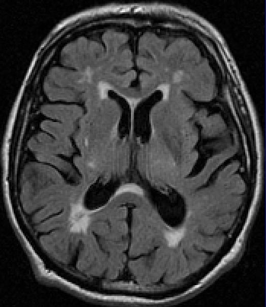
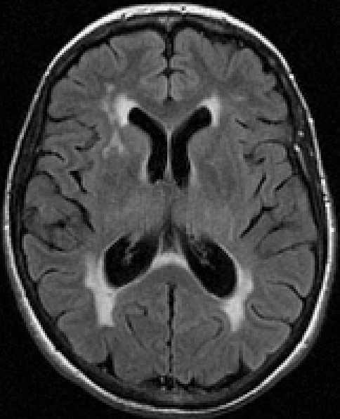
7 Fazekas Scale - Examples There are two rows of images in each slide, each showing 3 standard slices, from inferior to superior. The T2-weighted images are on the top row with the FLAIR images below. T2 inferior T2 medial T2 superior FLAIR inferior FLAIR medial FLAIR superior The consensus expert ratings for each scale are shown below the images. WMH can be assessed on either T2 or FLAIR images. They are easier to assess on FLAIR, in which an inversion recovery sequence suppresses signal from free water (for example in the ventricles). Note that some of the cases have also have a focal lesion, which are pointed out (see arrows). Focal lesions should be ignored for the purposes of rating white matter lesions.


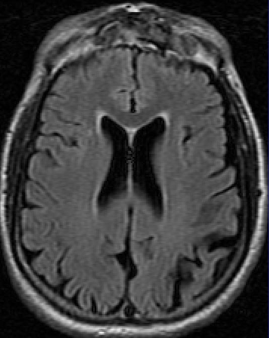


8 1 Fazekas Rating: PVH 1 / DWMH 0 Comments: This slide demonstrates the lowest level of white matter lesions. NOT ELIGIBLE FOR PRESERVE


9 2 Fazekas Rating: PVH 1 / DWMH 1 NOT ELIGIBLE FOR PRESERVE


10 3 Fazekas Rating: PVH 1 / DWMH 1 NOT ELIGIBLE FOR PRESERVE


11 4 Fazekas Rating: PVH 1 / DWMH 1 NOT ELIGIBLE FOR PRESERVE


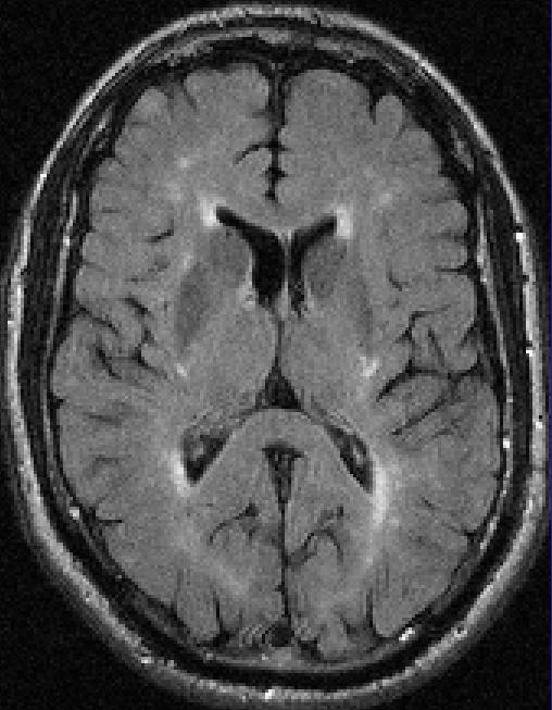


12 5 Fazekas Rating: PVH 2 / DWMH 1 ELIGIBLE FOR PRESERVE


13 6 Fazekas Rating: PVH 2 / DWMH 2 ELIGIBLE FOR PRESERVE


14 7 Fazekas Rating: PVH 3 / DWMH 2 ELIGIBLE FOR PRESERVE


15 8 Fazekas Rating: PVH 3 / DWMH 3 ELIGIBLE FOR PRESERVE


16 9 Fazekas Rating: PVH 3 / DWMH 3 ELIGIBLE FOR PRESERVE


17 10 Fazekas Rating: PVH 3 / DWMH 3 ELIGIBLE FOR PRESERVE Comments: This case has a focal lesion; a lacunar infarct in the posterior limb of the left internal capsule (arrow) which you should ignore. Note that there are also numerous enlarged perivascular spaces the tiny white dots in the basal ganglia on the T2-weighted images (circled on the right side).


18 11 Fazekas Rating: PVH 3 / DWMH 3 ELIGIBLE FOR PRESERVE Comments: In this case, the white matter is diffusely abnormal throughout the cerebral hemispheres. The case demonstrates the highest possible level of white matter lesions.
19 The following slides show FLAIR images only


20 12 Fazekas Rating: PVH 0 / DWMH 0 NOT ELIGIBLE FOR PRESERVE


21 13 Fazekas Rating: PVH 0 / DWMH 0 NOT ELIGIBLE FOR PRESERVE


22 14 Fazekas Rating: PVH 1 / DWMH 0 NOT ELIGIBLE FOR PRESERVE


23 15 Fazekas Rating: PVH 2 / DWMH 1 ELIGIBLE FOR PRESERVE


24 16 Fazekas Rating: PVH 3 / DWMH 2 ELIGIBLE FOR PRESERVE


25 17 Fazekas Rating: PVH 2 / DWMH 2 ELIGIBLE FOR PRESERVE


26 18 Fazekas Rating: PVH 2 / DWMH 3 ELIGIBLE FOR PRESERVE


27 19 Fazekas Rating: PVH 3 / DWMH 3 ELIGIBLE FOR PRESERVE
28 Rating Hippocampal (Medial Temporal) Atrophy This only applies if you are entering the patient on the basis of Vascular Cognitive impairment
29 Why do we want to do this? When patients are entered on the basis of vascular cognitive impairment (VCI) we are trying to exclude patients with Alzheimer s disease, rather than VCI, as a cause for their cognitive impairment A hallmark of Alzheimer s is hippocampal atrophy
30 PRESERVE Rating Hippocampal (Medial Temporal) Atrophy Rate on both left and right sides Use coronal T1 weighted MR and look through a number of slices (covering approx 20mm) starting at the anterior hippocampus (immediately posterior to the amygdala)
31 Visual ratings of MTL atrophy - a visual scale Width of choroid fissure Width of temporal horn Hippocampal height 0 N N N 1 N N Scheltens, 1992 To be eligible for PRESERVE the patient must have a score of 0 or 1 on both sides If the score is above 2 on either the left or right side they are not eligible Scheltens P, Leys D, Barkhof F, Huglo D, Weinstein HC, Vermersch P, et al. Atrophy of medial temporal lobes on MRI in "probable" Alzheimer's disease and normal ageing: diagnostic value and neuropsychological correlates. Journal of Neurology, Neurosurgery & Psychiatry. 1992;55(10):
32 Examples
33 A MTA 0 ELIGIBLE FOR PRESERVE
34 B MTA 0 ELIGIBLE FOR PRESERVE
35 C MTA 0 ELIGIBLE FOR PRESERVE

36 D MTA 0 ELIGIBLE FOR PRESERVE
37 A MTA 1 ELIGIBLE FOR PRESERVE
38 B MTA 1 ELIGIBLE FOR PRESERVE
39 C MTA 1 ELIGIBLE FOR PRESERVE

40 D MTA 1 ELIGIBLE FOR PRESERVE

41 A MTA 2 NOT ELIGIBLE FOR PRESERVE
42 B MTA 2 NOT ELIGIBLE FOR PRESERVE
43 C MTA 2 NOT ELIGIBLE FOR PRESERVE
44 D MTA 2 NOT ELIGIBLE FOR PRESERVE
45 A MTA 3 NOT ELIGIBLE FOR PRESERVE
46 B MTA 3 NOT ELIGIBLE FOR PRESERVE
47 C MTA 3 NOT ELIGIBLE FOR PRESERVE

48 D MTA 3 NOT ELIGIBLE FOR PRESERVE
49 A MTA 4 NOT ELIGIBLE FOR PRESERVE
50 B MTA 4 NOT ELIGIBLE FOR PRESERVE

51 C MTA 4 NOT ELIGIBLE FOR PRESERVE
52 D MTA 4 NOT ELIGIBLE FOR PRESERVE
A Neuropsychiatric, Neuroradiological, and Neuropsychological Profile of a Cohort of Patients with Vascular Dementia
 A Neuropsychiatric, Neuroradiological, and Neuropsychological Profile of a Cohort of Patients with Vascular Dementia Moises Gaviria, MD University of Illinois at Chicago Advocate Christ Medical Center
Acute stroke. Ischaemic stroke. Characteristics. Temporal classification. Clinical features. Interpretation of Emergency Head CT
 Ischaemic stroke Characteristics Stroke is the third most common cause of death in the UK, and the leading cause of disability. 80% of strokes are ischaemic Large vessel occlusive atheromatous disease
brain MRI for neuropsychiatrists: what do you need to know
 brain MRI for neuropsychiatrists: what do you need to know Christoforos Stoupis, MD, PhD Department of Radiology, Spital Maennedorf, Zurich & Inselspital, University of Bern, Switzerland c.stoupis@spitalmaennedorf.ch
Neuro-Imaging in dementia: using MRI in routine work-up Prof. Philip Scheltens
 Neuro-Imaging in dementia: Philip Scheltens Alzheimer Center VU University Medical Center Amsterdam The Netherlands 1 Outline of talk Current guidelines Imaging used to exclude disease Specific patterns
Essentials of Clinical MR, 2 nd edition. 14. Ischemia and Infarction II
 14. Ischemia and Infarction II Lacunar infarcts are small deep parenchymal lesions involving the basal ganglia, internal capsule, thalamus, and brainstem. The vascular supply of these areas includes the
Pediatric MS MRI Study Methodology
 General Pediatric MS MRI Study Methodology SCAN PREPARATION axial T2-weighted scans and/or axial FLAIR scans were obtained for all subjects when available, both T2 and FLAIR scans were scored. In order
ORIGINAL CONTRIBUTION. How Complex Interactions of Ischemic Brain Infarcts, White Matter Lesions, and Atrophy Relate to Poststroke Dementia
 ORIGINAL CONTRIBUTION How Complex Interactions of Ischemic Brain Infarcts, White Matter Lesions, and Atrophy Relate to Poststroke Dementia Tarja Pohjasvaara, MD, PhD; Riitta Mäntylä, MD; Oili Salonen,
정진일 외 : 일과성 허혈성 발작의 확산 강조MR영상
 17 A B Fig. 1. A 71 year-old male patient was complained of abrupt motor weakness of extremities, lasting about 3-4 hours. He was diagnosed as TIA (transient ischemic attack) with full recovery of neurologic
MRI of Pathological Aging Brain
 MRI of Pathological Aging Brain Yukio Miki Department of Radiology, Osaka City University A variety of pathological changes occur in the brain with aging, and many of these changes can be identified by
1 MS Lesions in T2-Weighted Images
 1 MS Lesions in T2-Weighted Images M.A. Sahraian, E.-W. Radue 1.1 Introduction Multiple hyperintense lesions on T2- and PDweighted sequences are the characteristic magnetic resonance imaging (MRI) appearance
Blood Supply. Allen Chung, class of 2013
 Blood Supply Allen Chung, class of 2013 Objectives Understand the importance of the cerebral circulation. Understand stroke and the types of vascular problems that cause it. Understand ischemic penumbra
Patient with vertigo, dizziness and depression
 Clinical Case - Test Yourself Neuro/Head and Neck Radiology Patient with vertigo, dizziness and depression Michael Mantatzis, Paraskevi Argyropoulou, Panos Prassopoulos Radiology Department, Democritus
Acute Ischaemic Stroke
 Acute Ischaemic Stroke CT or MR SCA READIG FORM SCA ID: DATE OF READIG: SCA QUALIT: Good Moderate Poor Comment: READER ID: TPE OF SCA: CT: Without contrast: With contrast: MR: Diffusion: Perfusion ote
Visual Rating Scale Reference Material. Lorna Harper Dementia Research Centre University College London
 Visual Rating Scale Reference Material Lorna Harper Dementia Research Centre University College London Background The reference materials included in this document were compiled and used in relation to
Neuroimaging predictors of death and dementia in a cohort of older stroke survivors
 RESEARCH PAPER Neuroimaging predictors of death and dementia in a cohort of older stroke survivors Michael J Firbank, Louise M Allan, Emma J Burton, Robert Barber, John T O Brien, Raj N Kalaria Institute
Cerebral small vessel disease
 Cerebral small vessel disease What is it? What are the clinical syndromes? How do we diagnose it? What is the pathophysiology? New insights from genetics? Possible therapies? Small Vessel disease Changes
Silent Cerebral Strokes: Clinical Outcomes and Management
 Silent Cerebral Strokes: Clinical Outcomes and Management Nagaendran Kandiah Senior Consultant Neurologist, National Neuroscience Institute, Singapore Clinician Scientist, National Medical Research Council,
Pearls and Pitfalls in Neuroradiology of Cerebrovascular Disease The Essentials with MR and CT
 Pearls and Pitfalls in Neuroradiology of Cerebrovascular Disease The Essentials with MR and CT Val M. Runge, MD Wendy R. K. Smoker, MD Anton Valavanis, MD Control # 823 Purpose The focus of this educational
The central nervous system
 Sectc.qxd 29/06/99 09:42 Page 81 Section C The central nervous system CNS haemorrhage Subarachnoid haemorrhage Cerebral infarction Brain atrophy Ring enhancing lesions MRI of the pituitary Multiple sclerosis
Title:Cognitive profile in patients with a first-ever lacunar infarct with and without silent lacunes: a comparative study.
 Author's response to reviews Title:Cognitive profile in patients with a first-ever lacunar infarct with and without silent lacunes: a comparative study. Authors: Lorena Blanco-Rojas (l.blanco.rojas@copc.cat)
Cerebral white matter lesions associations with Aβ isoforms and amyloid PET
 Supplementary information Cerebral white matter lesions associations with Aβ isoforms and amyloid PET Danielle van Westen, MD PhD 1, *, Daniel Lindqvist, MD PhD,, Kaj Blennow, MD PhD,, Lennart Minthon,
[(PHY-3a) Initials of MD reviewing films] [(PHY-3b) Initials of 2 nd opinion MD]
![[(PHY-3a) Initials of MD reviewing films] [(PHY-3b) Initials of 2 nd opinion MD]](/thumbs/89/98619893.jpg "[(PHY-3a) Initials of MD reviewing films] [(PHY-3b) Initials of 2 nd opinion MD]") 2015 PHYSICIAN SIGN-OFF (1) STUDY NO (PHY-1) CASE, PER PHYSICIAN REVIEW 1=yes 2=no [strictly meets case definition] (PHY-1a) CASE, IN PHYSICIAN S OPINION 1=yes 2=no (PHY-2) (PHY-3) [based on all available
2015 PHYSICIAN SIGN-OFF (1) STUDY NO (PHY-1) CASE, PER PHYSICIAN REVIEW 1=yes 2=no [strictly meets case definition] (PHY-1a) CASE, IN PHYSICIAN S OPINION 1=yes 2=no (PHY-2) (PHY-3) [based on all available
NEURO IMAGING OF ACUTE STROKE
 1 1 NEURO IMAGING OF ACUTE STROKE ALICIA RICHARDSON, MSN, RN, ACCNS-AG, ANVP-BC WENDY SMITH, MA, RN, MBA, SCRN, FAHA LYNN HUNDLEY, APRN, CNRN, CCNS, ANVP-BC 2 2 1 DISCLOSURES Alicia Richardson: Stryker
W hite matter high intensity lesions (WML) on T2
 on T2") 576 PAPER Significance of white matter high intensity lesions as a predictor of stroke from arteriolosclerosis H Yamauchi, H Fukuda, C Oyanagi... See end of article for authors affiliations... Correspondence
576 PAPER Significance of white matter high intensity lesions as a predictor of stroke from arteriolosclerosis H Yamauchi, H Fukuda, C Oyanagi... See end of article for authors affiliations... Correspondence
Medical Neuroscience Tutorial Notes
 Medical Neuroscience Tutorial Notes Blood Supply to the Brain MAP TO NEUROSCIENCE CORE CONCEPTS 1 NCC1. The brain is the body's most complex organ. LEARNING OBJECTIVES After study of the assigned learning
Published July 10, 2014 as /ajnr.A4046
 Published July 10, 2014 as 10.3174/ajnr.A4046 ORIGINAL RESEARCH BRAIN Preoperative Prognostic Value of MRI Findings in 108 Patients with Idiopathic Normal Pressure Hydrocephalus J. Virhammar, K. Laurell,
Fig.1: A, Sagittal 110x110 mm subimage close to the midline, passing through the cingulum. Note that the fibers of the corpus callosum run at a
 Fig.1 E Fig.1:, Sagittal 110x110 mm subimage close to the midline, passing through the cingulum. Note that the fibers of the corpus callosum run at a slight angle are through the plane (blue dots with
How to read the report
 Dear Client, This is your second opinion report. How to read the report 1. Always consult your findings with your doctor. 2. Please bear in mind that the report is based only on the information you provide
Vague Neurological Conditions
 Vague Neurological Conditions Dr. John Lefebre, MD, FRCPC Chief Regional Medical Director Europe, India, South Africa, Middle East and Turkey Canada 2014 2 3 4 Agenda Dr. John Lefebre, M.D., FRCPC 1. TIA
Yin-Hui Siow MD, FRCPC Director of Nuclear Medicine Southlake Regional Health Centre
 Yin-Hui Siow MD, FRCPC Director of Nuclear Medicine Southlake Regional Health Centre Today Introduction to CT Introduction to MRI Introduction to nuclear medicine Imaging the dementias The Brain ~ 1.5
NACC Vascular Consortium. NACC Vascular Consortium. NACC Vascular Consortium
 NACC Vascular Consortium NACC Vascular Consortium Participating centers: Oregon Health and Science University ADC Rush University ADC Mount Sinai School of Medicine ADC Boston University ADC In consultation
ageing: diagnostic value and neuropsychological
 Journal of Neurology, Neurosurgery, and Psychiatry 1992;55:967-972 Atrophy of medial temporal lobes on MRI in "probable" Alzheimer's disease and normal ageing: diagnostic value and neuropsychological correlates
NEURO IMAGING 2. Dr. Said Huwaijah Chairman of radiology Dep, Damascus Univercity
 NEURO IMAGING 2 Dr. Said Huwaijah Chairman of radiology Dep, Damascus Univercity I. EPIDURAL HEMATOMA (EDH) LOCATION Seventy to seventy-five percent occur in temporoparietal region. CAUSE Most likely caused
Cerebrovascular Disorders. Blood, Brain, and Energy. Blood Supply to the Brain 2/14/11
 Cerebrovascular Disorders Blood, Brain, and Energy 20% of body s oxygen usage No oxygen/glucose reserves Hypoxia - reduced oxygen Anoxia - Absence of oxygen supply Cell death can occur in as little as
Stroke/TIA. Tom Bedwell
 Stroke/TIA Tom Bedwell tab1g11@soton.ac.uk The Plan Definitions Anatomy Recap Aetiology Pathology Syndromes Brocas / Wernickes Investigations Management Prevention & Prognosis TIAs Key Definitions Transient
LIMBIC SYSTEM. Dr. Amani A. Elfaki Associate Professor Department of Anatomy
 LIMBIC SYSTEM Dr. Amani A. Elfaki Associate Professor Department of Anatomy Learning Objectives Define the limbic system Identify the parts of the limbic system Describe the circulation of the limbic system
Cover Page. The handle holds various files of this Leiden University dissertation.
 Cover Page The handle http://hdl.handle.net/1887/35771 holds various files of this Leiden University dissertation. Author: Palm, Walter Miguel Title: Ventricular dilatation in aging and dementia Issue
Amyotrophic lateral sclerosis (ALS) is a progressive neurodegenerative
 is a progressive neurodegenerative") ORIGINAL RESEARCH E. Matsusue S. Sugihara S. Fujii T. Kinoshita T. Nakano E. Ohama T. Ogawa Cerebral Cortical and White Matter Lesions in Amyotrophic Lateral Sclerosis with Dementia: Correlation with MR
ORIGINAL RESEARCH E. Matsusue S. Sugihara S. Fujii T. Kinoshita T. Nakano E. Ohama T. Ogawa Cerebral Cortical and White Matter Lesions in Amyotrophic Lateral Sclerosis with Dementia: Correlation with MR
Moyamoya Disease A Vasculopathy and an Uncommon Cause of Recurrent Cerebrovascular Accidents
 Moyamoya Disease A Vasculopathy and an Uncommon Cause of Recurrent Cerebrovascular Accidents Yasmin S. Hamirani, Md 1 *, Mohammad Valikhani, Md 2, Allison Sweney, Ms Iii 2, Hafsa Khan, Md 2, Mohammad Pathan,
Visual T2 Hyperintensity Of Medial Lemniscus Predicts Presence Of Small Vessel Disease
 Visual T2 Hyperintensity Of Medial Lemniscus Predicts Presence Of Small Vessel Disease Poster No.: C-1460 Congress: ECR 2013 Type: Scientific Exhibit Authors: M. Hakky 1, K. Erbay 1, E. Brewer 1, R. French
TIA AND STROKE. Topics/Order of the day 1. Topics/Order of the day 2 01/08/2012
 Charles Ashton Medical Director TIA AND STROKE Topics/Order of the day 1 What Works? Clinical features of TIA inc the difference between Carotid and Vertebral territories When is a TIA not a TIA TIA management
Index. aneurysm, 92 carotid occlusion, 94 ICA stenosis, 95 intracranial, 92 MCA, 94
 A ADC. See Apparent diffusion coefficient (ADC) Aneurysm cerebral artery aneurysm, 93 CT scan, 93 gadolinium, 93 Angiography, 13 Anoxic brain injury, 25 Apparent diffusion coefficient (ADC), 7 Arachnoid
Cerebral malaria: MR imaging spectrum
 Cerebral malaria: MR imaging spectrum Poster No.: C-2705 Congress: ECR 2010 Type: Educational Exhibit Topic: Neuro Authors: P. S. Naphade, M. D. Agrawal, S. S. Sankhe, K. M. Siva, B. K. Jain; Mumbai/IN
MRI and differential diagnosis in patients suspected of having MS
 Andrea Falini Italy MRI and differential diagnosis in patients suspected of having MS IMPROVING THE PATIENT S LIFE THROUGH MEDICAL EDUCATION www.excemed.org Outline of presentation - Diagnostic criteria
Stroke School for Internists Part 1
 Stroke School for Internists Part 1 November 4, 2017 Dr. Albert Jin Dr. Gurpreet Jaswal Disclosures I receive a stipend for my role as Medical Director of the Stroke Network of SEO I have no commercial
Differential diagnosis of Frontotemporal Dementia FTLD using visual rating scales
 Differential diagnosis of Frontotemporal Dementia FTLD using visual rating scales Poster No.: C-0491 Congress: ECR 2016 Type: Scientific Exhibit Authors: S. Manouvelou 1, G. ANYFANTAKIS 2, V. Koutoulidis
The most common cause of dementia is Alzheimer disease.
 ORIGINAL RESEARCH A.J. Bastos-Leite J.H. van Waesberghe A.L. Oen W.M. van der Flier P. Scheltens F. Barkhof Hippocampal Sulcus Width and Cavities: Comparison Between Patients with Alzheimer Disease and
PARA210 SUMMARY Hyperglycaemia (DKA & HHS) Brain & Nervous System Anatomy & Physiology Degenerative Neurological Disorders
 Brain & Nervous System Anatomy & Physiology Degenerative Neurological Disorders") PARA210 SUMMARY Page Topic 01-03 Diabetes Mellitus 04-05 Hyperglycaemia (DKA & HHS) 06-13 Toxicology 14-18 12 Lead ECG 19-21 Brain & Nervous System Anatomy & Physiology 22-24 Degenerative Neurological
PARA210 SUMMARY Page Topic 01-03 Diabetes Mellitus 04-05 Hyperglycaemia (DKA & HHS) 06-13 Toxicology 14-18 12 Lead ECG 19-21 Brain & Nervous System Anatomy & Physiology 22-24 Degenerative Neurological
CNS Imaging. Dr Amir Monir, MD. Lecturer of radiodiagnosis.
 CNS Imaging Dr Amir Monir, MD Lecturer of radiodiagnosis www.dramir.net Types of radiological examinations you know Plain X ray X ray with contrast GIT : barium (swallow, meal, follow through, enema) ERCP
ISCHEMIC STROKE IMAGING
 ISCHEMIC STROKE IMAGING ผศ.พญ พญ.จ ร ร ตน ธรรมโรจน ภาคว ชาร งส ว ทยา คณะแพทยศาสตร มหาว ทยาล ยขอนแก น A case of acute hemiplegia Which side is the abnormality, right or left? Early Right MCA infarction
Introduction to the Central Nervous System: Internal Structure
 Introduction to the Central Nervous System: Internal Structure Objective To understand, in general terms, the internal organization of the brain and spinal cord. To understand the 3-dimensional organization
Cerebral hemisphere. Parietal Frontal Occipital Temporal
 Cerebral hemisphere Sulcus / Fissure Central Precental gyrus Postcentral gyrus Lateral (cerebral) Parieto-occipital Cerebral cortex Frontal lobe Parietal lobe Temporal lobe Insula Amygdala Hippocampus
Slide 1. Slide 2. Slide 3. Tomography vs Topography. Computed Tomography (CT): A simplified Topographical review of the Brain. Learning Objective
: A simplified Topographical review of the Brain. Learning Objective") Slide 1 Computed Tomography (CT): A simplified Topographical review of the Brain Jon Wheiler, ACNP-BC Slide 2 Tomography vs Topography Tomography: A technique for displaying a representation of a cross
Slide 1 Computed Tomography (CT): A simplified Topographical review of the Brain Jon Wheiler, ACNP-BC Slide 2 Tomography vs Topography Tomography: A technique for displaying a representation of a cross
Vascular Cognitive Impairment-- NEUROPATHOLOGIC ISSUES. VCI vs. IVD/DEMENTIA with VASCULAR DISEASE (IVD) advanced pathology
 advanced pathology") Vascular Cognitive Impairment-- NEUROPATHOLOGIC ISSUES VCI vs. IVD/DEMENTIA with VASCULAR DISEASE (IVD) advanced pathology HANDLING the BRAIN at AUTOPSY: What to FIX vs. what to FREEZE? --no need to be
Vascular Cognitive Impairment-- NEUROPATHOLOGIC ISSUES VCI vs. IVD/DEMENTIA with VASCULAR DISEASE (IVD) advanced pathology HANDLING the BRAIN at AUTOPSY: What to FIX vs. what to FREEZE? --no need to be
Pathologies of postchiasmatic visual pathways and visual cortex
 Pathologies of postchiasmatic visual pathways and visual cortex Optic radiation: anatomy Pathologies of the postchiamsatic visual pathways and visual cortex Characterized by homonymous hemianopsia. This
Discovering the hippocampus with cranial-ct.
 Discovering the hippocampus with cranial-ct. Poster No.: C-0378 Congress: ECR 2018 Type: Educational Exhibit Authors: F. Pozo Piñon, A. B. Barba Arce, E. herrera romero, V. 1 2 3 1 3 3 Fernández Lobo,
Imaging in Epilepsy. Nucharin Supakul, MD Ramathibodi Hospital, Mahidol University August 22, 2015
 Imaging in Epilepsy Nucharin Supakul, MD Ramathibodi Hospital, Mahidol University August 22, 2015 Nothing to disclose Outline Role of Imaging and pitfalls Imaging protocol Case scenarios Clinical & Electrophysiologic
Magnetic resonance imaging (MRI) has the potential to
 has the potential to") Frequency and Location of Microbleeds in Patients With Primary Intracerebral Hemorrhage Gudrun Roob, MD; Anita Lechner, MD; Reinhold Schmidt, MD; Erich Flooh, MSc; Hans-Peter Hartung, MD; Franz Fazekas,
Frequency and Location of Microbleeds in Patients With Primary Intracerebral Hemorrhage Gudrun Roob, MD; Anita Lechner, MD; Reinhold Schmidt, MD; Erich Flooh, MSc; Hans-Peter Hartung, MD; Franz Fazekas,
Edinburgh Research Explorer
 Edinburgh Research Explorer Stroke subtype, vascular risk factors and total MRI brain small vessel disease burden Citation for published version: Staals, J, Makin, S, Doubal, F, Dennis, M & Wardlaw, J
Edinburgh Imaging Academy online distance learning courses. Neuroanatomy
 Neuroanatomy Semester 1 / Autumn 10 credits (IMSc) / 20 Credits (N14R) Each Course is composed of Modules & Activities. Modules: Major Lobes and Fissures IMSc NI4R MIAA Ventricles and CSF IMSc NI4R MIAA
SWI including phase and magnitude images
 On-line Table: MRI imaging recommendation and summary of key features Sequence Pathologies Visible Key Features T1 volumetric high-resolution whole-brain reformatted in axial, coronal, and sagittal planes
M agnetic resonance imaging (MRI) studies have
 studies have") PAPER Enlarged perivascular spaces are associated with cognitive function in healthy elderly men A M J MacLullich, J M Wardlaw, K J Ferguson, J M Starr, J R Seckl, I J Deary... See Editorial Commentary,
PAPER Enlarged perivascular spaces are associated with cognitive function in healthy elderly men A M J MacLullich, J M Wardlaw, K J Ferguson, J M Starr, J R Seckl, I J Deary... See Editorial Commentary,
BRAIN STEM AND CEREBELLUM..
 Lecture Title: BRAIN STEM AND CEREBELLUM.. (CNS Block, Radiology) Dr. Hamdy Hassan Ass.Prof. Consultant Radiology Department KKHU King Saud University Lecture Objectives.. Students at the end of the lecture
Telencephalon (Cerebral Hemisphere)
") Telencephalon (Cerebral Hemisphere) OUTLINE The Cortex - Lobes, Sulci & Gyri - Functional Subdivisions - Limbic Lobe & Limbic System The Subcortex - Basal Ganglia - White Matter (Internal Capsule) - Relations
Telencephalon (Cerebral Hemisphere) OUTLINE The Cortex - Lobes, Sulci & Gyri - Functional Subdivisions - Limbic Lobe & Limbic System The Subcortex - Basal Ganglia - White Matter (Internal Capsule) - Relations
Keep Imaging Simple: An Introduction To Neuroimaging
 Keep Imaging Simple: An Introduction To Neuroimaging Meghan Elkins, OD, FAAO Please silence all mobile devices and remove items from chairs so others can sit. Unauthorized recording of this session is
Blood Supply of the CNS
 Blood Supply of the CNS Lecture Objectives Describe the four arteries supplying the CNS. Follow up each artery to its destination. Describe the circle of Willis and its branches. Discuss the principle
CEREBRUM. Dr. Jamila EL Medany
 CEREBRUM Dr. Jamila EL Medany Objectives At the end of the lecture, the student should be able to: List the parts of the cerebral hemisphere (cortex, medulla, basal nuclei, lateral ventricle). Describe
Department of Neurology, Soonchunhyang University Bucheon Hospital, Bucheon, Korea
 Print ISSN 1738-1495 / On-line ISSN 2384-0757 Dement Neurocogn Disord 2015;14(4):158-162 / http://dx.doi.org/10.12779/dnd.2015.14.4.158 ORIGINAL ARTICLE DND The Clinical Characteristics according to the
Zhenyu Jia, MD,* Wasif Mohammed, MD,* Yiru Qiu, MD, Xunning Hong, MD,* and Haibin Shi, MD, PhD*
 Hypertension Increases the Risk of Cerebral Microbleed in the Territory of Posterior Cerebral Artery: A Study of the Association of Microbleeds Categorized on a Basis of Vascular Territories and Cardiovascular
CT and MR findings of systemic lupus erythematosus involving the brain: Differential diagnosis based on lesion distribution
 CT and MR findings of systemic lupus erythematosus involving the brain: Differential diagnosis based on lesion distribution Poster No.: C-2723 Congress: ECR 2010 Type: Educational Exhibit Topic: Neuro
Diagnostic improvement from average image in acute ischemic stroke
 Diagnostic improvement from average image in acute ischemic stroke N. Magne (1), E.Tollard (1), O. Ozkul- Wermester (2), V. Macaigne (1), J.-N. Dacher (1), E. Gerardin (1) (1) Department of Radiology,
Leukoaraiosis new concepts and modern imaging
 DOI: https://doi.org/10.5114/pjr.2018.74344 Received: 09.05.2017 Accepted: 29.06.2017 Published: 15.02.2018 http://www.polradiol.com Review paper Leukoaraiosis new concepts and modern imaging Marta Marek
Prof. Saeed Abuel Makarem & Dr.Sanaa Alshaarawy
 Prof. Saeed Abuel Makarem & Dr.Sanaa Alshaarawy 1 Objectives By the end of the lecture, you should be able to: Describe the anatomy and main functions of the thalamus. Name and identify different nuclei
NEURORADIOLOGY DIL part 4
 NEURORADIOLOGY DIL part 4 Strokes and infarcts K. Agyem MD, G. Hall MD, D. Palathinkal MD, Alexandre Menard March/April 2015 OVERVIEW Introduction to Neuroimaging - DIL part 1 Basic Brain Anatomy - DIL
Marc Norman, Ph.D. - Do Not Use without Permission 1. Cerebrovascular Accidents. Marc Norman, Ph.D. Department of Psychiatry
 Cerebrovascular Accidents Marc Norman, Ph.D. Department of Psychiatry Neuropsychiatry and Behavioral Medicine Neuropsychology Clinical Training Seminar 1 5 http://www.nlm.nih.gov/medlineplus/ency/images/ency/fullsize/18009.jpg
Case Report Transient Global Amnesia as the First Clinical Symptom for Malignant B-Cell Lymphoma with Central Nervous System Involvement
 Case Reports in Neurological Medicine Volume 2015, Article ID 191709, 4 pages http://dx.doi.org/10.1155/2015/191709 Case Report Transient Global Amnesia as the First Clinical Symptom for Malignant B-Cell
The distribution of plaques in the cerebrum in
 J. Neurol. Neurosurg. Psychiat., 1962, 25, 315 The distribution of plaques in the cerebrum in multiple sclerosis BETTY BROWNELL AND J. TREVOR HUGHES From the Departments of Neurology and Pathology, Radcliffe
Benign brain lesions
 Benign brain lesions Diagnostic and Interventional Radiology Hung-Wen Kao Department of Radiology, Tri-Service General Hospital, National Defense Medical Center Computed tomography Hounsfield unit (HU)
FILE / PERIVENTRICULAR MICROVASCULAR ISCHEMIC CHANGES EBOOK
 07 June, 2018 FILE / PERIVENTRICULAR MICROVASCULAR ISCHEMIC CHANGES EBOOK Document Filetype: PDF 365.11 KB 0 FILE / PERIVENTRICULAR MICROVASCULAR ISCHEMIC CHANGES EBOOK I recently had a MRI last week with
An Introduc+on to Stroke
 An Introduc+on to Stroke Elizabeth Huntoon MS, MD Assistant Professor Department of Physical Medicine and Rehabilita>on Vanderbilt University School of Medicine Defini+on Sudden focal neurologic deficit
Biological Bases of Behavior. 3: Structure of the Nervous System
 Biological Bases of Behavior 3: Structure of the Nervous System Neuroanatomy Terms The neuraxis is an imaginary line drawn through the spinal cord up to the front of the brain Anatomical directions are
Cognitive impairment and MRI correlates in the elderly patients with type 2 diabetes mellitus
 Age and Ageing 2007; 36: 164 170 doi:10.1093/ageing/afl180 The Author 2007. Published by Oxford University Press on behalf of the British Geriatrics Society. All rights reserved. For Permissions, please
Cerebro-vascular stroke
 Cerebro-vascular stroke CT Terminology Hypodense lesion = lesion of lower density than the normal brain tissue Hyperdense lesion = lesion of higher density than normal brain tissue Isodense lesion = lesion
Stroke: clinical presentations, symptoms and signs
 Stroke: clinical presentations, symptoms and signs Professor Peter Sandercock University of Edinburgh EAN teaching course Burkina Faso 8 th November 2017 Clinical diagnosis is important to Ensure stroke
Patients with Alzheimer s disease with multiple microbleeds: relation with cerebrospinal fluid biomarkers and cognition 2.4
 Patients with Alzheimer s disease with multiple microbleeds: relation with cerebrospinal fluid biomarkers and cognition Chapter Abstract Background and Purpose: Microbleeds (MBs) are commonly observed
CT - Brain Examination
 CT - Brain Examination Submitted by: Felemban 1 CT - Brain Examination The clinical indication of CT brain are: a) Chronic cases (e.g. headache - tumor - abscess) b) ER cases (e.g. trauma - RTA - child
MR Imaging with the CCSVI or Haacke protocol
 MR Imaging with the CCSVI or Haacke protocol Reports from the Haacke protocol are often made available to the patients. The report consists of four major components: 1. anatomical images of major neck
Clinical Research on Treating Senile Dementia by Combining Acupuncture with Acupoint-Injection
 Clinical Research on Treating Senile Dementia by Combining Acupuncture with Acupoint-Injection Yemeng Chen, M.D. Acupuncture Department, Huashan Hospital Shanghai Medical University, Shanghai 20040, P.
T he concept of vascular cognitive covers a
 28 PAPER Cognitive profile of subcortical ischaemic vascular disease H Jokinen, H Kalska, R Mäntylä, T Pohjasvaara, R Ylikoski, M Hietanen, O Salonen, M Kaste, T Erkinjuntti... J Neurol Neurosurg Psychiatry
Patologie infiammatorie encefaliche e midollari
 Patologie infiammatorie encefaliche e midollari Maria Laura Stromillo Department of Medicine, Surgery and Neuroscience Inflammatory disorders of the CNS NMOSD ADEM Multiple Sclerosis Neuro-Myelitis Optica
INTRACEREBRAL HAEMORRHAGE:
 INTRACEREBRAL HAEMORRHAGE: WHAT IS THE CAUSE? Prof. Charlotte Cordonnier Head, Department of neurology & stroke centre Director, Lille haemorrhagic stroke research program Lille University Hospital France
Applications in Disease
 Applications in Disease Semester 1 / Autumn 10 Credits Each Course is composed of Modules & Activities. Modules: Systematic review methodology IMSc NI4R Neurosurgery IMSc NI4R Stroke NI4R Applied MR in
Regional and Lobe Parcellation Rhesus Monkey Brain Atlas. Manual Tracing for Parcellation Template
 Regional and Lobe Parcellation Rhesus Monkey Brain Atlas Manual Tracing for Parcellation Template Overview of Tracing Guidelines A) Traces are performed in a systematic order they, allowing the more easily
Automated Identification of Neoplasia in Diagnostic Imaging text reports
 Automated Identification of Neoplasia in Diagnostic Imaging text reports "This work has been funded in whole or in part with Federal funds from the National Cancer Institute, National Institutes of Health,
Magnetic resonance imaging in multiple sclerosis
 APRIL 2002 103 Magnetic resonance imaging in multiple sclerosis Beside the ability to detect the Olga Ciccarelli and David H. Miller NMR Unit, Institute of Neurology, University College London, Queen Square,
Visual rating and volumetry of the medial temporal lobe on magnetic resonance imaging in dementia: a comparative study
 630 Department of linical Neuroscience, Occupational Therapy and Elderly are Research, Karolinska Institute, Huddinge University Hospital L-O Wahlund P Julin P Scheltens Department of Statistics, Stockholm
Role of imaging (images) in my practice. Dr P Senthur Nambi Consultant Infectious Diseases
 in my practice. Dr P Senthur Nambi Consultant Infectious Diseases") Role of imaging (images) in my practice Dr P Senthur Nambi Consultant Infectious Diseases Medical images: My thoughts Images are just images Subject to the intellect of the interpreter View it in conjuction
Role of imaging (images) in my practice Dr P Senthur Nambi Consultant Infectious Diseases Medical images: My thoughts Images are just images Subject to the intellect of the interpreter View it in conjuction
Small Vessel Stroke. Domenico Inzitari Careggi University Hospital Florence (Italy)
") Small Vessel Stroke Domenico Inzitari Careggi University Hospital Florence (Italy) Topics Lacunar stroke The small vessel conundrum Small and large Conclusions Fisher s lacunar syndromes Pure motor hemiparesis
Small Vessel Stroke Domenico Inzitari Careggi University Hospital Florence (Italy) Topics Lacunar stroke The small vessel conundrum Small and large Conclusions Fisher s lacunar syndromes Pure motor hemiparesis