Imaging in Epilepsy. Nucharin Supakul, MD Ramathibodi Hospital, Mahidol University August 22, 2015
|
|
|
- Hugh Hunt
- 5 years ago
- Views:
Transcription


1 Imaging in Epilepsy Nucharin Supakul, MD Ramathibodi Hospital, Mahidol University August 22, 2015
2 Nothing to disclose
3 Outline Role of Imaging and pitfalls Imaging protocol Case scenarios
4 Clinical & Electrophysiologic diagnosis Identify and Locate Structural Abnormality


5 IMAGING IN EPILEPSY: Structural/Anatomical vs Physiologic CT SPECT Conventional MRI PET MEG fmri MRS DTI

6 IMAGING: conventional study Identify and Locate Structural Abnormality Visible abnormalities Invisible abnormalities Too subtle to identify: microdysgenesis/ molecular or chemical abnormities MRI pitfalls: widespread abnormalities, multiple lesions, dual pathologies

7 MEDICATION Fail medication Identify localization related epilepsy Surgery

8 MEDICATION Fail medication Identify localization related epilepsy Negative conventional MRI Functional Imaging: Combine physiologic data: SPECT, PET, DTI Higher Magnet field


9

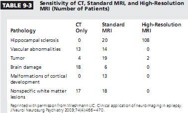
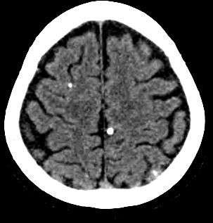
10 Sensitivity MRI 95% vs CT 32%
11

12 Future

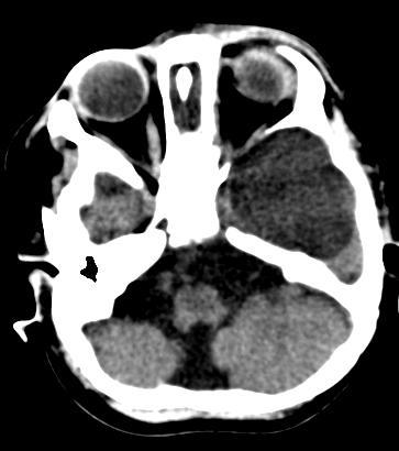
13 CT EMERGENCY situation
14 Role of Imaging Pre-surgery Identify structural abnormality Plan for surgery Help confirm epileptogenicity Relationship with eloquent areas Predict resectivity and Prognostication Post-surgery Evaluate residual lesion Surveillance
15 MRI protocol
16 Ideal Imaging Distinguish abnormal from normal -> High resolution Tell etiology/nature of abnormality -> Good Characterization Allow assessment of relationship with eloquent structures -> Functional/Microstructural derangement Evaluate epileptogenicity -> Physiologic data
17 Good clinical history and EEG findings High magnet field Appropriate protocol How to maximize MRI sensitivity? Good technologist Experienced radiologist
18 Seizure protocol Contrast vs non contrast Standard sequences Sagittal T1 Axial T2, FLAIR, DWI Coronal T2 GRE/SWI (axial or coronal) Seizure sequences: Coronal T2, FLAIR through hippocampi Coronal T1 3D SPGR through hippocampi T1 3D SPGR T1W whole brain ± 3D FLAIR with 3 planes reformation 1-2 mm thickness


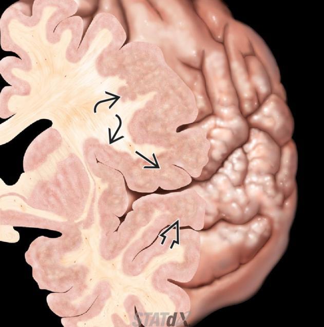
19 T1 3D SPGR Gray matter dark
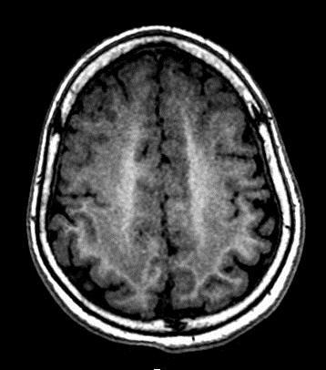
20

21 Imaging pitfalls Widespread abnormalities Multiple lesions Dual pathologies
22 Case scenarios
23 Etiologies/Epileptogenic Substrates Identifiable with MRI PEDIATRIC Congenital Malformation Inborn-error of metabolism Mesial temporal sclerosis Birth-related/ post trauma ADULT Vascular (Stroke, AVM, cavernoma) Tumor (primary and mets) Prior brain injury Mesial temporal sclerosis Infection Neoplasm Vascular (malformation) Neurocutaneous syndrome

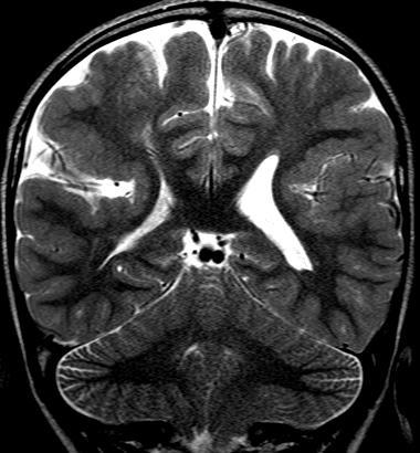
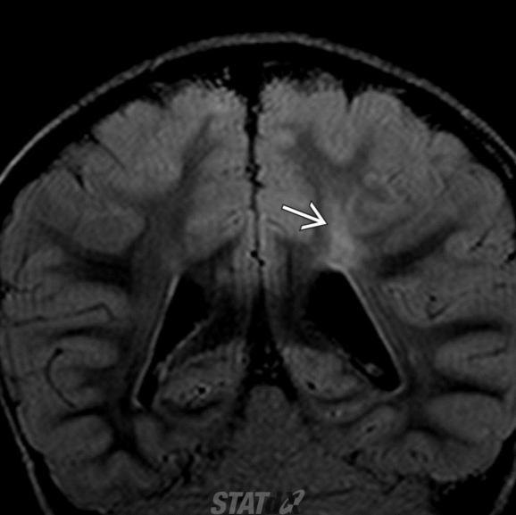
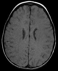
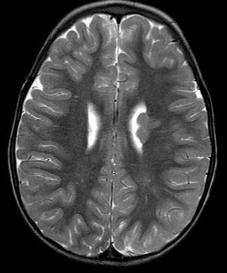
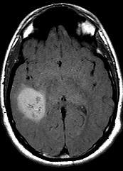
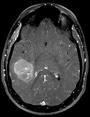
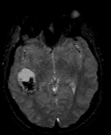
24 Cortically based tumor
25 Cortical malformation
26 Polymicrogyria Pachygyria: thick, smooth cortex Polymicrogyria: small cobblestone or micronodular appearing gyri


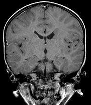
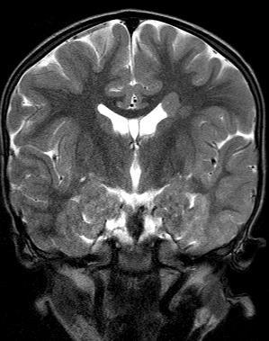
27 Cortical Dysplasia


28 Schizencephaly CSF cleft extending to ventricular epedyma GM lined Associated with absent septum pellucidum and septo-optic dysplasia
29 Heterotropia: nodular type

30 Heterotropia: Band heterotropia


31 Hippocampal Sclerosis 1 st sign: abnormal T2 hyperintensity, hippocampal volume loss/atrophy, obscuration of internal architecture 2 nd signs: ipsilateral fornix and mammillary body atrophy, enlarged ipsilateral temporal horn and choroidal fissure
")

 Associated with")

32 Dysembryoplastic Neuroepithelial Tumor (DNET) High epileptogenicity Cortically-based tumor Medial temporal lobe (MCC location) Associated with cortical dysplasia Imaging: Bubbly corticallybased tumor No enhancement or calcification

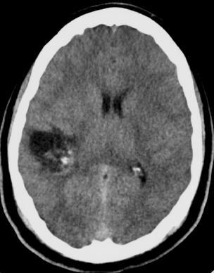

33 Oligodendroglioma Partially calcified subcortical/cortical mass in middle-aged adult
 Mental")

 Subepedymal giant cell")
34 Tuberous sclerosis Clinical triad Facial angiofibromas (90%) Mental retardation (50-80%) Seizure (80-90%) Classic Radiographic Findings: Cortical tuber Subependymal nodule (<1.3 cm) Subepedymal giant cell astrocytoma (> 1.3 cm)
 Hemiparesis (30-66%) Imaging findings Ipsilateral to port wine stain")

35 Sturge Weber syndrome Clinical presentation Port wine stain (CN V1 98%) Seizure (75-90%) Hemiparesis (30-66%) Imaging findings Ipsilateral to port wine stain Gyral/subcortical white matter calcifications (tram-track calcification) hemispheric brain atrophy Serpentine leptomeningeal enhancement Engorged/enlarged enhancing choroid plexi


36 AVM
37 Summary
38 Role of Imaging Pre-surgery Identify structural abnormality Plan for surgery Help confirm epileptogenicity Relationship with eloquent areas Predict resectivity and Prognostication Post-surgery Evaluate residual lesion Surveillance
39 Seizure protocol Contrast vs non contrast Standard sequences Sagittal T1 Axial T2, FLAIR, DWI Coronal T2 GRE/SWI (axial or coronal) Seizure sequences: Coronal T2, FLAIR through hippocampi Coronal T1 3D SPGR through hippocampi T1 3D SPGR T1W whole brain ± 3D FLAIR with 3 planes reformation 1-2 mm thickness
40 Thank you
Imaging of Pediatric Epilepsy MRI. Epilepsy: Nonacute Situation
 Imaging of Pediatric Epilepsy Epilepsy: Nonacute Situation MR is the study of choice Tailor MR study to suspected epileptogenic zone Temporal lobe Extratemporal A. James Barkovich, MD University of California
Imaging for Epilepsy Diagnosis December 2, 2011
 Imaging for Epilepsy Diagnosis December 2, 2011 Samuel Wiebe, MD University of Calgary Canada American Epilepsy Society Annual Meeting Disclosure University of Calgary Hopewell Professorship of Clinical
Magnetic resonance imaging findings and Spectrum of Etiologies in children epilepsy
 Magnetic resonance imaging findings and Spectrum of Etiologies in children epilepsy Poster No.: C-2262 Congress: ECR 2015 Type: Educational Exhibit Authors: B. Alami, L. Tazi, Z. Traoré, M. Y. Alaoui Lamrani,
3T MRI imaging approach to pediatric epileptic seizures:
 3T MRI imaging approach to pediatric epileptic seizures: Poster No.: C-1886 Congress: ECR 2016 Type: Educational Exhibit Authors: J. S. Alaín, E. M. DE LUCAS, J. C. Quintero Rivera, C. Pérez 1 2 3 3 3
PORT WINE STAINS AND STURGE-WEBER SYNDROME
 PORT WINE STAINS AND STURGE-WEBER SYNDROME Ong Hian Tat It is important for general practitioners to recognize cutaneous port-wine stains as these could signify important association with Sturge Weber
Pharmacoresistant temporal lobe epilepsy - a diagnostic performance of standardized MRI protocol in detection of epileptogenic lesion
 Pharmacoresistant temporal lobe epilepsy - a diagnostic performance of standardized MRI protocol in detection of epileptogenic lesion Poster No.: C-2226 Congress: ECR 2013 Type: Scientific Exhibit Authors:
High spatial resolution reveals excellent detail in pediatric neuro imaging
 Publication for the Philips MRI Community Issue 46 2012/2 High spatial resolution reveals excellent detail in pediatric neuro imaging Achieva 3.0T with 32-channel SENSE Head coil has become the system
Magnetic Resonance Imaging of Mesial Temporal Sclerosis (MTS): What radiologists ought to know?
: What radiologists ought to know?") Magnetic Resonance Imaging of Mesial Temporal Sclerosis (MTS): What radiologists ought to know? Poster No.: C-0856 Congress: ECR 2012 Type: Educational Exhibit Authors: P. Singh, G. Mittal, R. Kaur, K.
Magnetic Resonance Imaging of Mesial Temporal Sclerosis (MTS): What radiologists ought to know? Poster No.: C-0856 Congress: ECR 2012 Type: Educational Exhibit Authors: P. Singh, G. Mittal, R. Kaur, K.
MALFORMATIONS OF CORTICAL DEVELOPMENT: A PICTORIAL REVIEW
 MALFORMATIONS OF CORTICAL DEVELOPMENT: A PICTORIAL REVIEW Padmaja K. Naidu, M.D. Usha D. Nagaraj, M.D. William T. O Brien, Sr., D.O. AOCR Annual Conference 2018 Scottsdale, Arizona @CincyKidsRad facebook.com/cincykidsrad
Hamartomas and epilepsy: clinical and imaging characteristics
 Seizure 2003; 12: 307 311 doi:10.1016/s1059 1311(02)00272-8 Hamartomas and epilepsy: clinical and imaging characteristics B. DIEHL, R. PRAYSON, I. NAJM & P. RUGGIERI Departments of Neurology, Pathology
Invasive Evaluation for Epilepsy Surgery Lesional Cases NO DISCLOSURES. Mr. Johnson. Seizures at 29 Years of Age. Dileep Nair, MD Juan Bulacio, MD
 Invasive Evaluation for Epilepsy Surgery Lesional Cases NO DISCLOSURES Dileep Nair, MD Juan Bulacio, MD Mr. Johnson Seizures at 29 Years of Age Onset of seizures at 16 years of age bed wetting episodes
Neuroimaging in Investigation of Patients With Epilepsy Fernando Cendes, MD, PhD
 Review Article Neuroimaging in Investigation of Patients With Epilepsy Fernando Cendes, MD, PhD ABSTRACT Purpose of Review: This review discusses the MRI and functional imaging findings in patients with
How do we evaluate patients before epilepsy surgery?
 How do we evaluate patients before epilepsy surgery? Yotin Chinvarun, MD Chaiyos Khongkhatithum, MD How do we evaluate patients before epilepsy surgery? Chaiyos Khongkhatithum, MD Division of Neurology
SWI including phase and magnitude images
 On-line Table: MRI imaging recommendation and summary of key features Sequence Pathologies Visible Key Features T1 volumetric high-resolution whole-brain reformatted in axial, coronal, and sagittal planes
The Changing Surgical Landscape in Kids
 The Changing Surgical Landscape in Kids December 7, 2013 Howard L. Weiner, MD NYU Langone Medical Center American Epilepsy Society Annual Meeting Disclosure none American Epilepsy Society 2013 Annual Meeting
brain MRI for neuropsychiatrists: what do you need to know
 brain MRI for neuropsychiatrists: what do you need to know Christoforos Stoupis, MD, PhD Department of Radiology, Spital Maennedorf, Zurich & Inselspital, University of Bern, Switzerland c.stoupis@spitalmaennedorf.ch
Case 7391 Intraventricular Lesion
 Case 7391 Intraventricular Lesion Bastos Lima P1, Marques C1, Cabrita F2, Barbosa M2, Rebelo O3, Rio F1. 1Neuroradiology, 2Neurosurgery, 3Neuropathology, Coimbra University Hospitals, Portugal. University
Malformations of cortical development. Clinical and imaging findings.
 Malformations of cortical development. Clinical and imaging findings. Poster No.: C-2086 Congress: ECR 2012 Type: Educational Exhibit Authors: I. Alba de Caceres, B. García-Castaño, L. Ibañez, E. Roa,
Neuroimaging in Epilepsy. Ted Passe, MD Neuroradiology Mayo Clinic Rochester, MN
 Neuroimaging in Epilepsy Ted Passe, MD Neuroradiology Mayo Clinic Rochester, MN Objectives Imaging modalities in epilepsy Anatomic CT/MRI Functional MRS, SPECT, PET, fmri, MSI, DTI Intra-op MRI Characteristic
Malformations of cortical development. Clinical and imaging findings.
 Malformations of cortical development. Clinical and imaging findings. Poster No.: C-2086 Congress: ECR 2012 Type: Educational Exhibit Authors: I. Alba de Caceres, B. García-Castaño, L. Ibañez, E. Roa 1
Case reports functional imaging in epilepsy
 Seizure 2001; 10: 157 161 doi:10.1053/seiz.2001.0552, available online at http://www.idealibrary.com on Case reports functional imaging in epilepsy MARK P. RICHARDSON Medical Research Council Fellow, Institute
CASE OF THE WEEK PROFESSOR YASSER METWALLY
 CASE OF THE WEEK PROFESSOR YASSER METWALLY CLINICAL PICTURE CLINICAL PICTURE: CLINICAL PICTURE: A 6 years old male patient presented clinically with intractable complex partial seizure. The child is mentally
EGI Clinical Data Collection Form Cover Page
 EGI Clinical Data Collection Form Cover Page Please find enclosed the EGI Clinical Data Form for my patient. This form was completed by: On (date): _ Page 1 of 14 EGI Clinical Data Form Patient Name: Date
Imaging of tuberous sclerosis complex
 Imaging of tuberous sclerosis complex Poster No.: C-0388 Congress: ECR 2015 Type: Educational Exhibit Authors: M. V. Vu#kovi#, N. Menkovic, A. Petkovic, S. Ognjanovic, 1 2 1 1 1 1 1 1 J. Markov, M. Ilic,
Magnetic Resonance Imaging of the Temporal Lobe: Normal Anatomy and Diseases
 Canadian Association of Radiologists Journal 65 (2014) 148e157 Neuroradiology / Neuroradiologie Magnetic Resonance Imaging of the Temporal Lobe: Normal Anatomy and Diseases www.carjonline.org Alla Khashper,
Brain Imaging. IC calcifications. Mamdouh mahfouz MD
 Brain Imaging IC calcifications www.ssregypt.com Mamdouh mahfouz MD mamdouh.m5@gmail.com CT Hyper dense [ more than100 HU ] MRI Low signal in T1 and T2 WIs [non mobile protons] Exceptions Minute calcifications
Dr H. Gharebaghian MD Neurologist Department of Neurology Kermanshah Faculty of Medicine
 Dr H. Gharebaghian MD Neurologist Department of Neurology Kermanshah Faculty of Medicine Definitions Seizures are transient events that include symptoms and/or signs of abnormal excessive hypersynchronous
Acute Management of Seizures
 Acute Management of Seizures KURT HECOX M.D. PH.D. CHIEF OF PEDIATRIC NEUROLOGY BAUMAN ENDOWED CHAIR IN PEDIATRIC EPILEPSY Outline Management Principles Categorizing the event Key elements to the history
Advanced Imaging Techniques MRI, PET, SPECT, ESI-MSI, DTI December 8, 2013
 Advanced Imaging Techniques MRI, PET, SPECT, ESI-MSI, DTI December 8, 2013 Robert C. Knowlton, MD, MSPH University of California San Francisco Seizure Disorders Surgical Program American Epilepsy Society
Multimodal Imaging in Extratemporal Epilepsy Surgery
 Open Access Case Report DOI: 10.7759/cureus.2338 Multimodal Imaging in Extratemporal Epilepsy Surgery Christian Vollmar 1, Aurelia Peraud 2, Soheyl Noachtar 1 1. Epilepsy Center, Dept. of Neurology, University
Hemimegalencephaly without seizures: report of a case and review of literature
 Romanian Neurosurgery Volume XXXI Number 3 2017 July-September Article Hemimegalencephaly without seizures: report of a case and review of literature Agrawal Atul, Dutta Gautam, Singh Daljit, Sachdeva
Challenges for multivariate and multimodality analyses in "real life" projects: Epilepsy
 Challenges for multivariate and multimodality analyses in "real life" projects: Epilepsy Susanne Mueller M.D. Center for Imaging of Neurodegenerative Diseases Background: Epilepsy What is epilepsy? Recurrent
Epilepsy Surgery, Imaging, and Intraoperative Neuromonitoring: Surgical Perspective
 Epilepsy Surgery, Imaging, and Intraoperative Neuromonitoring: Surgical Perspective AC Duhaime, M.D. Director, Pediatric Neurosurgery, Massachusetts General Hospital Professor, Neurosurgery, Harvard Medical
RADIOLOGY TEACHING CONFERENCE
 RADIOLOGY TEACHING CONFERENCE John Athas, MD Monica Tadros, MD Columbia University, College of Physicians & Surgeons Department of Otolaryngology- Head & Neck Surgery September 27, 2007 CT SCAN IMAGING
Neurocutaneous Syndromes. Phakomatoses
 Neurocutaneous Syndromes Phakomatoses Financial Disclosures I have NO SIGNIFICANT FINANCIAL, GENERAL, OR OBLIGATION INTERESTS TO REPORT Neurocutaneous Syndomes Definition Entities Diagnosis/ Presentation
Epilepsy in children: Review of the main causes detectable by MRI
 Epilepsy in children: Review of the main causes detectable by MRI Poster No.: C-2182 Congress: ECR 2014 Type: Educational Exhibit Authors: A. A. S. M. D. Santos, T. C. R. S. SANTOS, A. Monteiro ; 1 1 1
Epilepsy, a common chronic neurological disorder, is a
 10 SUPPLEMENT TO Journal of the association of physicians of india august 2013 VOL. 61 Epilepsy: Diagnostic Evaluation JMK Murthy* Epilepsy, a common chronic neurological disorder, is a potentially treatable
Index. aneurysm, 92 carotid occlusion, 94 ICA stenosis, 95 intracranial, 92 MCA, 94
 A ADC. See Apparent diffusion coefficient (ADC) Aneurysm cerebral artery aneurysm, 93 CT scan, 93 gadolinium, 93 Angiography, 13 Anoxic brain injury, 25 Apparent diffusion coefficient (ADC), 7 Arachnoid
Magnetic Resonance Imaging of the Brain in Adults Presenting With New Onset Seizures Pannag Desai K N 1, Ravi N 2
 Magnetic Resonance Imaging of the Brain in Adults Presenting With New Onset Seizures Pannag Desai K N 1, Ravi N 2 1 (Department of Radiodiagnosis, BMC&RI, India) 2 (Department of Radiodiagnosis, BMC&RI,
Pediatric Neuroimaging in Epilepsy. Bhagwan Moorjani, MD, FAAP, FAAN Hope Neurologic Center La Quinta, CA
 Pediatric Neuroimaging in Epilepsy Bhagwan Moorjani, MD, FAAP, FAAN Hope Neurologic Center La Quinta, CA Neuroimaging in Childhood Neuroimaging issues are distinct from adults Sedation/anesthesia Motion
Introduction to Neurosurgical Subspecialties:
 Introduction to Neurosurgical Subspecialties: Pediatric Neurosurgery Brian L. Hoh, MD 1 and Gregory J. Zipfel, MD 2 1 University of Florida, 2 Washington University Pediatric Neurosurgery Pediatric neurosurgeons
What is the Relationship Between Arachnoid Cysts and Seizure Foci?
 Epilepsin, 38( 10):1098-1102, 1997 Lippincott-Raven Publishers, Philadelphia 0 International League Against Epilepsy What is the Relationship Between Arachnoid Cysts and Seizure Foci? Santiago Arroyo and
Magnetic Resonance Imaging. Basics of MRI in practice. Generation of MR signal. Generation of MR signal. Spin echo imaging. Generation of MR signal
 Magnetic Resonance Imaging Protons aligned with B0 magnetic filed Longitudinal magnetization - T1 relaxation Transverse magnetization - T2 relaxation Signal measured in the transverse plane Basics of MRI
Gross Organization I The Brain. Reading: BCP Chapter 7
 Gross Organization I The Brain Reading: BCP Chapter 7 Layout of the Nervous System Central Nervous System (CNS) Located inside of bone Includes the brain (in the skull) and the spinal cord (in the backbone)
Imaging of intractable paediatric epilepsy
 Page 1 of 10 Imaging of intractable paediatric epilepsy Authors: Sanjay Prabhu 1 Nasreen Mahomed 2 Affiliations: 1 Department of Radiology, Boston Children s Hospital, Harvard Medical School, United States
Cerebral MRI as an important diagnostic tool in temporal lobe epilepsy
 Cerebral MRI as an important diagnostic tool in temporal lobe epilepsy Poster No.: C-2190 Congress: ECR 2014 Type: Educational Exhibit Authors: A. Puiu, D. Negru; Iasi/RO Keywords: Neuroradiology brain,
5/22/2009. Pediatric Neurosurgery Pediatric Neurology Neuroradiology Neurophysiology Neuropathology Neuropsychology
 Current Surgical Treatment Strategies for the Management of Pediatric Epilepsy University of California, San Francisco Department of Neurological Surgery San Francisco, California Kurtis Ian Auguste, M.D.
J Neurol Neurosurg Psychiatry 2005; 76(Suppl III):iii2 iii10. doi: /jnnp RAY COMPUTED TOMOGRAPHY
:iii2 iii10. doi: /jnnp RAY COMPUTED TOMOGRAPHY") iii2 See end of article for authors affiliations Correspondence to: Professor John S Duncan, Department of Clinical and Experimental Epilepsy, Institute of Neurology, Queen Square, University College London,
iii2 See end of article for authors affiliations Correspondence to: Professor John S Duncan, Department of Clinical and Experimental Epilepsy, Institute of Neurology, Queen Square, University College London,
Surgical Modalities for Epilepsy Treatment. C.J. Bui, MD, FAANS Ochsner Neuroscience Symposium 2016
 Surgical Modalities for Epilepsy Treatment C.J. Bui, MD, FAANS Ochsner Neuroscience Symposium 2016 Conflict of Interest Nothing to disclose Surgical Modalities Diagnostic Subdural Grids Depth Electrodes
Candidates for Epilepsy Surgery. Presurgical Evaluation. Presurgical Evaluation. Presurgical Evaluation. Presurgical Evaluation 8/27/2017
 PresurgicalEpilepsy Eval: A multidisciplinary approach to intractable epilepsy Tayard Desudchit MD Faculty Of Medicine Chulalongkorn U. Candidates for Epilepsy Surgery Persistent seizures despite appropriate
PRESERVE: How intensively should we treat blood pressure in established cerebral small vessel disease? Guide to assessing MRI scans
 PRESERVE: How intensively should we treat blood pressure in established cerebral small vessel disease? Guide to assessing MRI scans Inclusion Criteria Clinical syndrome Patients must have clinical evidence
Beyond Mesial Temporal Sclerosis: Optimizing MRI Evaluation in Focal Epilepsy
 BRAIN CME ABBREVIATIONS KEY Beyond Mesial Temporal Sclerosis: Optimizing MRI Evaluation in Focal Epilepsy Elliott R. Friedman, MD, and Nitin Tandon, MD CME Credit The American Society of Neuroradiology
Attenuation value in HU From -500 To HU From -10 To HU From 60 To 90 HU. From 200 HU and above
 Brain Imaging Common CT attenuation values Structure Air Fat Water Brain tissue Recent hematoma Calcifications Bone Brain edema and infarction Normal liver parenchyma Attenuation value in HU From -500
Cortical malformations and epilepsy: role of MR imaging
 Cortical malformations and epilepsy: role of MR imaging Poster No.: C-0921 Congress: ECR 2013 Type: Scientific Exhibit Authors: S. Jerbi, O. Bradai, S. Haj Slimene, Y. Abdelhafidh, H. HAMZA; Mahdia/TN
Neuroimaging in Epileptic Disorders
 Neuroimaging in Epileptic Disorders 9 José Augusto Bragatti Hospital de Clínicas de Porto Alegre Universidade Federal do Rio Grande do Sul Brazil 1. Introduction Epilepsy is one of the most common neurologic
Utility of double inversion recovery MRI in paediatric epilepsy
 Utility of double inversion recovery MRI in paediatric epilepsy Bruno Soares, Emory University Samuel G Porter, Emory University Amit Saindane, Emory University Seena Dehkharghani, Emory University Nilesh
9/30/2016. Advances in Epilepsy Surgery. Epidemiology. Epidemiology
 Advances in Epilepsy Surgery George Jallo, M.D. Director, Institute for Brain Protection Sciences Johns Hopkins All Children s Hospital St Petersburg, Florida Epidemiology WHO lists it as the second most
Adult-Onset Neurologic Dysfunction Associated with Cortical Malformations
 AJNR Am J Neuroradiol 20:1037 1043, June/July 1999 Adult-Onset Neurologic Dysfunction Associated with Cortical Malformations Woo Ho Cho, David Seidenwurm, and A. James Barkovich BACKGROUND AND PURPOSE:
NEURORADIOLOGY Part I
 NEURORADIOLOGY Part I Vörös Erika University of Szeged Department of Radiology SZEGED BRAIN IMAGING METHODS Plain film radiography Ultrasonography (US) Computer tomography (CT) Magnetic resonance imaging
Neuroimaging in pediatric seizures
 International Journal of Research in Medical Sciences Sahdev R et al. Int J Res Med Sci. 2017 Jan;5(1):295-299 www.msjonline.org pissn 2320-6071 eissn 2320-6012 Original Research Article DOI: http://dx.doi.org/10.18203/2320-6012.ijrms20164566
ESOR COURSES FOR EDiR 2018
 ESOR COURSES FOR EDiR 2018 Education in partnership ESOR Courses for EDiR ESOR Courses for EDiR Course information In 2018, ESOR is continuing to organise foundation courses to prepare/assist those entering
Presurgical Evaluation before Epilepsy Surgery
 Presurgical Evaluation before Epilepsy Surgery Epilepsy Course for Neurology Resident 2015 Kanjana Unnwongse- Wehner, MD Prasat Neurological Epilepsy Center Facts About Epilepsy & Surgery Localization-related
Epilepsy and Brain Tumor. Apasri Lusawat M.D. Pediatric Neurology Prasat Neurological Institute
 Epilepsy and Brain Tumor Apasri Lusawat M.D. Pediatric Neurology Prasat Neurological Institute Outline Overview Pathogenesis : Tumor-> epilepsy Etiology of Epilepsy, age EPILEPSY AND BRAIN TUMORS CNS neoplasms
Epilepsy surgery is an increasingly recognized therapeutic
 J Neurosurg Pediatrics 14:68 80, 2014 AANS, 2014 Magnetic resonance imaging abnormalities in the resection region correlate with histopathological type, gliosis extent, and postoperative outcome in pediatric
Refractory focal epilepsy: findings by MRI.
 Refractory focal epilepsy: findings by MRI. Doctors Nicolás Sgarbi, Osmar Telis Clinical Radiology Department Hospital de Clínicas Montevideo- Uruguay ABSTRACT Epilepsy is one of the most frequent neurological
Keep Imaging Simple: An Introduction To Neuroimaging
 Keep Imaging Simple: An Introduction To Neuroimaging Meghan Elkins, OD, FAAO Please silence all mobile devices and remove items from chairs so others can sit. Unauthorized recording of this session is
The Asymmetric Mamillary Body: Association with Medial Temporal Lobe Disease Demonstrated with MR
 The Mamillary Body: Association with Medial Temporal Lobe Disease Demonstrated with MR Alexander C. Mamourian, Lawrence Rodichok, and Javad Towfighi PURPOSE: To determine whether mamillary body atrophy
An Unusual Kind Of Traumatic Intracranial Hemorrhage: Post Traumatic Bleed Into The Schizencephalic Cleft
 ISPUB.COM The Internet Journal of Radiology Volume 8 Number 2 An Unusual Kind Of Traumatic Intracranial Hemorrhage: Post Traumatic Bleed Into The Schizencephalic J T, V Rajendran, E Devarajan Citation
PRESURGICAL EVALUATION. ISLAND OF COS Hippocrates: On the Sacred Disease. Disclosure Research-Educational Grants. Patients with seizure disorders
 PRESURGICAL EVALUATION Patients with seizure disorders Gregory D. Cascino, MD Mayo Clinic Disclosure Research-Educational Grants Mayo Foundation Neuro Pace, Inc. American Epilepsy Society American Academy
Epilepsy and malformations of the cerebral cortex
 Abnormalities of cortical development and epilepsy Epileptic Disord 2003; 5 (Suppl 2): S 9 S 26 Epilepsy and malformations of the cerebral cortex Renzo Guerrini, Federico Sicca, Lucio Parmeggiani Institute
We are IntechOpen, the world s leading publisher of Open Access books Built by scientists, for scientists. International authors and editors
 We are IntechOpen, the world s leading publisher of Open Access books Built by scientists, for scientists 4,100 116,000 120M Open access books available International authors and editors Downloads Our
NEURORADIOLOGY Angela Lignelli, MD
 Neuroradiology NEURORADIOLOGY Angela Lignelli, MD Plain radiographs CT MRI Cerebral Angiogram Myelograms Neuroradiology Computerized Axial Tomography (CT) CT without and with contrast CTA CT angiogram
NEURORADIOLOGY Angela Lignelli, MD
 NEURORADIOLOGY Angela Lignelli, MD Neuroradiology Plain radiographs CT MRI Cerebral Angiogram Myelograms 1 Neuroradiology Computerized Axial Tomography (CT) CT without and with contrast CTA CT angiogram
Is DTI Increasing the Connectivity Between the Magnet Suite and the Clinic?
 Current Literature In Clinical Science Is DTI Increasing the Connectivity Between the Magnet Suite and the Clinic? Spatial Patterns of Water Diffusion Along White Matter Tracts in Temporal Lobe Epilepsy.
Introduction to Neurosurgical Subspecialties:
 Introduction to Neurosurgical Subspecialties: Functional Neurosurgery Brian L. Hoh, MD 1 and Gregory J. Zipfel, MD 2 1 University of Florida, 2 Washington University Functional Neurosurgery Functional
Pediatric CNS Tumors. Disclosures. Acknowledgements. Introduction. Introduction. Posterior Fossa Tumors. Whitney Finke, MD
 Pediatric CNS Tumors Disclosures Whitney Finke, MD Neuroradiology Fellow PGY-6 University of Utah Health Sciences Center Salt Lake City, Utah None Acknowledgements Introduction Nicholas A. Koontz, MD Luke
RASMUSSEN S ENCEPHALITIS - A CASE REPORT. Dr. Suchithra.J DNB PG RAILWAY HOSPITAL
 RASMUSSEN S ENCEPHALITIS - A CASE REPORT Dr. Suchithra.J DNB PG RAILWAY HOSPITAL Master Naveen Kumar, 7/Mch, first born of nonconsanguinous marriage presented to us with Left hemiparesis Seizures on L
Detection of Leptomeningeal CNS Metastases in Children
 Detection of Leptomeningeal CNS Metastases in Children Noah D. Sabin, M.D. Julie H. Harreld M.D. Kathleen J. Helton M.D. Zoltan Patay M.D., Ph.D. St. Jude Children s Research Hospital Memphis, TN Leptomeningeal
Physiological Markers of Pharmacoresistant Epilepsy December 2, 2011
 Physiological Markers of Pharmacoresistant Epilepsy December 2, 2011 Jerome Engel, Jr., MD, PhD Director of the Seizure Disorder Center The Jonathan Sinay Distinguished Professor of Neurology, Neurobiology,
Joana Ramalho, MD C. Ryan Miller, MD, PhD
 Joana Ramalho, MD C. Ryan Miller, MD, PhD Case 1 3 month old baby girl Presented with new onset of seizures Newborn. Questionable blurring of the gray-white junction within the right occipital lobe. Findings
The American Approach to Depth Electrode Insertion December 4, 2012
 The American Approach to Depth Electrode Insertion December 4, 2012 Jonathan Miller, MD Director, Epilepsy Surgery University Hospitals Case Medical Center/Case Western Reserve University Cleveland, Ohio
Applicable Neuroradiology
 For the Clinical Neurology Clerkship LSU Medical School New Orleans Amy W Voigt, MD Clerkship Director Introduction The field of Radiology first developed following the discovery of X-Rays by Wilhelm Roentgen
MR evaluation of the spectrum of pathologies affecting the hippocampus
 MR evaluation of the spectrum of pathologies affecting the hippocampus Poster No.: C-0590 Congress: ECR 2017 Type: Educational Exhibit Authors: S. Mantripragada, R. Gupta, J. Modi, J. P. sharma ; Gurgaon/
CT - Brain Examination
 CT - Brain Examination Submitted by: Felemban 1 CT - Brain Examination The clinical indication of CT brain are: a) Chronic cases (e.g. headache - tumor - abscess) b) ER cases (e.g. trauma - RTA - child
Neuromodulation in Epilepsy. Gregory C. Mathews, M.D., Ph.D.
 Neuromodulation in Epilepsy Gregory C. Mathews, M.D., Ph.D. Disclosure There are no disclosures to share with regards to this presentation. Epilepsy Basics What is epilepsy? Partial versus generalized
Discovering the hippocampus with cranial-ct.
 Discovering the hippocampus with cranial-ct. Poster No.: C-0378 Congress: ECR 2018 Type: Educational Exhibit Authors: F. Pozo Piñon, A. B. Barba Arce, E. herrera romero, V. 1 2 3 1 3 3 Fernández Lobo,
HEAD AND NECK IMAGING. James Chen (MS IV)
") HEAD AND NECK IMAGING James Chen (MS IV) Anatomy Course Johns Hopkins School of Medicine Sept. 27, 2011 OBJECTIVES Introduce cross sectional imaging of head and neck Computed tomography (CT) Review head
HEAD AND NECK IMAGING James Chen (MS IV) Anatomy Course Johns Hopkins School of Medicine Sept. 27, 2011 OBJECTIVES Introduce cross sectional imaging of head and neck Computed tomography (CT) Review head
Malformations of cortical development
 Abnormalities of cortical development and epilepsy Epileptic Disord 2003; 5 (Suppl 2): S 59 S 66 Morphological neuroimaging of malformations of cortical development Ludovico D Incerti Department of Neuroradiology,
PET and SPECT in Epilepsy
 PET and SPECT in Epilepsy 12.6.2013 William H Theodore MD Chief, Clinical Epilepsy Section NINDS NIH Bethesda MD American Epilepsy Society Annual Meeting Disclosures Entity DIR NINDS NIH Elsevier Individual
SWISS SOCIETY OF NEONATOLOGY. A rare cause of neonatal seizures
 SWISS SOCIETY OF NEONATOLOGY A rare cause of neonatal seizures October 2006 2 Hagmann C, Robertson NJ, Centre for Perinatal Brain Research, Institute for Women s Health, University College London, London
Prenatal Prediction of The Neurologically Impaired Neonate By Ultrasound
 Prenatal Prediction of The Neurologically Impaired Neonate By Ultrasound Robert H. Debbs, D.O.,F.A.C.O.O.G. Professor of OB-GYN Perelman School of Medicine, University of Pennsylvania Director, Pennsylvania
Diagnosing TSC by Making Clinical Connections
 Diagnosing TSC by Making Clinical Connections TSC = tuberous sclerosis complex. Diagnosing tuberous sclerosis complex: MORE CLUES Definite Diagnosis of Tuberous Sclerosis Complex (TSC) Possible Diagnosis
Table of Contents: SKULL AND BRAIN. Scalp, Skull. Anatomically Based Differentials. Skull Normal Variants. Scalp Mass, Child.
 Table of Contents: SKULL AND BRAIN Scalp, Skull Skull Normal Variants Scalp Mass, Child Scalp Mass, Adult Congenital Anomalies of Skull Base Sellar/Parasellar Mass With Skull Base Invasion "Hair on End"
Interictal High Frequency Oscillations as Neurophysiologic Biomarkers of Epileptogenicity
 Interictal High Frequency Oscillations as Neurophysiologic Biomarkers of Epileptogenicity December 10, 2013 Joyce Y. Wu, MD Associate Professor Division of Pediatric Neurology David Geffen School of Medicine
Pediatric Brain Tumors Pre, Intra & Post Op Evaluation and Management. Timothy M. George, MD, FACS, FAAP
 Pediatric Brain Tumors Pre, Intra & Post Op Evaluation and Management Timothy M. George, MD, FACS, FAAP PEDIATRIC BRAIN TUMORS BACKGROUND: Incidence: Third most common pediatric tumor type (leukemia, neuroblastoma,
Epilepsy Surgery: Who should be considered? How will patients do? Bassel Abou-Khalil, M.D.
 Epilepsy Surgery: Who should be considered? How will patients do? Bassel Abou-Khalil, M.D. Disclosures none Self-assessment questions Q1- Which qualify for drug resistance in focal epilepsy? A. Failure
Group, University Department of Clinical Neurology, Queen Square, London WC1N 3BG, UK
 braini0204 Brain (1997), 120, 339 377 INVITED REVIEW Imaging and epilepsy John S. Duncan Epilepsy Research Group, University Department of Clinical Neurology, National Hospital for Neurology and Neurosurgery,
The central nervous system
 Sectc.qxd 29/06/99 09:42 Page 81 Section C The central nervous system CNS haemorrhage Subarachnoid haemorrhage Cerebral infarction Brain atrophy Ring enhancing lesions MRI of the pituitary Multiple sclerosis
NEURO IMAGING OF ACUTE STROKE
 1 1 NEURO IMAGING OF ACUTE STROKE ALICIA RICHARDSON, MSN, RN, ACCNS-AG, ANVP-BC WENDY SMITH, MA, RN, MBA, SCRN, FAHA LYNN HUNDLEY, APRN, CNRN, CCNS, ANVP-BC 2 2 1 DISCLOSURES Alicia Richardson: Stryker
EGI Clinical Data Collection Form Cover Page
 EGI Clinical Data Collection Form Cover Page Please find enclosed the EGI Clinical Data Form for my patient. This form was completed by: On (date): Page 1 of 15 Patient Name: Date of Birth: MM/DD/YYYY