Salivary Glands tumors. Pr Cécile Badoual Service Pr Cécile d anatomo-pathologie Hôpital HEGP, Européen Paris G Pompidou
|
|
|
- Pamela Richard
- 5 years ago
- Views:
Transcription


1 Salivary Glands tumors Pr Cécile Badoual Service Pr Cécile d anatomo-pathologie Badoual Hôpital HEGP, Européen Paris G Pompidou
2 Tumours of the salivary glands Huge number of histological subtype : OMS classification 2017 contains 11 subtypes of benign lesion and 19 subtypes of carcinomas. 2/3 are benign 1/3 are malignant. Salivary glands carcinoma are rare and represent a range of 3 to 5 % of the head and neck carcinoma, with a incidence <1/ habitants. Peak incidence at years, and a mean around 45 years. Female predominance. Carcinoma generally classified in 3 groups : low grade, intermediate grade and high grade of malignancy.
3 Tumours of the salivary glands The etiology of salivary gland neoplasms remains unknown. Growing evidence that certain environmental factors such as radiation, viruses, diet could be envolved Certain occupational exposures may increase the risk of developing tumors of the salivary glands. Specific genetic abnormalities have recently been well characterized. Su YX, Ann Surg Oncol. 2015; Spiro RH. Head Neck Surg. 1986; Who classification 2005
4 Tumours of the salivary glands HPV infection role still debated Epstein-Barr virus (EBV) associated with lymphoepithelial carcinoma of the salivary gland in the Asian population. No evidence of a causal role of EBV in other primary salivary gland neoplasms. No positive signal by in situ hybridization for EBV RNA in 42 benign salivary gland neoplasms. Other viruses including human herpesvirus-8, and cytomegalovirus do not appear to have any etiologic role in salivary gland neoplasms. Hühns M, Biomed Res Int. 2015; Veit JA, Anticancer Res. 2015; Pollock AM, J Laryngol Otol. 1999
5 What s up? Clinical strategy for diagnosis WHO classification. New one? Molecular/genetics and histochemical findings Salivary glands tumor and chilwood Place of new therapeutics
6 Methods to detect a salivary gland lesion Ponction with needle Frozen section Definitive histology The radiologists can give a diffusion and perfusion coefficient to help the diagnosis
7 Frozen section Methods Frozen tissue quick staining Answer in less than 10 minutes Problems Orientation diagnose Alteration of the tissue sample examination Indications Control of one of the surgical border Guidance of the surgery Modification of the surgery


8


9 Fine needle aspiration Useful for small tumors First orientation for the diagnosis Expert ++
10 Fine needle aspiration cytology and frozen section in the diagnosis of malignant parotid tumours? Evaluation of frozen section diagnosis in 721 parotid gland lesions Badoual C, Histopathology 2006
11 Fine needle aspiration cytology and frozen section in the diagnosis of malignant parotid tumours? Badoual C, Evaluation of frozen section diagnosis in 721 parotid gland lesions. Histopathology 2006 Fakhary N, J Oral Maxillofac Surg (138 tumors)
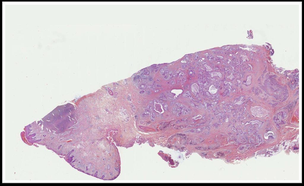
12 Macroscopy


13
14 07h3226 parotid

15

16
17
18 Immunohistological profile cytokératin Smooth muscle actin Caldesmon/ calponin S100 Epithelial cells Myoepithelial cells Ki67, cerb2, ckit (ADK), ER, PR, AR (ductal carcinoma) CK19 (acinic), Dog-1(acinic), mammaglobin (secretory carcinoma)

19 CK7/CAM5.2 (+/-) Acinar cells Vimentin (+/-) CK7/CAM5.2 CK7/CAM5.2 CK7/CAM5.2 Claponin/SMA/myosin, CK7/CAM5.2, P63,S100, vimentin

20
21 Cases 2C and 2L

22

23

24
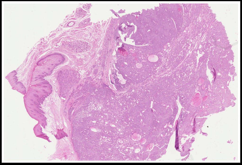
25 Pleomorphic adenoma Clinical characteristics Most frequent benign tumor, 80% over benign lesions Mean age 45 yo. Slow development Childhood, 5-15 yo. During childhood boys >girls conversely adult age. Incidence 2,4 to 3,5 for Main salivary glands and all the others glands like palate, lachrymal, laryngeal, bronchus.
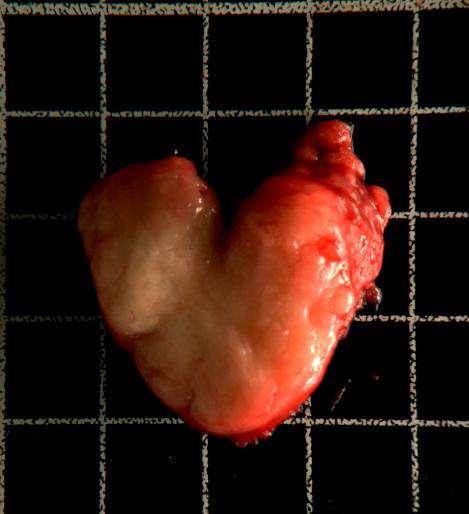
26 Pleomorphic adenoma Clinical features and macroscopy : Parotid: under skin well circumscribed mass, mobile, painless, no facial nerve weakness or dysesthesia. round or oval shape, surrounded by a capsule. others sites: not well delimited, no capsule. Surface light tan to grey. Cystic changes can been seen. Haemorrhage and infarction can been seen.


27
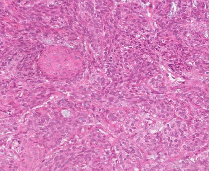
28 Pleomorphic adenoma Histological description: Typically, two types of tumor cells Epithelial cells, tubes, glands, isolated, mucinous, sebaceous or oncocytic metaplasia Myoepithelial spindle cells, epithelioïd, clear cells, plasmocytoïd. Atypias mostly for hypercellular adenoma. Foci of squamous metaplasia secondary to ischemic necrosis General architecture, mixture of different aspects avec with solid nests, tubes but also epithelialmyoepithelial or pseudo cylindromatous.
29
30
31
32
33 Pseudo cylindromatous shape
34 Epithelial-myoepithelial
35 Pleomorphic adenoma Histological feature: Mostly abondant stroma, chondroïd, myxoïd, osteoïd, adipocyte like, even fibrous. Not abondant, if hypercellular adenoma. Calcifications and cristalloïds rich in tyrosine. Rarely, adipocytes with stroma adipocytic metaplasia. Vascular emboli, capsular effractions, are not necessary associated with malignant transformation.
36
37 squamous metaplasia Tyrosine cristalloïd Adipocytic metaplasia
38 CD34
39 Pleomorphic adenoma Immunohisstochemistry Molecular biology 70% : chromosomic aberrations : 8q12 rearrangement (targeted gene PLAG1), rearrangement 12q13-15 (targeted gene HMGA2) coding for chromatine protein. Not well circonscribed pleomorphic adenomas and well delimited adenoid cystic carcinomas! Minor salivary glands biopsies ( no interface between tumor tissu /sane tissu ) Cytological Ponctions
40 Nasale cavity
41 Nasal cavity
42 AE1/AE3 S100
43 inferior lip
44 Inferior lip GEPSO External auditory canal
45 Pleomorphic adenoma Histological description: Typically, two types of tumor cells Epithelial cells, tubes, glands, isolated or in. Mucinous, sebaceous or oncocytic metaplasia Myoepithelial spindle cells, epithelioïd, clear cells, plasmocytoïd. Atypies surtout si adénome hypercellulaire. Zones de métaplasie malpighienne. Foyers de nécrose ischémique à l origine. L architecture générale, mélange d aspect divers et variés, avec des massifs, des tubes mais aussi des aspects épithélio-myoépithéliaux ou pseudo cylindromateux.

46

47

48

49


50 Architecture pseudo cylindromateuse

51 Architecture épithéliale-myoépithéliale
52 Adénome pléomorphe Aspects histologiques : Stroma le plus souvent abondant, chondroïde, myxoïde, ostéoïde, adipocyte like, fibreux, voire squirreux. Peu abondant, si adénome hypercellulaire. Calcifications et cristalloïdes riches de tyrosine. Plus rarement, adipocytes avec métaplasie lipomateuse du stroma. Des emboles vasculaires, ainsi que effractions de capsule, ne préjugent pas nécessairement d une transformation maligne.

53


54 Métaplasie malpighienne Cristalloïdes riches en tyrosine Métaplasie adipocytaire


55 CD34
56 Pleomorphic adenoma Immunohisstochemistry Molecular biology 70% : chromosomic aberrations : 8q12 rearrangement (targeted gene PLAG1), rearrangement 12q13-15 (targeted gene HMGA2) coding for chromatine protein. Not well circonscribed pleomorphic adenomas and well delimited adenoid cystic carcinomas! Minor salivary glands biopsies ( no interface between tumor tissu /sane tissu ) Cytological Ponctions

57 Cloison nasale


58 Fosse nasale


59 AE1/AE3 S100

60 inferior lip


61 Inferior lip GEPSO External auditory canal
62 Pleomorphic adenoma Evolution : The two major risks are recurrences or transformation Frequent recurrences. Multiple nodules and of very variable size. Complete surgical excision, if this is possible. Radiation therapy may be offered. Increased volume, appearance of pain, facial paralysis, will lead to a diagnosis of malignancy. Notion of metastatic pleomorphic adenoma discussed

63

64

65

66


67 Biopsie de rein
68 PLAG1 (1) gene fusions involving the PLAG1 and HMGA2 oncogenes are specific for benign pleomorphic adenomas. PLAG1 encodes a developmentally regulated DNAbinding zinc finger protein that is part of a family of cell cycle progression-related proteins. Rearrangement of PLAG1 in aproximatively 50 % of the pleomorphic adenomas Prognosis++
69 Usefulness of MYB/PLAG Not well circonscribed pleomorphic adenoma / well delimited adenoid cystic carcinoma! PLAG1 persistant after a transformation of a pleomorphic adenoma Fine needle aspiration Prognostic implication
70 A translocation-generated network of oncogenic gene fusions in salivary gland tumors. The multiple translocation target genes MYB,HMGA2, and PLAG1 are indicated in red. ACC adenoid cystic carcinoma, MEC mucoepidermoid carcinoma, HCCC hyalinizing clear Stenman G, Head Neck Pathol. 2013
71 Cases 2D and 2D.. Woman19 yo parotid lesions since the childhood with recent increase of the volume

72

73

74

75

76
77
78

79
80 Pleomorphic adenoma Evolution : Pleomorphous ex-adenoma carcinoma: pleomorphic adenoma with transformed tumor zones. Aggressiveness: infiltration of the capsule, of the glandular parenchyma, adipocytic tissue or peri-nervous sheaths + cytological criteria Most often high-grade (ductal) carcinoma but, all histological types of carcinomas described. The tumor type and grade of the carcinomatous contingent must be specified because they also participate in the evaluation of the prognosis.

81

82

83

84

85

86

87


88
89 pleomorphic ex-adenoma carcinomas Capsular invasion determinant for the prognosis Clearly proved that according to the extensions pleomorphic ex-adenoma carcinomas have a totally different prognosis: New 2017 WHO classification Strictly enclosed excellent prognosis almost comparable to the pleomorphic adenoma one. Capsular invasion, minimal invasion (invasion outside the capsule <4-6 mm) more favorable prognosis, Invasion out of the capsule> 4-6 mm, prognosis more reserved. Massively invasive> 8 mm prognosis consistently pejorative


90
91 Cas 2A Woman lesion superficial lobule of the left parotid since 2 ans

92

93

94

95
96 Basal cell adenoma (tubulo-trabecular) Morphologically monotonic proliferation of small basaloid cells with occasional inner ductal epithelial cells forming nests and cords From a phenotypic point of view these cells are of essentially basal type (P63) but also show myoepithelial (AML) and luminal (ACE) differentiation. 6% of benign tumors of the salivary glands Sixth decade Parotid 80%> oral mucosa> under maxillary Firm tumor well limited usually <3 cm
97 Histopathology Tumour mixture of solid, tubular, trabecular and membranous patterns Basaloïd cells with scant cytoplasm, indistinct cell borders, and round to oval nuclei and may show peripheral palissading Pas d implication clinique sauf pour le type membraneux (recurrence 25%) Transformation in basal cell adenocarcinoma rare but can occur Often multi-nodular.so recurrence are likely to be more frequente

98 Small cells with pale cytoplasm P63 +, Pancytokeratin + More or less basophilic nuclei according to tumors interface with the stroma: palisade arrangement of the nuclei Squamous differenciation
 Palisadic")
99 Solid subtype: Nest with varyious size and shape Scarce stroma (never chondromatous nor myxoid) Palisadic arrangement


100 Tubular and trabecular pattern

101 Membranous basal cells adenoma Membranous mostly associated with recurrent and is sometimes associated with skin tumors

102
103 Basal cells adenocarcinoma? Rare ++, yo Same distribution as adenoma: parotid Diagnosis especially because of infiltration and invasion Beware of the often multinodular membranous adenoma) Atypia, inconstant mitosis Low grade lesion: 37% recurrence and lymph node metastasis 8%, distant metastases 4%
104 Cases 2B

105

106

107
108 Canalicular adenoma Benign tumor composed of monomorphous epithelial ductal cells Well delineated and lobulated Correlation with basal cell adenomas? Special clinical behaviour: upper lip in almost 80% of cases from phenotypic point of view pure epithelial tumor: no positive myoepithelial AML contingent

109
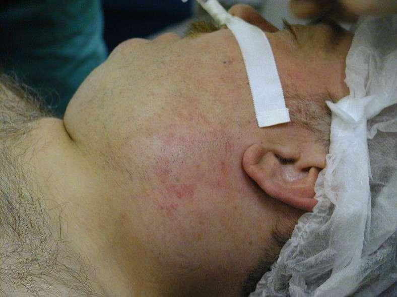
110 Cases 2E Man 46 yo parotid lesion since 8 months, retracted skin

111

112 07H3126 parotid
113

114

115
116 Mucoepidermoid Carcinoma (MEC) Localisation : parotid et minor salivary glands Histology : 3 types of cells mostly assembled in nest: mucus producing, squamoid and intermediate cells. Differential diagnosis : pleomorphic adenoma, acinic carcinoma, myoepithelial carcinoma, metastases of renal cell carcinoma Prognosis : uncertain, with recurrence of 25% and 10% of metastasis Molecular genetics: t(11;19) fusion gene MECT1-MAML2 for 60-70% MEC. Inactivation pathway Notch, favorable outcome Evolution : slow (recurrence after 7 to 10 years)
117 Mucoepidermoid Carcinoma (CME) Grading (OMS 2005) Aspect histologique Score Cystic component < 20% 2 Neural invasion 2 Necrosis 3 4 mitoses or more /10 hpf 3 Anaplasia 4 Grade Low 0-4 Intermediate 5-6 High > No OMS 2005 grading
118 Mucoepidermoid Carcinoma (MEC) - CME low grade : Cystic, mucinous cell-rich and well circonscribe -CME intermediate grade :More solid and less circonscribe, diversity of appearances including mucin extravasation - CME high grade : One or more of the features : nuclear anaplasia, necrosis, increased mitotic rate, and perineural, lymphovascular and bony invasion -
 Translocation MECT1- MAML2 No translocation Survival mean > 10 years 1,6 years Mean age 48 years 76 years apparition Tumor size 1 3 Tumor grade low and intermediate grade High and")
119 MECT1-MAML2 translocation associated with favorable prognosis MECT1: CREB mediated transcription MAML2:mastermind-like gene family: voie Notch normal FISH MAML2 «split» Behboudi and coll. (Cancer 2006) Translocation MECT1- MAML2 No translocation Survival mean > 10 years 1,6 years Mean age 48 years 76 years apparition Tumor size 1 3 Tumor grade low and intermediate grade High and intermediate grade High grade associated with low survival CDKN2A methylation or deletion Sethala Am J Surg Pathol 2010
120 A translocation-generated network of oncogenic gene fusions in salivary gland tumors. The multiple translocation target genes MYB,HMGA2, and PLAG1 are indicated in red. ACC adenoid cystic carcinoma, MEC mucoepidermoid carcinoma, HCCC hyalinizing clear Stenman G, Head Neck Pathol. 2013

121

122

123


124 Low grade mucoepidermoid carcinoma


125 intermediate grade mucoepidermoid carcinoma


126 High grade mucoepidermoid carcinoma


127 Alcian Blue PAS


128 CK5/6 CK5/6 Cerb2 P63 CK7
129 Cases 2M woman 63 yo painless parotid lesion

130

131

132


133 Oncocytic mucoepidermoïd carcinoma MEC very close the oncocytoma features Infiltration and mucinous cells


134 KI67
135 Oncocytoma Benign tumor proliferation consitued exclusively of oncocytic cells Parotid 95% > 70 years Single nodule, well limited, +/- encapsulated, red brown, from 1 to 7 cm Single nodule, well limited, +/- encapsulated, red brown, from 1 to 7 cm Sometimes, main nodule in a multinodular context
136

137
138 Case 2F

139

140

141
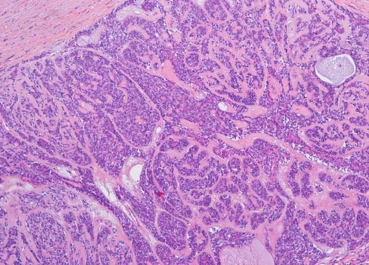
142 (ex cylindrome in french) Frequently painful Adenoid cystic carcinoma Localisation : parotid, submandibular gland, minor salivary glands (palate, tongue, lip, lachrymal glands, lip, buccal mucosa..) Microscopy: well circumscribed but not encapsulated, invariably infiltrative Histopathology : Two main cell types : epithelial and myoepithelial. No atypia. Rare mitosis. Perineural invasion+++. Stroma hyalinized or myxoid Diagnostic différentiel : adenocarcinoma, pleomporphic adenoma, basal cell adenoma, basal cell adenocarcinoma
143 Adenoid cystic carcinoma - patterns 3 mains patterns Tubular well formed ducts and tubules. Inner epithelial cells and outer myoepithelial Cribriform, most frequent nests of cells with cylindromatous microcystic spaces. Solid or basaloid type sheets of uniform basaloid cells Tumors can be composite or present a predominant pattern.
144 Adenoid cystic carcinoma Prognostic: influenced by numerous clinical considerations (size, localisation, pattern..), slow evolution, Recurrences after 7-8 years. Late mestastases. No official grading but some authors propose : - grade 1 : Tubular or cribriform pattern - grade 2 : less than 30%solid pattern - grade 3 : solid predominant pattern Szanto PA Cancer 1984 No grade in the 2017 WHO classification


145


146


147


148 PAS CK7 AML S100 SMA


149 Ckit RO REGF RP

150 Molecular biology of the acinic carcinoma Translocation between Chromosomes 6q and 9q with the creation of a MYB-NFIB chimeric fusion in about 50% cases

151 Wysocki PT Oncotarget 2016

152
153 A translocation-generated network of oncogenic gene fusions in salivary gland tumors. The multiple translocation target genes MYB,HMGA2, and PLAG1 are indicated in red. ACC adenoid cystic carcinoma, MEC mucoepidermoid carcinoma, HCCC hyalinizing clear Stenman G, Head Neck Pathol. 2013
154 Conclusion interesting +++ for mucoepidermoid carcinomas: diagnosis (difficult variants, fine needle biopsies) prognosis (intermediate grades) Differential diagnosis :pleomorphic adenomas and adenoid cystic carcinomas +++ Help to define malignancy
155 Cases 2G Man 47 yo parotid lesion

156

157
158 Cases 2N woman 63 yo smoker oral cavity

159

160

161
162 Epithelial-myoepithelial carcinoma (clear cell adenoma, cell clear ductal adenocarcinoma= Parotid ++ (80%) Macroscopy: usually plurinodular with incomplete capsule. Often necrosis Histology: double cellular population layering numerous glandular formations - epithelial: internal (cytokeratin +) - myoepithelial: external (S100 +, AML)
163 Epithelial-myoepithelial carcinoma (clear cell adenoma, cell clear ductal adenocarcinoma= Differential diagnosis: acinic cell carcinoma, mucoepidermoid carcinoma, sebaceous carcinoma, metastasis of renal clear cell carcinoma Prognosis: low grade of malignancy, recurrence, rare metastases

164

165

166

167

168

169
170 Case 2H

171

172

173
174 Acinic cell carcinoma 6 % of the salivary glands tumor and 5-15 % among malignant salivary glands tumor. Most often in parotid Histology :large, polygonal cells, few atypia. Cytoplasm contain granules (PAS-diastase+) vacuolated cells contain clear cytoplasm with vacuoles numerous pattern (acinar, ductal, inflammatory infiltration) Hyalinization associated with a bad prognosis Rarely mimic thyroid pattern
175 4 different cells Vacuolated Clear Oncocytic Hobnail 3 different patterns: Microcystic Solid Follicular Acinic cell carcinoma Prognosis: dependant on the size, the surgery. No grading Recurrence in 25% cases but metastasis are rare (10%).

176

177

178

179

180


181 Dog1 and SOX10: cellules acinic cell apical staining and cells coming from the intercalar duct Head Neck Pathol Dec;10(4):

182

183
184 Case 2I

185

186

187

188
189 Secretory carcinoma (MASC) Recently described, rare salivary gland tumor that relates the morphology and genetics of an equally rare malignancy of the breast, secretory carcinoma (SC) Previously classified as acinic cell carcinoma ("zymogen poor", intercalated cell predominant variant), mucoepidermoid carcinoma and adenocarcinoma, NOS Specific cytogenetic characteristic: t(12; 15)(q13;q25): ETV6-NTRK3 translocation, demonstrated by either FISH or PCR
190 Secretory carcinoma (MASC) Typically it is a disease of young male patients but occurs in a wide age range (21-75), with a mean of 46 years Pediatric cases have been Sex ration :1 Parotid gland (up to 70%); lips, hard palate, submandibular glands. More frequent in nonparotid sites compared to acinic cell carcinoma
191 Secretory carcinoma (MASC) Overall slowly growing, painless tumor, occasional extracapsular extension and perineural invasion Infrequent local recurrences Rare metastatic dissemination to cervical lymph nodes, pleura, pericardium and lungs Broad range of clinical behaviours, from indolent to aggressive Currently there is no way to predict which tumours will behave aggressively Higher incidence of regional lymph node involvement than acinic cell carcinomas Treatment Local excision, radiation therapy in select cases Molecular targeted gene therapy currently investigated
192 Secretory carcinoma (MASC) Gross description Most frequently solitary, well circumscribed, nonencapsulated or multinodular mass Brown or gray in color and rubbery in texture Variable sizes, from 0.2 cm to 5.5 cm Histologic)description Prominent "bubbly" low power aspect Cystic, tubular, solid or papillary architecture Intermediate size cells with eosinophilic / amphophilic vacuolated cytoplasm; absence of zymogen granules Low grade, bland and pale nuclei, some with prominent nucleoli Intraluminal or intracellular colloid-like material with a "bubbly" appearance May have mucinous differentiation May have perineural invasion No extensive necrosis and very low mitotic activity Unusual feature: foci of high grade / dedifferentiation with large nests and comedonecrosis

193 Lame 32: tumeur de la parotide, femme de 42 ans

194 Lame 32

195 Lame 32

196 Lame 32

197 Lame 32

198 Lame 34

199 Lame 34

200 Lame 34
201 Case 2J
202

203

204

205

206

207

208
209 Polymorphous adenocarcinoma Oral cavity ++, (26% of carcinomas of the oral cavity) Macroscopy: firm mass, fairly well limited, unencapsulated, yellowish, multi-lobed, nodules of different size (mean size = 2.1 cm) Histology: ++ Feature: cellular -monomorphism: (small to medium cells) architectural polymorphism, - infiltrating cells, (lobules, islets, columns) in adjacent tissue or salivary parenchyma
210 Immunohistochemistry: CK7+, ps100+,bcl2+, Mammaglobin+, p63+, p40-, DOG1+/-. Pronostic : low grade of malignancy, but recurrent, rare metastases
211 CATS : Cribriform Adenocarcinoma of the Tongue and other minor Salivary glands posterior third of the tongue Synchronous local node metastases: 70% of cases. No distant metastasis excellent prognosis. average age 54 years (5 to 85 years)
212 One other new entity? Tongue Cytology very close the papillary carcinoma of the thyroid (thyroglobulin and TTF1-) lymph node metastases +++ gene Fusion PRKD1, 2 ou 3
213 Case 2K

214

215

216

217
218 Salivary duct carcinoma Aggressiveness ++ High level of malignancy 9% of the salivary glands tumor 4M /1F Parotid, submandibular, minor salivary gland Similar to intraductal and infiltrating mammary duct carcinoma Cribriform growth pattern, comedonecrosis, atypia++mitosis. De novo (53%) or ex pleomorphic adenoma (47%) AE1/AE3+, S100-, Ki67++, RA+, CerB2+

219

220


221 RA CK7 RA KI67 CerB2


222 GCDFP15 P63 Mamaglobin

223 Dalin M Cancers 2017,9:17
Salivary Glands 3/7/2017
 Salivary Glands 3/7/2017 Goals and objectives Focus on the entities unique to H&N Common board type facts Information for your future practice Salivary Glands Salivary Glands Major gland. Paratid. Submandibular.
04/09/2018. Salivary Gland Pathology in the Molecular Era Old Friends, Old Foes, & New Acquaintances
 Salivary Gland Pathology in the Molecular Era Old Friends, Old Foes, & New Acquaintances Jennifer L. Hunt, MD, MEd Aubrey J. Hough Jr, MD, Endowed Professor of Pathology Chair of Pathology and Laboratory
Update in Salivary Gland Pathology. Benjamin L. Witt University of Utah/ARUP Laboratories February 9, 2016
 Update in Salivary Gland Pathology Benjamin L. Witt University of Utah/ARUP Laboratories February 9, 2016 Objectives Review the different appearances of a selection of salivary gland tumor types Establish
Salivary gland neoplasms: an update 29th Annual Meeting of Arab Division of the International Academy of Pathology MUSCAT, OMAN 2017
 Salivary gland neoplasms: an update 29th Annual Meeting of Arab Division of the International Academy of Pathology MUSCAT, OMAN 2017 Dr Mary Toner Consultant Pathologist St James Hospital Trinity College
Oncocytic-Appearing Salivary Gland Tumors. Oncocytic, Cystic, Mucinous, and High Grade Salivary Gland Tumors SALIVARY GLAND FNA: PART II
 William C. Faquin, MD, PhD Professor of Pathology Harvard Medical School Director of Head and Neck Pathology Massachusetts Eye and Ear Massachusetts General Hospital SALIVARY GLAND FNA: PART II Oncocytic,
Lesions Mimicking Adenoid Cystic Carcinoma. Diagnostic Problems in Salivary Gland Pathology An Update 5/29/2009
 Diagnostic Problems in Salivary Gland Pathology An Update Lesions Mimicking Adenoid Cystic Carcinoma Stacey E. Mills, M.D. W.S. Royster Professor of Pathology Director of Surgical and Cytopathology University
Objectives. Salivary Gland FNA: The Milan System. Role of Salivary Gland FNA 04/26/2018
 Salivary Gland FNA: The Milan System Dr. Jennifer Brainard Section Head Cytopathology Cleveland Clinic Objectives Introduce the Milan System for reporting salivary gland cytopathology Define cytologic
Review and Updates of Immunohistochemistry in Selected Salivary Gland and Head and Neck Tumors
 Review and Updates of Immunohistochemistry in Selected Salivary Gland and Head and Neck Tumors. Monophasic tumors : myoepithelioma, acinic cell carcinoma, and salivary duct carcinoma. Biphasic tumors includes
Mammary analogue secretory carcinoma of salivary gland A case report of new entity
 Case Report Mammary analogue secretory carcinoma of salivary gland A case report of new entity Vaibhav Bhika Bari 1*, Sandhya Unmesh Bholay 2 1 Assistant Professor, 2 Associate Professor Rajiv Gandhi Medical
PRELIMINARY CYTOLOGIC DIAGNOSIS: Suspicious for Acinic Cell Carcinoma. Cell Block: Immunohistochemical Studies CYTOLOGIC DIAGNOSIS:
 1 PRELIMINARY CYTOLOGIC DIAGNOSIS: Suspicious for Acinic Cell Carcinoma. Cell Block: Immunohistochemical Studies GCDFP-15 S-100 CYTOLOGIC DIAGNOSIS: Consistent with mammary analogue secretory carcinoma.
Molecular Diagnostics of Head and Neck Tumors Justin A. Bishop, M.D. Associate Professor of Pathology The Johns Hopkins University Baltimore, Maryland
 Molecular Diagnostics of Head and Neck Tumors Justin A. Bishop, M.D. Associate Professor of Pathology The Johns Hopkins University Baltimore, Maryland Two Main Topics Molecular insights in salivary gland
My Journey into the World of Salivary Gland Sebaceous Neoplasms
 My Journey into the World of Salivary Gland Sebaceous Neoplasms Douglas R. Gnepp Warren Alpert Medical School at Brown University Rhode Island Hospital Pathology Department Providence RI Asked to present
Los Angeles Society Of Pathologists Dr. Shobha Castelino Prabhu
 Los Angeles Society Of Pathologists Dr. Shobha Castelino Prabhu Loma Linda University Medical Center June 12, 2007 CASE 1 76 year-old gentleman Status post right parotidectomy 1 year ago for a rare tumor
PLEOMORPHIC ADENOMA ( BENIGN MIXED TUMOR )
") ( BENIGN MIXED TUMOR ) Grossly, the tumor is freely movable, solid, sometimes lobulated and occasionally cystic. If recurrent, multinodular masses are common. Histologically, within a fibrous capsule,
( BENIGN MIXED TUMOR ) Grossly, the tumor is freely movable, solid, sometimes lobulated and occasionally cystic. If recurrent, multinodular masses are common. Histologically, within a fibrous capsule,
4/17/2015. Case 1. A 37 year old man with a 2.2 cm solitary left thyroid mass.
 Case 1 A 37 year old man with a 2.2 cm solitary left thyroid mass. Case 1 Case 1 1 Case 1: Diagnosis? A. Benign B. Atypia of undetermined significance/follicular lesion of undetermined significance C.
A 60-year old Man with Left Jaw Mass. Simon Chiosea, MD University of Pittsburgh medical Center 3/15/2016
 ACCME/Disclosures The USCAP requires that anyone in a position to influence or control the content of CME disclose any relevant financial relationship WITH COMMERCIAL INTERESTS which they or their spouse/partner
WHO 2017: New Classification of Head and Neck Tumours
 WHO 2017: New Classification of Head and Neck Tumours Ilmo Leivo Department of Pathology University of Turku & Turku University Hospital FINLAND Suomen IAP Aulanko 18.05.2018 WHO 2017 New entities Simplification
ARIZONA SOCIETY OF PATHOLOGISTS 13 TH APRIL 2013 HEAD AND NECK CYTOPATHOLOGY. F ZAHRA ALY, MD, PhD
 ARIZONA SOCIETY OF PATHOLOGISTS 13 TH APRIL 2013 HEAD AND NECK CYTOPATHOLOGY F ZAHRA ALY, MD, PhD The main areas sites amenable for cytopathology include lymph nodes, thyroid, major salivary glands especially
Evening Specialty Conference: Cytopathology
 : Cytopathology N. Paul Ohori, M.D. University of Pittsburgh Medical Center Disclosure of Relevant Financial Relationships Disclosure of Relevant Financial Relationships USCAP requires that all planners
Enterprise Interest None
 Enterprise Interest None What are triple negative breast cancers? A synopsis of their histological patterns Ian Ellis Molecular Medical Sciences, University of Nottingham Department of Histopathology,
Oncocytic carcinoma: A rare malignancy of the parotid gland
 ISPUB.COM The Internet Journal of Pathology Volume 8 Number 2 Oncocytic carcinoma: A rare malignancy of the parotid gland K Mardi, J Sharma Citation K Mardi, J Sharma.. The Internet Journal of Pathology.
FNA OF SALIVARY GLANDS: A PRACTICAL APPROACH
 FNA OF SALIVARY GLANDS: A PRACTICAL APPROACH FNA of Salivary Glands: Challenges Wide range of neoplastic and non-neoplastic lesions Cytological overlap between the different benign and malignant tumors
Salivary Gland FNA ATYPICAL : Criteria and Controversies
 Salivary Gland FNA ATYPICAL : Criteria and Controversies W.C. Faquin, M.D., Ph.D. Director, Head and Neck Pathology Massachusetts General Hospital Massachusetts Eye and Ear Infirmary Harvard Medical School
Notice of Faculty Disclosure
 California Society of Pathology Diagnostic Problems in Surgical Pathology December 2015 Case 2 Laura C. Collins, M.D. Associate Professor of Pathology Associate Director of Anatomic Pathology Beth Israel
Salivary Gland Neoplasms. Napa Valley Pathology Conference Silverado Resort & Spa May 18, /22/2018. Salivary Gland Neoplasms Outline
 Salivary Gland Neoplasms Napa Valley Pathology Conference Silverado Resort & Spa May 18, 2018 Bruce M. Wenig, MD Moffitt Cancer Center Tampa, FL Salivary Gland Neoplasms Outline Basic concepts of salivary
Differential Diagnosis of Oral Masses. Palatal Lesions
 Differential Diagnosis of Oral Masses Palatal Lesions Palatal Masses Periapical Abscess Torus Palatinus Mucocele Lymphoid Hyperplasia Adenomatous Hyperplasia Benign Salivary Neoplasms Malignant Salivary
Salivary Gland Neoplasms. Salivary Gland Neoplasms Outline 4/16/2018. MUSC Pathology Multi-Specialty Course Kiawah Island, SC April 19, 2018
 Salivary Gland Neoplasms MUSC Pathology Multi-Specialty Course Kiawah Island, SC April 19, 2018 Bruce M. Wenig, MD Moffitt Cancer Center Tampa, FL Salivary Gland Neoplasms Outline Basic concepts of salivary
Papillary Lesions of the breast
 Papillary Lesions of the breast Emad Rakha Professor of Breast Pathology The University of Nottingham Papillary lesions of the breast are a heterogeneous group of disease, which are characterised by neoplastic
DISCUSSION: PLGA accounts for about 2% of all salivary gland tumours and occurs almost exclusively in the minor salivary glands.
 SWELLING ON THE HARD PALATE PRESENTING AS POLYMORPHOUS LOW GRADE ADENOCARCINOMA: A AND REVIEW OF LITERATURE Swapnil D. Chandekar 1, Sunita S. Dantkale 2, Rahul R. Narkhede 3, Snehal V. Chavhan 4, Khushboo
Fine-Needle Aspiration Cytology of Low-Grade Cribriform Cystadenocarcinoma with Many Psammoma Bodies of the Salivary Gland
 The Korean Journal of Pathology 2013; 47: 481-485 CASE STUDY Fine-Needle Aspiration Cytology of Low-Grade Cribriform Cystadenocarcinoma with Many Psammoma Bodies of the Salivary Gland Ji Yun Jeong Dongbin
USCAP 2012: Companion Meeting of the AAOOP. Update on lacrimal gland neoplasms: Molecular pathology of interest
 USCAP 2012: Companion Meeting of the AAOOP Vancouver BC, Canada, March 17, 2012 Update on lacrimal gland neoplasms: Molecular pathology of interest Valerie A. White MD, MHSc, FRCPC Department of Pathology
DIAGNOSTIC SLIDE SEMINAR: PART 1 RENAL TUMOUR BIOPSY CASES
 DIAGNOSTIC SLIDE SEMINAR: PART 1 RENAL TUMOUR BIOPSY CASES Dr. Andrew J. Evans MD, PhD, FACP, FRCPC Consultant in Genitourinary Pathology University Health Network, Toronto, ON Case 1 43 year-old female,
Case year old female presented with asymmetric enlargement of the left lobe of the thyroid
 Case 4 22 year old female presented with asymmetric enlargement of the left lobe of the thyroid gland. No information available relative to a prior fine needle aspiration biopsy. A left lobectomy was performed.
Case 4 Diagnosis 2/21/2011 TGB
 Case 4 22 year old female presented with asymmetric enlargement of the left lobe of the thyroid gland. No information available relative to a prior fine needle aspiration biopsy. A left lobectomy was performed.
Salivary gland tumor cytologic and histologic correlation: Algorithmic and risk stratification based approaches
 Salivary gland tumor cytologic and histologic correlation: Algorithmic and risk stratification based approaches Christopher C. Griffith, MD, PhD Raja R. Seethala, MD 1. Salivary gland tumor cytology: A
Educational Cases EQA November T.J. Palmer Raigmore Hospital Inverness
 Educational Cases EQA November 2013 T.J. Palmer Raigmore Hospital Inverness Case 2 Clinical Details Dob 11 February 1951 PMH: 1964 Extraction of 45 aet 13 yr 1966 Cyst between 44 and 46 enucleated 1973
Salivary Gland Pathology
 IN THE NAME OF GOD Salivary Gland Pathology CHAPTER 11 Dr.kheirandish Oral and maxillofacial pathology Sialadenosis Adenomatoid Hyperplasia of the Minor Salivary Glands Necrotizing Sialometaplasia Pleomorphic
04/10/2018. Intraductal Papillary Neoplasms Of Breast INTRADUCTAL PAPILLOMA
 Intraductal Papillary Neoplasms Of Breast Savitri Krishnamurthy MD Professor of Pathology Deputy Division Head The University of Texas MD Anderson Cancer Center 25 th Annual Seminar in Pathology Pittsburgh,
Pleomorphic adenoma of breast - a case report and distinction with metaplastic carcinoma D Gupta, S Agrawal, N Trivedi, A Tewari
 of breast - a case report and distinction with metaplastic carcinoma D Gupta, S Agrawal, N Trivedi, A Tewari Introduction, also known as mixed tumour, is a benign tumour which typically presents as a painless,
(CYLINDROMA) ATLAS OF HEAD AND NECK PATHOLOGY ADENOID CYSTIC CARCINOMA
 ATLAS OF HEAD AND NECK PATHOLOGY ADENOID CYSTIC CARCINOMA") (CYLINDROMA) This malignant tumor is poorly encapsulated and while seemingly well defined within the affected gland, there is usually infiltration of surrounding tissue on closer examination. The cut surface
(CYLINDROMA) This malignant tumor is poorly encapsulated and while seemingly well defined within the affected gland, there is usually infiltration of surrounding tissue on closer examination. The cut surface
Polymorphous Low-Grade. December 5 th, 2008
 Polymorphous Low-Grade Adenocarcinoma December 5 th, 2008 Epidemiology Represents 2 nd or 3 rd most common minor salivary gland malignancy (17-26%) 1 st mucoepidermoid carcinoma Rare in reported Asian
Salivary Gland Cytology
 Salivary Gland Cytology Diagnostic challenges and potential pitfalls Tarik M. Elsheikh, MD Professor and Medical Director Anatomic Pathology Cleveland Clinic FNA Salivary Gland Lesions Indications Distinguish
DISORDERS OF THE SALIVARY GLANDS Neoplasms Dr.M.Baskaran Selvapathy S IV
 DISORDERS OF THE SALIVARY GLANDS Neoplasms Dr.M.Baskaran Selvapathy S IV NEOPLASMS A) Epithelial I. Benign Pleomorphic adenoma( Mixed tumour) Adenolymphoma (Warthin s tumour) Oxyphil adenoma (Oncocytoma)
Papillary Lesions of the Breast A Practical Approach to Diagnosis. (Arch Pathol Lab Med. 2016;140: ; doi: /arpa.
 Papillary Lesions of the Breast A Practical Approach to Diagnosis (Arch Pathol Lab Med. 2016;140:1052 1059; doi: 10.5858/arpa.2016-0219-RA) Papillary lesions of the breast Span the spectrum of benign,
Breast pathology. 2nd Department of Pathology Semmelweis University
 Breast pathology 2nd Department of Pathology Semmelweis University Breast pathology - Summary - Benign lesions - Acute mastitis - Plasma cell mastitis / duct ectasia - Fat necrosis - Fibrocystic change/
Case 1. ACCME/Disclosure. Clinical History. Dr. Mulligan has nothing to disclose
 Breast Evening Specialty Conference USCAP, 2016 Case 1 Anna Marie Mulligan University Health Network, Toronto University of Toronto ACCME/Disclosure Dr. Mulligan has nothing to disclose Clinical History
Salivary gland cytology. Salivary gland cytology. Triage helps the clinician. Salivary gland tumors. Diagnostic difficulties
 Salivary gland cytology Salivary Gland Cytology Pınar Fırat, MD Professor of Pathology İ.U. İstanbul Faculty of Medicine Çapa, İstanbul It is a reliable diagnostic test However, definitive subclassification
BSD 2015 Case 19. Female 21. Nodule on forehead. The best diagnosis is:
 BSD 2015 Case 19 Female 21. Nodule on forehead. The best diagnosis is: A. mixed tumour of skin B. porocarcinoma C. nodular hidradenoma D. metastatic adenocarcinoma BSD 2015 Case 19 Female 21 Nodule on
PSA. HMCK, p63, Racemase. HMCK, p63, Racemase
 Case 1 67 year old male presented with gross hematuria H/o acute prostatitis & BPH Urethroscopy: small, polypoid growth with a broad base emanating from the left side of the verumontanum Serum PSA :7 ng/ml
Presentation material is for education purposes only. All rights reserved URMC Radiology Page 1 of 98
 Presentation material is for education purposes only. All rights reserved. 2011 URMC Radiology Page 1 of 98 Radiology / Pathology Conference February 2011 Brooke Koltz, Cytopathology Resident Presentation
Slide seminar. Asist. Prof. Jože Pižem, MD, PhD Institute of Pathology Medical Faculty, University of Ljubljana
 Slide seminar Asist. Prof. Jože Pižem, MD, PhD Institute of Pathology Medical Faculty, University of Ljubljana Case 5 A 57-year-old man with a dermal/subcutaneous lesion on the scalp, which was interpreted
Rare Breast Tumours. 1. Breast Tumours. 1.1 General Results. 1.2 Incidence
 Rare Breast Tumours 1. Breast Tumours 1.1 General Results Table 1. Epithelial Tumours of Breast: Incidence, Trends, Survival Flemish Region 2001-2010 Incidence Trend Survival Females EAPC Relative survival
Carcinoma mammario: le istologie non frequenti. Valentina Guarneri Università di Padova IOV-IRCCS
 Carcinoma mammario: le istologie non frequenti Valentina Guarneri Università di Padova IOV-IRCCS Histological diversity of breast adenocarcinomas Different histological types are defined according to specific
EQA Circulation 43 Educational Cases
 EQA Circulation 43 Educational Cases E1-E2 Monica Agarwal Monklands Hospital E1 38 yrs male Submandibular gland tumour E1 Formal excision following diagnosis of poorly differentiated carcinoma on core
Salivary gland Workshop Trondheim 31th may 2012
 Salivary gland Workshop Trondheim 31th may 2012 Peter Jebsen cytopathologist Oslo University Hospital Rikshospitalet Anna Bofin ass. Professor St. Olavs Hospital, Trondheim Drying artifacts Lymfocytes
POLYMORPHOUS LOW GRADE ADENOCARCINOMA - CASE REPORT AND REVIEW OF LITERATURE
 POLYMORPHOUS LOW GRADE ADENOCARCINOMA - CASE REPORT AND REVIEW OF LITERATURE S.Sunil 1 B.S. Sreenivasan 2 Jisha Titus 3 Soma Susan 4 Jubin Thomas 4 Antony George 4 Devi Gopakumar 5 1 Reader, 2 Professor,
Proliferative Epithelial lesions of the Breast. Sami Shousha, MD, FRCPath Charing Cross Hospital & Imperial College, London
 Proliferative Epithelial lesions of the Breast Sami Shousha, MD, FRCPath Charing Cross Hospital & Imperial College, London Amman, November2013 Proliferative Epithelial Lesions of the Breast Usual type
Case Scenario 1: Thyroid
 Case Scenario 1: Thyroid History and Physical Patient is an otherwise healthy 80 year old female with the complaint of a neck mass first noticed two weeks ago. The mass has increased in size and is palpable.
57th Annual HSCP Spring Symposium 4/16/2016
 An Unusual Malignant Spindle Cell Lesion to Involve the Breast Erinn Downs-Kelly, D.O. Associate Professor of Pathology University of Utah & ARUP Laboratories No disclosures Case 39 y/o female with no
TYPES and FREQUENCY of SALIVARY GLAND TUMORS in MAJOR and MINOR. Karl Donath Department of Oral Pathology (Director:Prof. Dṛ Dr.
 TYPES and FREQUENCY of SALIVARY GLAND TUMORS in MAJOR and MINOR SALIVARY GLANDS Karl Donath Department of Oral Pathology (Director:Prof. Dṛ Dr. Karl Donath) University of Hamburg, Salivary gland tumors
Papillary Lesions of the Breast
 Papillary Lesions of the Breast Laura C. Collins, M.D. Associate Professor of Pathology Associate Director, Division of Anatomic Pathology Beth Israel Deaconess Medical Center and Harvard Medical School
ARTHUR PURDY STOUT SOCIETY COMPANION MEETING: DIFFICULT NEW DIFFERENTIAL DIAGNOSES IN PROSTATE PATHOLOGY. Jonathan I. Epstein.
 1 ARTHUR PURDY STOUT SOCIETY COMPANION MEETING: DIFFICULT NEW DIFFERENTIAL DIAGNOSES IN PROSTATE PATHOLOGY Jonathan I. Epstein Professor Pathology, Urology, Oncology The Reinhard Professor of Urological
AGGRESSIVE VARIANTS OF PAPILLARY THYROID CARCINOMA DIAGNOSIS AND PROGNOSIS
 AGGRESSIVE VARIANTS OF PAPILLARY THYROID CARCINOMA DIAGNOSIS AND PROGNOSIS PAPILLARY THYROID CARCINOMA Clinical Any age Microscopic to large Female: Male= 2-4:1 Radiation history Lymph nodes Prognosis
Disclosures. Parathyroid Pathology. Objectives. The normal parathyroid 11/10/2012
 Disclosures Parathyroid Pathology I have nothing to disclose Annemieke van Zante MD/PhD Assistant Professor of Clinical Pathology Associate Chief of Cytopathology Objectives 1. Review the pathologic features
Normal thyroid tissue
 Thyroid Pathology Overview Normal thyroid tissue Normal thyroid tissue with follicles filled with colloid. Thyroid cells form follicles, spheres of epithelial cells (always single layered in health, usually
Disclosure. Relevant Financial Relationship(s) None. Off Label Usage None MFMER slide-1
 None. Off Label Usage None MFMER slide-1") Disclosure Relevant Financial Relationship(s) None Off Label Usage None 2013 MFMER slide-1 Case Presentation A 43 year old male, with partial nephrectomy for a right kidney mass 2013 MFMER slide-2 2013
Disclosure Relevant Financial Relationship(s) None Off Label Usage None 2013 MFMER slide-1 Case Presentation A 43 year old male, with partial nephrectomy for a right kidney mass 2013 MFMER slide-2 2013
Case Report Endocrine Mucin-Producing Sweat Gland Carcinoma, a Histological Challenge
 Hindawi Volume 2017, Article ID 6343709, 4 pages https://doi.org/10.1155/2017/6343709 Case Report Endocrine Mucin-Producing Sweat Gland Carcinoma, a Histological Challenge Mary Anne Brett, Samih Salama,
A Cutaneous Facial Mass Identified as the New Entity Mammary Analogue Secretory Carcinoma of Probable Salivary Gland Origin
 A Cutaneous Facial Mass Identified as the New Entity Mammary Analogue Secretory Carcinoma of Probable Salivary Gland Origin Scott W. Binder, MD Professor and Senior Vice Chair Chief, Dermatopathology Geffen/UCLA
PROSTATIC ADENOCARCINOMA: DIAGNOSTIC CRITERIA AND IMPORTANT MIMICKERS PROSTATIC ADENOCARCINOMA: DIAGNOSTIC CRITERIA
 PROSTATIC ADENOCARCINOMA: DIAGNOSTIC CRITERIA AND IMPORTANT MIMICKERS PROSTATIC ADENOCARCINOMA: DIAGNOSTIC CRITERIA 1 A good H & E helps! ADENOCARCINOMA DIAGNOSTIC CRITERIA Relatively uniform proliferation
Triple Negative Breast Cancer
 Triple Negative Breast Cancer Prof. Dr. Pornchai O-charoenrat Division of Head-Neck & Breast Surgery Department of Surgery Faculty of Medicine Siriraj Hospital Breast Cancer Classification Traditional
Low-grade cribriform cystadenocarcinoma arising from a minor salivary gland: a case report
 145 Journal of Oral Science, Vol. 58, No. 1, 145-149, 2016 Case Report Low-grade cribriform cystadenocarcinoma arising from a minor salivary gland: a case report Masashi Kimura 1), Shinji Mii 2), Shinichi
Gross appearance of nodular hyperplasia in material obtained from suprapubic prostatectomy. Note the multinodular appearance and the admixture of
 Tiền liệt tuyến Tiền liệt tuyến Gross appearance of nodular hyperplasia in material obtained from suprapubic prostatectomy. Note the multinodular appearance and the admixture of solid and microcystic areas.
Basement membrane in lobule.
 Bahram Memar, MD Basement membrane in lobule. Normal lobule-luteal phase Normal lobule-follicular phase Lactating breast Greater than 95% are adenocarcinomas in situ carcinomas and invasive carcinomas.
ACCME/Disclosures. Cribriform Lesions of the Prostate. Case
 Cribriform Lesions of the Prostate Ming Zhou, MD, PhD Departments of Pathology and Urology New York University Langone Medical Center New York, NY Ming.Zhou@NYUMC.ORG ACCME/Disclosures The USCAP requires
Mody. AIS vs. Invasive Adenocarcinoma of the Cervix
 Common Problems in Gynecologic Pathology Michael T. Deavers, M.D. Houston Methodist Hospital, Houston, Texas Common Problems in Gynecologic Pathology Adenocarcinoma in-situ (AIS) of the Cervix vs. Invasive
Slide Seminar of the Head and Neck Session of the European Congress of Pathology Bilbao, Spain, 2018.
 Slide Seminar of the Head and Neck Session of the European Congress of Pathology Bilbao, Spain, 2018. Prof Sulen Sarioglu, MD Dokuz Eylul University Faculty of Medicine Department of Pathology Graduate
Desmoplastic Melanoma R/O BCC. Clinical Information. 74 y.o. man with lesion on left side of neck r/o BCC
 R/O BCC Sabine Kohler, M.D. Professor of Pathology and Dermatology Dermatopathology Service Stanford University School of Medicine Clinical Information 74 y.o. man with lesion on left side of neck r/o
Case 2. Dr. Sathima Natarajan M.D. Kaiser Permanente Medical Center Sunset
 Case 2 Dr. Sathima Natarajan M.D. Kaiser Permanente Medical Center Sunset History 24 year old male presented with a 3 day history of right flank pain, sharp in nature Denies fever, chills, hematuria or
G3.02 The malignant potential of the neoplasm should be recorded. CG3.02a
 G3.02 The malignant potential of the neoplasm should be recorded. CG3.02a Conventional adrenocortical neoplasm. Each of the below parameters is scored 0 when absent and 1 when present. 3 or more of these
Carcinoma ex Pleomorphic Adenoma (CXPA)-A rare parotid malignancy
-A rare parotid malignancy") Indian Journal of Mednodent and Allied Sciences, pp- 54-58 Indian journals.com Case Report Carcinoma ex Pleomorphic Adenoma (CXPA)-A rare parotid malignancy Vani Padmaja GJ 1 *, Sireesha A 2, Sunderi Devi
Indian Journal of Mednodent and Allied Sciences, pp- 54-58 Indian journals.com Case Report Carcinoma ex Pleomorphic Adenoma (CXPA)-A rare parotid malignancy Vani Padmaja GJ 1 *, Sireesha A 2, Sunderi Devi
Glycogen-rich adenocarcinoma in the lower lip: report of a case with particular emphasis on differential diagnosis
 Open Journal of Stomatology, 2011, 1, 109-113 doi:10.4236/ojst.2011.13017 Published Online September 2011 (http://www.scirp.org/journal//). Glycogen-rich adenocarcinoma in the lower lip: report of a case
Cytokeratin immunoprofile of primary and metastatic adenoid cystic carcinoma of salivary glands: a report of two cases
 Article / Clinical Case Report Cytokeratin immunoprofile of primary and metastatic adenoid cystic carcinoma of salivary glands: a report of two cases Cibele Pidorodeski Nagano a, Cláudia Malheiros Coutinho-Camillo
See the latest estimates for new cases of salivary gland cancers in the US and what research is currently being done.
 About Salivary Gland Cancer Overview and Types If you have been diagnosed with salivary gland cancer or are worried about it, you likely have a lot of questions. Learning some basics is a good place to
5/22/2017. An Aggressive Nasopharyngeal Tumor. Case History
 An Aggressive Nasopharyngeal Tumor Head & Neck/Endocrine Evening Specialty Conference Martin Bullock, MD, FRCPC Dalhousie University, Halifax, Nova Scotia Case History 52-year-old male, 6 month history
Synonyms. Nephrogenic metaplasia Mesonephric adenoma
 Nephrogenic Adenoma Synonyms Nephrogenic metaplasia Mesonephric adenoma Definition Benign epithelial lesion of urinary tract with tubular, glandular, papillary growth pattern Most frequently in the urinary
International Journal of Pharma and Bio Sciences CHROMOPHOBE VARIANT OF RENAL CELL CARCINOMA MASQUARDING AS RENAL ONCOCYTOMA ON CYTOLOGY.
 Case Report Pathology International Journal of Pharma and Bio Sciences ISSN 0975-6299 CHROMOPHOBE VARIANT OF RENAL CELL CARCINOMA MASQUARDING AS RENAL ONCOCYTOMA ON CYTOLOGY. DR.MAMATHA K*, DR. ARAKERI
Neoplasia 2018 Lecture 2. Dr Heyam Awad MD, FRCPath
 Neoplasia 2018 Lecture 2 Dr Heyam Awad MD, FRCPath ILOS 1. List the differences between benign and malignant tumors. 2. Recognize the histological features of malignancy. 3. Define dysplasia and understand
3/28/2017. Head and Neck/Endocrine Pathology Specialty Conference Case 4 Raja R. Seethala, M.D. University of Pittsburgh Medical Center
 Head and Neck/Endocrine Pathology Specialty Conference Case 4 Raja R. Seethala, M.D. University of Pittsburgh Medical Center Disclosure of Relevant Financial Relationships Disclosure of Relevant Financial
Pitfalls in thyroid tumor pathology. Prof.Valdi Pešutić-Pisac MD, PhD
 Pitfalls in thyroid tumor pathology Prof.Valdi Pešutić-Pisac MD, PhD Too many or... Tumour herniation through a torn capsule simulating capsular invasion fibrous capsule with a sharp discontinuity, suggestive
CN 925/15 History. Microscopic Findings
 CN 925/15 History 78 year old female. FNA indeterminate lesion right thyroid lobe. Previous THY1C (UK) Bethesda category 1 cyst fluid. Ultrasound showed part solid/cystic changes, indeterminate in nature
Basaloid neoplasms of the head and neck. Basaloid SCC. Clinico-pathologic features 5/5/11. Basaloid Tumors Head and Neck
 Basaloid neoplasms of the head and neck Richard Jordan DDS PhD FRCPath Professor & Director UCSF Oral Pathology Laboratory University of California San Francisco Basaloid Tumors Head and Neck Basaloid
They Do Look Alike : Mimics of Prostate Cancer in Biopsy Samples
 They Do Look Alike : in Biopsy Samples Gladell P. Paner, MD Departments of Pathology and Surgery (Urology) University of Chicago, IL USA Gladell.paner@uchospitals.edu Benign in Needle Biopsy 1. Benign
Protocol for the Examination of Biopsy Specimens From Patients With Invasive Carcinoma of the Breast
 Protocol for the Examination of Specimens From Patients With Invasive Carcinoma of the Breast Version: BreastInvasive 1.0.0.0 Protocol Posting Date: February 2019 Accreditation Requirements The use of
Diagnostically Challenging Cases in Gynecologic Pathology
 Diagnostically Challenging Cases in Gynecologic Pathology Eric C. Huang, M.D., Ph.D. Department of Pathology and Laboratory Medicine University of California, Davis Medical Center Case 1 Presentation 38
Recent advances in breast cancers
 Recent advances in breast cancers Breast cancer is a hetrogenous disease due to distinct genetic alterations. Similar morphological subtypes show variation in clinical behaviour especially in response
Rare Presentation Of Adenoidcystic Carcinoma Of External Auditory Canal With Subcutaneous Metastasis In Temporal Region
 ISPUB.COM The Internet Journal of Otorhinolaryngology Volume 13 Number 2 Rare Presentation Of Adenoidcystic Carcinoma Of External Auditory Canal With Subcutaneous Metastasis In Temporal Region S Kaushik,
Papillary Lesions of the Breast: WHO Update
 Papillary Lesions of the Breast: WHO Update Stuart J. Schnitt, M.D. Department of Pathology Beth Israel Deaconess Medical Center and Harvard Medical School Boston, MA, USA Papillary Lesions of the Breast
An Alphabet Soup of Thyroid Neoplasms
 Overall Objectives An Alphabet Soup of Thyroid Neoplasms Lester D. R. Thompson www.lester-thompson.com What is the current management of papillary carcinoma? What are the trends and what can we do differently?
05/07/2018. Types of challenges. Challenging cases in uterine pathology. Case 1 ` 65 year old female Post menopausal bleeding Uterine Polyp
 Types of challenges Challenging cases in uterine pathology Nafisa Wilkinson Gynaecological Pathologist UCLH London Lack of complete history often, NO clinical history at all! Cases from other centres often
Non Small Cell Lung Cancer Histopathology ד"ר יהודית זנדבנק
 Non Small Cell Lung Cancer Histopathology ד"ר יהודית זנדבנק 26.06.09 Lecture outlines WHO histological classification Macro/Micro assessment Early diagnosis Minimal pathology Main subtypes SCC, AdCa, LCLC