Kathleen Finnegan MS MT(ASCP)SHCM
|
|
|
- Nathan Dawson
- 5 years ago
- Views:
Transcription

1 Kathleen Finnegan MS MT(ASCP)SHCM
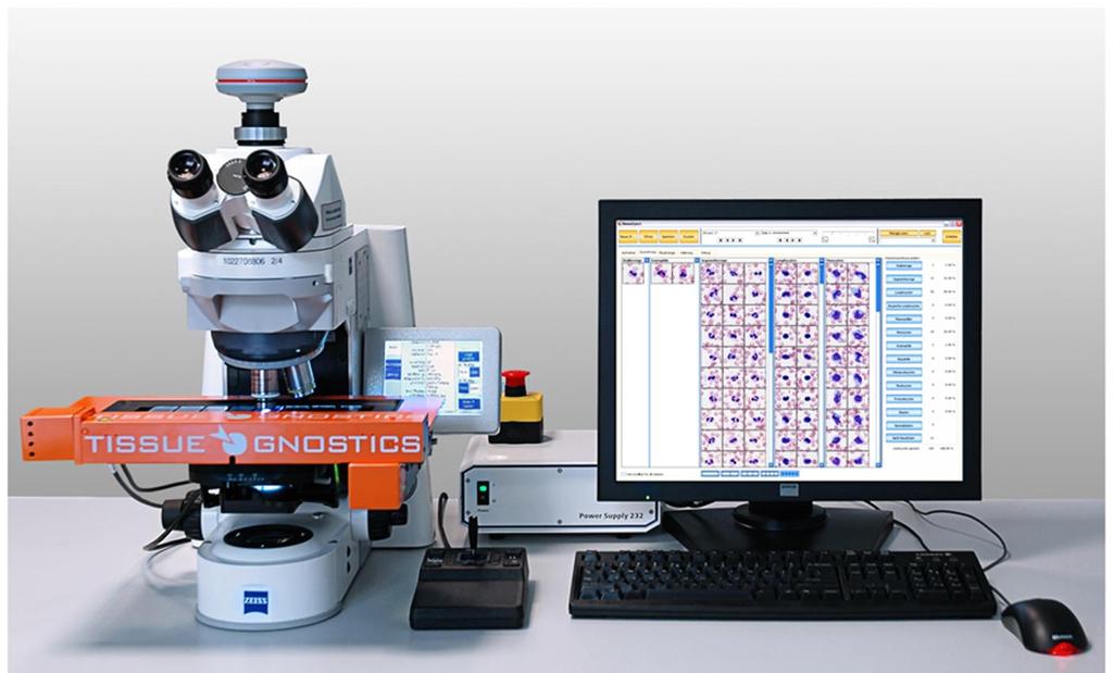
2 Discuss the history of hematology automation and digital differentials. Discuss the HemoFAXS Hematology Analysis System by Tissue Gnostics. Review automated microscopy analysis with the use of case studies.
3 1953 Wallace Coulter and the Coulter Principle 1960 Hematologic Evaluations were performed manually 1961 Coulter Electronics Inc, Hialeah, Florida 1970 s Vigorous competition began 1980 s Reporting out 7-parameter blood count and a three part differential 1980 s Introduction of flow based analyzers
4 Next 20 years grew in complexity 1988 a slide maker was introduced 1990 s the five part differential and multi parameters Early 2000 s the fully automated Reticulocyte was introduced 2011 Bloodhound Integrated Hematology System: Blood printing
5 For over 100 hundred years have manually viewed blood smears with the use of a microscope 1962: First television based image analyzer utilizing microscopic pictures which were developed by Metals Research. 1976: A cytology study conducted using a microscope, TV camera, an automatic cell finder, and a servo driven computer controlled stage is conducted. 1982: Automated Intelligence Microscopy (AIM) is coined with the advent of the Yellow IRIS urinalysis workstation. 1986: Hirox Co Ltd develops early digital microscope lens in Tokyo, Japan. 2000: Launch of Cellavision s Diffmaster Octavia in Europe. 2011: Bloodhound and a digital differential.
6
7 Automated cell differential to detect cell nucleus and cytoplasm Measurement parameters are: Morphology Color Differences Granularity Cell Nuclei Cytoplasm

8 3 Mega Pixel Color Camera 10 x and 100X Oil objectives Zeiss AxioImager Z2 Microscope 8 Motorized Slide Insert


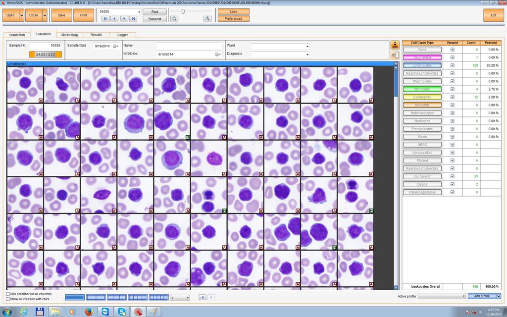
9 10X 10X Slide ID is scanned, digitized and presented with a control image Monolayer is localized Leukocytes are localized within the identified monolayer
 HemoFAXS detects the predefined number of")
 in the same process Insert Slides Acquisition & Classification")
10 Image acquisition and preclassification HemoFAXS searches automatically for the optimal working area (monolayer) HemoFAXS detects the predefined number of Leukocytes (10x) HemoFAXS acquires morphology pictures for morphological evaluation (10x) in the same process Insert Slides Acquisition & Classification Validation Reporting


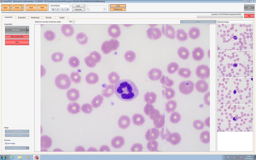
11 Morphology 10 x morphology images Morphology comments for Thrombocytes, Erythrocytes and Leukocytes 100 x morphology images Easy one-click scoring Insert Slides Acquisition & Classification Validation Reporting
12


13 Image acquisition and preclassification The image of each leukocyte is localised and acquired with a 100x oil objective The image analysis algorithms segment the cellular compartments to acquire the dataset Insert Slides Acquisition & Classification Validation Reporting


14 Classification into leukocyte classes Insert Slides Acquisition & Classification Validation Reporting


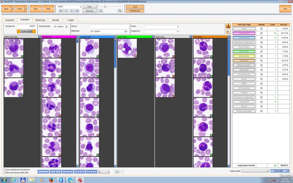
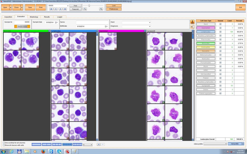
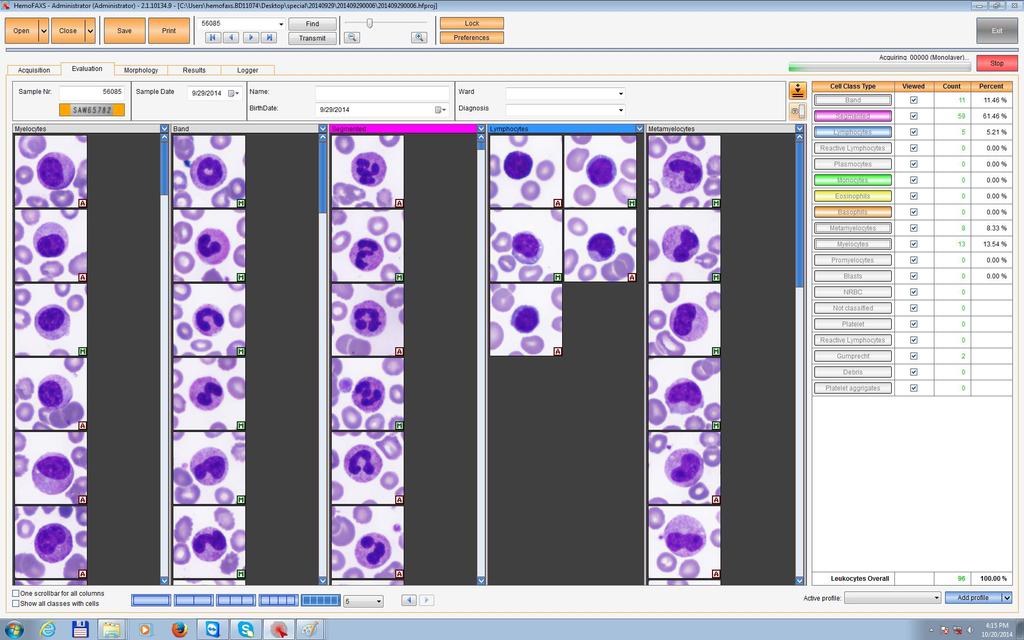
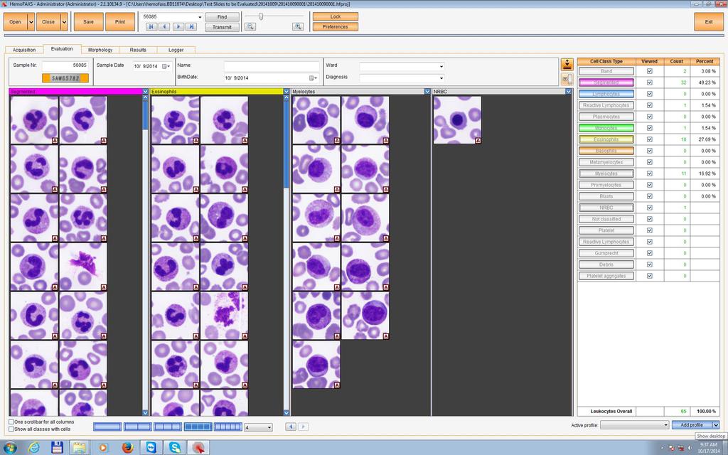
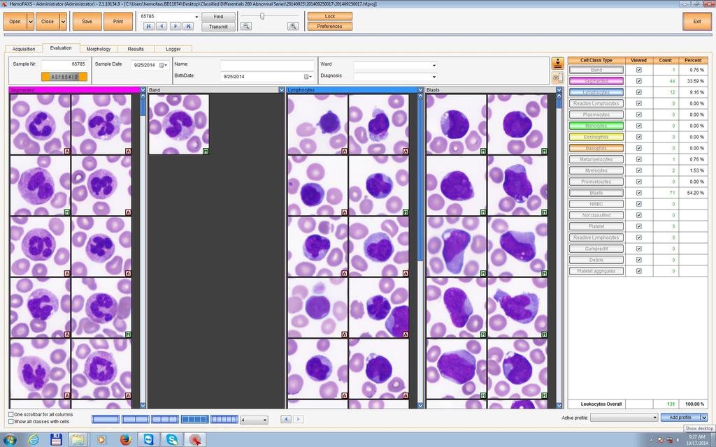
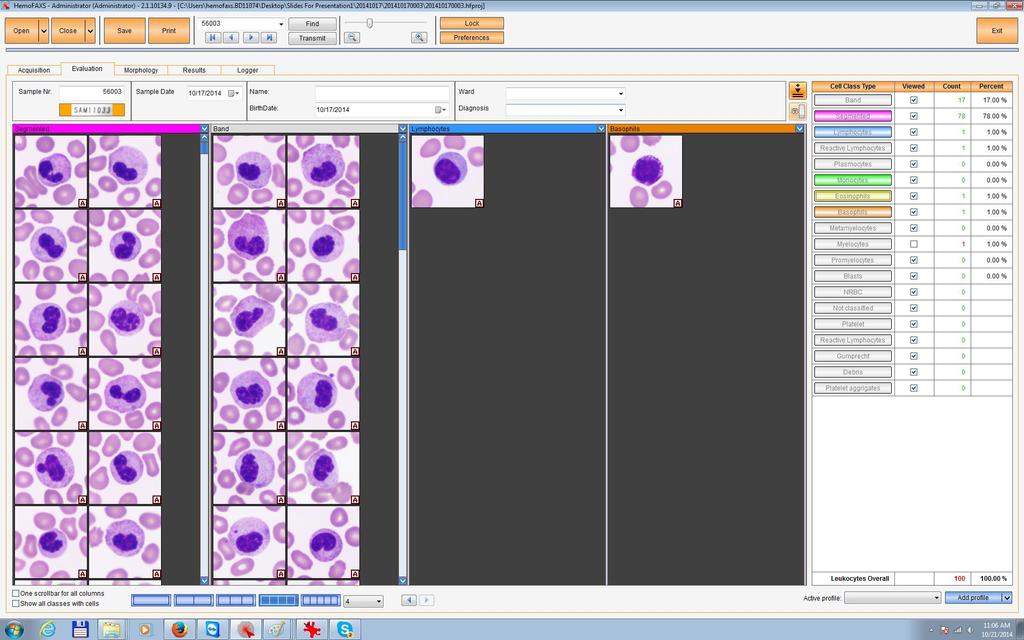
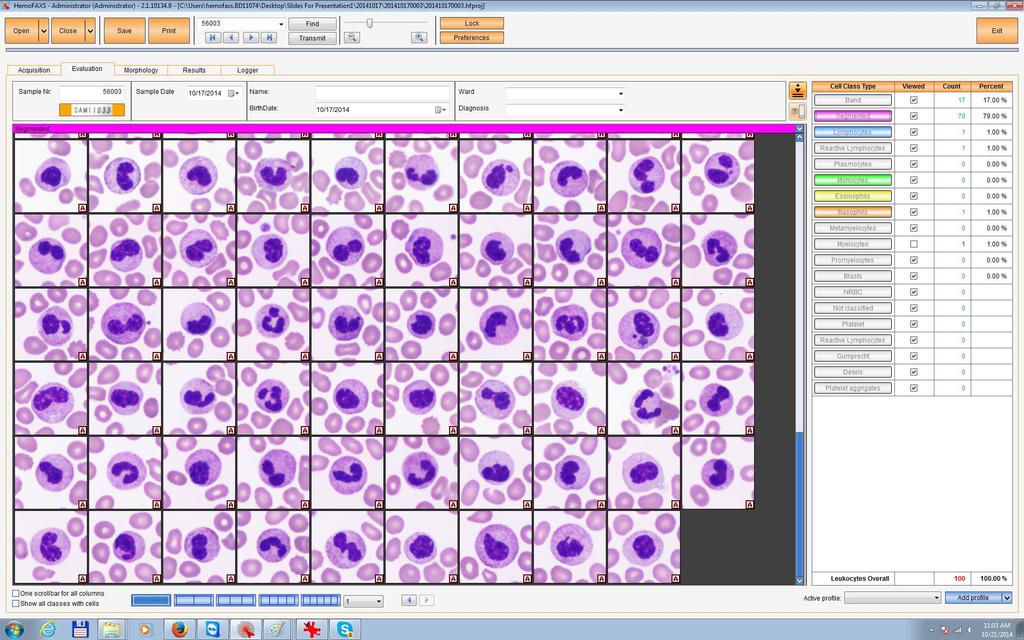
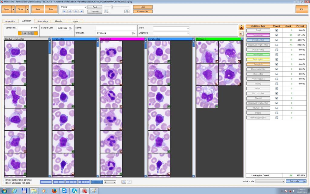
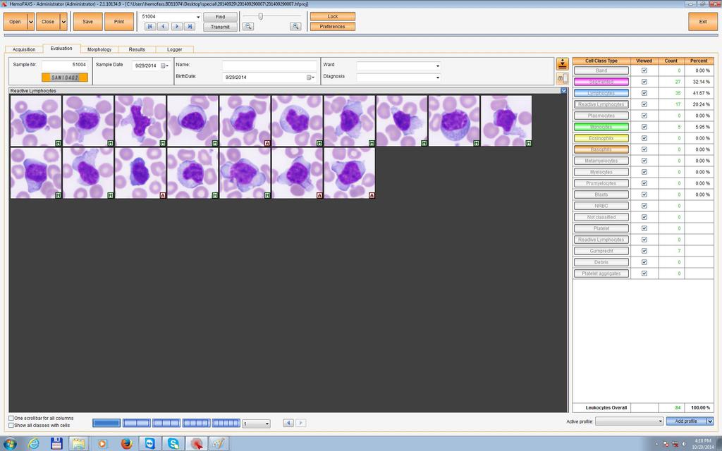
15 Evaluation Slide ID Patient data fields Differential count and percentages 100 x cell images displayed in columns Insert Slides Acquisition & Classification Validation Reporting


16 Optimal Visualisation Up to 10 columns can be visualized Insert Slides Acquisition & Classification Validation Reporting

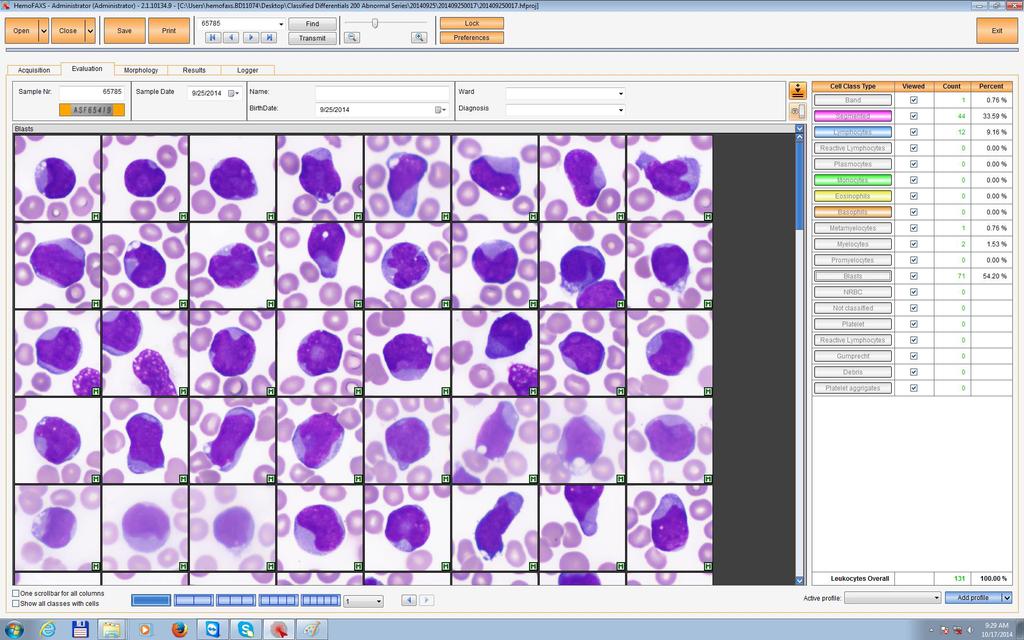
17 All cell images displayed Number and Percent Insert Slides Acquisition & Classification Validation Reporting
18
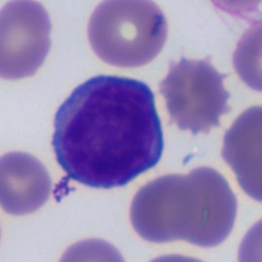
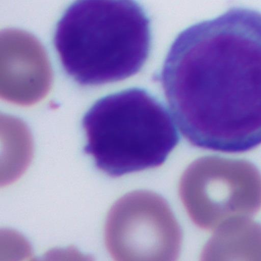
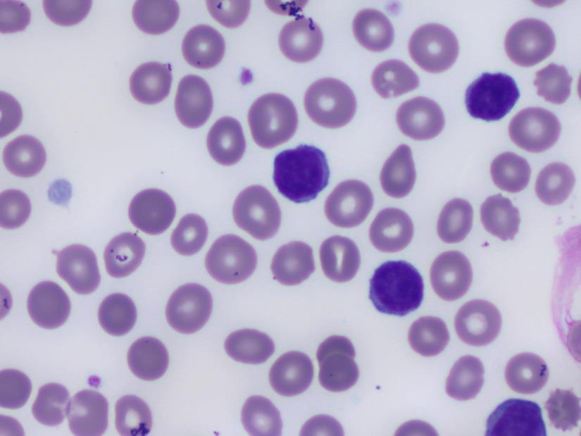
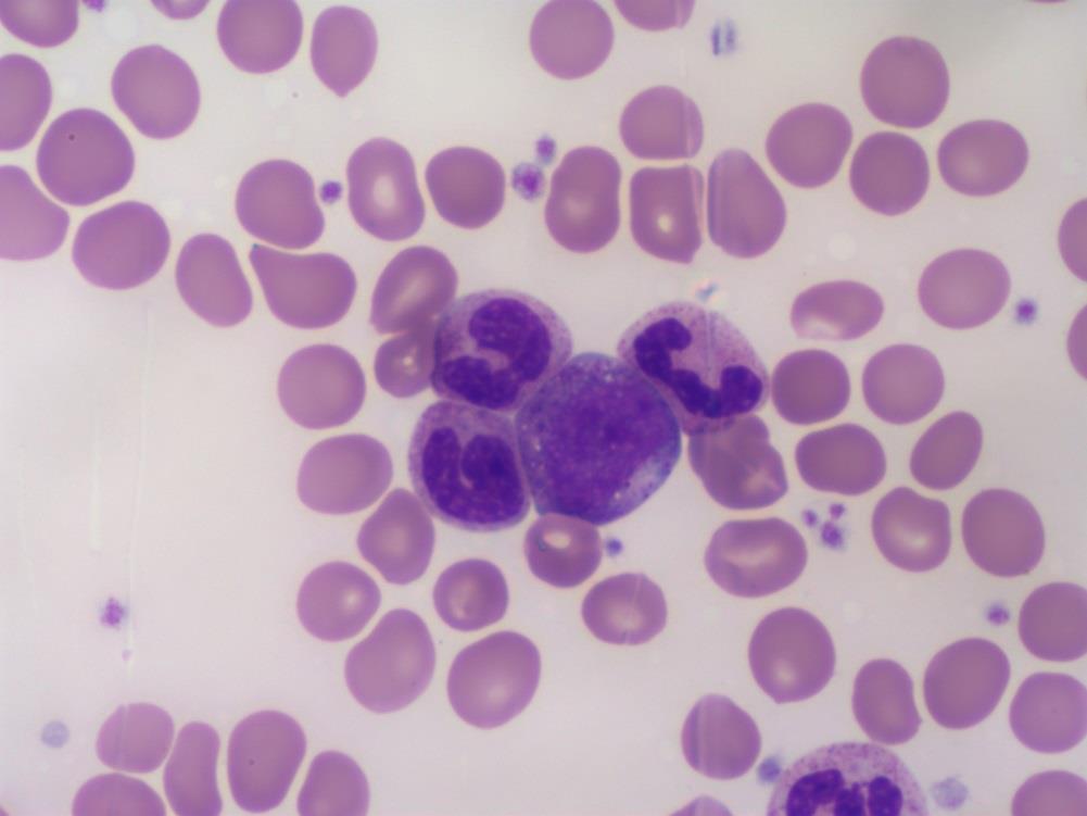
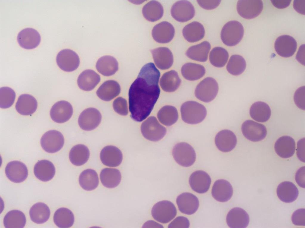
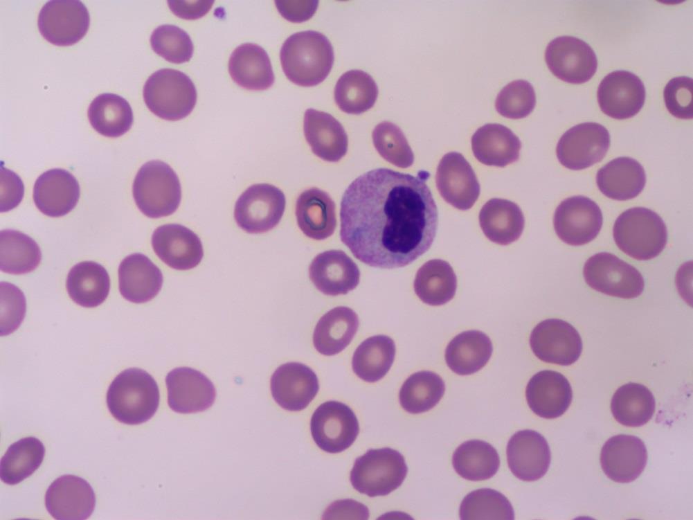
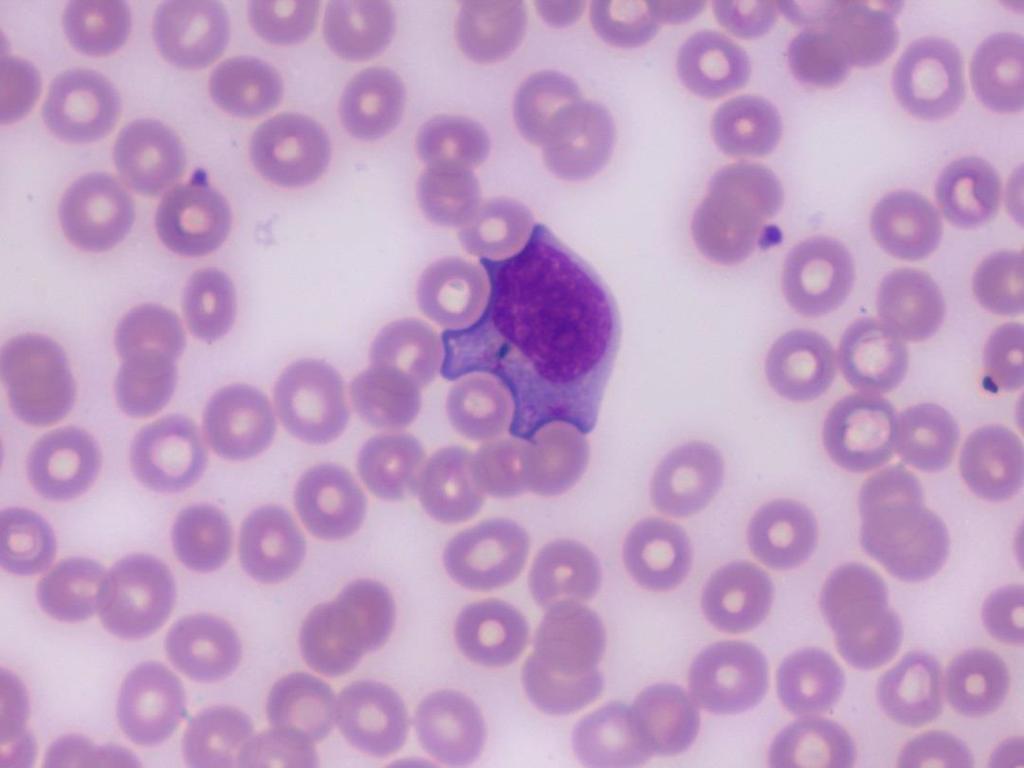
19 Patient History: A 84 year old female was admitted to University Hospital with complaints of exhaustion, heart palpitations and dehydration.
20 WBC RBC 2.44 HGB 8.2 HCT 26.6 MCV 109 MCH 33.6 MCHC 30.8 RDW 20.6 PLT 76 MPV 11.2
21
22

23
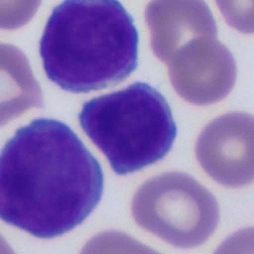
24 Symptoms of long duration May be asymptomatic for a long time Mostly mature cell forms found Usually organ involvement Platelets are normal to increased Anemia develops later on
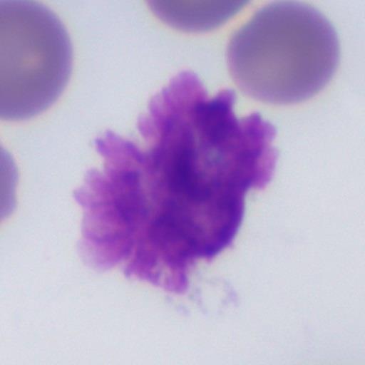
25 Majority involve the B Lymphocyte 75% Usually seen in the older population Male Commonly picked up by chance Accumulation of small mature lymphocytes Smudge cells: very fragile lymphocytes Platelet count normal N/N Anemia Progressive anemia and thrombocytopenia
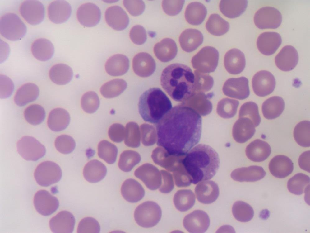
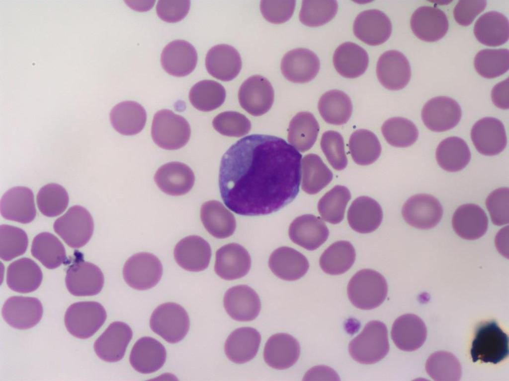
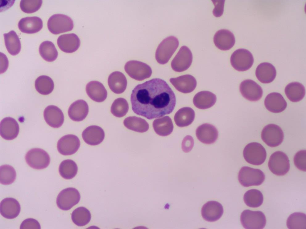
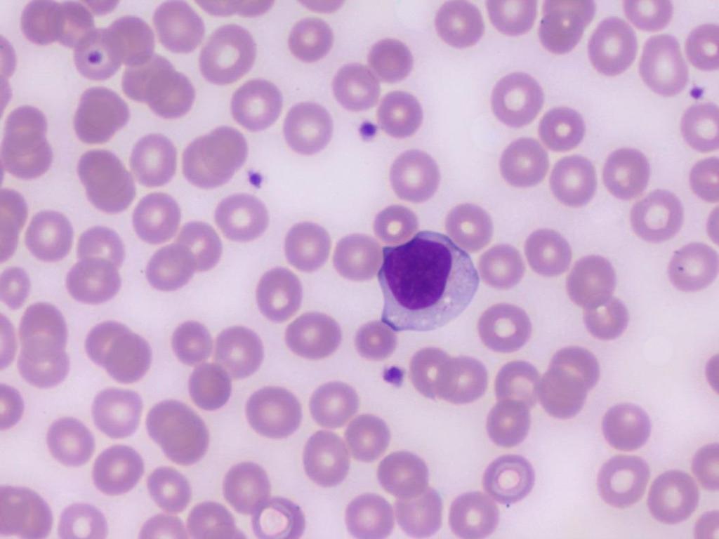
26 Patient History A 63 year old female was admitted to the hospital for fatigue and anemia. She had been feeling poorly over the course of several weeks. Additional complaints were abdominal fullness.
27 WBC 22.7 RBC 2.39 HGB 7.7 HCT 24.9 MCV MCH 32.2 MCHC 30.9 RDW 18.7 PLT 113 MPV 11.0
28
29
30
31 Clonal stem cell disorder Marked leukocytosis with all stages of granulocytic maturation Primarily disease of adults rare in children Hepatosplenomegaly Thrombocytosis is common
 Philadelphia Chromosome arm of 22 is translocated to 9, positive in 90% of")
32 WBC count ranges from 100, ,000 Differential: complete spectrum Increase Eos and Baso N/N Anemia Increased platelets, giant and fragments Bone Marrow: hypercellular 25:1 BCR/ABL gene or fusion gene 95% cases (necessary for pathogenesis) Philadelphia Chromosome arm of 22 is translocated to 9, positive in 90% of the cases

33 BCR: Breakpoint Cluster Region DNA fragment localized on chromosome 22 Defines the chromosomal break ABL: protein possessing increased tyrosine kinase activity
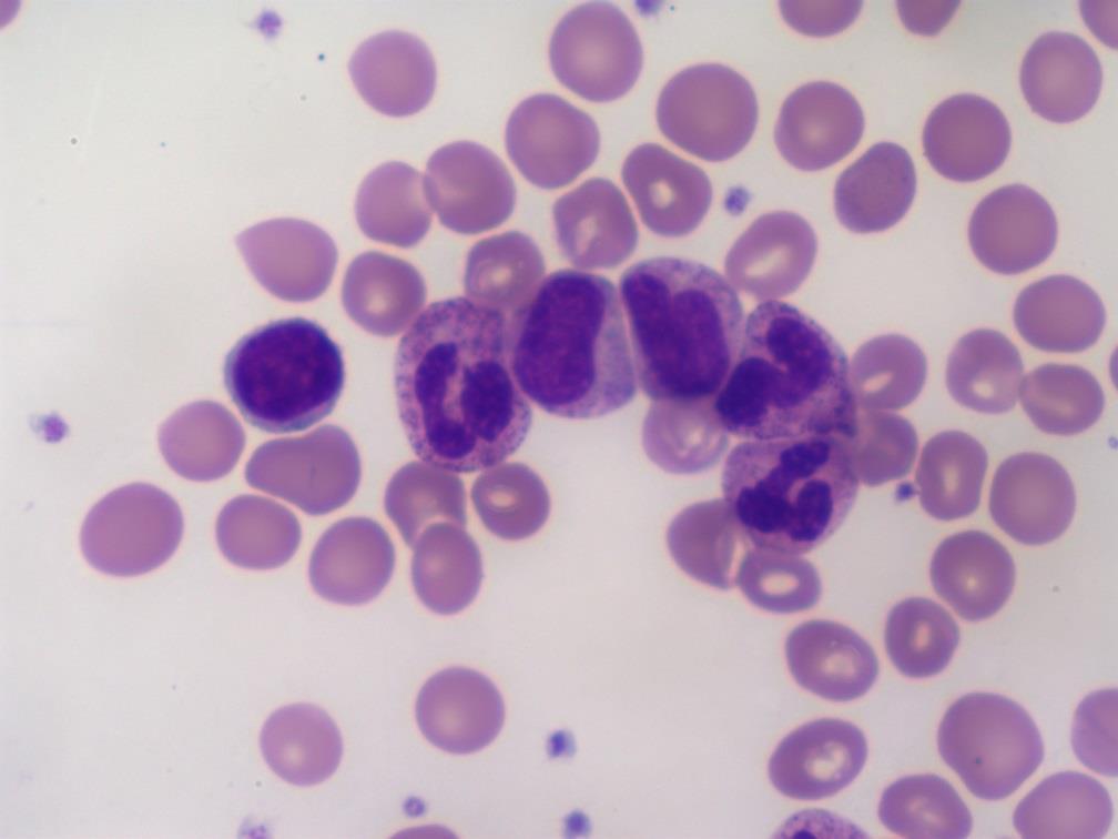
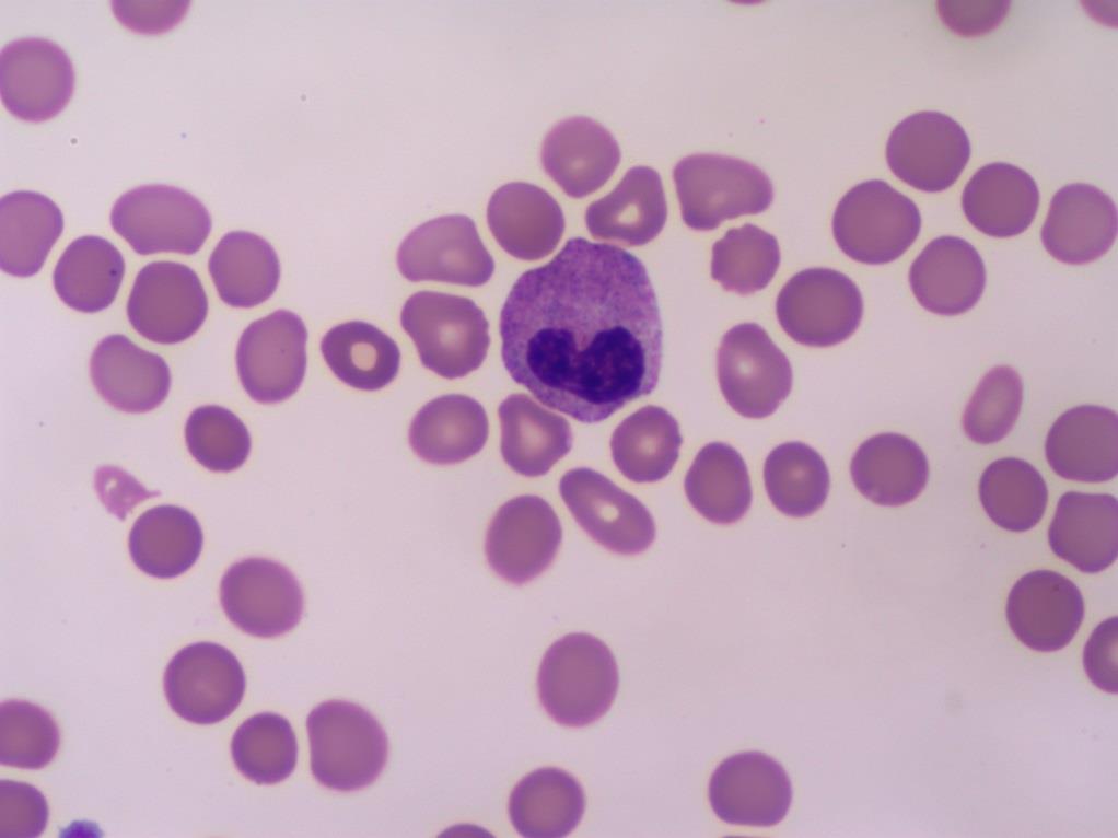
34 Patient History A 43 year old male admitted to the Emergency Room for a progressive infection and stagnant healing of a molar tooth extraction.
35 WBC 27.7 RBC 2.96 HGB 8.4 HCT 24.6 MCV 83.1 MCH 28.4 MCHC 34.1 RDW 14.5 PLT 32 MPV 11.9
36
37
38
39 Short Duration Onset sudden Many immature cells in the bone marrow and peripheral blood > 30% Blasts in the bone marrow (FAB) >20% Blasts in the bone marrow (WHO) Platelet count usually decreased N/N Anemia

40 L1- Lymphocytic, childhood Small uniform lymphoblasts L2- Lymphocytic Adult Large pleomorphic lymphoblasts L3- Burkitts, poor prognosis Deeply basophilic and vacuolated

41 Precursor B lymphoblastic Leukemia/Lymphoma Precursor B cell Pre B - ALL B - ALL Precursor T-Lymphoblastic Leukemia/Lymphoma
42 Frequent found in adults (64%) Blasts are large, twice the size of L1 Chromatin is fine to course condensed, nuclear clefting Nucleoli present, usually single and large Blasts are confused with M1
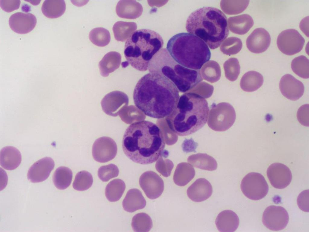
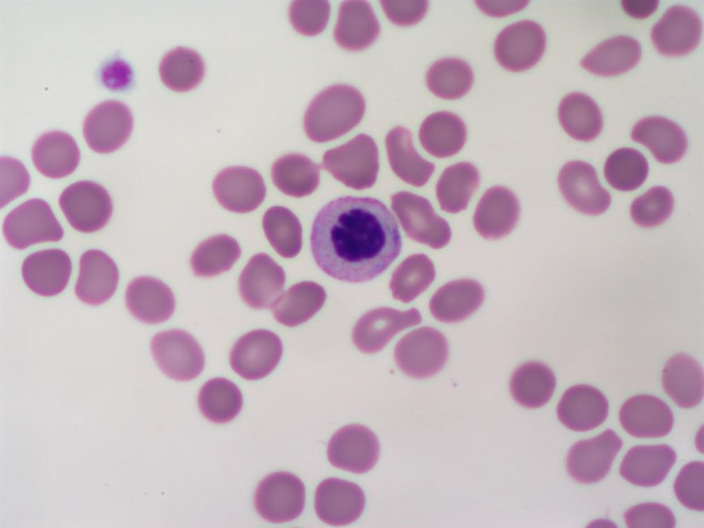
43 Patient History A 42 year old female was being seen by her physician for treatment of a bacterial infection and a progressive anemia.
44 WBC 3.63 RBC 2.72 HGB 7.8 HCT 24.3 MCV 89.3 MCH 28.7 MCHC 32.1 RDW 15.6 PLT 90 MPV 11.6
45
46
47
48 Benign inherited autosomal dominant Neutrophil nucleus does not segment beyond the bi - lobed stage Peanut or dumbbell shape Heavy chromatin clumping distinguishes from bands Mutant defect in the lamin B receptor gene Controls the shape of the nucleus 70-90% neutrophils affected The cell functions normal
49 Heterozygous State Nuclear chromatin densely clumped, dark and coarse 55-93% show bilobed nucleus Homozygous Nucleus is often single, round or slightly indented Can be eccentrically placed in the granulocyte with no nuclear segmentation Dense chromatin pattern Acquired Leukemoid reactions during severe bacterial infections, HIV, TB, MDS, MPN s
50 Patient History A 19 year old male is seen by his physician for flu like symptoms. Major complaints was progressive fatigue, low grade fever, swollen lymph nodes and a sore throat. He had been feeling poorly for several weeks.
51 WBC 9.2 RBC 4.27 HGB 12.4 HCT 36.9 MCV 86.4 MCH 29.0 MCHC 33.6 RDW 14.6 PLT 320 MPV 9.7
52
53
54
55 Young Adult : yrs Epstein - Barr virus Transmission: oral through saliva (most common) Relative and Absolute Lymphocytosis Reactive Lymph > 50% lymphs Splenomegaly: 50-75% of the cases Hepatomegaly: 25% of the cases Positive Mono Spot/ Heterophile Antibody
56 Incubation period 11 days Low grade fever Pharyngitis (sore throat) Enlarged lymph nodes Hematological Complications: auto hemolytic anemia Rare complication: hepatitis Convalescence: few weeks to months

57 HemoFAXS is in the process of running the clinical trials for FDA clearance
EDUCATIONAL COMMENTARY BLOOD CELL IDENTIFICATION
 EDUCATIONAL COMMENTARY BLOOD CELL IDENTIFICATION Educational commentary is provided through our affiliation with the American Society for Clinical Pathology (ASCP). To obtain FREE CME/CMLE credits click
EDUCATIONAL COMMENTARY DISTINGUISHING MORPHOLOGIC LOOK-ALIKES
 EDUCATIONAL COMMENTARY DISTINGUISHING MORPHOLOGIC LOOK-ALIKES Educational commentary is provided through our affiliation with the American Society for Clinical Pathology (ASCP). To obtain FREE CME/CMLE
Blood Cell Identification Graded
 BCP-21 Blood Cell Identification Graded Case History The patient is a 37-year-old female with a history of multiple sickle cell crises. She now presents with avascular necrosis of the left hip. Laboratory
EDUCATIONAL COMMENTARY MORPHOLOGIC CHANGES IN PERIPHERAL BLOOD CELLS
 EDUCATIONAL COMMENTARY MORPHOLOGIC CHANGES IN PERIPHERAL BLOOD CELLS Educational commentary is provided through our affiliation with the American Society for Clinical Pathology (ASCP). To obtain FREE CME/CMLE
Myeloproliferative Disorders - D Savage - 9 Jan 2002
 Disease Usual phenotype acute leukemia precursor chronic leukemia low grade lymphoma myeloma differentiated Total WBC > 60 leukemoid reaction acute leukemia Blast Pro Myel Meta Band Seg Lymph 0 0 0 2
Guide to the 1-3 Minute Blood Film Microscopic Review: Why and How?
 Guide to the 1-3 Minute Blood Film Microscopic Review: Why and How? Dennis B. DeNicola, DVM, PhD, DACVP Chief Veterinary Educator IDEXX Laboratories, Inc. Westbrook, ME USA Adjunct Professor of Veterinary
Beyond the CBC Report: Extended Laboratory Testing in the Evaluation for Hematologic Neoplasia Disclosure
 Beyond the CBC Report: Extended Laboratory Testing in the Evaluation for Hematologic Neoplasia Disclosure I am receiving an honorarium from Sysmex for today s presentation. 1 Determining the Etiology for
Lymphoma Tumor Board Quiz! Laboratory Hematology: Basic Cell Morphology
 Lymphoma Tumor Board Quiz! Laboratory Hematology: Basic Cell Morphology CABOT RINGS Cabot rings in a patient with hemolytic anemia. Cabot ring (red arrow) and Howell-Jolly body (blue arrow). Observed in
EDUCATIONAL COMMENTARY MORPHOLOGIC ABNORMALITIES IN LEUKOCYTES
 EDUCATIONAL COMMENTARY MORPHOLOGIC ABNORMALITIES IN LEUKOCYTES Educational commentary is provided through our affiliation with the American Society for Clinical Pathology (ASCP). To obtain FREE CME/CMLE
Collect and label sample according to standard protocols. Gently invert tube 8-10 times immediately after draw. DO NOT SHAKE. Do not centrifuge.
 Complete Blood Count CPT Code: CBC with Differential: 85025 CBC without Differential: 85027 Order Code: CBC with Differential: C915 Includes: White blood cell, Red blood cell, Hematocrit, Hemoglobin, MCV,
MECHANISMS OF HUMAN DISEASE: LABORATORY SESSIONS LYMPHOMA. April 16, 2008
 MECHANISMS OF HUMAN DISEASE: LABORATORY SESSIONS LYMPHOMA April 16, 2008 FACULTY COPY GOAL: Learn the appearance of normal peripheral blood elements and lymph nodes. Recognize abnormal peripheral blood
Group of malignant disorders of the hematopoietic tissues characteristically associated with increased numbers of white cells in the bone marrow and
 Group of malignant disorders of the hematopoietic tissues characteristically associated with increased numbers of white cells in the bone marrow and / or peripheral blood Classified based on cell type
ADx Bone Marrow Report. Patient Information Referring Physician Specimen Information
 ADx Bone Marrow Report Patient Information Referring Physician Specimen Information Patient Name: Specimen: Bone Marrow Site: Left iliac Physician: Accession #: ID#: Reported: 08/19/2014 - CHRONIC MYELOGENOUS
WBCs Disorders 1. Dr. Nabila Hamdi MD, PhD
 WBCs Disorders 1 Dr. Nabila Hamdi MD, PhD ILOs Compare and contrast ALL, AML, CLL, CML in terms of age distribution, cytogenetics, morphology, immunophenotyping, laboratory diagnosis clinical features
Hematology 101. Cindy Rogers, MT(ASCP) Diagnostics System Specialist
 Diagnostics System Specialist") Hematology 101 Cindy Rogers, MT(ASCP) Diagnostics System Specialist More Acronyms...» CBC» RBC» HGB» HCT» WBC» MPV» PLT» RDW» DIFF» H&H» Complete Blood Count» Red Blood Cell» Hemoglobin» Hematocrit» White
Hematology 101 Cindy Rogers, MT(ASCP) Diagnostics System Specialist More Acronyms...» CBC» RBC» HGB» HCT» WBC» MPV» PLT» RDW» DIFF» H&H» Complete Blood Count» Red Blood Cell» Hemoglobin» Hematocrit» White
Full Blood Count analysis Is a 3 part-diff good enough? Dr Marion Münster, Sysmex South Africa
 Full Blood Count analysis Is a 3 part-diff good enough? Dr Marion Münster, Sysmex South Africa The Role of the FBC in clinical decision making History Examination Investigations Decision 70% FBC Laboratory
74y old Female with chronic elevation of Platelet count. August 18, 2005 Faizi Ali, MD Hematopathology Fellow
 74y old Female with chronic elevation of Platelet count August 18, 2005 Faizi Ali, MD Hematopathology Fellow Clinical History Patient is a 74y old otherwise healthy Caucasian female with no major complaint
Hematopathology Lab. Third year medical students
 Hematopathology Lab Third year medical students Objectives Identify the lesion Know the specific name of the lesion Know associated disease Know relevant pathologic background Spherocytes: appear small,
Hemopoiesis and Blood
 Hemopoiesis and Blood Blood Cells o o o Erythrocytes Leukocytes Thrombocytes Function o Transport nutrients and wastes throughout the bloodstream, fight foreign antigens and blood coagulation. Location
Proper Slide Preparation
 Hematology Essentials: A Foundation for WBC Review Using Case Studies Christine Hinz, MS, MLS(ASCP) CM Proper Slide Preparation smooth, homogenous film 1/2 to 3/4 the slide length straight feather edge
EDUCATIONAL COMMENTARY DIFFERENTIATING IMMATURE PERIPHERAL BLOOD CELLS
 Educational commentary is provided through our affiliation with the American Society for Clinical Pathology (ASCP). To obtain FREE CME/CMLE credits click on Continuing Education on the left side of the
VETERINARY HEMATOLOGY ATLAS OF COMMON DOMESTIC AND NON-DOMESTIC SPECIES COPYRIGHTED MATERIAL SECOND EDITION
 VETERINARY HEMATOLOGY ATLAS OF COMMON DOMESTIC AND NON-DOMESTIC SPECIES SECOND EDITION COPYRIGHTED MATERIAL CHAPTER ONE HEMATOPOIESIS GENERAL FEATURES All blood cells have a finite life span, but in normal
3. Blood Cell Histograms:
 LECTURE MODULE 6c: ELECTRONIC CELL COUNTING PART III 3. Blood Cell Histograms: a. The Coulter cell counters today provides size distributions of the cellular content: 1) volume given in µm 3 or fl vs relative
CELL-DYN Strength in Technology, Proven Reliability. Optical WBC Technology. Patented M.A.P.S.S. Differential. Multiple Technologies
 CELL-DYN 3700 Strength in Technology, Proven Reliability Optical WBC Technology Patented M.A.P.S.S. Differential Multiple Technologies CELL-DYN 3700 Multiple Technologies One Superior Result Multiple Technologies
Blood Cell Identification Graded
 Blood Cell Identification Graded Case History The patient is a 20-year-old female with sickle cell disease who presents with bilateral leg pain for 3 days. She is scheduled to have bilateral hip and leg
Hematopathology Case Study
 www.medfusionservices.com Hematopathology Case Study CV3515-14 JUNE Clinical Presentation: Clinical Information: A 42 year old male with history of chronic myelogenous leukemia (CML) presents with an elevated
Differential Blood Smear H3
 Verein für Association pour le Associazione per il medizinische Qualitätskontrolle contrôle de qualité médical controllo di qualità medico Report Differential Blood Smear H3 MQ 2015-4 MQ, Institut für
Complete Blood Count (CBC) Assist.Prof. Filiz BAKAR ATEŞ
 Assist.Prof. Filiz BAKAR ATEŞ") Complete Blood Count (CBC) Assist.Prof. Filiz BAKAR ATEŞ The complete blood count (CBC) is one of the most common blood test used. It analyzes the three major types of cells in blood 1. red blood cells,
Complete Blood Count (CBC) Assist.Prof. Filiz BAKAR ATEŞ The complete blood count (CBC) is one of the most common blood test used. It analyzes the three major types of cells in blood 1. red blood cells,
Interpreting the CBC. Robert Miller PA Assistant Professor of Clinical Pediatrics and Family Medicine USC Keck School of Medicine Retired
 Interpreting the CBC Robert Miller PA Assistant Professor of Clinical Pediatrics and Family Medicine USC Keck School of Medicine Retired The CBC 3 Cell Lines RBCs WBCs Platelets Assess general health Make
7 Omar Abu Reesh. Dr. Ahmad Mansour Dr. Ahmad Mansour
 7 Omar Abu Reesh Dr. Ahmad Mansour Dr. Ahmad Mansour -Leukemia: neoplastic leukocytes circulating in the peripheral bloodstream. -Lymphoma: a neoplastic process in the lymph nodes, spleen or other lymphatic
Blood Cell Identification Graded
 Blood Cell Identification Graded Case History The patient was a five-day-old girl with an elevated unconjugated bilirubin and a weakly positive direct antiglobulin test (DAT). Her CBC showed: WBC = 11.0
Participants Identification No. % Evaluation. Mitotic figure Educational Erythrocyte precursor, abnormal 1 0.
 Cell Identification Mitotic figure 212 99.5 Educational Erythrocyte precursor, abnormal BMD-02 The arrowed cell is a mitotic figure. It was correctly identified by 99.5% of the participants. A cell containing
Myelodysplastic syndrome (MDS) & Myeloproliferative neoplasms
 & Myeloproliferative neoplasms") Myelodysplastic syndrome (MDS) & Myeloproliferative neoplasms Myelodysplastic syndrome (MDS) A multipotent stem cell that can differentiate into any of the myeloid lineage cells (RBCs, granulocytes, megakaryocytes)
Myelodysplastic syndrome (MDS) & Myeloproliferative neoplasms Myelodysplastic syndrome (MDS) A multipotent stem cell that can differentiate into any of the myeloid lineage cells (RBCs, granulocytes, megakaryocytes)
Heme 9 Myeloid neoplasms
 Heme 9 Myeloid neoplasms The minimum number of blasts to diagnose acute myeloid leukemia is 5% 10% 20% 50% 80% AML with the best prognosis is AML with recurrent cytogenetic abnormality AML with myelodysplasia
A Look Into the Determination of Cell Morphology in Hematology in the 21 st Century. Ramon Simon-Lopez, MD Global Scientific Director Beckman Coulter
 A Look Into the Determination of Cell Morphology in Hematology in the 21 st Century Ramon Simon-Lopez, MD Global Scientific Director Beckman Coulter Is cell morphology important? AML M7 CLL CD5 CD19 NHL
COMPANY OR UNIVERSITY
 CONTRIBUTOR NAME Daniel Heinrich, DVM CONTRIBUTOR EMAIL dheinric@umn.edu COAUTHORS Jed Overmann, DVM, DACVP; Davis Seelig DVM, PhD, DACVP & Matthew Sturos, DVM COMPANY OR UNIVERSITY University of Minnesota
Polycthemia Vera (Rubra)
") Polycthemia Vera (Rubra) Polycthemia Vera (Rubra) Increased red cells Clonal Myeloid lineages also increased 2-13 cases per million Mean age: 60 years Sites of Involvement Bone marrow Peripheral blood
Polycthemia Vera (Rubra) Polycthemia Vera (Rubra) Increased red cells Clonal Myeloid lineages also increased 2-13 cases per million Mean age: 60 years Sites of Involvement Bone marrow Peripheral blood
Acute Lymphoblastic Leukaemia
 Acute Lymphoblastic Leukaemia Terri Boyer 17 th October 2006 Overview Disease information: Aetiology of ALL proposed theory, contributing factors Symptoms Complications Diagnostic approaches - morphology
Disclosures/COI. Cases in Hematopathology. Outline. Heme Path Findings Not to Miss. Normal Peripheral Smear 6/30/2016
 Disclosures/COI Cases in Hematopathology Vamsi Kota Assistant Professor Department of Hematology & Medical Oncology Leukemia/BMT I have no disclosures or conflicts of interest regarding this presentation.
Complete Blood Count PSI AP Biology
 Complete Blood Count PSI AP Biology Name: Objective Students will examine how the immunological response affects molecules in the blood. Students will analyze three complete blood counts and create diagnoses
Blood DLC, Retic count, PCV, Hb and ESR. Dr. Tamara Alqudah
 Blood DLC, Retic count, PCV, Hb and ESR Dr. Tamara Alqudah Differential Leukocyte Count (DLC) There are 5 main types of WBCs: 1. Neutrophils: 40-80% 2. Eosinophils: 1-6 % 3. Basophils: < 1-2% 4. Lymphocytes:
The Complete Blood Count
 The Complete Blood Count (Cartesian Thinking at Its Best) A SEM Image of Normal Human Blood Laurie Larsson February 22, 2010 Anatomy and Philology II Dr. Danil Hammoudi Introduction A complete blood count
HEMATOLOGIC MORPHOLOGY- AECOM HEMATOLOGY COURSE
 Log Out Help current login :lcytryn@montefiore.org HEMATOLOGIC MORPHOLOGY- AECOM HEMATOLOGY COURSE Lawrence Cytryn, M.D. - Course Director 1998 Edward Burns, M.D. Images used by permission within AECOM
By Dr. Mohamed Saad Daoud
 By Dr. Mohamed Saad Daoud Part I Introduction Types of White Blood Cells Genesis of the White Blood Cells Life Span of the White Blood Cells Dr. Mohamed Saad Daoud 2 Leucocytes Introduction: Infectious
Hematopathology Case Study
 Hematopathology Case Study AMP Outreach Course 2009 AMP Annual Meeting John Greg Howe Ph.D. Department of Laboratory Medicine Yale University School of Medicine November 19, 2009 HISTORY Case History An
Hematology Unit Lab 2 Review Material
 Objectives Hematology Unit Lab 2 Review Material - 2018 Laboratory Instructors: 1. Assist students during lab session Students: 1. Review the introductory material 2. Study the case histories provided
Hematology 101. Blanche P Alter, MD, MPH, FAAP Clinical Genetics Branch Division of Cancer Epidemiology and Genetics Bethesda, MD
 Hematology 101 Blanche P Alter, MD, MPH, FAAP Clinical Genetics Branch Division of Cancer Epidemiology and Genetics Bethesda, MD Hematocrits Plasma White cells Red cells Normal, Hemorrhage, IDA, Leukemia,
Hematology Unit Lab 1 Review Material
 Hematology Unit Lab 1 Review Material - 2018 Objectives Laboratory instructors: 1. Assist students during lab session Students: 1. Review the introductory material 2. Study the case histories provided
Myelodysplastic Syndromes: Everyday Challenges and Pitfalls
 Myelodysplastic Syndromes: Everyday Challenges and Pitfalls Kathryn Foucar, MD kfoucar@salud.unm.edu Henry Moon lecture May 2007 Outline Definition Conceptual overview; pathophysiologic mechanisms Incidence,
Pathology. #11 Acute Leukemias. Farah Banyhany. Dr. Sohaib Al- Khatib 23/2/16
 35 Pathology #11 Acute Leukemias Farah Banyhany Dr. Sohaib Al- Khatib 23/2/16 1 Salam First of all, this tafreegh is NOT as long as you may think. If you just focus while studying this, everything will
The patient had a mild splenomegaly but no obvious lymph node enlargement. The consensus phenotype obtained from part one of the exercise was:
 Case History An 86 year old male was admitted to hospital with chest infection. Haematological examination subsequently revealed the following: Hb- 11.0 g/dl; WBC- 67.1 x 10^9/l; PLT- 99 x10^9/l; RBC-
Incorporating Differentials Into Every Complete Blood Count. Paige Flowers, LVT Dogwood Veterinary Internal Medicine
 Incorporating Differentials Into Every Complete Blood Count Paige Flowers, LVT Dogwood Veterinary Internal Medicine Complete Blood Count Diagnostic performed to evaluate the quantity and morphology of
HISTOLOGY VIRTUAL LABORATORY BLOOD AND LYMPHATICS SYSTEM
 HISTOLOGY VIRTUAL LABORATORY BLOOD AND LYMPHATICS SYSTEM Login: http://histopath.westernu.edu Histology Atlas AND Virtual Histology links. I. HEMATOLOGY - PERIPHERAL BLOOD Purpose: To be able to identify
Disorders of Blood Cells & Blood Coagulation
 Disorders of Blood Cells & Blood Coagulation HIHIM 409 WBC count RBC count WBC differential Hemoglobin (HGB) Hematocrit (HCT) % of volume occupied by RBCs CBC Red cell indices Mean cell volume (MCV) average
CELL-DYN 3700 Strength in Technology, Proven Reliability
 CELL-DYN 3700 Strength in Technology, Proven Reliability Optical WBC Technology Patented MAPSS Differential Multiple Technologies CELL-DYN 3700 Multiple Technologies One Superior Result Multiple Technologies
CHAPTER:4 LEUKEMIA. BY Mrs. K.SHAILAJA., M. PHARM., LECTURER DEPT OF PHARMACY PRACTICE, SRM COLLEGE OF PHARMACY 8/12/2009
 LEUKEMIA CHAPTER:4 1 BY Mrs. K.SHAILAJA., M. PHARM., LECTURER DEPT OF PHARMACY PRACTICE, SRM COLLEGE OF PHARMACY Leukemia A group of malignant disorders affecting the blood and blood-forming tissues of
Interpreting Hematology Scatter-Plots; One Cancer Center s Keys to Seeing the BIG Picture
 Interpreting Hematology Scatter-Plots; One Cancer Center s Keys to Seeing the BIG Picture Barbara L. Burch, MHA MT (ASCP) Laboratory Manager New York University Clinical Cancer Center Disclosure Ms Burch
Myeloid neoplasms. Early arrest in the blast cell or immature cell "we call it acute leukemia" Myoid neoplasm divided in to 3 major categories:
 Myeloid neoplasms Note: Early arrest in the blast cell or immature cell "we call it acute leukemia" Myoid neoplasm divided in to 3 major categories: 1. AML : Acute myeloid leukemia(stem cell with myeloid
Almost any suspected tumor can be aspirated easily and safely. Some masses are more risky to aspirate including:
 DOES THIS PATIENT HAVE CANCER? USING IN-HOUSE CYTOLOGY TO HELP YOU MAKE THIS DIAGNOSIS. Joyce Obradovich, DVM, Diplomate, ACVIM (Oncology) Animal Cancer & Imaging Center, Canton, Michigan Almost every
HENATOLYMPHOID SYSTEM THIRD YEAR MEDICAL STUDENTS- UNIVERSITY OF JORDAN AHMAD T. MANSOUR, MD. Part 4 MYELOID NEOPLASMS
 HENATOLYMPHOID SYSTEM THIRD YEAR MEDICAL STUDENTS- UNIVERSITY OF JORDAN AHMAD T. MANSOUR, MD Part 4 MYELOID NEOPLASMS Introduction: o Myeloid neoplasms are divided into three major categories: o Acute
10/30/2015. XN Case Studies: Every Picture Tells a Story
 XN Case Studies: Every Picture Tells a Story Jill Crist MT(ASCP)Field Product Specialist OBJECTIVES Explain how scattergrams and histogram pictures can provide great insight into abnormal hematology samples
DR SUDHIR MEHTA MD,MNAMS,FICP. Senior Professor & Head Medical Unit SMS Medical College & Hospital Jaipur
 DR SUDHIR MEHTA MD,MNAMS,FICP Senior Professor & Head Medical Unit SMS Medical College & Hospital Jaipur s.smehta@hotmail.com CBC..What is the Utility of performing this basic Hematology Test? 10/31/2010
MS.4/ Acute Leukemia: AML. Abdallah Al Abbadi.MD.FRCP.FRCPath Feras Fararjeh MD
 MS.4/ 27.02.2019 Acute Leukemia: AML Abdallah Al Abbadi.MD.FRCP.FRCPath Feras Fararjeh MD Case 9: Acute Leukemia 29 yr old lady complains of fever and painful gums for 1 week. She developed easy bruising
Paper ID: ART
 analytical process. Delayed sample analysi changes of measured parameters co interpretation of results. Pre-analytical variables, such as stor temperature affect the measurement parameters collected in
Non-Hodgkin lymphomas (NHLs) Hodgkin lymphoma )HL)
 Hodgkin lymphoma )HL)") Non-Hodgkin lymphomas (NHLs) Hodgkin lymphoma )HL) Lymphoid Neoplasms: 1- non-hodgkin lymphomas (NHLs) 2- Hodgkin lymphoma 3- plasma cell neoplasms Non-Hodgkin lymphomas (NHLs) Acute Lymphoblastic Leukemia/Lymphoma
Non-Hodgkin lymphomas (NHLs) Hodgkin lymphoma )HL) Lymphoid Neoplasms: 1- non-hodgkin lymphomas (NHLs) 2- Hodgkin lymphoma 3- plasma cell neoplasms Non-Hodgkin lymphomas (NHLs) Acute Lymphoblastic Leukemia/Lymphoma
WBCs Disorders. Dr. Nabila Hamdi MD, PhD
 WBCs Disorders Dr. Nabila Hamdi MD, PhD ILOs Compare and contrast ALL, AML, CLL, CML in terms of age distribution, cytogenetics, morphology, immunophenotyping, laboratory diagnosis clinical features and
Blood: Functions. Liquid connective tissue 3 general functions 1. Transportation. 2. Regulation. 3. Protection
 Blood Elements Lecture Objectives List blood components. Classify formed elements of blood. Discuss the scientific basis of the above classification. Describe the basic structure of erythrocytes and criteria
Easy Trick to Spot Leukemia for Pediatricians
 Easy Trick to Spot Leukemia for Pediatricians Piya Rujkijyanont, MD Division of Hematology-Oncology Department of Pediatrics Phramongkutklao Hospital Most Common Pediatric Cancers Age 0-14 Leukemia 32%
MYELOPROLIFARATIVE NEOPLASMS. Dr. Hasan Fahmawi, MRCP(UK), FRCP(Edin).
, FRCP(Edin).") MYELOPROLIFARATIVE NEOPLASMS Dr. Hasan Fahmawi, MRCP(UK), FRCP(Edin). These are a group of chronic conditions characterised by clonal proliferation of marrow precursor cells. PRV, essential thrombocyathaemia,
MYELOPROLIFARATIVE NEOPLASMS Dr. Hasan Fahmawi, MRCP(UK), FRCP(Edin). These are a group of chronic conditions characterised by clonal proliferation of marrow precursor cells. PRV, essential thrombocyathaemia,
Bone marrow aspiration as the initial diagnostic tool in the diagnosis of leukemia - A case study
 Original Research Article Bone marrow aspiration as the initial diagnostic tool in the diagnosis of leukemia - A case study Priyanka Poonam 1*, N.K. Bariar 2 1 Tutor, Department of Pathology, Patna Medical
Blood Cell Identification: 2011-B Mailing: Acute Myeloid Leukemia (AML)
") Please Note: To view the Figures and Images contained within this education activity in color, access the electronic version of the reading. CASE HISTORY This peripheral blood smear is from a 51-year-old
Please Note: To view the Figures and Images contained within this education activity in color, access the electronic version of the reading. CASE HISTORY This peripheral blood smear is from a 51-year-old
Standard Operating Procedure
 Subject Differential: Counting and Morphology Index Number Lab-5074 Section Laboratory Subsection Hematology Automated Laboratory Category Departmental Contact Moldenhauer, Emily C Last Revised 11/28/2017
Chronic Idiopathic Myelofibrosis (CIMF)
") Chronic Idiopathic Myelofibrosis (CIMF) CIMF Synonyms Agnogenic myeloid metaplasia Myelosclerosis with myeloid metaplasia Chronic granulocytic-megakaryocytic myelosis CIMF Megakaryocytic proliferation
Chronic Idiopathic Myelofibrosis (CIMF) CIMF Synonyms Agnogenic myeloid metaplasia Myelosclerosis with myeloid metaplasia Chronic granulocytic-megakaryocytic myelosis CIMF Megakaryocytic proliferation
Megakaryocyte or Precursor, Normal
 Precursor, Normal SYNONYMS none VITAL STATISTICS size...20-160 µm in diameter N:C ratio...varible, depending on maturation of cell; early forms have a high N:C rato which decreases as cell matures and
Done By : WESSEN ADNAN BUTHAINAH AL-MASAEED
 Done By : WESSEN ADNAN BUTHAINAH AL-MASAEED Acute Myeloid Leukemia Firstly we ll start with this introduction then enter the title of the lecture, so be ready and let s begin by the name of Allah : We
CH 11 Blood OUTLINE: Functions of Blood Composition of Blood Blood Cell Disorders Blood Types Blood Clotting Functions of Blood Transportation
 1 CH 11 Blood OUTLINE: Functions of Blood Composition of Blood Blood Cell Disorders Blood Types Functions of Blood Transportation Protection Regulation ph Temperature Composition of Blood Plasma: liquid
Patterns of Lymphoid Neoplasia in Peripheral Blood. Leon F. Baltrucki, M.D. Leon F. Baltrucki, M.D. Disclosure
 Patterns of Lymphoid Neoplasia in Peripheral Blood Leon F. Baltrucki, M.D. Leon F. Baltrucki, M.D. Disclosure Dr Baltrucki has received an honorarium for his participation as a faculty presenter in this
Does Morphology Matter in 2017
 Does Morphology Matter in 2017 ISLH May 2017 Kathryn Foucar Distinguished Professor Emerita kfoucar@salud.unm.edu Objectives Recognize unique RBC and WBC abnormalities in non-neoplastic disorders Learn
Formation of Blood Cells
 Hematopoiesis Lecture Objectives Name organs responsible for hematopoiesis in the fetus. List the developmental stages of hematopoiesis both prenatally and postnatally. Outline the major steps of post
2013 Pathology Student
 About this guide If you re reading this introduction, it means you are probably either a) covering hematopathology in your pathology class right now, or b) studying for boards. Either way, you ve come
Taking The Fear Out of Abnormal CBC s Problems of Production, Destruction or loss
 Taking The Fear Out of Abnormal CBC s Problems of Production, Destruction or loss Joanne Eddington, MN, FNP, AOCN Providence Oncology and Hematology Care Clinic - Eastside Blood Cell Abnormalities Abnormalities
Hematology Case Conference 11/26/02
 Hematology Case Conference 11/26/02 Clinical History A 28-year-old man with a history of alcohol and intravenous drug use presented with delirium tremens, fever, and progressive anemia. Physical examination
2007 Workshop of Society for Hematopathology & European Association for Hematopathology Indianapolis, IN, USA Case # 228
 2007 Workshop of Society for Hematopathology & European Association for Hematopathology Indianapolis, IN, USA Case # 228 Vishnu V. B Reddy, MD University of Alabama at Birmingham Birmingham, AL USA 11/03/07
MORPHOLOGY OF BONE MARROW ASPIRATES. Dr.Prasanna N Kumar Head Department of Pathology, Oman Medical College, Oman
 MORPHOLOGY OF BONE MARROW ASPIRATES Dr.Prasanna N Kumar Head Department of Pathology, Oman Medical College, Oman BONE MARROW ASPIRATION Sites Sternum Anterior or posterior iliac spines Aspiration from
namib la UnIVERSITY OF SCIEnCE AnD TECHnOLOGY FACULTY OF HEALTH AND APPLIED SCIENCES DEPARTMENT OF HEALTH SCIENCES
 namib la UnIVERSITY OF SCIEnCE AnD TECHnOLOGY FACULTY OF HEALTH AND APPLIED SCIENCES DEPARTMENT OF HEALTH SCIENCES QUALIFICATION: BACHELOR OF BIOMEDICAL SCIENCES QUALIFICATION CODE: SOBBMS LEVEL: 6 COURSE
Extramedullary precursor T-lymphoblastic transformation of CML at presentation
 Extramedullary precursor T-lymphoblastic transformation of CML at presentation Neerja Vajpayee, Constance Stein, Bernard Poeisz & Robert E. Hutchison Clinical History 30 year old man presented to the emergency
Continuing Education Questions
 FOCUS: INTERPRETING THE COMPLETE BLOOD COUNT Continuing Education Questions SUMMER 2017 1. A methodical approach to CBC interpretation that is aimed at medical laboratory professionals differs from one
Leukocytes (White Blood Cells)
") Leukocytes (White Blood Cells) As we, leukocytes ( white blood cells ) are of two types : 1- Granulocytes :have granules in the cytoplasm and include neutrophils, eosinophils and basophils. 2- Agranulocytes:
Leukocytes (White Blood Cells) As we, leukocytes ( white blood cells ) are of two types : 1- Granulocytes :have granules in the cytoplasm and include neutrophils, eosinophils and basophils. 2- Agranulocytes:
MS.4/ 1.Nov/2015. Acute Leukemia: AML. Abdallah Abbadi
 MS.4/ 1.Nov/2015. Acute Leukemia: AML Abdallah Abbadi Case 9: Acute Leukemia 29 yr old lady complains of fever and painful gums for 1 week. She developed easy bruising and hemorrhagic spots on her trunk
PNH Glossary of Terms
 AA Absolute neutrophil count Alendronate Allergen ALT Anemia Antibodies Anticoagulant Anticoagulation Antigen Antithymocyte globulin (ATG) Aplastic Aplastic anemia Band Bilirubin Blast cells Bone marrow
Clinical implications for decreased lymphocytes (lymphopenia) o Corticosteroid therapy, adrenocortical hyperfunction, stress, shock
 o Corticosteroid therapy, adrenocortical hyperfunction, stress, shock") Learning Objectives At the completion of this program, the participants will be able to: 1. Identify the components of the CBC and Differential and their clinical implications. 2. Identify normal pediatric
Learning Objectives At the completion of this program, the participants will be able to: 1. Identify the components of the CBC and Differential and their clinical implications. 2. Identify normal pediatric
HumaCount 5D. Outperforming 5-part Hematology System. CoreLab DX. Hematology
 HumaCount 5D Outperforming 5-part Hematology System Direct Capillary Process by OptimalCount Technology Distinct 5-part Differentiation Definite Immature Cell Count (LIC, ALY) CoreLab DX Hematology 5-part
Pathology of Hematopoietic and Lymphoid tissue
 CONTENTS Pathology of Hematopoietic and Lymphoid tissue White blood cells and lymph nodes Quantitative disorder of white blood cells Reactive lymphadenopathies Infectious lymphadenitis Tumor metastasis
Notes for the 2 nd histology lab
 Notes for the 2 nd histology lab Note : Please refer to the slides and see the morphological characteristics of each cell, as the practical exam will be in the form of figures. SLIDE #2 Erythropoiesis
Participants Identification No. % Evaluation. Mitotic figure Educational Erythrocyte precursor, abnormal/
 Cell Identification BMD-09 Participants Identification No. % Evaluation Mitotic figure 233 96.7 Educational Erythrocyte precursor, abnormal/ 4 1.7 Educational dysplastic nuclear features Erythrocyte precursor
Peripheral Blood Smear: Diagnostic Clues and Algorithms
 Transcript Details This is a transcript of a continuing medical education (CME) activity accessible on the ReachMD network. Additional media formats for the activity and full activity details (including
INFECTION/ INFLAMMATION
 HAEMATOLOGY OCTOBER 2017* WHITE PAPER INFECTION/ INFLAMMATION Novel haematological parameters for rapidly monitoring the immune system response Patients with inflammatory disease are common on hospital
Juvenile Myelomonocytic Leukemia (JMML)
") Juvenile Myelomonocytic Leukemia (JMML) JMML: Definition Monoclonal hematopoietic disorder of childhood characterized by proliferation of the granulocytic and monocytic lineages Erythroid and megakaryocytic
Juvenile Myelomonocytic Leukemia (JMML) JMML: Definition Monoclonal hematopoietic disorder of childhood characterized by proliferation of the granulocytic and monocytic lineages Erythroid and megakaryocytic
Differential Blood Smear H3
 Verein für Association pour le Associazione per il medizinische Qualitätskontrolle contrôle de qualité médical controllo di qualità medico Report Differential Blood Smear H3 MQ 2018-1 MQ, Institut für
XN series. Case interpretation. Gebruikersdag Vlaanderen- 6 oktober 2016
 XN series Case interpretation Gebruikersdag Vlaanderen- 6 oktober 2016 Fluorescence flow cytometry RET channel PLT-F channel WDF channel WPC channel WNR channel Case 1 Case 1: Initial measurement Patient