Case Report Malignancy within a Tail Gut Cyst: A Case of Retrorectal Carcinoid Tumour
|
|
|
- Christina Price
- 5 years ago
- Views:
Transcription
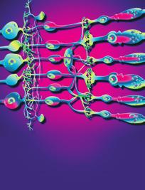
1 Case Reports in Surgery, Article ID , 4 pages Case Report Malignancy within a Tail Gut Cyst: A Case of Retrorectal Carcinoid Tumour A. A. Abukar, 1 B. J. Parcell, 2 C. B. Lim, 2 P. V. Patil, 2 A. Ramsanahie, 1 F. Carey, 3 R. J. C. Steele, 2 and M. A. Thaha 1 1 Academic Surgical Unit, Blizard Institute, National Centre for Bowel Research & Surgical Innovations, Queen Mary University of London and The Royal London Hospital, Barts Health NHS Trust, London E1 2AT, UK 2 Department of Surgery & Molecular Oncology, Ninewells Hospital & Medical School, University of Dundee, Dundee DD1 9SY, UK 3 Department of Molecular & Cellular Pathology, Ninewells Hospital & Medical School, University of Dundee, Dundee DD1 9SY, UK Correspondence should be addressed to M. A. Thaha; m.a.thaha@qmul.ac.uk Received 3 August 2014; Accepted 28 October 2014; Published 13 November 2014 Academic Editor: Akihiro Nakajo Copyright 2014 A. A. Abukar et al. This is an open access article distributed under the Creative Commons Attribution License, which permits unrestricted use, distribution, and reproduction in any medium, provided the original work is properly cited. Purpose. Tailgut cysts with malignant transformation are rare entities. We discuss the diagnostic strategy and treatment of a malignancy within a tailgut cyst. Methods. In this study we report on the case of a 61-year-old man with a malignant neuroendocrine tumour arising within a tailgut cyst and an overview of the literature emphasising the histopathological characteristics and differential diagnosis. Results. Our patient presented with lower back pain, rectal pain, and increased urgency of defecation. MRI scan and CT-guided biopsy on histological analysis revealed a diagnosis of carcinoid tumour of the presacral space. The patient subsequently underwent an abdominoperineal excision of the rectum. Conclusions. This case highlights the importance of tailgut cysts as a differential diagnosis of presacral masses. It is a rare congenital lesion developing from remnants of the embryonic postanal gut and is predominantly benign in nature. Approximately half of cases remain asymptomatic; therefore, diagnosis is often delayed. Magnetic resonance imaging is the investigation of choice and an awareness of the possibility of malignant potential is critical to avoiding missed diagnosis and subsequent morbidity. Complete surgical excision allows accurate diagnosis, confirmation of oncological clearance, and prevention of mortality. 1. Introduction Tailgut cysts are rare developmental cysts, which occur in the presacral region. They have also been described as retrorectal cysts, cyst of postanal intestine, retrorectal cystic hamartomas, simple cysts, mucus secreting cysts, and myoepithelial hamartoma of the rectum [1 3]. It is rare for carcinoid tumours to be present in the presacral region. If this occurs, it is usually due to direct or metastatic spread from a primary rectal tumour [4 6]. Approximately 23 cases of malignant transformation in tailgut cysts have been reported in the literature, of which eleven have been carcinoid tumours. They are mainly reported in middle-aged women, are usually asymptomatic, and are found during routine physical examination or at childbirth; therefore, diagnosis is often delayed. We describe a case in which a 61-year-old man presented with a threeweek history of dull aching pain in his lower back, rectal pain, and increased frequency of defecation. Following abdominoperineal excision of the rectum, the presence of a tailgut cyst was established. Arising within this was a low grade epithelial neoplasm with morphological and immunohistochemical evidence of neuroendocrine differentiation. 2. Case Report A61-year-oldmanpresentedwithahistoryofdullaching pain in his lower spine, rectal pain, and increased urgency of defecation lasting for three weeks. Abdominal examination and digital rectal examination were unremarkable. A barium enema showed extrinsic compression of the lower rectum


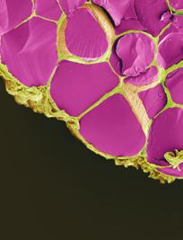
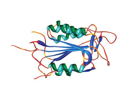
2 2 Case Reports in Surgery from a presumed low pelvic mass and two persistent filling defects within the descending colon. Colonoscopy showed a pedunculated polyp at 50 cm. Biopsies of this polyp confirmedittobeatubulovillousadenomawithmilddysplasia. The rectal mucosa was normal. CT-guided biopsy of the presacral mass initially suggested a prostate cancer. However, on review of the histology and scan, a carcinoid tumour of the presacral space was diagnosed. An MRI of the pelvis carried out showed a loculated mass cm, proximal to the lower rectum. The rectal mucosa was found to be intact whilsttheserosalsurfacewasinferiorlybreachedandin direct contact with the mass. The mass displaced the inferior aspect of levator ani laterally and there was evidence of direct extension into muscle on the left side. Staging CT scan did not show any evidence of distant metastases. The patient underwent abdominoperineal excision of the rectum. Microscopy of the CT-guided biopsies of the presacral mass showed fibrous tissue extensively infiltrated by an epithelial neoplasm. The tumour was moderately to poorly differentiated and had a focally cribriform growth pattern. Despite being described as moderately to poorly differentiated, the tumour had a low Ki-67 index with no necrosis or other features of poor prognosis. Immunohistochemical staining of the tumour cells strongly expressed prostatic acid phosphatase and showed weak patchy CD56 positivity. Furthermore, it revealed strong and diffuse positivity for neuroendocrine markers: synaptophysin and chromogranin. The tumour cells also coexpressed the cytokeratin MNF116 andshowedpatchyae1/3positivity. Macroscopically, the surgical resection specimen showed atumourmassmeasuring mm which was attached posteriorly to the rectum in the midline, close to the distal margin. The tumour surface had a grey-white appearance, and yellow discoloured areas were present (Figure 1). Distally, there were areas of cystic changes within the tumour mass. The tumour involved the muscle of the anal canal but not the mucosa. Microscopic sections of the tumour revealed ahomogenousneoplasmthatwasmadeupofauniform population of round cells that had slightly granular nuclei (Figure 2). The cells were arranged in trabeculae and islands with a prominent vascular connective tissue stroma. In some areas, the tumour intermingled with smooth muscle bundles from around the anal canal. The morphological features suggested a neuroendocrine (carcinoid) tumour (Figure 3). This was confirmed by immunohistochemistry which showed that neoplastic cells expressed both chromogranin and synaptophysin. The cysts described inferiorly were lined by columnar mucinous epithelium, which strongly expressed cytokeratins 7 and 19 but was negative for cytokeratin 20. There was a subpopulation of neuroendocrine cells within the cyst lining showing expression of chromogranin and synaptophysin and thus confirming the diagnosis of a carcinoid tumour arising from a retrorectal cystic hamartoma (tailgut cyst). There was no cytological evidence of high grade malignancy; however, the tumour did reach circumferential margin at many points. Proximal lymph nodes from the mesorectum showed no evidence of malignancy. A NM MIBG scan also ruled out distant metastasis. Figure1:Macroscopicappearanceoftumourwithinthetailgutcyst. Figure 2: Characteristic microscopic appearance of carcinoid tumour. 3. Discussion Tailgut cysts are rare congenital retrorectal lesions. They are thought to develop from the remnant of the tailgut (postanal gut), a primitive gut which is temporarily present at the caudal portion of the embryo [1]. During days 28 35, the embryo possessesatruetailandthisislargestonthe35thdayof gestation [1, 3]. On the 56th day of gestation, the anus develops above this tail, and by the 8th week the tailgut regresses [1]. It is believed that if this tailgut persists, tailgut cysts may occur [1, 3]. The retrorectal (presacral) space is bounded anteriorly by the rectum and posteriorly by the sacrum [1, 3]. Differential diagnoses of masses in this area include teratomas, dermoid cyst, anal gland cyst, anterior sacral meningocele, and duplication cyst (enterogenous cyst) [1]. Tailgut cysts are found in adults and children but are predominantly reported in middle aged women with a female to male ratio of 3: 1[1]. Approximately 50% of patients with tailgut cysts are asymptomatic and are only identified by routine physical examination or at childbirth, and diagnosis is therefore often delayed [1]. Symptoms may be due to mass effects causing abdominal pain, rectal bleeding, tenesmus, and urinary frequency [1, 6, 7]. Tailgut cysts sometimes result in complications such as bleeding, infection, and rarely
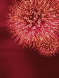
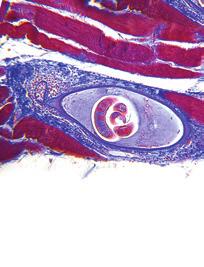
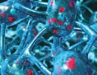
![Case Reports in Surgery 3 Figure 3: Immunohistochemical staining (columnar lined cells expressing both chromogranin and synaptophysin). malignant degeneration [8, 9].](/docs-images/96/128538002/images/3-0.jpg "Infection may be misdiagnosed as a pilonidal cyst, recurrent retrorectal abscess, or anorectal fistula [1]. Magnetic resonance imaging is the investigation of choice to image tailgut cysts [10].")
3 Case Reports in Surgery 3 Figure 3: Immunohistochemical staining (columnar lined cells expressing both chromogranin and synaptophysin). malignant degeneration [8, 9]. Infection may be misdiagnosed as a pilonidal cyst, recurrent retrorectal abscess, or anorectal fistula [1]. Magnetic resonance imaging is the investigation of choice to image tailgut cysts [10]. Tailgut cysts can result in morbidity and mortality if the mass is not suspected and surgery is not carried out [1]. Tailgut cysts are treated optimally with complete surgical excision, which allows accurate diagnosis and treatment [1]. On macroscopic examination of excised specimens tailgut cysts are typically well circumscribed [1]. These cysts may be multicystic or multiloculated masses with adherent surrounding fibroadipose tissue [1, 8]. The cysts may contain translucent mucoid fluid or clear serous fluid and may be lined by a wide range of epithelia such as stratified squamous type, stratified columnar type, transitional type, cuboidal type, mucinous or ciliated pseudostratified columnar, and gastric type [1, 2, 7, 11]. The majority of cysts contain focal well-formed disorganised bundles of bland smooth muscle cell in their walls [1]. They may develop carcinoid tumours, adenocarcinoma, and germ cell tumours [1]. Carcinoid tumours are thought to develop from neuroendocrine cells present in the glandular epithelium of the tailgut cyst (these cells were prominent in the residual cyst in our case) and can be classified according to their architectural arrangement suchastrabecular,glandular,undifferentiated,insular,and mixed [1]. The main differential diagnosis is teratoma and duplication cyst. Our patient had a cyst which did not contain any skin adnexal structures, or mesenchymal tissues, for instance bone and cartilage, excluding the possibility that the cyst was a teratoma. A distinctive feature of duplication cysts is a two-layer smooth muscle bundle wall containing a nerve plexus [11]; this was not identified in our patient. Enteric cysts often show epithelial expression of cytokeratin 20 (absent in our patient). Diagnosis based on biopsy can be difficult as biopsy specimens commonly contain inflamed fibrous tissue with no epithelia or only one type of epithelium, making it difficult to determine whether the mass is a tailgut cyst or another type of developmental cyst [1].Malignant foci may beveryfocalandthesampleshouldbeexaminedcarefully. Additionally, malignant cells can spill into the peritoneal cavity during biopsy [6]. In this case, the morphological features of this tumour initially suggested a possible diagnosis of prostate carcinoma. Carcinoid tumours may have a similar histological appearance to prostate cancer and rectal carcinoids may express the marker prostatic acid phosphatase [12, 13]. However, our patient had no clinical evidence of a prostatic mass and PSA was not elevated. In addition, on radiological investigation, the patient s presacral mass also appeared to be separate from the prostate. This case highlights the importance of tailgut cysts as a differential diagnosis of presacral masses as malignant transformation can occur and result in mortality if surgery is not carried out [1, 6]. Diagnosing tailgut cysts is difficult as their existence is not well recognised, clinical examination may be difficult, and symptoms may mimic other more commonly occurring lesions such as perianal fistulas and abscesses. Consent The patient concerned gave his informed consent regarding inclusion in this case report. Conflict of Interests The authors declare that they have no conflict of interests. Authors Contribution All authors contributed to the paper (writing/critical comments/revisions). References [1] A. R. Prasad, M. B. Amin, T. L. Randolph, C. S. Lee, and C. K. Ma, Retrorectal cystic hamartoma: report of 5 cases with malignancy arising in 2, Archives of Pathology and Laboratory Medicine,vol.124,no.5,pp ,2000. [2] B.M.HjermstadandE.B.Helwig, Tailgutcysts.Reportof53 cases, American Clinical Pathology,vol.89,no.2,pp , [3] V. Sriganeshan and J. B. Alexis, A 37-year-old woman with a presacral mass. Tailgut cyst (retrorectal cystic hamartoma), Archives of Pathology & Laboratory medicine,vol.130,no.5,pp. e77 e78, [4] M. G. Horenstein, R. A. Erlandson, D. M. Gonzalez-Cueto, and J. Rosai, Presacral carcinoid tumors: report of three cases and review of the literature, The American Surgical Pathology,vol.22,no.2,pp ,1998. [5] B. H. Federspiel, A. P. Burke, L. H. Sobin, and K. M. Shekitka, Rectal and colonic carcinoids. A clinicopathologic study of 84 cases, Cancer,vol.65,no.1,pp ,1990. [6] D.E.Song,J.K.Park,B.Hur,andJ.Y.Ro, Carcinoidtumor arising in a tailgut cyst of the anorectal junction with distant metastasis: a case report and review of the literature, Archives of Pathology and Laboratory Medicine, vol.128,no.5,pp , [7] N. Mourra, S. Caplin, R. Parc, and J.-F. Flejou, Presacral neuroendocrine carcinoma developed in a tailgut cyst: report of a case, Diseases of the Colon and Rectum, vol.46,no.3,pp , 2003.
4 4 Case Reports in Surgery [8] H.Dahan,L.Arrivé, D. Wendum, H. D. Le Pointe, H. Djouhri, and J.-M. Tubiana, Retrorectal developmental cysts in adults: clinical and radiologic-histopathologic review, differential diagnosis, and treatment, Radiographics, vol. 21, no. 3, pp , [9] W. L. Campbell and M. Wolff, Retrorectal cysts of developmental origin, The American Roentgenology Radium Therapy and Nuclear Medicine,vol.117,no.2,pp ,1973. [10] A. Mathieu, R. Chamlou, F. Le Moine, C. Maris, J. Van de Stadt, and I. Salmon, Tailgut cyst associated with a carcinoid tumor: case report and review of the literature, Histology and Histopathology,vol.20,no.4,pp ,2005. [11] S.-H. Jang, K.-S. Jang, Y.-S. Song et al., Unusual prerectal location of a tailgut cyst: a case report, World Gastroenterology,vol.12,no.31,pp ,2006. [12] E. D. Davidson and W. S. McDougal, Elevated serum acid phosphatase levels with rectal carcinoid tumor, Gastroenterology, vol. 70, no. 1, pp , [13] L. H. Sobin, B. M. Hjermstad, I. A. Sesterhenn, and E. B. Helwig, Prostatic acid phosphatase activity in carcinoid tumors, Cancer, vol. 58, no. 1, pp , 1986.


5 MEDIATORS of INFLAMMATION The Scientific World Journal Gastroenterology Research and Practice Diabetes Research International Endocrinology Immunology Research Disease Markers Submit your manuscripts at BioMed Research International PPAR Research Obesity Ophthalmology Evidence-Based Complementary and Alternative Medicine Stem Cells International Oncology Parkinson s Disease Computational and Mathematical Methods in Medicine AIDS Behavioural Neurology Research and Treatment Oxidative Medicine and Cellular Longevity
Case Report Carcinoid Tumors Arising in Tailgut Cysts May Be Associated with Estrogen Receptor Status: Case Report and Review of the Literature
 www.ijcep.com/ijcep709009 Case Report Carcinoid Tumors Arising in Tailgut Cysts May Be Associated with Estrogen Receptor Status: Case Report and Review of the Literature John J. Liang 1, Sadir Alrawi 2,
Retrorectal cystic hamartomas are uncommon lesions
 Retrorectal Cystic Hamartoma Report of 5 Cases With Malignancy Arising in 2 Anil R. Prasad, MD; Mahul B. Amin, MD; Todd L. Randolph, MD; Chong S. Lee, MD; Chan K. Ma, MD Background. Retrorectal cystic
Case Report A Rare Cutaneous Adnexal Tumor: Malignant Proliferating Trichilemmal Tumor
 Case Reports in Medicine Volume 2015, Article ID 742920, 4 pages http://dx.doi.org/10.1155/2015/742920 Case Report A Rare Cutaneous Adnexal Tumor: Malignant Proliferating Trichilemmal Tumor Omer Alici,
Case history: Figure 1. H&E, 5x. Figure 2. H&E, 20x.
 1 Case history: A 49 year-old female presented with a 5 year history of chronic anal fissure. The patient s past medical history is otherwise unremarkable. On digital rectal examination there was a very
Retrorectal tumors: report of two cases
 Journal of College of Medical Sciences-Nepal, 2012, Vol-8, No-4, 46-50 Case Report Retrorectal tumors: report of two cases P Kafle, 1 Praveen CR, 2 S Kumar, 3 BN Patowary, 4 N Maharjan, 1 S Shah, 1 S Agrawal
Case Report Five-Year Survival after Surgery for Invasive Micropapillary Carcinoma of the Stomach
 Case Reports in Surgery Volume 2013, Article ID 560712, 4 pages http://dx.doi.org/10.1155/2013/560712 Case Report Five-Year Survival after Surgery for Invasive Micropapillary Carcinoma of the Stomach Shigeo
Kidney Case 1 SURGICAL PATHOLOGY REPORT
 Kidney Case 1 Surgical Pathology Report February 9, 2007 Clinical History: This 45 year old woman was found to have a left renal mass. CT urography with reconstruction revealed a 2 cm medial mass which
Case Report Müllerian Remnant Cyst as a Cause of Acute Abdomen in a Female Patient with Müllerian Agenesis: Radiologic and Pathologic Findings
 Volume 2016, Article ID 6581387, 4 pages http://dx.doi.org/10.1155/2016/6581387 Case Report üllerian Remnant Cyst as a Cause of Acute Abdomen in a Female Patient with üllerian Agenesis: Radiologic and
ailgut cyst or retrorectal cystic
 bdominal Imaging Yang et al. MRI of Tailgut Cyst Dal Mo Yang 1 Chul Hi Park 1 Wook Jin 1 Suk Ki Chang 1 Jee Eun Kim 1 Soo Jin Choi 1 Dong Hae Jung 2 Yang DM, Park CH, Jin W, et al. Received June 5, 2004;
Case Report Intracranial Capillary Hemangioma in the Posterior Fossa of an Adult Male
 Case Reports in Radiology Volume 2016, Article ID 6434623, 4 pages http://dx.doi.org/10.1155/2016/6434623 Case Report Intracranial Capillary Hemangioma in the Posterior Fossa of an Adult Male Jordan Nepute,
COLORECTAL CARCINOMA
 QUICK REFERENCE FOR HEALTHCARE PROVIDERS MANAGEMENT OF COLORECTAL CARCINOMA Ministry of Health Malaysia Malaysian Society of Colorectal Surgeons Malaysian Society of Gastroenterology & Hepatology Malaysian
Case Report Features of the Atrophic Corpus Mucosa in Three Cases of Autoimmune Gastritis Revealed by Magnifying Endoscopy
 Volume 2012, Article ID 368160, 4 pages doi:10.1155/2012/368160 Case Report Features of the Atrophic Corpus Mucosa in Three Cases of Autoimmune Gastritis Revealed by Magnifying Endoscopy Kazuyoshi Yagi,
Note: The cause of testicular neoplasms remains unknown
 - In the 15- to 34-year-old age group, they are the most common tumors of men. - Tumors of the testis are a heterogeneous group of neoplasms that include: I. Germ cell tumors : 95%; all are malignant.
American Journals of Cancer Case Reports. A Rare Case of Rectal Metastasis from Sarcomatoid Variant of Urothelial Carcinoma: A Case Report
 American Journals of Cancer Case Reports Lin JYJ et al. American Journals of Cancer Case Reports 2014, 3:1-5 http://ivyunion.org/index.php/ajccr Page 1 of 5 Vol 3 Article ID 20140539, 5 pages Case Report
Tailgut cyst associated with a carcinoid tumor: case report and review of the literature
 Histol Histopathol (2005) 20: 1065-1069 http://www.hh.um.es Histology and Histopathology Cellular and Molecular Biology Tailgut cyst associated with a carcinoid tumor: case report and review of the literature
Management of an Appendiceal Mass - Approach to acute presentation of appendiceal neoplasms
 Management of an Appendiceal Mass - Approach to acute presentation of appendiceal neoplasms Dr. Claudia LY WONG, Department of Surgery, Kwong Wah Hospital Joint Hospital Surgical Grand Round Presentation,
Case Report A Case of Cystic Basal Cell Carcinoma Which Shows a Homogenous Blue/Black Area under Dermatoscopy
 Volume 20, Article ID 450472, 4 pages doi:0.55/20/450472 Case Report A Case of Cystic Basal Cell Carcinoma Which Shows a Homogenous Blue/Black Area under Dermatoscopy Akihiro Yoneta, Kohei Horimoto, Keiko
8. The polyp in the illustration can be described as (circle all that apply) a. Exophytic b. Pedunculated c. Sessile d. Frank
 a. Exophytic b. Pedunculated c. Sessile d. Frank") Quiz 1 Overview 1. Beginning with the cecum, which is the correct sequence of colon subsites? a. Cecum, ascending, splenic flexure, transverse, hepatic flexure, descending, sigmoid. b. Cecum, ascending,
Quiz 1 Overview 1. Beginning with the cecum, which is the correct sequence of colon subsites? a. Cecum, ascending, splenic flexure, transverse, hepatic flexure, descending, sigmoid. b. Cecum, ascending,
Brief History. Identification : Past History : HTN without regular treatment.
 Brief History Identification : Name : 陳 x - Admission : 94/10/06 Gender : male Age : 75 y/o Chief Complaint : Urinary difficulty for months. Past History : HTN without regular treatment. Brief History
R. F. Falkenstern-Ge, 1 S. Bode-Erdmann, 2 G. Ott, 2 M. Wohlleber, 1 and M. Kohlhäufl Introduction. 2. Histology
 Case Reports in Oncological Medicine Volume 2013, Article ID 167585, 4 pages http://dx.doi.org/10.1155/2013/167585 Case Report Late Lung Metastasis of a Primary Eccrine Sweat Gland Carcinoma 10 Years after
Case Report Ovarian Metastasis from Lung Cancer: A Rare Entity
 Case Reports in Obstetrics and Gynecology Volume 2013, Article ID 378438, 4 pages http://dx.doi.org/10.1155/2013/378438 Case Report Ovarian Metastasis from Lung Cancer: A Rare Entity Huseyin Cengiz, Fükrü
Case Report A Case of Primary Submandibular Gland Oncocytic Carcinoma
 Case Reports in Otolaryngology Volume 2013, Article ID 384238, 4 pages http://dx.doi.org/10.1155/2013/384238 Case Report A Case of Primary Submandibular Gland Oncocytic Carcinoma Kunihiko Tokashiki, Kiyoaki
ONCOLOGY. Csaba Bödör. Department of Pathology and Experimental Cancer Research november 19., ÁOK, III.
 ONCOLOGY Csaba Bödör Department of Pathology and Experimental Cancer Research 2018. november 19., ÁOK, III. bodor.csaba1@med.semmelweis-univ.hu ONCOLOGY Characteristics of Benign and Malignant Neoplasms
Case Report Tumor-to-Tumor Metastasis: Lung Carcinoma Metastasizing to Thyroid Neoplasms
 Case Reports in Pathology Volume 2015, Article ID 153932, 5 pages http://dx.doi.org/10.1155/2015/153932 Case Report Tumor-to-Tumor Metastasis: Lung Carcinoma Metastasizing to Thyroid Neoplasms Shiuan-Li
performed to help sway the clinician in what the appropriate diagnosis is, which can substantially alter the treatment of management.
 Hello, I am Maura Polansky at the University of Texas MD Anderson Cancer Center. I am a Physician Assistant in the Department of Gastrointestinal Medical Oncology and the Program Director for Physician
Case Report Perforation of an Occult Carcinoma of the Prostate as a Rare Differential Diagnosis of Subcutaneous Emphysema of the Leg
 Case Reports in Orthopedics Volume 2016, Article ID 5430637, 5 pages http://dx.doi.org/10.1155/2016/5430637 Case Report Perforation of an Occult Carcinoma of the Prostate as a Rare Differential Diagnosis
Case Report Pulmonary Hilar Tumor: An Unusual Presentation of Sclerosing Hemangioma
 Volume 2016, Article ID 8919012, 4 pages http://dx.doi.org/10.1155/2016/8919012 Case Report Pulmonary Hilar Tumor: An Unusual Presentation of Sclerosing Hemangioma Jui-Hung Hung, Ching Hsueh, Chiung-Ying
Case Report Isolated Enteric Cyst in the Neck
 Case Reports in Otolaryngology, Article ID 597813, 4 pages http://dx.doi.org/10.1155/2014/597813 Case Report Isolated Enteric Cyst in the Neck Amit Mahore, 1 Raghvendra Ramdasi, 1 Palak Popat, 2 Shilpa
R. J. L. F. Loffeld, 1 P. E. P. Dekkers, 2 and M. Flens Introduction
 ISRN Gastroenterology Volume 213, Article ID 87138, 5 pages http://dx.doi.org/1.1155/213/87138 Research Article The Incidence of Colorectal Cancer Is Decreasing in the Older Age Cohorts in the Zaanstreek
[A RESEARCH COORDINATOR S GUIDE]
![[A RESEARCH COORDINATOR S GUIDE]](/thumbs/88/117127924.jpg "[A RESEARCH COORDINATOR S GUIDE]") 2013 COLORECTAL SURGERY GROUP Dr. Carl J. Brown Dr. Ahmer A. Karimuddin Dr. P. Terry Phang Dr. Manoj J. Raval Authored by Jennifer Lee A cartoon about colonoscopies. 1 [A RESEARCH COORDINATOR S GUIDE]
2013 COLORECTAL SURGERY GROUP Dr. Carl J. Brown Dr. Ahmer A. Karimuddin Dr. P. Terry Phang Dr. Manoj J. Raval Authored by Jennifer Lee A cartoon about colonoscopies. 1 [A RESEARCH COORDINATOR S GUIDE]
In-situ and invasive carcinoma of the colon in patients with ulcerative colitis
 Gut, 1972, 13, 566-570 In-situ and invasive carcinoma of the colon in patients with ulcerative colitis D. J. EVANS AND D. J. POLLOCK From the Departments of Pathology, Royal Postgraduate Medical School
Case Report Overlap of Acute Cholecystitis with Gallstones and Squamous Cell Carcinoma of the Gallbladder in an Elderly Patient
 Case Reports in Surgery Volume 2015, Article ID 767196, 4 pages http://dx.doi.org/10.1155/2015/767196 Case Report Overlap of Acute Cholecystitis with Gallstones and Squamous Cell Carcinoma of the Gallbladder
-The cause of testicular neoplasms remains unknown
 - In the 15- to 34-year-old age group, they are the most common tumors of men. - include: I. Germ cell tumors : (95%); all are malignant. II. Sex cord-stromal tumors: from Sertoli or Leydig cells; usually
Case Report Denosumab Chemotherapy for Recurrent Giant-Cell Tumor of Bone: A Case Report of Neoadjuvant Use Enabling Complete Surgical Resection
 Case Reports in Oncological Medicine Volume 2013, Article ID 496351, 4 pages http://dx.doi.org/10.1155/2013/496351 Case Report Denosumab Chemotherapy for Recurrent Giant-Cell Tumor of Bone: A Case Report
Papillary Lesions of the Breast A Practical Approach to Diagnosis. (Arch Pathol Lab Med. 2016;140: ; doi: /arpa.
 Papillary Lesions of the Breast A Practical Approach to Diagnosis (Arch Pathol Lab Med. 2016;140:1052 1059; doi: 10.5858/arpa.2016-0219-RA) Papillary lesions of the breast Span the spectrum of benign,
Disorders of Cell Growth & Neoplasia. Histopathology Lab
 Disorders of Cell Growth & Neoplasia Histopathology Lab Paul Hanna April 2010 Case #84 Clinical History: 5 yr-old, West Highland White terrier. skin mass from axillary region. has been present for the
Surgical management of a retro-rectal cystic hamartoma (tailgut cyst) using a trans-rectal approach: a case report and review of the literature
 using a trans-rectal approach: a case report and review of the literature") Kildušis and Samalavičius Journal of Medical Case Reports 2014, 8:11 JOURNAL OF MEDICAL CASE REPORTS CASE REPORT Surgical management of a retro-rectal cystic hamartoma (tailgut cyst) using a trans-rectal
Kildušis and Samalavičius Journal of Medical Case Reports 2014, 8:11 JOURNAL OF MEDICAL CASE REPORTS CASE REPORT Surgical management of a retro-rectal cystic hamartoma (tailgut cyst) using a trans-rectal
Mandana Moosavi 1 and Stuart Kreisman Background
 Case Reports in Endocrinology Volume 2016, Article ID 6471081, 4 pages http://dx.doi.org/10.1155/2016/6471081 Case Report A Case Report of Dramatically Increased Thyroglobulin after Lymph Node Biopsy in
Case Report PET/CT Imaging in Oncology: Exceptions That Prove the Rule
 Case Reports in Oncological Medicine Volume 2013, Article ID 865032, 4 pages http://dx.doi.org/10.1155/2013/865032 Case Report PET/CT Imaging in Oncology: Exceptions That Prove the Rule M. Casali, 1 A.
Case Report Multiple Giant Cell Tumors of Tendon Sheath Found within a Single Digit of a 9-Year-Old
 Case Reports in Orthopedics Volume 2016, Article ID 1834740, 4 pages http://dx.doi.org/10.1155/2016/1834740 Case Report Multiple Giant Cell Tumors of Tendon Sheath Found within a Single Digit of a 9-Year-Old
Case Report Cytomegalovirus Colitis with Common Variable Immunodeficiency and Crohn s Disease
 Volume 2015, Article ID 348204, 4 pages http://dx.doi.org/10.1155/2015/348204 Case Report Cytomegalovirus Colitis with Common Variable Immunodeficiency and Crohn s Disease Betül Ünal, Cumhur Ebrahim BaGsorgun,
Endoscopic Ultrasonography Assessment for Ampullary and Bile Duct Malignancy
 Diagnostic and Therapeutic Endoscopy, Vol. 3, pp. 35-40 Reprints available directly from the publisher Photocopying permitted by license only (C) 1996 OPA (Overseas Publishers Association) Amsterdam B.V.
Case Report Renal Cell Carcinoma Metastatic to Thyroid Gland, Presenting Like Anaplastic Carcinoma of Thyroid
 Case Reports in Urology Volume 2013, Article ID 651081, 4 pages http://dx.doi.org/10.1155/2013/651081 Case Report Renal Cell Carcinoma Metastatic to Thyroid Gland, Presenting Like Anaplastic Carcinoma
Case Report Synchronous Bilateral Solid Papillary Carcinomas of the Breast
 Case Reports in Surgery Volume 2013, Article ID 812129, 4 pages http://dx.doi.org/10.1155/2013/812129 Case Report Synchronous Bilateral Solid Papillary Carcinomas of the Breast Noriko Yoshimura, 1 Shigeru
Case Report Two Cases of Small Cell Cancer of the Maxillary Sinus Treated with Cisplatin plus Irinotecan and Radiotherapy
 Case Reports in Otolaryngology Volume 2013, Article ID 893638, 4 pages http://dx.doi.org/10.1155/2013/893638 Case Report Two Cases of Small Cell Cancer of the Maxillary Sinus Treated with Cisplatin plus
Salivary Glands 3/7/2017
 Salivary Glands 3/7/2017 Goals and objectives Focus on the entities unique to H&N Common board type facts Information for your future practice Salivary Glands Salivary Glands Major gland. Paratid. Submandibular.
University Journal of Pre and Para Clinical Sciences
 ISSN 2455 2879 Volume 2 Issue 1 2016 Metaplastic carcinoma breast a rare case report Abstract : Metaplastic carcinoma of the breast is a rare malignancy with two distinct cell lines described as a breast
3/27/2017. Pulmonary Pathology Specialty Conference. Disclosure of Relevant Financial Relationships. Clinical History:
 Pulmonary Pathology Specialty Conference Saul Suster, M.D. Medical College of Wisconsin Disclosure of Relevant Financial Relationships USCAP requires that all planners (Education Committee) in a position
Research Article Stromal Expression of CD10 in Invasive Breast Carcinoma and Its Correlation with ER, PR, HER2-neu, and Ki67
 SAGE-Hindawi Access to Research International Breast Cancer Volume 20, Article ID 47957, 4 pages doi:0.406/20/47957 Research Article Stromal Expression of CD0 in Invasive Breast Carcinoma and Its Correlation
ISPUB.COM. Anal gland adenocarcinoma. A Alhumidi, M Dababo, M Hamodat INTRODUCTION CASE REPORT
 ISPUB.COM The Internet Journal of Pathology Volume 9 Number 2 A Alhumidi, M Dababo, M Hamodat Citation A Alhumidi, M Dababo, M Hamodat.. The Internet Journal of Pathology. 2008 Volume 9 Number 2. Abstract
Cancers of unknown primary : Knowing the unknown. Prof. Ahmed Hossain Professor of Medicine SSMC
 Cancers of unknown primary : Knowing the unknown Prof. Ahmed Hossain Professor of Medicine SSMC Definition Cancers of unknown primary site (CUPs) Represent a heterogeneous group of metastatic tumours,
S1.04 Principal clinician. G1.01 Comments. G2.01 *Specimen dimensions (prostate) S2.02 *Seminal vesicles
 S2.02 *Seminal vesicles") Prostate Cancer Histopathology Reporting Proforma (Radical Prostatectomy) Includes the International Collaboration on Cancer reporting dataset denoted by * Family name Given name(s) Date of birth Sex Male
Prostate Cancer Histopathology Reporting Proforma (Radical Prostatectomy) Includes the International Collaboration on Cancer reporting dataset denoted by * Family name Given name(s) Date of birth Sex Male
IMAGING GUIDELINES - COLORECTAL CANCER
 IMAGING GUIDELINES - COLORECTAL CANCER DIAGNOSIS The majority of colorectal cancers are diagnosed on colonoscopy, with some being diagnosed on Ba enema, ultrasound or CT. STAGING CT chest, abdomen and
Case Report Metastatic Malignant Melanoma of Parotid Gland with a Regressed Primary Tumor
 Case Reports in Otolaryngology Volume 2016, Article ID 5393404, 4 pages http://dx.doi.org/10.1155/2016/5393404 Case Report Metastatic Malignant Melanoma of Parotid Gland with a Regressed Primary Tumor
Colonic Polyp. Najmeh Aletaha. MD
 Colonic Polyp Najmeh Aletaha. MD 1 Polyps & classification 2 Colorectal cancer risk factors 3 Pathogenesis 4 Surveillance polyp of the colon refers to a protuberance into the lumen above the surrounding
Colon and Rectum: 2018 Solid Tumor Rules
 2018 SEER Solid Tumor Manual 2018 KCR SPRING TRAINING Colon and Rectum: 2018 Solid Tumor Rules 1 Colon and Rectum Solid Tumor Rules Separate sections for: Introduction Changes from 2007 MP/H rules Equivalent
Research Article Predictions of the Length of Lumbar Puncture Needles
 Computational and Mathematical Methods in Medicine, Article ID 732694, 5 pages http://dx.doi.org/10.1155/2014/732694 Research Article Predictions of the Length of Lumbar Puncture Needles Hon-Ping Ma, 1,2
DATA STANDARDS AND QUALITY CONTROL MEMORANDUM DSQC #
 DATA STANDARDS AND QUALITY CONTROL MEMORANDUM DSQC #2006-01 CATEGORY: CLARIFICATION SUBJECT: RESCINDMENT - DSQC MEMORANDUM 2002-08 Coding Complex Morphologic Diagnoses (revised 8/02) EFFECTIVE: For Cases
GOBLET CELL CARCINOID. Hanlin L. Wang, MD, PhD University of California Los Angeles
 GOBLET CELL CARCINOID Hanlin L. Wang, MD, PhD University of California Los Angeles Disclosure of Relevant Financial Relationships USCAP requires that all planners (Education Committee) in a position to
GOBLET CELL CARCINOID
 GOBLET CELL CARCINOID Hanlin L. Wang, MD, PhD University of California Los Angeles Disclosure of Relevant Financial Relationships USCAP requires that all planners (Education Committee) in a position to
04/10/2018. Intraductal Papillary Neoplasms Of Breast INTRADUCTAL PAPILLOMA
 Intraductal Papillary Neoplasms Of Breast Savitri Krishnamurthy MD Professor of Pathology Deputy Division Head The University of Texas MD Anderson Cancer Center 25 th Annual Seminar in Pathology Pittsburgh,
Radiology Pathology Conference
 Radiology Pathology Conference Nadia F. Yusaf, M.D. PGY-3 1/29/2010 Presentation material is for education purposes only. All rights reserved. 2010 URMC Radiology Page 1 of 90 Case 1 60 year- old man presents
Select problems in cystic pancreatic lesions
 Disclosure Select problems in cystic pancreatic lesions Five Prime Therapeutics shareholder Adicet Bio shareholder Bristol-Meyer Squibb advisory board grace.kim@ucsf.edu Pancreatic cystic lesions Intraductal
11/21/13 CEA: 1.7 WNL
 Case Scenario 1 A 70 year-old white male presented to his primary care physician with a recent history of rectal bleeding. He was referred for imaging and a colonoscopy and was found to have adenocarcinoma.
Bilateral Renal Angiomyolipomas with Invasion of the Renal Vein: A Case Report
 Case Study TheScientificWorldJOURNAL (2008) 8, 145 148 TSW Urology ISSN 1537-744X; DOI 10.1100/tsw.2008.29 Bilateral Renal Angiomyolipomas with Invasion of the Renal Vein: A Case Report C. Blick, N. Ravindranath,
Clinical Study Primary Malignant Tumours of Bone Following Previous Malignancy
 Sarcoma Volume 2008, Article ID 418697, 4 pages doi:10.1155/2008/418697 Clinical Study Primary Malignant Tumours of Bone Following Previous Malignancy J. T. Patton, S. M. M. Sommerville, and R. J. Grimer
Case Report Osteoclastic Giant Cell Rich Squamous Cell Carcinoma of the Uterine Cervix: A Case Report and Review of the Literature
 Case Reports in Pathology, Article ID 415328, 4 pages http://dx.doi.org/10.1155/2014/415328 Case Report Osteoclastic Giant Cell Rich Squamous Cell Carcinoma of the Uterine Cervix: A Case Report and Review
Neoplasia part I. Dr. Mohsen Dashti. Clinical Medicine & Pathology nd Lecture
 Neoplasia part I By Dr. Mohsen Dashti Clinical Medicine & Pathology 316 2 nd Lecture Lecture outline Review of structure & function. Basic definitions. Classification of neoplasms. Morphologic features.
Case Report Postoperative Megarectum in an Adult Patient with Imperforate Anus and Rectourethral Fistula
 Case Reports in Gastrointestinal Medicine Volume 2015, Article ID 613926, 4 pages http://dx.doi.org/10.1155/2015/613926 Case Report Postoperative Megarectum in an Adult Patient with Imperforate Anus and
Case Report Primary Neuroendocrine Carcinoma of Ocular Adnexa
 Volume 2013, Article ID 281351, 4 pages http://dx.doi.org/10.1155/2013/281351 Case Report Primary Neuroendocrine Carcinoma of Ocular Adnexa Daisuke Yamanouchi, 1 Toshiyuki Oshitari, 1 Yosuke Nakamura,
Case Report IgG4-Related Nasal Pseudotumor
 Case Reports in Otolaryngology Volume 2015, Article ID 749890, 4 pages http://dx.doi.org/10.1155/2015/749890 Case Report IgG4-Related Nasal Pseudotumor L. K. Døsen, 1 P. Jebsen, 2 B. Dingsør, 3 and R.
Normal endometrium: A, proliferative. B, secretory.
 Normal endometrium: A, proliferative. B, secretory. Nội mạc tử cung Nội mạc tử cung Cyclic changes in endometrium.. Approximate relationship of useful microscopic changes. Arias-Stella reaction in endometrial
Research Article A Clinicopathological Analysis of Soft Tissue Sarcoma with Telangiectatic Changes
 Sarcoma Volume 2015, Article ID 740571, 5 pages http://dx.doi.org/10.1155/2015/740571 Research Article A Clinicopathological Analysis of Soft Tissue Sarcoma with Telangiectatic Changes Hiroshi Kobayashi,
Bio & 241 A&P Unit 1 / Lecture 3
 Bio & 241 A&P Unit 1 / Lecture 3 Tissues All body tissues arise from three fundamental embryonic tissues. Endoderm: forms epithelial tissues lining internal organs such as the GI tract Mesoderm: connective
Cutaneous metastases. Thaddeus Mully. University of California, San Francisco Professor, Departments of Pathology and Dermatology
 Cutaneous metastases Thaddeus Mully University of California, San Francisco Professor, Departments of Pathology and Dermatology DISCLOSURE OF RELATIONSHIPS WITH INDUSTRY Thaddeus Mully Course C005 Essential
Case Report Complex Form Variant of Dysembryoplastic Neuroepithelial Tumor of the Cerebellum
 Case Reports in Pathology Volume 2012, Article ID 718651, 4 pages doi:10.1155/2012/718651 Case Report Complex Form Variant of Dysembryoplastic Neuroepithelial Tumor of the Cerebellum Jesús Vaquero, 1,
Gross appearance of peritoneal cysts. They have a thin, translucent wall and contain a clear fluid.
 Gross appearance of peritoneal cysts. They have a thin, translucent wall and contain a clear fluid. So-called multicystic benign mesothelioma. A, Gross appearance. So-called multicystic benign mesothelioma.
Other Sites. Table 2 Continued. MPH Rules 11/8/07. NAACCR Webinar Series 1
 MPH s 11/8/07 Other s 1 Table 2 Continued Use this two-page table to select combination histology codes. Compare the terms in the diagnosis to the terms in Columns 1 and 2. If the terms match, code the
Synonyms. Nephrogenic metaplasia Mesonephric adenoma
 Nephrogenic Adenoma Synonyms Nephrogenic metaplasia Mesonephric adenoma Definition Benign epithelial lesion of urinary tract with tubular, glandular, papillary growth pattern Most frequently in the urinary
Solitary Contralateral Adrenal Metastases after Nephrectomy for Renal Cell Carcinoma
 Original Report ISSN 1537-744X; DOI 10.1100/tsw.2004.39 Solitary Contralateral Adrenal after Nephrectomy for Renal Cell Carcinoma Nikolaos Antoniou, M.D. and Demetrios Karanastasis, M.D. General Hospital
Prenatal and Neonatal MRI of Sacrococcygeal Teratoma With Surgical Correlation
 Prenatal and Neonatal MRI of Sacrococcygeal Teratoma With Surgical Correlation Ali Mahmood, M.D., and Nadia F. Mahmood, M.D. Citation: Mahmood A, Mahmood NF. Prenatal and Neonatal MRI of Sacrococcygeal
Case Scenario 1: Thyroid
 Case Scenario 1: Thyroid History and Physical Patient is an otherwise healthy 80 year old female with the complaint of a neck mass first noticed two weeks ago. The mass has increased in size and is palpable.
Case Report Uncommon Mixed Type I and II Choledochal Cyst: An Indonesian Experience
 Case Reports in Surgery Volume 2013, Article ID 821032, 4 pages http://dx.doi.org/10.1155/2013/821032 Case Report Uncommon Mixed Type I and II Choledochal Cyst: An Indonesian Experience Fransisca J. Siahaya,
Principles of Surgical Oncology. Winnie Achilles Tierklinik Hollabrunn Lastenstrasse Hollabrunn
 Principles of Surgical Oncology Winnie Achilles Tierklinik Hollabrunn Lastenstrasse 2 2020 Hollabrunn boexi@gmx.de The first surgery provides the best chance for a cure in an animal with a tumor Clinical
Muco-epidermoid tumours of the anal canal
 J. clin. Path. (1963), 16, 200 Muco-epidermoid tumours of the anal canal B. C. MORSON AND H. VOLKSTADT From the Research Department, St. Mark's Hospital, London SYNOPSIS The pathology of 21 cases of muco-epidermoid
Mody. AIS vs. Invasive Adenocarcinoma of the Cervix
 Common Problems in Gynecologic Pathology Michael T. Deavers, M.D. Houston Methodist Hospital, Houston, Texas Common Problems in Gynecologic Pathology Adenocarcinoma in-situ (AIS) of the Cervix vs. Invasive
Neoplasia 2018 Lecture 2. Dr Heyam Awad MD, FRCPath
 Neoplasia 2018 Lecture 2 Dr Heyam Awad MD, FRCPath ILOS 1. List the differences between benign and malignant tumors. 2. Recognize the histological features of malignancy. 3. Define dysplasia and understand
Ectopic Prostatic Tissue in the Rectum: A Case Report 직장에발생된이소성전립선조직 : 증례보고
 Case Report pissn 1738-2637 / eissn 2288-2928 https://doi.org/10.3348/jksr.2017.76.2.91 Ectopic Prostatic Tissue in the Rectum: A Case Report 직장에발생된이소성전립선조직 : 증례보고 Myung Jin Seol, MD 1, Kyung Hee Noh,
Case Report Late Simultaneous Metastasis of Renal Cell Carcinoma to the Submandibular and Thyroid Glands Seven Years after Radical Nephrectomy
 International Otolaryngology Volume 2010, Article ID 698014, 4 pages doi:10.1155/2010/698014 Case Report Late Simultaneous Metastasis of Renal Cell Carcinoma to the Submandibular and Thyroid Glands Seven
Case 1307 Mesothelial cysts
 Case 1307 Mesothelial cysts Vinhais S, Monteiro M, Cunha TM INSTITUTO PORTUGUÊS DE ONCOLOGIA de Francisco Gentil de LISBOA Section: Gastro-Intestinal Imaging Published: 2001, Nov. 23 Patient: 44 year(s),
Surgical Management of Neuroendocrine Tumors of the Gut. Richard Hodin MD Professor of Surgery Massachusetts General Hospital Harvard Medical School
 Surgical Management of Neuroendocrine Tumors of the Gut Richard Hodin MD Professor of Surgery Massachusetts General Hospital Harvard Medical School Sites of GI Carcinoid Tumors Small intestine 44% Rectum
ANAL CANAL DUPLICATION (ACD) is the most
 is the most") Anal Canal Duplication in Infants By Soon-Ok Choi and Woo-Hyun Park Daegu, Korea Background/Purpose: Anal canal duplication (ACD) is the most distal and the least frequent digestive duplication. A review
Anal Canal Duplication in Infants By Soon-Ok Choi and Woo-Hyun Park Daegu, Korea Background/Purpose: Anal canal duplication (ACD) is the most distal and the least frequent digestive duplication. A review
COLON AND RECTUM SOLID TUMOR RULES ABSTRACTORS TRAINING
 COLON AND RECTUM SOLID TUMOR RULES ABSTRACTORS TRAINING COLON AND RECTUM SOLID TUMOR RULES Separate sections for: Introduction Changes from 2007 MP/H rules Equivalent Terms Terms that are NOT Equivalent
Neoplasia literally means "new growth.
 NEOPLASIA Neoplasia literally means "new growth. A neoplasm, defined as "an abnormal mass of tissue the growth of which exceeds and is uncoordinated with that of the normal tissues and persists in the
B. Cystic Teratoma: Refer to virtual microscope slide p_223 ovary, teratoma and compare to normal virtual microscope slide 086 ovary.
 LAB 2: THE CONNECTIVE TISSUE AND EPITHELIUM The focus of this week s lab will be pathology of connective tissue and epithelium. The lab will introduce you to the four basic tissue types: epithelium, connective
COLORECTAL CANCER FAISALGHANISIDDIQUI MBBS; FCPS; PGDIP-BIOETHICS; MCPS-HPE
 COLORECTAL CANCER FAISALGHANISIDDIQUI MBBS; FCPS; PGDIP-BIOETHICS; MCPS-HPE PROFESSOR OF SURGERY & DIRECTOR, PROFESSIONAL DEVELOPMENT CENTRE J I N N A H S I N D H M E D I C A L U N I V E R S I T Y faisal.siddiqui@jsmu.edu.pk
Images In Gastroenterology
 Images In Gastroenterology Thong-Ngam D, et al. THAI J GASTROENTEROL 2005 Vol. 6 No. 2 May - Aug. 2005 105 Imaging of Gastrointestinal Stromal Tumors Pornpim Fuangtharnthip, M.D. Narumol Hargroove, M.D.
S1.04 PRINCIPAL CLINICIAN G1.01 COMMENTS S2.01 SPECIMEN LABELLED AS G2.01 *SPECIMEN DIMENSIONS (PROSTATE) S2.03 *SEMINAL VESICLES
 S2.03 *SEMINAL VESICLES") Prostate Cancer Histopathology Reporting Proforma (Radical Prostatectomy) Includes the International Collaboration on Cancer reporting dataset denoted by * Family name Given name(s) Date of birth Indigenous
Prostate Cancer Histopathology Reporting Proforma (Radical Prostatectomy) Includes the International Collaboration on Cancer reporting dataset denoted by * Family name Given name(s) Date of birth Indigenous
Case Report Unicystic Ameloblastoma with Mural Proliferation Managed by Conservative Treatment
 Case Reports in Pathology Volume 2016, Article ID 3089540, 4 pages http://dx.doi.org/10.1155/2016/3089540 Case Report Unicystic Ameloblastoma with Mural Proliferation Managed by Conservative Treatment
EDUCATIONAL CASES E1 & E2. Natasha Inglis 20/03/15
 EDUCATIONAL CASES E1 & E2 Natasha Inglis 20/03/15 CASE E1 79 year old female Rectum. Altemeier operation Histology Superficial erosions and mucosal congestion volcano lesion and pseudomembrane formation