|
|
|
- Brandon Harrison
- 5 years ago
- Views:
Transcription
1
2
3
4
5
6
7
8
9
10
11
12
13
14
15
16 Intracanalicular (0 mm) 10 mm 20 mm 30 mm


17 Translabyrinthine approach Retrosigmoid approach Middle Fossa Approach
18 Otology & Neurotology 24: , Otology & Neurotology, Inc. The Effect of Age on Acoustic Neuroma Surgery Outcomes John S. Oghalai, James L. Buxbaum, Lawrence H. Pitts, and Robert K. Jackler Departments of Otolaryngology Head and Neck Surgery and Neurosurgery, University of California San Francisco. San Francisco, California, U.S.A. Objectives: To ascertain the effect of age on hearing preservation, facial nerve outcome, and complication rates after acoustic neuroma surgery. Study Design: Retrospective chart review. Two study arms were used: a comparison of the authors oldest patients with their youngest patients (extremes of age arm) and an analysis of all middle fossa surgical procedures (middle fossa arm). Setting: Tertiary referral center Patients: Total of 329 patients. For the extremes of age arm, 205 patients were studied in two cohorts with 150 older patients (>60 years) compared with 55 younger patients (<40 years). The approaches included 21 middle fossa (MF), 38 retrosigmoid (RS), and 91 translabyrinthine (TL) procedures in the older group versus 25 MF, 17 RS, and 13 TL in the younger. For the middle fossa arm, there were 170 patients (age range years) who underwent the MF approach for an attempt at hearing preservation. Main Outcome Measures: Hearing preservation was defined as the maintenance of either class A or class B hearing (AAO- HNS class). Good facial nerve outcome was considered the maintenance of either grade 1 or 2 (House-Brackmann scale). Cerebrospinal fluid leak rates and other postoperative complications were also tabulated. Results: After adjustment for tumor size and surgical approach using multiple logistic regression analysis, the extremes of age study arm demonstrated that there is a lower chance of preserving good hearing in older patients (p 0.048, odds ratio 0.30). Age was not associated with a difference in the rate of good facial nerve outcome (p 0.2). There was a trend toward slightly higher rates of cerebrospinal fluid leak in the older patient group (p 0.07) but no difference in the rate of other complications (p 0.9). The middle fossa study arm, after adjustment for tumor size and surgical approach, demonstrated that older patient age is associated with a lower rate of preservation of good hearing (p 0.01, O.R ). There was no association between age and good facial outcome (p 0.7). Conclusions: Older patient age lowers the chance of hearing preservation but does not affect facial outcomes. There is a trend toward a higher rate of cerebrospinal fluid leak in older patients, but no increased risk of other complications. Key Words: Vestibular schwannoma Acoustic neuroma Middle fossa approach Hearing preservation Skull base surgery. Otol Neurotol 24: , Acoustic neuromas (AN) in young and middle-aged patients are usually treated surgically. The rates of hearing preservation, facial nerve function, and complications after tumor resection in the general population have been well described by multiple retrospective reviews (1 5). However, because of perceived increases in surgical risk and shorter expected patient life span, the trend in managing these tumors in patients over 60 years old has been toward nonoperative strategies. Consequently, the literature on AN in the elderly tends to focus more on operative versus nonoperative management (6 11). Because the aging baby boomer cohort has improved health and a lengthened life expectancy in comparison with Address correspondence and reprint requests to Dr. John S. Oghalai, Department of Otolaryngology Head and Neck Surgery, 400 Parnassus Avenue, Suite A-730, University of California San Francisco, San Francisco, CA , U.S.A.; oghalai@itsa.ucsf.edu earlier generations (12 14), the role of age in the surgical management of AN needs to be evaluated carefully. Functional outcomes are particularly important in this active and vibrant patient population (15). It is possible, however, that older age may diminish the ability of the cochlear or the facial nerve to tolerate the surgical manipulation necessary to resect an AN. We sought to ascertain the effect of age on hearing preservation, facial nerve outcome, and complication rates after AN surgery. MATERIALS AND METHODS Study Design and Patient Population Our retrospective study of 329 patients included two arms to evaluate for an effect of age on AN surgical outcomes. The first was a comparison of our oldest patients versus our youngest patients (extremes of age study arm). This was because we hypothesized that any major differences in surgical outcomes 473
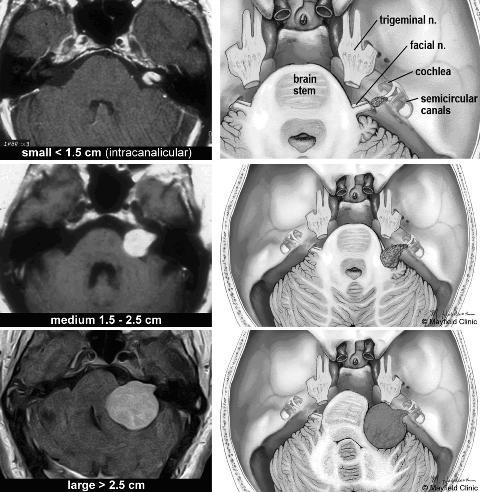
19 474 J. S. OGHALAI ET AL. based on age would be most easily detectable using patients at the extremes of age. The second was an analysis of all middle fossa surgery outcomes (middle fossa arm). This study arm was used to evaluate for subtle effects of age on hearing and facial nerve outcomes, because the middle fossa route is our usual approach for hearing conservation surgery. The extremes of age study arm was a retrospective study comparing older and younger patients (total n 205). All patients over 60 years of age who underwent surgical excision of an AN were compared with those patients under than 40 years of age. The details of the two groups of patients are given in Table 1. The expected patient life span was determined from an actuarial table (16). Tumor size was measured as millimeters in the cerebellopontine angle (CPA). Tumors wholly confined to the internal auditory canal were categorized as intracanalicular (0 mm). The average tumor size in the patients undergoing the middle fossa approach was much smaller than that in the patients undergoing the retrosigmoid approach. This reflects our philosophy of using the middle fossa approach as our primary strategy toward hearing preservation, while reserving the retrosigmoid approach only for hearing conservation attempts for tumors measuring 15 to 25 mm in the CPA (17). Older patients undergoing the translabyrinthine approach also tended to have larger tumors, because our usual policy for older patients with small tumors and poor hearing is to either monitor them with serial magnetic resonance imaging or treat them with stereotactic radiotherapy. The middle fossa study arm was a retrospective review of all patients who underwent the middle fossa approach for resection of an AN, regardless of age. This included patients at the age extremes as well as those 40 to 60 years old (total n 170, Table 1). This larger patient population permitted us to study the effect of patient age on hearing preservation with greater power than did the extremes of age study arm. The average patient age was 45 ± 10 years (mean ± SD), and the average tumor size was 5±6mm(mean ± SD) in the CPA. Main Outcome Measures Good hearing preservation was defined as postoperative AAO-HNS class A or B hearing (18). We did not include patients in this analysis who had class C or D hearing preoperatively. We measured postoperative hearing at least 3 months after surgery. Good postoperative facial function was considered House-Brackmann grade 1 or 2 (19). We did not include patients in this analysis who had grade 3 or worse facial function preoperatively. Postoperative facial weakness was not tabulated unless it was present 1 year after surgery. Postoperative complications were also tabulated. Cerebrospinal fluid (CSF) leaks were identified separately from other complications. This included all postoperative leaks identified, whether they required lumbar subarachnoid drain and/or reoperation. Fewer than 5% of patients with leaks required operative intervention (20). Statistical Analysis SPSS (version 10.0; SPSS, Inc., Chicago) was used for statistical analysis. All averages are reported as mean ± standard deviation. We used the two-tailed t test to compare continuous variables (patient age and tumor size). Multiple logistic regression analysis was performed to account for confounding variables in the comparison of discrete variables (hearing preservation, good facial function, CSF leak, other complications). This technique was also used for the continuous variables to verify the t test results. A stepwise entry methodology was used. For all analyses, statistical significance was determined if p RESULTS Extremes of Age Study Arm This first arm was designed to compare patients at the extremes of age for each of the three surgical approaches to identify major differences that might be associated with patient age. The main outcome measures are presented in Table 2. The rate of hearing preservation was lower in the older patient group than in the younger patient group for both the middle fossa (29% vs. 60%) and the retrosigmoid (8% vs. 15%) approaches. Multiple logistic regression analysis was performed to identify an effect of age on hearing preservation, while accounting for surgical approach and tumor size. This demonstrated that patients older than 60 years were less likely to have TABLE 1. Patient population stratified by surgical approach and age group Surgical approach Group N Age (yr) Expected lifespan (yr) Tumor size a (mm in CPA) Study arm Middle fossa <40 yr ± 7 46 ± 6 5 ± 6 Extremes of age, [15 39] [40 62] [0 18] middle fossa yr ± 5 30 ± 2 5 ± 6 Middle fossa [41 59] [22 39] [0 18] >60 yr ± 4 17 ± 3 7 ± 5 Extremes of age, [60 76] [11 22] [0 18] middle fossa Retrosigmoid <40 yr ± 6 46 ± 6 21 ± 3 Extremes of age [20 39] [40 58] [15 25] >60 yr ± 6 16 ± 4 21 ± 11 Extremes of age [60 89] [5 22] [0 50] Translabyrinthine <40 yr ± 5 45 ± 5 22 ± 15 Extremes of age [22 38] [41 57] [0 40] >60 yr ± 6 16 ± 4 20 ± 12 Extremes of age [60 86] [6 22] [0 45] All values are mean ± standard deviation. The range is in brackets. Expected lifespan was calculated from actuarial data (25). a Intracanalicular tumors recorded as 0 mm in the cerebellopontine angle. Otology & Neurotology, Vol. 24, No. 3, 2003
20 EFFECT OF AGE ON ACOUSTIC NEUROMA OUTCOMES 475 TABLE 2. Outcome measures stratified by surgical approach and age group Surgical approach Group Good hearing (%) Good facial function (%) CSF leak (%) Rate of other complications (%) Other complications Middle fossa <40 yr 15/25 (60) 24/25 (96) 4/25 (16) 0/25 (0) yr a 55/121 (46) 115/124 (93) 12/124 (10) 1/124 (0) Deep venous thrombosis, treated with inferior vena cava filter (1) >60 yr 5/17 (29) 16/16 (100) 5/21 (24) 0/21 (0) Retrosigmoid <40 yr 2/13 (15) 16/16 (100) 1/16 (6) 0/16 (0) >60 yr 1/12 (8) 25/28 (89) 6/38 (16) 1/38 (3) Postoperative communicating hydrocephalus that resolved spontaneously (1) Translabyrinthine <40 yr n/a 12/13 (92) 0/13 (0) 0/16 (0) >60 yr n/a 64/74 (87) 14/91 (15) 6/91 (7) Wound infections that resolved with local wound care and antibiotics (3), noncommunicating hydrocephalus secondary to intraventricular hemorrhage that required ventriculoperitoneal shunt (1), diplopia (1), postoperative myocardial infarction with good recovery (1) The number of patients in each category may be less than the total number of patients studied. Only patients with preoperative AAO-HNS class A or B hearing or preoperative House-Brackmann grade 1 or 2 facial function were included in the analysis. a This middle age group of patients was not use for the extremes of age study arm, but was included in the middle fossa study arm. CSF, cerebrospinal fluid; n/a, not applicable. preserved good hearing than those younger than 40 years (p 0.048, odds ratio 0.30). The rate of good postoperative facial function did not seem to be dramatically different between the older and the younger age groups for any of the surgical approaches (87 100%). Multiple logistic regression analysis accounting for surgical approach and tumor size demonstrated that there was no effect of age grouping on facial outcomes (p 0.2). There was a trend toward slightly higher rates of CSF leak (0 15%) in the older patient group (p 0.07), but there was no statistically significant difference in the rate of other complications (0 7%, p 0.8) between the age extremes. Middle Fossa Study Arm This second arm was a review of all patients who underwent the middle fossa approach. It was designed to identify subtle associations of age with hearing and facial nerve outcomes in our primary hearing preservation approach. First, we compared the average patient age and tumor size for patients in whom we were and were not able to preserve hearing and maintain good facial nerve function (Fig. 1.). Patients in whom hearing was preserved were younger than those in whom hearing was not preserved (A, preserved: 48 ± 11 years, n 75 vs. not preserved: 52 ± 9 years, n 88, p 0.01). Additionally, there was a trend toward patients with a smaller tumor size having an improved rate of hearing preservation (B, preserved: 4±5mm,n 74 vs. not preserved: 6 ± 6 mm, n 88, p 0.08). Neither younger patient age (C, good facial function: 50 ± 10 years, n 155 vs. poor facial function: 51 ± 7 years, n 10, p 0.8) nor tumor size (D, good facial function: 5±6mm,n 154 vs. poor facial function: 6 ± 5 mm, n 10, p 0.7) were correlated with good facial function. Because larger tumor size can be associated with lower hearing preservation rates and worse facial nerve outcomes, it is important to distinguish between the effects of patient age and the effects of tumor size. Multiple logistic regression analysis was then performed to account for tumor size as a confounding variable. This revealed that older age was associated with poorer rates of hearing preservation (p 0.01; odds ratio 1.044). This means that the odds of hearing preservation decreases by 4.4% per year. However, older age was not an independent predictor of poor facial function (p 0.7). DISCUSSION Both arms of the study demonstrate that older patients have a decreased rate of hearing preservation when tumor size was controlled for. Additionally, there was also a trend toward higher CSF leak rates in older patients, but there was no statistically significant effect of age on other complications or facial nerve function. However, our facial function results may be biased toward better preservation rates in older patients because we typically are less aggressive in trying to achieve total tumor removal in this population. We may leave a small bit of tumor on the facial nerve to reduce trauma and subsequent morbidity. A near-total resection means leaving a scrap of tumor no larger than 25 mm 2 (5 5 mm) and 2 mm thick, and a subtotal resection means leaving any larger piece. It is likely that without our altered surgical technique for older patients, there would have been a significantly reduced rate of good postoperative facial Otology & Neurotology, Vol. 24, No. 3, 2003
21 476 J. S. OGHALAI ET AL. FIG. 1. The effect of patient age and tumor size on surgical outcomes with the middle fossa approach. Patients in whom hearing (A, B) or good facial nerve function (C, D) were able to be preserved were compared with those in whom they could not be preserved. The box and whisker plots demonstrate the 25th and 75th percentiles of the samples (bottom and top of the boxes), the medians (line in the middle of the boxes), and the extent of the sample (whiskers above and below the boxes). Outliers more than 1.5 times the interquartile range away from the top or bottom of the box are identified (o). Younger patient age (A, 48 ± 11 years,n=75, vs. 52 ± 9 years, n=88,p = 0.01) was correlated with hearing preservation. There was a trend toward hearing preservation in patients with smaller tumors (B,4 ± 5 mm,n=74,vs.6±6mm,n=88, p = 0.08). The number of patients is less than the total number of patients studied because only patients with preoperative AAO-HNS class A or B hearing were analyzed. Neither younger patient age (C, 50 ± 10 years, n = 155, vs. 51 ± 7 years, n=10,p = 0.8) nor tumor size (D, 5±6mm,n=154, vs. 6±5mm,n=10,p = 0.7) was correlated with good facial function. function (as there was with the rate of hearing preservation). We did not have enough patients to perform a quantitative analysis of the effect of the completeness of surgical resection on hearing and facial nerve function. Our hearing and facial nerve preservation findings concur with and expand on those of other investigators. Brackmann et al. (3) found that the average age of patients who lost hearing postoperatively with the middle fossa approach was slightly higher than in patients who did not, although the difference was not significant. Ramsay and Luxford (6) found similar postoperative facial nerve function in patients older than 70 years and younger than 70 years of age, all of whom were treated with the translabyrinthine approach. Certainly, there is a generalized increased risk of surgical complications in older patients in all types of surgery. This is predominantly due to the steady decline in organ system functioning during aging (21). One might reasonably suspect that complications during AN surgery should occur more frequently in older patients. However, older patient age has not been shown to be associated with increased complications in AN surgery (22,23). Our data support these studies, in that we saw no effect of older age on complication rates. The middle fossa approach is thought to be more technically challenging in older patients because of thinning and adhesions of the dura. Indeed, patient age greater than 60 years has been suggested as a relative contraindication to the middle fossa approach (24,25). Our philosophy has been to base our surgical approach on tumor size, location, and residual hearing, and we do not believe that older patient age is a contraindication to the middle fossa approach. Meticulous surgical technique in raising the craniectomy bone flap, suture repair of any dural tears, and careful bipolar cautery of any bleeding vessels is critical to the successful prevention of technical complications. Also larger tumor size is probably not associated with an increased rate of CSF leak (20). The dilemma in treating older patients is whether to remove a small AN to preserve potential hearing loss in the years to come. Our philosophy on the management of AN in patients older than 60 years is to treat with either surgery or stereotactic radiation only if the patient has a large tumor with substantial brainstem compression, has progressive worsening of symptoms (particularly failing balance), or demonstrates tumor growth at a rate that will eventually lead to serious brainstem compression within the patient s predicted life span. By far the majority of older patients with tumors smaller than 2 cm in the CPA undergo a period of observation with serial magnetic resonance imaging to determine tumor growth rate, before intervention strategies are decided on. The risk of tumor progression and hearing loss (with either no treatment or stereotactic radiation) and the risk of the development of a radiation-induced malignancy (with stereotactic radiation) during an expected life span of 15 to 20 years should be weighed against the risks and benefits of tumor removal in consultation with the patient. Otology & Neurotology, Vol. 24, No. 3, 2003
22 EFFECT OF AGE ON ACOUSTIC NEUROMA OUTCOMES 477 REFERENCES 1. Slattery WH 3rd, Brackmann DE, Hitselberger W. Middle fossa approach for hearing preservation with acoustic neuromas. Am J Otol 1997;18: Irving RM, Jackler RK, Pitts LH. Hearing preservation in patients undergoing vestibular schwannoma surgery: comparison of middle fossa and retrosigmoid approaches. J Neurosurg 1998;88: Brackmann DE, Owens RM, Friedman RA, et al. Prognostic factors for hearing preservation in vestibular schwannoma surgery. Am J Otol 2000;21: Holsinger FC, Coker NJ, Jenkins HA. Hearing preservation in conservation surgery for vestibular schwannoma. Am J Otol 2000; 21: Wiet RJ, Mamikoglu B, Odom L, et al. Long-term results of the first 500 cases of acoustic neuroma surgery. Otolaryngol Head Neck Surg 2001;124: Ramsay HA, Luxford WM. Treatment of acoustic tumors in elderly patients: is surgery warranted? J Laryngol Otol 1993;107: Silverstein H, Rosenberg SI, Flanzer JM, et al. An algorithm for the management of acoustic neuromas regarding age, hearing, tumor size, and symptoms. Otolaryngol Head Neck Surg 1993;108: Pulec JL. Acoustic neuroma surgery in geriatric patients. Ear Nose Throat J 1999;78:429 30, 33 6, passim. 9. Rosenberg SI. Natural history of acoustic neuromas. Laryngoscope 2000;110: Shin YJ, Fraysse B, Cognard C, et al. Effectiveness of conservative management of acoustic neuromas. Am J Otol 2000;21: Perry BP, Gantz BJ, Rubinstein, JT. Acoustic neuromas in the elderly. Otol Neurotol 2001;22: Schneider EL, Guralnik JM. The aging of America: impact on health care costs. JAMA 1990;263: Cornman JM, Kingson ER. Trends, issues, perspectives, and values for the aging of the baby boom cohorts. Gerontologist 1996; 36: Tuljapurkar S, Li N, Boe C. A universal pattern of mortality decline in the G7 countries. Nature 2000;405: Robine JM, Romieu I, Cambois E. Health expectancy indicators. Bull World Health Organ 1999;77: Center for Disease Control. National Vital Statistics Report. Available at: Statistics, NCfH. 17. Satar B, Jackler RK, Oghalai JS, et al. Risk benefit analysis of using the middle fossa approach for acoustic neuromas with >10 mm cerebellopontine angle component. Laryngoscope 2002;112: Committee on Hearing and Equilibrium guidelines for the evaluation of hearing preservation in acoustic neuroma (vestibular schwannoma). American Academy of Otolaryngology-Head Neck Surgery Foundation, Inc. Otolaryngol Head Neck Surg 1995;113: House JW, Brackmann DE. Facial nerve grading system. Otolaryngol Head Neck Surg 1985;93: Becker SS, Jackler RK, Pitts LH. Cerebrospinal fluid leak after acoustic neuroma surgery: a comparison of the translabyrinthine, middle fossa, and retrosigmoid approaches. Otol Neurol 2003;24: Zenilman, ME. Preoperative assessment of the elderly patient. In: Cameron JL, ed. Current Surgical Therapy. 5th ed. St. Louis: C.V. Mosby, 1995: Samii M, Tatagiba M, Matthies C. Acoustic neurinoma in the elderly: factors predictive of postoperative outcome. Neurosurgery 1992;31:615 9; discussion Slattery WH 3rd, Francis S, House KC. Perioperative morbidity of acoustic neuroma surgery. Otol Neurotol 2001;22: Coker NJ, Jenkins HA. Atlas of Otologic Surgery. Philadelphia: W.B. Saunders, Centers for Disease Control. National Vital Statistics Report. Available at: htm. Accessed October Otology & Neurotology, Vol. 24, No. 3, 2003
23 The fate of the tumor remnant after less-than-complete acoustic neuroma resection DOV C. BLOCH, MD, JOHN S. OGHALAI, MD, ROBERT K. JACKLER, MD, MONICA OSOFSKY, MD, and LAWRENCE H. PITTS, MD, San Francisco, California OBJECTIVES: We sought to determine the recurrence rate after near-total and subtotal resection of acoustic neuroma. STUDY DESIGN, SETTING, AND PATIENTS: We conducted a retrospective chart review of a total of 79 patients: 50 with near-total resections (remnant <25 mm 2 and <2 mm thick) and 29 with subtotal resections (any larger remnant). Surgical approach included 5 middle fossa, 17 retrosigmoid, and 57 translabyrinthine. MAIN OUTCOME MEASURES: Recurrence was defined as documented tumor growth by serial imaging or the recommendation for further treatment after a single scan. No recurrence was defined as no visible tumor on imaging for a minimum follow-up time of 3 years or tumor remnants that remained unchanged on serial scans (mean, 5-year follow-up). RESULTS: Fifty-two patients were included in the study group. Recurrences were seen in 1 (3%) of 33 patients who had a near-total resection compared with 6 (32%) of 19 patients who had a subtotal resection. After adjustment for follow-up time and large tumor size, the odds ratio for recurrence was 12 times larger for subtotal than for near-total resections (P 0.033). All recurrences were seen following the translabyrinthine approach in the midcerebellopontine angle. None were encountered in the internal auditory canal. The mean time interval from surgery to the detection of a recurrence was 3 years (range, 1 to 5 years). From the Departments of Otolaryngology Head and Neck Surgery (Drs Bloch, Oghalai, Jackler, Osofsky, and Pitts) and Neurosurgery (Drs Pitts and Jackler), University of California San Francisco. Presented at the Annual Meeting of the American Academy of Otolaryngology Head and Neck Surgery, San Diego, CA, September 22-25, Reprint requests: John S. Oghalai, MD, Department of Otorhinolaryngology and Communicative Sciences, One Baylor Plaza, NA102, Houston, TX 77030; , jso@bcm.tmc.edu /$30.00 Copyright 2004 by the American Academy of Otolaryngology Head and Neck Surgery Foundation, Inc. doi: /s (03) CONCLUSIONS: The recurrence rate when performing a near-total resection is low but is substantially higher with a subtotal resection. Recurrences can be detected within the first 5 postoperative years. We recommend near-total resection in any patient if needed to preserve neural integrity. Subtotal resection is best avoided whenever possible; however, adjunctive treatment with stereotactic radiotherapy may be considered. (Otolaryngol Head Neck Surg 2004;130: ) The goals of acoustic neuroma (AN) surgery are tumor control and preservation of cranial nerves, including the facial nerve and at times the auditory nerve. Balancing the priority of these goals is influenced by other considerations such as patient age and life expectancy, overall health and comorbidities, and tumor adherence and size. Microsurgical gross total tumor removal is by far the most common result of AN surgery. Less-than-complete resection most commonly involves an intraoperative decision to leave tumor remnants behind as a concession to neural integrity. Elderly or debilitated patients may have a subtotal removal planned preoperatively to minimize the morbidity and mortality associated with a more extensive and lengthy procedure. The aim of the present study was to determine the risk of tumor recurrence with incomplete AN resection. METHODS Seventy-nine patients with incomplete AN resections from 1978 to 1999 were identified, accounting for less than 10% of the patients who underwent AN surgery at our institution. At the University of California San Francisco (UCSF), a team composed of a neurotologist and a neurosurgeon perform the procedure. A retrospective chart review was performed from records kept by both departments and entered into a computerized database. The study protocol was approved by the UCSF Committee on Human Research. Clinical parameters, including patient age, gender, symptoms, surgical approach, and postopera- 104
24 Otolaryngology Head and Neck Surgery Volume 130 Number 1 BLOCH et al 105 Fig 1. (A) The 2 types of incomplete tumor resection strategies. Near-total resections were defined by tumor remnants no greater than 25 mm 2 and 2 mm thick; subtotal resections included anything larger. (B) Surgical dissection of the most adherent area of the tumor off of the facial nerve. tive complications, were recorded. Tumor size, based on the greatest dimension within the cerebellopontine angle (CPA), was obtained from preoperative imaging data. The surgeons notes were examined to ascertain the indication for performing an incomplete resection whenever possible. As opposed to defining residual tumor as a percentage of the original tumor, near-total resections were defined by tumor remnants no greater than 25 mm 2 and 2 mm thick; subtotal resections included anything larger 1 (Fig 1). Facial nerve outcomes with a minimum of 1-year follow-up were recorded, based on the House-Brackmann (HB) grading system. Our routine protocol for postoperative tumor surveillance consists of imaging at 1 and 3 years for tumors that have been totally removed. Incompletely resected tumors are followed annually with a gradual lengthening of the time between scans when there is no evidence of recurrence. Because UCSF is a tertiary referral center, some patients receiving longitudinal follow-up care at other institutions had different imaging regimens. The modality most com-
25 Otolaryngology Head and Neck Surgery 106 BLOCH et al January 2004 Table 1. Patient populations Near-total (n 50; 63%) Subtotal (n 29; 37%) Gender Male Female Tumor size in CPA,* mean SD (mm) (P 0.005) (range, 0 to 45) (range, 7 to 65) Surgical approach Translabyrinthine 34 (68%) 23 (79%) Mean (mm) 26 (range, 12 to 45) 31 (range, 7 to 65) Retrosigmoid 12 (24%) 5 (17%) Mean (mm) 22 (range, 0 to 37) 33 (range, 18 to 45) Middle fossa 4 (8%) 1 (4%) Mean (mm) 7 (range, 0 to 12) 15 Age Mean SD (yr) (P 0.10) (range, 21 to 78) (range, 34 to 81) *Largest diameter dimension in the CPA. Intracanalicular tumors recorded as 0 mm. CPA, Cerebellopontine angle. monly used was magnetic resonance imaging (MRI), although a few patients early in the study period had computed tomography (CT). All patients, except for 1, had an MRI study at some point in their follow-up. The results of all postoperative follow-up imaging were recorded to identify residual and/or recurrent tumor. No tumor recurrence was defined by the absence of visible tumor on imaging after a minimum follow-up of 3 years or if stable residual tumor was present. Stable residual tumor was defined as a tumor remnant that remained unchanged on serial imaging. Recurrent tumor was defined as tumor growth on serial imaging. Additionally, tumor recurrence was defined as when the patient underwent additional treatment (either stereotactic radiation or repeat surgery) after only a single scan because of the large tumor size. Statistical Analysis Stata (version 7.0; Stata Corp, College Station, TX), SPSS (version 10.0; SPSS, Inc, Chicago, IL), and StatXact (version 3.0; Cytel Software, Cambridge, MA) were used for statistical analysis. Fisher exact test and 2-sided t test were used to evaluate bivariate relationships. Logistic regression was performed on recurrence and good facial nerve function (HB I or II) to assess the influence of subtotal versus near-total resection, age, large tumor size ( 25 mm), and other predictors. Odds ratios were calculated and statistical significance was determined if P Logistic regression was followed by statistical analyses using exact stratified contingency table analysis to check the stability of the logistic regression results. RESULTS Patient Population There were 50 patients (63%) with near-total resections and 29 patients (37%) with subtotal resections (Table 1). The mean age was 59 years (range, 21 to 81 years). Patients who underwent subtotal resections tended to be older than the near-total group (mean, 62 versus 57 years; P 0.10). The mean tumor size was significantly greater in the subtotal resection group than in the near-total resection group (31 mm versus 24 mm, P 0.005). The predominant approach used in this group of patients was the translabyrinthine (TL), accounting for 72% of the operations. The average tumor size was greatest for the TL approach (26 mm) followed by the retrosigmoid (RS) (22 mm) and the middle fossa (MF) (7 mm) approaches. A comparison between the near-total and subtotal groups showed no significant difference in terms of gender, approach, and follow-up period. Postoperative complications are presented in Table 2. The cerebellar peduncular infarcts
26 Otolaryngology Head and Neck Surgery Volume 130 Number 1 BLOCH et al 107 Table 2. Complications Complication Near-total (n 50) Subtotal (n 29) Cerebrospinal fluid leak* 8 1 Meningitis (aseptic) 2 1 Wound infection 3 0 Cerebellar peduncular infarct 2 0 Deaths 0 0 *Two required surgery, and 7 were treated with a lumbar drain. were areas of distal anterior inferior cerebellar artery ischemia (with minimal symptomatology), not major strokes. 2 There were no known tumorrelated deaths, but there were 4 unrelated deaths seen on long-term follow-up. Indications for Incomplete Resection Although all patients had an incomplete resection to preserve facial nerve integrity, there were additional indications for this technique in a few patients. One patient had a near-total resection after experiencing bradyarrhythmias during dissection of the tumor from the brain stem. Four patients had planned incomplete resections for other reasons, including advanced age and medical comorbidities. Finally, 4 patients had subtotal resections after initial treatment with stereotactic radiotherapy failed. Of these, 3 patients had a ventriculoperitoneal shunt placed preoperatively to treat hydrocephalus and subsequently underwent a planned subtotal resection for brain stem decompression. The time interval between failed radiotherapy and operative intervention was from 3 to 17 months. Three of 4 of these patients had no evidence of recurrence at 3 years (the fourth patient was lost to follow-up). Facial Nerve Outcomes Good facial nerve function at 1 year postoperatively, defined as HB grade I or II, was seen in 57 (81%) of the 65 patients (Table 3). There were no detectable associations between good facial nerve function and tumor size (P 0.98), age (P 0.41), or approach (P 0.66). Logistic regression analysis demonstrated no statistically significant difference in good facial nerve function between the near-total and subtotal groups when controlling for age, tumor size, and operative approach (P 0.1). Radiographic Imaging Sixty-four patients had imaging available for at least 1 year postoperatively, with a mean radiographic follow-up period of 4.3 years (range, 1 to 13 years). Imaging revealed the residual tumor in approximately 20% of patients in the near-total group and 80% of patients in the subtotal group (Figs 2 and 3). The proportion of scans at each year that revealed tumor remained relatively stable over the study period. Recurrence Risk The risk of developing a tumor recurrence was determined in a subgroup of 52 patients who met the criteria for adequate follow-up, as described in Methods. The mean follow-up time for these patients was 5 years (maximum, 13 years). Recurrences were seen in 1 (3%) of 33 patients who had a near-total resection compared with 6 (32%) of 19 patients who had a subtotal resection. Multiple logistic regression analysis was then performed to adjust for differences in follow-up time and large tumor size ( 25 mm). From this analysis, the odds ratio for recurrence was found to be 12 times larger for subtotal than for near-total resections (P 0.033) (Fig 4). Seven patients had recurrent tumors (Table 4). All recurrences were seen following the TL approach (Fig 5). The locations of the recurrences were in the mid-cpa, and none were encountered in the internal auditory canal. The recurrent tumors were all large at the initial operation (mean, 37 mm; P 0.001); however, large size as a risk factor for recurrence was not significant when controlling for subtotal resections (P 0.32). There was no association between patient age and recurrence risk (P 0.19). The mean time interval from surgery to the detection of a recurrence was 3 years (range, 1 to 5 years). Stereotactic radiation treatment was recommended in 5 patients. The remaining 2 patients underwent a repeat incomplete resection with the RS approach (1 NT and 1 ST). DISCUSSION The rationale for using an incomplete tumor dissection technique is dictated by the risk/benefit ratio for the patient. Our data suggest that the risk of tumor recurrence after a near-total resection is
 23 (50%) 14 (30%) 8 (17%) 0 1 (2%) 0 Subtotal (n 21) 13 (54%) 7 (29%) 2 (8%) 1 (4%) 0 1 (4%) Fig 2.")
27 Otolaryngology Head and Neck Surgery 108 BLOCH et al January 2004 Table 3. Facial nerve function 1 year after surgery House-Brackmann grade I II III IV V VI Near-total (n 44) 23 (50%) 14 (30%) 8 (17%) 0 1 (2%) 0 Subtotal (n 21) 13 (54%) 7 (29%) 2 (8%) 1 (4%) 0 1 (4%) Fig 2. (A) Near-total residual (arrow) seen in mid-cpa on MRI 1 year after an RS resection. The patient had been previously treated at another institution with an RS resection and presented with brain stem compression and a recurrence involving the internal auditory canal. (B) Subtotal residual seen on MRI following a TL resection. A thin rind (arrow) is seen along the brain stem extending to the porus acousticus. quite low. Although we did not perform a randomized controlled trial to determine the degree of improvement in facial nerve outcomes, it seems logical to conclude that any further tumor removal would have compromised facial nerve function. Every patient in our study was selected because of a particularly high level of tumor adherence. Comparisons with stratified control patients who underwent a complete tumor resection would be inherently flawed, because these patients did not have tumors that were as difficult to dissect. In our experience, substantial adherence of the tumor to the facial nerve has a high risk of a poor facial nerve outcome. Near-total resection involves leaving a small rind of tumor to better preserve the facial nerve. Incomplete resection is helpful in preserving facial nerve integrity because as the tumor enlarges, it compresses the facial nerve and the tumor arachnoid mater plane is lost. 3 Difficulty in identifying a plane between the tumor and the facial nerve most commonly occurred just medial to the porus acousticus (Fig 1). The facial nerve, which follows the shape of the tumor, often becomes splayed and thinned in this region, possibly accounting for the adherence. Subtotal resection, in contrast, leads to a 12- time higher risk of recurrence. This may be explained merely by the larger initial tumor size in the subtotal group compared with the neartotal group (P 0.005), resulting in a more tenuous and adherent facial nerve. However, another explanation may be distinct variations in AN biological behavior. Although most tumors are slow growing (approximately 2 mm per year), 3,4 a small subset demonstrate rapid growth. 5 One patient illustrating an aggressive variant had a subtotal resection for a 25-mm tumor. She subsequently had 2 large symptomatic cystic recurrences after a 2- to 3-year interval. We use the subtotal resection technique with large tumors that have a large area of tumor stuck to the facial nerve and/or brain stem or to minimize other complications, particularly in elderly or debilitated patients. Rarely, an intraoperative event is the indication for a subtotal resection. 3 We favor a conservative approach
28 Otolaryngology Head and Neck Surgery Volume 130 Number 1 BLOCH et al 109 Fig 3. Frequency of visible tumor remnants on postoperative enhanced MRI. A cross-sectional plot of patients at 1-, 3-, and 3-year MRI. Roughly 20% of the patients with near-total tumor resections had visible tumor on follow-up MRI, as opposed to the 80% of patients with subtotal resections. Because not all patients followed the recommended protocol of an annual scan, the number of patients at each time frame varies. Most studies were MRI (1 year: 53 MRI, 2 CT; 3 years: 26 MRI, 2 CT; 3 years:32 MRI, 1 CT). The average follow-up time for the patients in the 3-year category was 6.3 years. Fig 4. Recurrence rates of near-total versus subtotal residuals (n 52). Recurrence was determined by documented growth or recommended treatment. No recurrence was defined by tumor that was stable on serial scans or not visible for a minimum of 3 years. The mean follow-up time was 5 years. There was a substantially higher risk of developing a recurrence if a subtotal resection (32%) was performed compared with a near-total resection (3%) (odds ratio, 12; P 0.033; details of multiple logistic regression analysis given in text). and often use the subtotal technique to treat patients for whom stereotactic radiation has failed. This is done to minimize the attendant risks engendered from radiation-induced changes. Microsurgery is often difficult due to inflammation and fibrosis that interfere with the identification of normal dissection planes, leading to poorer facial nerve outcomes. 6
 Initial tumor size (mm) Resection Approach Year recurred/treated Treatment Reason for retreatment ST NT 72 25 X TL 3/8 SR Growth continued")
29 Otolaryngology Head and Neck Surgery 110 BLOCH et al January 2004 Table 4. Characteristics of patients with recurrent tumors Age* (yr) Initial tumor size (mm) Resection Approach Year recurred/treated Treatment Reason for retreatment ST NT X TL 3/8 SR Growth continued (18 mm) X TL 5/5 SR X TL 5/5 MS Hydrocephalus (31 mm) X TL 3/5 SR Growth continued (21 mm) X TL 1/1 Proton therapy Hydrocephalus (40 mm) X TL 3/3 SR X TL 2/2 MS 30 mm, then developed another 35 mm recurrence (3 years later) *P 0.19; P 0.001; ST, Subtotal; NT, near-total; TL, translabyrinthine; SR, stereotactic radiation; MS, microsurgical (retrosigmoid approach used in both cases). Fig 5. (A) Near-total recurrence (arrow) seen on MRI 8 years after a TL resection. The tumor recurrence was detected on a 3-year scan and has continued to grow asymptomatically from 3 mm to 18 mm at 8 years after surgery (first patient, Table 4). (B) Subtotal recurrence (arrow) in the mid-cpa seen on MRI 3 years after a TL resection (sixth patient, Table 4). However, we have a particular concern about performing a subtotal resection in tumors with large cystic components. The cyst should not simply be marsupialized; the wall needs to be carefully microdissected from the surrounding normal structures and removed. A tendency exists for the residual cyst wall to reseal. Brain stem compression may result from the cyst recurrence even though the solid (neoplastic) residual has not grown. Stereotactic radiosurgery is not very effective in such cystic recurrences because it is not caused by rapid cell division. It is possible that radiosurgery may even stimulate further cyst expansion in such patients. Thus, a cystic recurrence likely necessitates reoperation. ANs do not have true capsules. The histopathology includes compressed neoplastic cells at the margin of the tumor surrounded by connective tissue only a few micrometers in thickness. 7 The entire mass contains viable tumor cells that carry the potential for recurrence. Thus, complete resection leaving only a small amount of capsule is
30 Otolaryngology Head and Neck Surgery Volume 130 Number 1 BLOCH et al 111 really incomplete tumor removal. Based on the volume of tumor and capsule remaining, it is best defined as a near-total or subtotal removal. Every effort should be made to remove as much of the tumor as possible while only leaving a small rind at areas with difficult planes of dissection. Limiting the gross volume of residual tumor has been shown to improve outcome. 8 One of the pitfalls in performing an incomplete resection occurs with the RS approach when tumors are truncated at the internal auditory canal without drilling out the intracanalicular component. We do not recommend this technique because these extensive residuals carry a high risk of recurrence. Indeed, even the need to perform a blind sweep of the fundus after a proper internal auditory canal drillout with the RS approach carries a higher incidence of tumor recurrence. 9 In contrast, all of the recurrences in our study were centered in the CPA and followed a TL resection. Because the majority of the near-total remnants and some of the subtotal remnants remained undetectable on serial follow-up imaging, we suspect that there was postoperative tumor regression. Other plausible explanations include tumor necrosis, detection limitations of the MRI, or an interruption of the blood supply preventing contrast enhancement. Residual remnants have been shown to be relatively avascular in a study looking at tumors managed with staged procedures. 3 Nonetheless, the bipolar electrocautery should be used to coagulate the remnant to cause further devascularization. The location of remnant appears to influence the recurrence risk. The blood supply of the internal auditory canal and brain stem may lead to an increased propensity for recurrence in these locations compared with remnants along the facial nerve in the mid-cpa that have a poor vascular supply. The mean time interval between surgery and tumor recurrence was 3 years (range, 1 to 5 years). All recurrences were seen on either the initial postoperative scan or serial scans that demonstrated growth. No patients had a clinical recurrence after an established quiescent period. This is contrasted by a study that revealed tumor growth in 17 patients after a median of 3.6 years with no evidence of tumor growth. 8 These conflicting observations may be explained by the use of lowersensitivity CT scanning for follow-up in the other study. Consistent with our findings was that once growth was identified, it continued unabated until institution of treatment. 8 AN recurrences are often asymptomatic due to the typical postoperative sequelae of hearing loss and vestibular ablation. It is imperative to follow these patients diligently with other diagnostic modalities that permit early detection and intervention before the need for urgent decompression surgery. The pre and post gadolinium enhanced T1-weighted MRI with fat saturation is the imaging of choice due to signal characteristics that allow the differentiation of postoperative scarring, blood, and fat graft from tumor. 10 Treatment options for a recurrent tumor include stereotactic radiosurgery or reoperation typically done via a different approach. In this series, all recurrences followed TL removals, presumably because this is the method typically used for tumors in the size range where less-than-complete removal is most likely to be chosen. When a reoperation is needed following the TL approach, we choose the RS method to avoid having to dissect the fat graft off of the facial nerve. Similarly, we use the TL approach to manage recurrences after other surgeons have left tumor (particularly in the internal auditory canal) during an RS approach to avoid the region of surgical scarring. CONCLUSION There has been an evolution in the management of ANs reflecting advances made in medical technology and an improved understanding of the natural progression of the disease. Historically, the goal of complete tumor removal took precedence over other considerations. Patients often presented with advanced disease, leading to unacceptable death rates. Today, high-quality imaging often leads to the diagnosis in patients with less severe symptoms and smaller tumors. A therapeutic dilemma arises when balancing the risk and benefits of a complete versus a less-than-complete resection of a benign tumor. Near-total resection has a low risk of recurrence and may be used in any patient as a concession to neural integrity. There is a substantially higher risk of recurrence with subtotal resections. Meticulous attention is needed to thin the tumor remnant as much as possible, limit
31 Otolaryngology Head and Neck Surgery 112 BLOCH et al January 2004 it to the CPA, and coagulate it with the electrocautery. The postoperative management should include vigilant surveillance with serial MRI to detect early recurrence. Detection of recurrent tumors was evident relatively early within the first 5 years after surgery. The authors extend their appreciation to Charles McCulloch, PhD, for statistical analysis. REFERENCES 1. Jackler RK. Atlas of neurotology and skull base surgery. St Louis: Mosby; 1996, p Hegarty JL, Jackler RK, Rigby PL, et al. Distal anterior inferior cerebellar artery syndrome after acoustic neuroma surgery. Otol Neurotol 2002;23: Comey CH, Jannetta PJ, Sheptak PE, et al. Staged removal of acoustic tumors: techniques and lessons learned from a series of 83 patients. Neurosurgery 1995;37:915-20; discussion Schessel DA, Nedzelski JM, Kassel EE, et al. Recurrence rates of acoustic neuroma in hearing preservation surgery. Am J Otol 1992;13: Bederson JB, von Ammon K, Wichmann WW, et al. Conservative treatment of patients with acoustic tumors. Neurosurgery 1991;28:646-50; discussion Battista RA, Wiet RJ. Stereotactic radiosurgery for acoustic neuromas: a survey of the American Neurotology Society. Am J Otol 2000;21: Kuo TC, Jackler RK, Wong K, et al. Are acoustic neuromas encapsulated tumors? Otolaryngol Head Neck Surg 1997;117: El-Kashlan HK, Zeitoun H, Arts HA, et al. Recurrence of acoustic neuroma after incomplete resection. Am J Otol 2000;21: Roberson JB Jr, Brackmann DE, Hitselberger WE. Acoustic neuroma recurrence after suboccipital resection: management with translabyrinthine resection. Am J Otol 1996;17: Battista RA, Bojrab DI, Wang AM. Evaluation of residual acoustic schwannoma using gadolinium-dtpa enhanced magnetic resonance imaging with the fat suppression technique. Am J Otol 1995;16:
32 Otology & Neurotology 24: , Otology & Neurotology, Inc. Is It Worthwhile to Attempt Hearing Preservation in Larger Acoustic Neuromas? *Philip D. Yates, * Robert K. Jackler, *Bulent Satar, * Lawrence H. Pitts, and *John S. Oghalai Departments of *Otolaryngology-Head and Neck Surgery and Neurologic Surgery, University of California, San Francisco, California, U.S.A. Objective: To determine the hearing outcome in patients undergoing surgery via the retrosigmoid approach for acoustic neuromas with a substantial component in the cerebellopontine angle. Study Design: Retrospective case review. Setting: Tertiary referral center. Patients: The medical records of all patients undergoing acoustic neuroma removal via the retrosigmoid approach at a tertiary referral center were retrospectively reviewed. Sixty-four patients with both cerebellopontine angle component 15 mm and preoperative audiometry of class A or B (American Academy of Otolaryngology Head and Neck Surgery) were identified. Main Outcome Measures: Postoperative average pure tone threshold and word recognition scores, categorized according to the classification of the American Academy of Otolaryngology Head and Neck Surgery, were used to assess hearing outcome. Results: Overall, only 6.3% (4 of 63) retained good hearing (class A or B) postoperatively. Hearing preservation rate in the smallest (15- to 19-mm) group was 17.6% (3 of 17), which was better than that for the larger groups. No successful hearing preservation was achieved in tumors with 25 mm cerebellopontine angle component (0 of 23). Conclusions: Surgeon and patient alike would always choose a hearing preservation technique if there was no potential for increased morbidity in making the attempt. When compared with the non hearing preservation translabyrinthine approach, the retrosigmoid approach had a higher incidence of persistent headache. In addition, efforts to conserve the auditory nerve prolong operating time, increase the incidence of postoperative vestibular dysfunction, and carry a slightly higher risk of tumor recurrence. Nevertheless, even though the probability of success is disappointingly small, when excellent hearing is present we favor offering the option of a hearing conservation attempt when the patient has been well informed of the pros and cons of the endeavor. Factors weighing against undertaking this effort include larger cerebellopontine angle component ( 25 mm), deep involvement of the fundus, wide erosion of the porus, and marginal residual hearing. Key Words: Acoustic neuroma (vestibular schwannoma) Hearing preservation Retrosigmoid approach. Otol Neurotol 24: , The three principal surgical approaches for acoustic neuroma (AN) removal are the translabyrinthine (TL), retrosigmoid (RS), and middle fossa (MF) approach. Only the latter two approaches provide the possibility of hearing preservation. The results of hearing preservation surgery have been shown to be superior for small tumors when the MF approach is used (1). However, the MF approach is only practicable in tumors with a limited cerebellopontine angle (CPA) extension. This leaves the RS approach as the only alternative when attempting to preserve hearing in larger ANs. Because tumor size is the most important variable influencing hearing outcome, the question arises as to whether hearing preservation surgery should still be attempted via the RS approach in patients with large tumors but serviceable hearing (2 5). Address correspondence and reprint requests to Robert K Jackler, M.D., University of California Hospital, 400 Parnassus Avenue, Ste. A-730, San Francisco, CA , U.S.A. rkj@itsa.ucsf.edu Tumor size itself should not be a criterion in choosing between the RS and TL approach, because both approaches provide adequate exposure, but the potential higher morbidity associated with the RS approach does need to be taken into account. This study was undertaken to determine the rate of hearing preservation in patients with ANs that have a substantial CPA component ( 15 mm) so that patients can be appropriately counseled before choosing between a RS and TL approach. PATIENT POPULATION This study is a retrospective review of the medical records of patients who have had surgical removal of a histologically confirmed AN in a single tertiary referral center between the years 1984 and Selection criteria for inclusion in the study were tumor removal via the RS approach, 15 mm CPA component, and preoperative hearing of class A or B. Because not every patient with a 15 mm AN and preoperative hearing 460
Management of Cerebrospinal Fluid Leaks After Vestibular Schwannoma Surgery
 Otology & Neurotology 32:1525Y1529 Ó 2011, Otology & Neurotology, Inc. Management of Cerebrospinal Fluid Leaks After Vestibular Schwannoma Surgery *Brannon D. Mangus, *Alejandro Rivas, Mi Jin Yoo, JoAnn
Revision Surgery for Vestibular Schwannomas
 528 Original Article Kevin A. Peng 1 Brian S. Chen 3 Mark B. Lorenz 2 Gregory P. Lekovic 1 Marc S. Schwartz 1 William H. Slattery 1 Eric P. Wilkinson 1 1 House Clinic, Los Angeles, California, United States
SPECIAL PAPER IN CELEBRATION OF PROF. YANG'S 50 YEARS CAREER IN MEDICINE
 JOURNAL OF OTOLOGY SPECIAL PAPER IN CELEBRATION OF PROF. YANG'S 50 YEARS CAREER IN MEDICINE ADVANCES IN SURGICAL TREATMENT OF ACOUSTIC NEUROMA HAN Dongyi,CAI Chaochan Acoustic Neuroma (AN) arises from
Acoustic Neuroma. Presenting Signs and Symptoms of an Acoustic Neuroma:
 Acoustic Neuroma An acoustic neuroma is a benign tumor which arises from the nerves behind the inner ear and which may affect hearing and balance. The incidence of symptomatic acoustic neuroma is estimated
Results of Surgery of Cerebellopontine angle Tumors
 Original Article Iranian Journal of Otorhinolaryngology, Vol. 27(1), Serial No.78, Jan 2015 Abstract Results of Surgery of Cerebellopontine angle Tumors Faramarz Memari 1, * Fatemeh Hassannia 1, Seyed
Antonio De la Cruz, MD 27th Alexandria International Combined ORL Congress Alexandria, Egypt April 8, 2009
 ACOUSTIC NEUROMA TREATMENT OPTIONS 2009 27 th Alexandria International Combined ORL Congress Alexandria, Egypt April 10, 2009 AntonioDelaCruz Cruz, MD House Ear Institute Los Angeles, California Antonio
Otolaryngologist s Perspective of Stereotactic Radiosurgery
 Otolaryngologist s Perspective of Stereotactic Radiosurgery Douglas E. Mattox, M.D. 25 th Alexandria International Combined ORL Conference April 18-20, 2007 Acoustic Neuroma Benign tumor of the schwann
With advances in microsurgical techniques and the. Staged resection of large vestibular schwannomas. Clinical article
 J Neurosurg 116:1126 1133, 2012 Staged resection of large vestibular schwannomas Clinical article Ahmed M. Raslan, M.D., 1 James K. Liu, M.D., 3 Sean O. McMenomey, M.D., 1,2 and Johnny B. Delashaw Jr.,
Internal Auditory Canal Involvement of Acoustic Neuromas: Surgical Correlates to Magnetic Resonance Imaging Findings
 Otology & Neurotology 22:92 96 200, Otology & Neurotology, Inc. Internal Auditory Canal Involvement of Acoustic Neuromas: Surgical Correlates to Magnetic Resonance Imaging Findings * Samuel H. Selesnick,
Temporal Lobe Cystic Collection and Associated Oedema: A Rare Complication of Translabyrinthine Resection of Vestibular Schwannoma
 Open Access Case Report DOI: 10.7759/cureus.2217 Temporal Lobe Cystic Collection and Associated Oedema: A Rare Complication of Translabyrinthine Resection of Vestibular Schwannoma Abdurrahman Raeiq 1 1.
The New England Journal of Medicine LONG-TERM OUTCOMES AFTER RADIOSURGERY FOR ACOUSTIC NEUROMAS
 LONG-TERM OUTCOMES AFTER RADIOSURGERY FOR ACOUSTIC NEUROMAS DOUGLAS KONDZIOLKA, M.D., L. DADE LUNSFORD, M.D., MARK R. MCLAUGHLIN, M.D., AND JOHN C. FLICKINGER, M.D. ABSTRACT Background Stereotactic radiosurgery
Dr. T. Venkat Kishan Asst. Prof Department of Radiodiagnosis
 Dr. T. Venkat Kishan Asst. Prof Department of Radiodiagnosis Schwannomas (also called neurinomas or neurilemmomas) constitute the most common primary cranial nerve tumors. They are benign slow-growing
Injury to the facial nerve is a common complication. Efficacy of facial nerve sparing approach in patients with vestibular schwannomas
 See the corresponding editorial in this issue, pp 915 916. J Neurosurg 115:917 923, 2011 Efficacy of facial nerve sparing approach in patients with vestibular schwannomas Clinical article Raqeeb Haque,
Evaluation of Variation in the Course of the Facial Nerve, Nerve Adhesion to Tumors, and Postoperative Facial Palsy in Acoustic Neuroma
 Original Article 39 Evaluation of Variation in the Course of the Facial Nerve, Nerve Adhesion to Tumors, and Postoperative Facial Palsy in Acoustic Neuroma Tetsuro Sameshima 1 Akio Morita 1 Rokuya Tanikawa
2. Department of Neurologic Surgery, Mayo Clinic, School of Medicine, Rochester, Minnesota,
 1 2 3 4 5 6 7 8 9 10 11 12 13 14 15 16 17 18 19 20 21 22 23 24 25 26 27 CONGRESS OF NEUROLOGICAL SURGEONS SYSTEMATIC REVIEW AND EVIDENCE-BASED GUIDELINE ON HEARING PRESERVATION OUTCOMES IN PATIENTS WITH
The Incidence of Cerebrospinal Fluid Leak after Vestibular Schwannoma Surgery
 Otology & Neurotology 25:387 393 2004, Otology & Neurotology, Inc. The Incidence of Cerebrospinal Fluid Leak after Vestibular Schwannoma Surgery Samuel H. Selesnick, Jeffrey C. Liu, Albert Jen, and Jason
Postoperative Nerve Injury and Recurrence in Surgical Treatment of Head and Neck Schwannomas
 Postoperative Nerve Injury and Recurrence in Surgical Treatment of Head and Neck Schwannomas SHU-HSIEN CHEN 1, PEI-YIEN TSAI 2, YEN-HUI 3 TSAI * AND CHIH-YING LIN 4 1 Institute of Health Industry Management
Hemorrhagic vestibular schwannoma: an unusual clinical entity Case report
 Neurosurg Focus 5 (3):Article 9, 1998 Hemorrhagic vestibular schwannoma: an unusual clinical entity Case report Dean Chou, M.D., Prakash Sampath, M.D., and Henry Brem, M.D. Departments of Neurological
Original Article. Emergency Department Evaluation of Ventricular Shunt Malfunction. Is the Shunt Series Really Necessary? Raymond Pitetti, MD, MPH
 Original Article Emergency Department Evaluation of Ventricular Shunt Malfunction Is the Shunt Series Really Necessary? Raymond Pitetti, MD, MPH Objective: The malfunction of a ventricular shunt is one
OTOLOGY. 1. BRIEF DESCRIPTION OF OTOLOGIC TRAINING Rotations that include otologic training are a component of each of the four years of training.
 OTOLOGY 1. BRIEF DESCRIPTION OF OTOLOGIC TRAINING Rotations that include otologic training are a component of each of the four years of training. Longwood Rotation PGY-2 through PGY-5 years o Clinic experience
Schwannoma of the intermediate nerve
 J Neurosurg 109:144 148, 2008 Schwannoma of the intermediate nerve Case report CHRISTIAN SCHELLER, M.D., 1 JENS RACHINGER, M.D., 1 JULIAN PRELL, M.D., 1 MALTE KORNHUBER, M.D., 2 AND CHRISTIAN STRAUSS,
Via Transmastoid Approach. Mitul Chaitan Bhatt. Maharashtra University Miraj India
 ISSN: 2250-0359 Volume 6 Issue 1 2016 Evaluation Of Patients Of Traumatic Facial Palsy Treated By Facial Nerve Decompression Via Transmastoid Approach Mitul Chaitan Bhatt Maharashtra University Miraj India
LONG-TERM FOLLOW-UP OF ACOUSTIC SCHWANNOMA RADIOSURGERY WITH MARGINAL TUMOR DOSES OF 12 TO 13 Gy
 doi:10.1016/j.ijrobp.2007.01.001 Int. J. Radiation Oncology Biol. Phys., Vol. 68, No. 3, pp. 845 851, 2007 Copyright 2007 Elsevier Inc. Printed in the USA. All rights reserved 0360-3016/07/$ see front
Editorial Manager(tm) for Neurosurgery Manuscript Draft. Manuscript Number:
 for Neurosurgery Manuscript Draft. Manuscript Number:") Editorial Manager(tm) for Neurosurgery Manuscript Draft Manuscript Number: Title: Evaluation of variation in the course of the facial nerve, nerve adhesion to tumors, and postoperative facial palsy in
Editorial Manager(tm) for Neurosurgery Manuscript Draft Manuscript Number: Title: Evaluation of variation in the course of the facial nerve, nerve adhesion to tumors, and postoperative facial palsy in
Comparison of Growth Patterns of Acoustic Neuromas With and Without Radiosurgery
 Otology & Neurotology 27:705 Y 712 Ó 2006, Otology & Neurotology, Inc. Comparison of Growth Patterns of Acoustic Neuromas With and Without Radiosurgery *Alex Battaglia, Bill Mastrodimos, and *Roberto Cueva
Intracanalicular acoustic neuroma: early surgery for preservation of hearing STEPHEN J. HAINES, M.D., AND SAMUEL C. LEVINE, M.D.
 J Neurosnrg 79:55-50, 99 Intracanalicular acoustic neuroma: early surgery for preservation of hearing STEPHEN J. HAINES, M.D., AND SAMUEL C. LEVINE, M.D. Center for Craniofacial and Skull Base Surgery,
Gamma knife radiosurgery for Koos grade 4 vestibular schwannomas
 Gamma knife radiosurgery for Koos grade 4 vestibular schwannomas David Mathieu MD FRCSC, Christian Iorio-Morin MD PhD, Fahd Al Subaie MD MSc FRCSC Division of neurosurgery, Université de Sherbrooke, Centre
Acoustic neuromas (or VSs) are one of the more
 are one of the more") Neurosurg Focus 33 (3):E15, 2012 Technical nuances of resection of giant (> 5 cm) vestibular schwannomas: pearls for success Charles G. Kulwin, M.D., and Aaron A. Cohen-Gadol, M.D., M.Sc. Goodman Campbell
Neurosurg Focus 33 (3):E15, 2012 Technical nuances of resection of giant (> 5 cm) vestibular schwannomas: pearls for success Charles G. Kulwin, M.D., and Aaron A. Cohen-Gadol, M.D., M.Sc. Goodman Campbell
The Best Candidates for Nerve-Sparing Stripping Surgery for Facial Nerve Schwannoma
 The Laryngoscope VC 2014 The American Laryngological, Rhinological and Otological Society, Inc. The Best Candidates for Nerve-Sparing Stripping Surgery for Facial Nerve Schwannoma Soon H. Park, MD; Jin
ACOUSTIC NEUROMAS. University of Florida ENT Clinic Patrick J. Antonelli, MD Matthew R. O Malley, MD
 ACOUSTIC NEUROMAS University of Florida ENT Clinic Patrick J. Antonelli, MD Matthew R. O Malley, MD Rev. 10.31.2011 ACOUSTIC TUMORS Acoustic tumors are non-malignant fibrous growths, originating from the
Submitted: Revised: Published:
 ORIGINAL ARTICLE ASIAN JOURNAL OF MEDICAL SCIENCES A study assessing the post operative outcome in patients of acoustic schwannoma operated through retrosigmoid approach at tertiary care institutions-
S urgical removal is the sole treatment for large acoustic
 453 PAPER Removal of large acoustic neurinomas (vestibular schwannomas) by the retrosigmoid approach with no mortality and minimal morbidity I Yamakami, Y Uchino, E Kobayashi, A Yamaura, N Oka... See end
Stereotactic radiosurgery in the management of acoustic neuromas associated with neurofibromatosis Type 2
 Stereotactic radiosurgery in the management of acoustic neuromas associated with neurofibromatosis Type 2 Brian R. Subach, M.D., Douglas Kondziolka, M.D., M. Sc., F.R.C.S.(C), L. Dade Lunsford, M.D., F.A.C.S.,
Stereotactic Radiosurgery/Fractionated Stereotactic Radiotherapy for Acoustic Neuroma (Vestibular Schwannomas)
") Strategic Commissioning Group West Midlands Commissioning Policy (WM/38) Stereotactic Radiosurgery/Fractionated Stereotactic Radiotherapy for Acoustic Neuroma (Vestibular Schwannomas) Version 1 July 2010
Strategic Commissioning Group West Midlands Commissioning Policy (WM/38) Stereotactic Radiosurgery/Fractionated Stereotactic Radiotherapy for Acoustic Neuroma (Vestibular Schwannomas) Version 1 July 2010
KEY WORDS vestibular schwannoma; Gamma Knife; stereotactic radiosurgery; microsurgery; facial nerve; hearing preservation
 clinical article J Neurosurg 125:1472 1482, 2016 A matched cohort comparison of clinical outcomes following microsurgical resection or stereotactic radiosurgery for patients with small- and medium-sized
Information for patients. Acoustic Neuroma. Neurosurgery: Neurosciences. Supported by
 Information for patients Acoustic Neuroma Neurosurgery: Neurosciences Supported by What is an Acoustic Neuroma You have been diagnosed as having an acoustic neuroma. An acoustic neuroma also known as a
Poor Outcomes in Head and Neck Non-Melanoma Cutaneous Carcinomas
 10 The Open Otorhinolaryngology Journal, 2011, 5, 10-14 Open Access Poor Outcomes in Head and Neck Non-Melanoma Cutaneous Carcinomas Kevin C. Huoh and Steven J. Wang * Head and Neck Surgery and Oncology,
Epidemiology And Treatment Of Cerebral Aneurysms At An Australian Tertiary Level Hospital
 ISPUB.COM The Internet Journal of Neurosurgery Volume 9 Number 2 Epidemiology And Treatment Of Cerebral Aneurysms At An Australian Tertiary Level Hospital A Granger, R Laherty Citation A Granger, R Laherty.
Outcome Evaluation of Chronic Subdural Hematoma Using Glasgow Outcome Score
 Outcome Evaluation of Chronic Subdural Hematoma Using Glasgow Outcome Score Mehdi Abouzari, Marjan Asadollahi, Hamideh Aleali Amir-Alam Hospital, Medical Sciences/University of Tehran, Tehran, Iran Introduction
MANAGEMENT OF ACOUSTIC NEUROMA. Mr Nigel Mendoza Consultant Neurosurgeon West London Neurosciences Centre Charing Cross Hospital
 MANAGEMENT OF ACOUSTIC NEUROMA Mr Nigel Mendoza Consultant Neurosurgeon West London Neurosciences Centre Charing Cross Hospital Acoustic Neuroma Vestibular Schwannoma Benign tumour that arises from the
Spatial Relationship between Vestibular Schwannoma and Facial Nerve on Three-dimensional T2-weighted Fast Spin-echo MR Images
 AJNR Am J Neuroradiol 21:810 816, May 2000 Spatial Relationship between Vestibular Schwannoma and Facial Nerve on Three-dimensional T2-weighted Fast Spin-echo MR Images Sabine Sartoretti-Schefer, Spyros
TABLES. Table 1: Imaging. Congress of Neurological Surgeons Author (Year) Description of Study Classification Process / Evidence Class
 Description of Study Classification Process / Evidence Class") TABLES Table 1: Imaging Kremer et al (2002) 2 Study Design: Prospective followed case series. Patient Population: Fifty adult patients with NFPA Study Description: Patients underwent MRI before surgery,
TABLES Table 1: Imaging Kremer et al (2002) 2 Study Design: Prospective followed case series. Patient Population: Fifty adult patients with NFPA Study Description: Patients underwent MRI before surgery,
Surgical treatment of large vestibular schwannomas (stages III and IV)
") European Annals of Otorhinolaryngology, Head and Neck diseases (2010) 127, 63 69 ORIGINAL CLINICAL RESEARCH Surgical treatment of large vestibular schwannomas (stages III and IV) S. Talfer a,, G. Dutertre
European Annals of Otorhinolaryngology, Head and Neck diseases (2010) 127, 63 69 ORIGINAL CLINICAL RESEARCH Surgical treatment of large vestibular schwannomas (stages III and IV) S. Talfer a,, G. Dutertre
External carotid blood supply to acoustic neurinomas
 External carotid blood supply to acoustic neurinomas Report of two cases HARVEY L. LEVINE, M.D., ERNEST J. FERmS, M.D., AND EDWARD L. SPATZ, M.D. Departments of Radiology, Neurology, and Neurosurgery,
Subtotal resection of vestibular schwannoma: Evaluation with Ki-67 measurement, magnetic resonance imaging, and long-term observation
 Clinical Report Subtotal resection of vestibular schwannoma: Evaluation with Ki-67 measurement, magnetic resonance imaging, and long-term observation Journal of International Medical Research 2017, Vol.
Neurosurgery 72[ONS Suppl 2]:ons103 ons115, 2013
![Neurosurgery 72[ONS Suppl 2]:ons103 ons115, 2013](/thumbs/96/127295777.jpg "Neurosurgery 72[ONS Suppl 2]:ons103 ons115, 2013") TUMOR Operative Nuances Contemporary Surgical Management of Vestibular Schwannomas: Analysis of Complications and Lessons Learned Over the Past Decade Yoichi Nonaka, MD, PhD* Takanori Fukushima, MD, DMSc*
TUMOR Operative Nuances Contemporary Surgical Management of Vestibular Schwannomas: Analysis of Complications and Lessons Learned Over the Past Decade Yoichi Nonaka, MD, PhD* Takanori Fukushima, MD, DMSc*
Skullbase Lesions. Skullbase Surgery Open vs endoscopic. Choice Of Surgical Approaches 12/28/2015. Skullbase Surgery: Evolution
 Skullbase Lesions Skullbase Surgery Open vs endoscopic Prof Asim Mahmood,FRCS,FACS,FICS,FAANS, Professor of Neurosurgery Henry Ford Hospital Detroit, MI, USA Anterior Cranial Fossa Subfrontal meningioma
What Is an Arteriovenous malformation (AVM)?
?") American Society of Neuroradiology What Is an Arteriovenous malformation (AVM)? From the Cerebrovascular Imaging and Intervention Committee of the American Heart Association Cardiovascular Council Randall
American Society of Neuroradiology What Is an Arteriovenous malformation (AVM)? From the Cerebrovascular Imaging and Intervention Committee of the American Heart Association Cardiovascular Council Randall
Capt. Nazim ATA Aerospace Medicine Specialist Turkish Air Force AAMIMO 2013
 F-15 Pilot with ACOUSTIC NEUROMA Capt. Nazim ATA Aerospace Medicine Specialist Turkish Air Force AAMIMO 2013 Disclosure Information 84 th Annual AsMA Scientific Meeting Nazim ATA I have no financial relationships
Conservative Management of Acoustic Neuroma
 ORIGINAL ARTICLE Conservative Management of Acoustic Neuroma Abdulrahman Al Sanosi, M.D., 1 Paul A. Fagan, M.D., F.R.C.S., F.R.A.C.S., 2 and Nigel D.W. Biggs, M.D., F.R.A.C.S. 2 ABSTRACT Aim of study:
Imaging of Hearing Loss
 Contemporary Imaging of Sensorineural Hearing Loss Imaging of Hearing Loss Discussion Outline (SNHL) Imaging Approaches Anatomic Relationships Lesions: SNHL KL Salzman, MD University of Utah School of
Facial nerve injury in acoustic neuroma (vestibular schwannoma) surgery: etiology and prevention
 surgery: etiology and prevention") Neurosurg Focus 5 (3):Article 4, 1998 Facial nerve injury in acoustic neuroma (vestibular schwannoma) surgery: etiology and prevention Prakash Sampath, M.D., Michael J. Holliday M.D., Henry Brem, M.D.,
Neurosurg Focus 5 (3):Article 4, 1998 Facial nerve injury in acoustic neuroma (vestibular schwannoma) surgery: etiology and prevention Prakash Sampath, M.D., Michael J. Holliday M.D., Henry Brem, M.D.,
NON MALIGNANT BRAIN TUMOURS Facilitator. Ros Taylor Advanced Neurosurgical Nurse Practitioner Southmead Hospital Bristol
 NON MALIGNANT BRAIN TUMOURS Facilitator Ros Taylor Advanced Neurosurgical Nurse Practitioner Southmead Hospital Bristol Neurosurgery What will be covered? Meningioma Vestibular schwannoma (acoustic neuroma)
DIRECT SURGERY FOR INTRA-AXIAL
 Kitakanto Med. J. (S1) : 23 `28, 1998 23 DIRECT SURGERY FOR INTRA-AXIAL BRAINSTEM LESIONS Kazuhiko Kyoshima, Susumu Oikawa, Shigeaki Kobayashi Department of Neurosurgery, Shinshu University School of Medicine,
The Classification of Posterior Petrous Meningiomas and Its Clinical Significance
 The Journal of International Medical Research 2009; 37: 949 957 [first published online as 37(3) 13] The Classification of Posterior Petrous Meningiomas and Its Clinical Significance FJ QU 1 *, XD ZHOU
ORIGINAL ARTICLE. Temporal Lobe Injury in Temporal Bone Fractures. imaging (MRI) to evaluate lesions of the temporal
 to evaluate lesions of the temporal") ORIGINAL ARTICLE Temporal Lobe Injury in Temporal Bone Fractures Richard M. Jones, MD; Michael I. Rothman, MD; William C. Gray, MD; Gregg H. Zoarski, MD; Douglas E. Mattox, MD Objective: To determine the
ORIGINAL ARTICLE Temporal Lobe Injury in Temporal Bone Fractures Richard M. Jones, MD; Michael I. Rothman, MD; William C. Gray, MD; Gregg H. Zoarski, MD; Douglas E. Mattox, MD Objective: To determine the
Year 2003 Paper two: Questions supplied by Tricia
 question 43 A 42-year-old man presents with a two-year history of increasing right facial numbness. He has a history of intermittent unsteadiness, mild hearing loss and vertigo but has otherwise been well.
Spartan Medical Research Journal
 Spartan Medical Research Journal Research at Michigan State University College of Osteopathic Medicine Volume 3 Number 2 Fall, 2018 Pages 113-122 Title: Correlation of Clinical Factors and Audiometric
Vestibular schwannomas (VSs) account for 6% 8%
 account for 6% 8%") clinical article J Neurosurg 125:1277 1282, 2016 Stability of hearing preservation and regeneration capacity of the cochlear nerve following vestibular schwannoma surgery via a retrosigmoid approach Christian
clinical article J Neurosurg 125:1277 1282, 2016 Stability of hearing preservation and regeneration capacity of the cochlear nerve following vestibular schwannoma surgery via a retrosigmoid approach Christian
Abstract Introduction Perioperative care of vestibular schwannoma (VS) patients is extremely
 patients is extremely") Original Article 297 A Cross-sectional Survey of the North American Skull Base Society on Vestibular Schwannoma, Part 2: Perioperative Practice Patterns of Vestibular Schwannoma in North America Jamie
Original Article 297 A Cross-sectional Survey of the North American Skull Base Society on Vestibular Schwannoma, Part 2: Perioperative Practice Patterns of Vestibular Schwannoma in North America Jamie
Rebecca J. Clark-Bash, R. EEG\EP T., CNIMeKnowledgePlus.net Page 1
 Navigating the Auditory Pathway: Technical & Physiological Impact on IOM Rebecca Clark-Bash, R. EEG\EP T, CLTM, CNIM, F.ASET, FASNM Faculty Rebecca Clark-Bash R. EEG\EP T., CLTM, CNIM, F.ASNM, F.ASET ASNM
Unilateral Vestibular Schwannomas in Childhood without Evidence of Neurofibromatosis: Experience of 10 Patients at a Single Institute
 DOI: 10.5137/1019-5149.JTN.16283-15.1 Received: 06.10.2015 / Accepted: 28.12.2015 Published Online: 27.05.2016 Original Investigation Unilateral Vestibular Schwannomas in Childhood without Evidence of
Spontaneous shrinkage of vestibular schwannoma
 OPEN ACCESS For entire Editorial Board visit : http://www.surgicalneurologyint.com Editor: James I. Ausman, MD, PhD University of California, Los Angeles, CA, USA Case Report Spontaneous shrinkage of vestibular
Title. Author(s) Takahashi, Haruo. Issue Date Right.
 Takahashi, Haruo. Issue Date Right.") NAOSITE: Nagasaki University's Ac Title Author(s) Citation A case with posterior fossa epiderm symptoms caused by insufficiency of usefulness of free DICOM image view Takasaki, Kenji; Kumagami, Hidetaka
NAOSITE: Nagasaki University's Ac Title Author(s) Citation A case with posterior fossa epiderm symptoms caused by insufficiency of usefulness of free DICOM image view Takasaki, Kenji; Kumagami, Hidetaka
Spontaneous regression of vestibular schwannomas after resection of contralateral tumor in neurofibromatosis Type 2
 J Neurosurg 112:158 162, 2010 Spontaneous regression of vestibular schwannomas after resection of contralateral tumor in neurofibromatosis Type 2 Report of 2 cases Kaj e ta n L. v o n Ec k a r d s t e
S tereotactic radiosurgery, whether delivered by a gamma
 1536 PAPER Gamma knife stereotactic radiosurgery for unilateral acoustic neuromas J G Rowe, M W R Radatz, L Walton, A Hampshire, S Seaman, A A Kemeny... See end of article for authors affiliations... Correspondence
diagnosis Temporal bone fractures: a clinical
 Archives of Emergency Medicine, 1988, 5, 146-150 Temporal bone fractures: a clinical diagnosis J. WALDRON & S. E. J. HURLEY Department of Ear Nose and Throat Surgery, St Mary's Hospital, London, England
PRINCESS MARGARET CANCER CENTRE CLINICAL PRACTICE GUIDELINES
 PRINCESS MARGARET CANCER CENTRE CLINICAL PRACTICE GUIDELINES CENTRAL NERVOUS SYSTEM MENINGIOMA CNS Site Group Meningioma Author: Dr. Norm Laperriere Date: February 20, 2018 1. INTRODUCTION 3 2. PREVENTION
Giant Pleomorphic Adenoma of the Parotid gland- A Case Report
 ISPUB.COM The Internet Journal of Otorhinolaryngology Volume 14 Number 1 Giant Pleomorphic Adenoma of the Parotid gland- A Case Report O M.E, U A.N, U Akpan, K J, I Bassey Citation O M.E, U A.N, U Akpan,
Research Article Predictions of the Length of Lumbar Puncture Needles
 Computational and Mathematical Methods in Medicine, Article ID 732694, 5 pages http://dx.doi.org/10.1155/2014/732694 Research Article Predictions of the Length of Lumbar Puncture Needles Hon-Ping Ma, 1,2
Pediatric and adult Chiari malformation Type I surgical series : a review of demographics, operative treatment, and outcomes
 PEDIATRICS literature review Pediatric and adult Chiari malformation Type I surgical series 1965 2013: a review of demographics, operative treatment, and outcomes aska arnautovic, bsc, 1 bruno splavski,
The translabyrinthine approach has been
 OPERATIVE NUANCES TRANSLABYRINTHINE APPROACH FOR ACOUSTIC NEUROMA John Diaz Day, M.D. Department of Neurosurgery, Drexel University College of Medicine, Philadelphia, Pennsylvania, and Allegheny General
Acoustic Neuroma: Treatment Planning
 Acoustic Neuroma: Treatment Planning Recorded on: June 12, 2012 Andrew J. Fishman, M.D. Otologist/Neurotologist and Skull Base Surgeon Northwestern Memorial Hospital Stacey Bock Please remember the opinions
Acoustic Neuroma (vestibular schwannoma) basic level
 basic level") Acoustic Neuroma (vestibular schwannoma) basic level Overview An acoustic neuroma is a tumor that grows from the nerves responsible for balance and hearing. More accurately called vestibular schwannoma,
Acoustic Neuroma (vestibular schwannoma) basic level Overview An acoustic neuroma is a tumor that grows from the nerves responsible for balance and hearing. More accurately called vestibular schwannoma,
Acoustic Neuroma (vestibular schwannoma)
") 1 2 Acoustic Neuroma (vestibular schwannoma) Overview An acoustic neuroma is a tumor that grows from the nerves responsible for balance and hearing. These tumors grow from the sheath covering the vestibulocochlear
1 2 Acoustic Neuroma (vestibular schwannoma) Overview An acoustic neuroma is a tumor that grows from the nerves responsible for balance and hearing. These tumors grow from the sheath covering the vestibulocochlear
Update on Pediatric Brain Tumors
 Update on Pediatric Brain Tumors David I. Sandberg, M.D. Director of Pediatric Neurosurgery & Associate Professor Dr. Marnie Rose Professorship in Pediatric Neurosurgery Pre-talk Questions for Audience
Cerebellopontine Angle Masses June 2004
 TITLE: Cerebellopontine Angle Masses SOURCE: Grand Rounds Presentation, UTMB, Dept. of Otolaryngology DATE: June 2, 2004 RESIDENT PHYSICIAN: Alan L. Cowan, MD FACULTY ADVISOR: Arun Gadre, MD SERIES EDITORS:
Neuroscience. Journal. Management of acoustic neuromas. P A L M E T T O H E A L T H Vol. 4 Issue 3 Summer 2018
 Neuroscience P A L M E T T O H E A L T H Vol. 4 Issue 3 Summer 2018 Journal Management of acoustic neuromas pg. 5 Palmetto Health-USC Huntington s Disease Clinic designated HDSA Center of Excellence pg.
Impact of Gamma Knife Radiosurgery on the neurosurgical management of skull-base lesions: The Combined Approach
 Radiosurgery as part of the neurosurgical armamentarium: Educational Symposium November 24 th 2011 Impact of Gamma Knife Radiosurgery on the neurosurgical management of skull-base lesions: The Combined
Kaitlin MacKay M.Cl.Sc. (AUD.) Candidate University of Western Ontario: School of Communication Sciences and Disorders
 Candidate University of Western Ontario: School of Communication Sciences and Disorders") 1 C ritical Review: Do adult cochlear implant (C I) recipients over 70 years of age experience similar speech perception/recognition gains postoperatively in comparison with adult C I recipients under
1 C ritical Review: Do adult cochlear implant (C I) recipients over 70 years of age experience similar speech perception/recognition gains postoperatively in comparison with adult C I recipients under
CSF Leaks. Abnormal communication between the subarachnoid space and the tympanomastoid space or nasal cavity. Presenting symptoms:
 CSF Leaks Steven Wright, M.D. Faculty Advisor: Matthew Ryan, M.D. The University of Texas Medical Branch Department of Otolaryngology Grand Rounds Presentation January 5, 2005 CSF Leaks Abnormal communication
Life after ARUBA: Management of Unruptured Brain Arteriovenous Malformations (AVMs)
") Life after ARUBA: Management of Unruptured Brain Arteriovenous Malformations (AVMs) Eric L. Zager, MD University of Pennsylvania Department of Neurosurgery No Disclosures Brain AVMs Incidence ~1 in 100,000
Life after ARUBA: Management of Unruptured Brain Arteriovenous Malformations (AVMs) Eric L. Zager, MD University of Pennsylvania Department of Neurosurgery No Disclosures Brain AVMs Incidence ~1 in 100,000
brain talk brain talk IRSA Inside: Definition 2 Radiosurgery: First Choice for Many 3 Management Algorithm 7 SRS and FSR 8 Patient Story 9 Scanning 10
 brain talk brain talk An Educational Publication Volume 9, Number 2 ISSN 1086-427X Acoustic Neuroma & Patient Choice Inside: Definition 2 : First Choice for Many 3 Management Algorithm 7 SRS and FSR 8
Anthony Sin, M.D. Department of Neurosurgery LSUHSC-S 1501 Kings Hwy Shreveport, LA
 Anthony Sin, M.D. Department of Neurosurgery LSUHSC-S 1501 Kings Hwy Shreveport, LA 71103 318-573-6906 318-675-6404 Education Emory University School of Medicine 1992-1996 Doctor of Medicine Johns Hopkins
The Natural History of Cerebellar Hemangioblastomas in von Hippel-Lindau Disease
 AJNR Am J Neuroradiol 24:1570 1574, September 2003 The Natural History of Cerebellar Hemangioblastomas in von Hippel-Lindau Disease Andrew Slater, Niall R. Moore, and Susan M. Huson BACKGROUND AND PURPOSE:
Case Report Intracranial Capillary Hemangioma in the Posterior Fossa of an Adult Male
 Case Reports in Radiology Volume 2016, Article ID 6434623, 4 pages http://dx.doi.org/10.1155/2016/6434623 Case Report Intracranial Capillary Hemangioma in the Posterior Fossa of an Adult Male Jordan Nepute,
Neurosurgery Sample Case
 120 Beulah Road, NE, Suite 200 Vienna, Virginia 22180 Toll Free: 800-336-0332 Fax: 703-255-6134 www.malpracticeexperts.com Neurosurgery Sample Case The patient had an intra-cerebral artery aneurysm operated
Selected radiosurgery cases from the Rotating Gamma Institute Debrecen, Hungary
 Selected radiosurgery cases from the Rotating Gamma Institute Debrecen, Hungary László Bognár M.D., Ph.D., József G. Dobai M.D., Gábor Csiky and Imre Fedorcsák M.D., Ph.D. Department of Neurosurgery, Medical
MOHS MICROGRAPHIC SURGERY: AN OVERVIEW
 MOHS MICROGRAPHIC SURGERY: AN OVERVIEW SKIN CANCER: Skin cancer is far and away the most common malignant tumor found in humans. The most frequent types of skin cancer are basal cell carcinoma, squamous
Disclosures. Neurological Manifestations of Von Hippel Lindau Syndrome. Objectives. Overview. None No conflicts of interest
 Neurological Manifestations of Von Hippel Lindau Syndrome ARNOLD B. ETAME MD, PhD NEURO-ONCOLOGY/NEUROSURGERY Moffitt Cancer Center Disclosures None No conflicts of interest VHL Alliance Annual Family
RADIOLOGY TEACHING CONFERENCE
 RADIOLOGY TEACHING CONFERENCE John Athas, MD Monica Tadros, MD Columbia University, College of Physicians & Surgeons Department of Otolaryngology- Head & Neck Surgery September 27, 2007 CT SCAN IMAGING
SURGICAL MANAGEMENT OF BRAIN TUMORS
 SURGICAL MANAGEMENT OF BRAIN TUMORS LIGIA TATARANU, MD, Ph D NEUROSURGICAL CLINIC, BAGDASAR ARSENI CLINICAL HOSPITAL BUCHAREST, ROMANIA SURGICAL INDICATIONS CONFIRMING HISTOLOGIC DIAGNOSIS REDUCING TUMOR
ORIGINAL ARTICLE. Computed Tomographic Staging and the Fate of the Dependent Sinuses in Revision Endoscopic Sinus Surgery
 Computed Tomographic Staging and the Fate of the Dependent es in Revision Endoscopic Surgery Neil Bhattacharyya, MD ORIGINAL ARTICLE Objectives: To determine the patterns of disease recurrence in chronic
Synovial cyst of spinal facet
 Case report CHUN C. KAO, M.D., STEFAN S. WINKLER, M.D., AND J. H. TURNER, M.D. Sections of Neurosurgery, Radiology, and Pathology, Madison Veterans Administration Hospital, and University of Wisconsin,
American Head and Neck Society - Journal Club Volume 9, December 2016
 - Table of Contents click the page number to go to the summary and full article link. Does resident participation influence otolaryngology-head and neck surgery morbidity and mortality? page 1 Risk of
Neurosurgery Review. Mudit Sharma, MD May 16 th, 2008
 Neurosurgery Review Mudit Sharma, MD May 16 th, 2008 Dr. Mudit Sharma, Neurosurgeon Manassas, Fredericksburg, Virginia http://www.virginiaspinespecialists.com Phone: 1-855-SPINE FIX (774-6334) Fundamentals
Acoustic Neuroma (vestibular schwannoma)
") Acoustic Neuroma (vestibular schwannoma) Overview An acoustic neuroma is a tumor that grows from the nerves responsible for balance and hearing. These tumors grow from the sheath covering the vestibulocochlear
Acoustic Neuroma (vestibular schwannoma) Overview An acoustic neuroma is a tumor that grows from the nerves responsible for balance and hearing. These tumors grow from the sheath covering the vestibulocochlear
Paraganglioma of the Skull Base. Ross Zeitlin, MD Medical College of Wisconsin Milwaukee, WI
 Paraganglioma of the Skull Base Ross Zeitlin, MD Medical College of Wisconsin Milwaukee, WI Case Presentation 63-year-old female presents with right-sided progressive conductive hearing loss for several
HIROSHI NAKAGUCHI, M.D., PH.D., TAKEO TANISHIMA, M.D., PH.D., Clinical Material and Methods
 J Neurosurg 93:791 795, 2000 Relationship between drainage catheter location and postoperative recurrence of chronic subdural hematoma after burr-hole irrigation and closed-system drainage HIROSHI NAKAGUCHI,
ELIZABETH CEDARS DR. KOREY HOOD Available September 29
 ELIZABETH CEDARS DR. KOREY HOOD Available September 29 Title and Investigators Optimizing Surgical Management of Thyroid Cancer: Using Surgeon-performed Ultrasound to Predict Extrathyroidal Extension of