Neuropathology Specialty Conference
|
|
|
- Johnathan Roberts
- 5 years ago
- Views:
Transcription
1 Neuropathology Specialty Conference March 22, 2010 Case 2 Rebecca Folkerth, MD Brigham and Women s Hospital Children s Hospital Harvard Medical School
2 Clinical History 18-gestational-week fetus found on prenatal ultrasound and MR to have: Cystic posterior fossa c/w Dandy-Walker malformation (DWM) Bilateral germinal matrix and intraventricular hemorrhage Hydrocephalus Amniocentesis with FISH for trisomies 13, 18, and 21 were normal Mother was 20-year-old G1P0, healthy


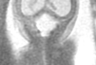
3 Fetal US - Coronal Fetal MR Coronal T2

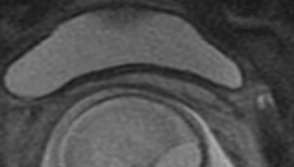
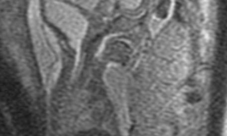
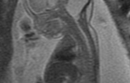
4 Fetal MR - Parasagittal Fetal MR - Parasagittal


5 View of vermis and IVth ventricle from dorsally and superiorly L cerebellar hemisphere Remnant of vermis Floor IVth vent. Clot in IVth vent., compressing R cerebellar hemisphere


6 View of IVth ventricle from dorsally and inferiorly L cerebellar hemisphere with calcification and old hem. Occipital bone Floor IVth vent Distorted R cerebellum


7 Coronal section of cerebral hemispheres Old and recent hemorrhage, and calcification, in floor of lateral ventricles, which are expanded


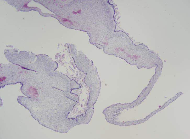
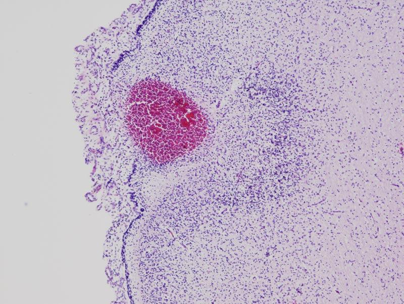
8 - Multifocal old and recent hemorrhage - *Focal cortical disruption and cystic degeneration * Sup. vermis Micro * IVth v. Pons


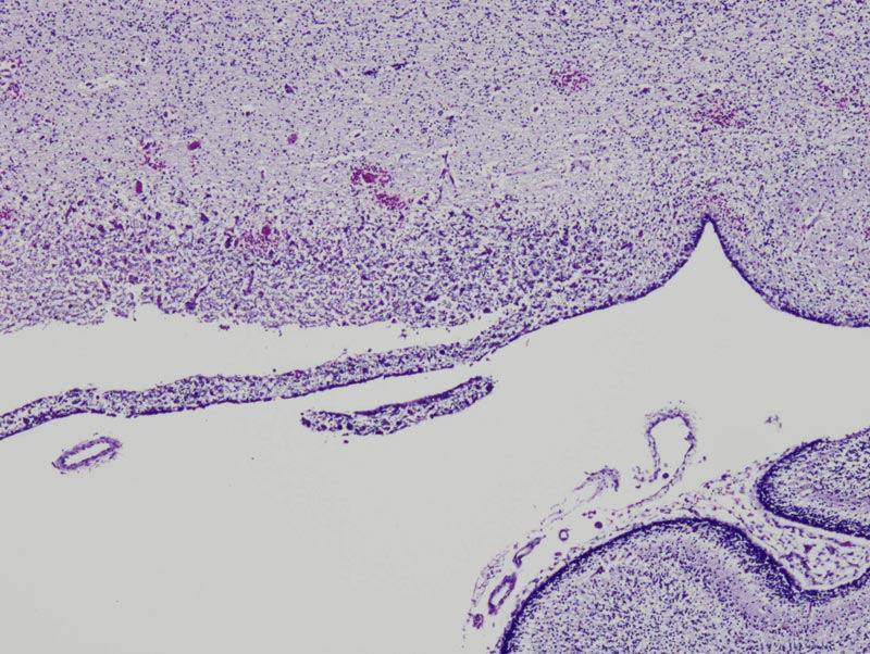
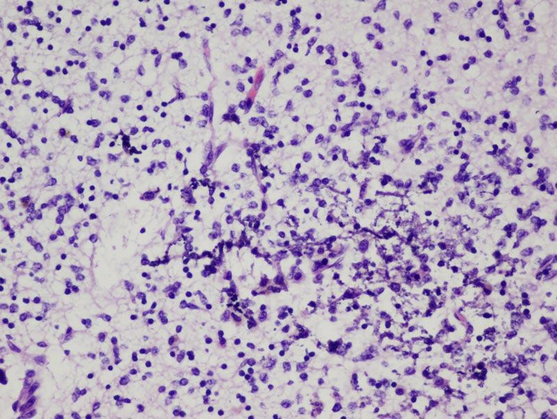
9 Focal cerebellar cortical disruption Focal cystic change in cerebellar cortex
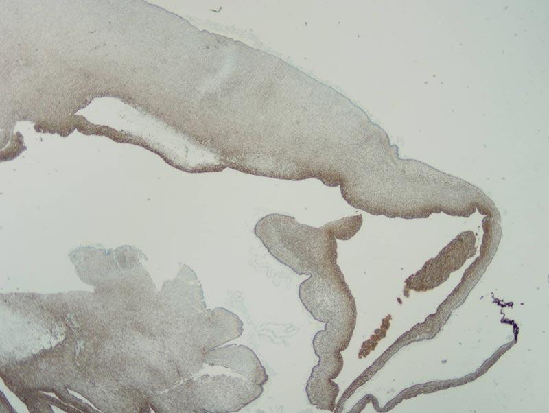
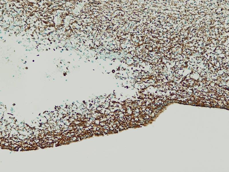
10 Cerebellar cx remnant GFAP
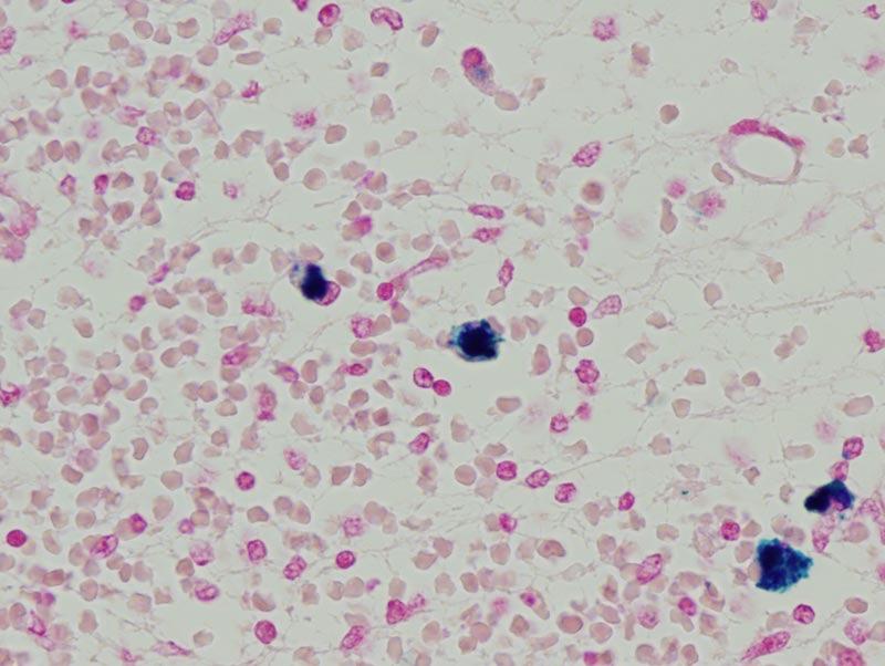
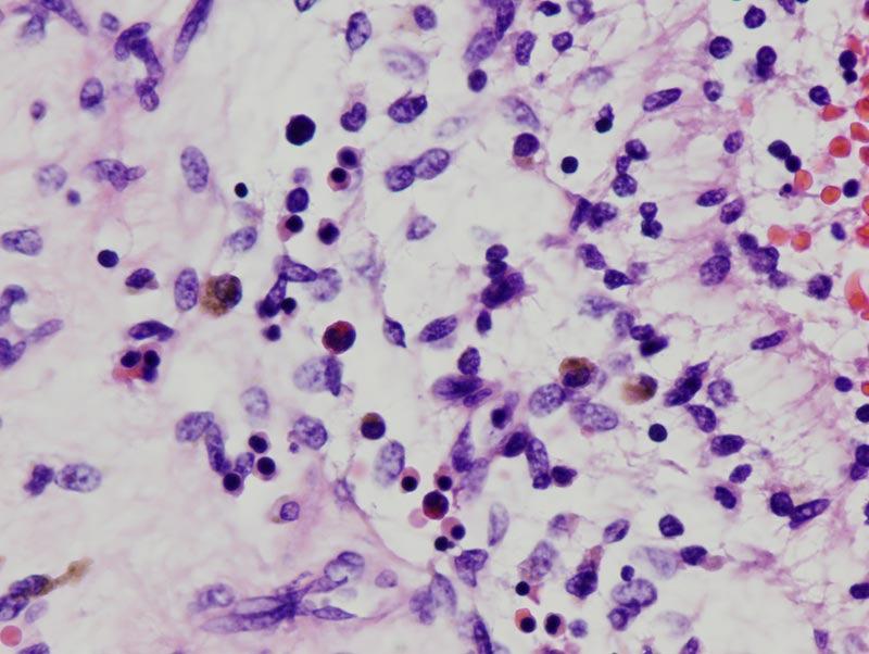
11 Cerebellar cx hemorrhage and mineralization Iron stain
12 Differential Diagnosis Dandy-Walker malformation (DWM; vermian aplasia/hypoplasia), with incidental intrauterine germinal matrix hemorrhage Disruptive lesion involving cerebellum, germinal matrix, and other sites Intrauterine infection (i.e., TORCH) Coagulopathy (e.g., Factor V Leiden deficiency, von Willebrand s disease)
13 Diagnosis Multifocal disruptive hemorrhagic lesions, with: Organizing germinal matrix hemorrhage with intraventricular extension (US grade III) Massive hydrocephalus due to aqueductal obstruction by hemorrhagic debris Acute and chronic ependymitis Organizing hemorrhage with cyst of cerebellar vermis, mimicking D-W Malformation Diffuse petechiae Epidural and subdural hemorrhages, acute and organizing Periventricular leukomalacia Gliosis of thalamus Special stains for toxo, HSV, VZV, CMV - negative
14 Why is this not DWM? DWM is a developmental constellation of: Absence of most or all of cerebellar vermis Cystic dilatation of the fourth ventricle Bowl-shaped expansion of the posterior fossa (indicating chronicity) Association with other CNS midline defects, cardiac and other systemic malformations, which tend to determine the prognosis Hemorrhage, mineralization, etc., are NOT present, and instead indicate disruption of a normally formed brain (= different pathogenesis) (Caveat: cannot absolutely exclude DWM, but not favored)
15 Pathology of typical DWM Note: NO evidence of hemorrhagic disruption


16 Pathogenesis of DWM may involve patterning defects
17 Autopsy in 2 of 3 cases showed organizing hemorrhage in posterior fossa, with tentorial, parenchymal, and subarachnoid hemorrhages Compression and atrophy or necrosis of adjacent structures resulted in cystic changes interpreted on prenatal imaging as developmental lesion (DWM, retrocerebellar dermoid cyst or arachnoid cyst) Presence of blood products in different compartments was key to recognition of disruptive process
18 Clinical implications Possible role of maternal/familial coagulopathy, with subsequent risk for future pregnancies Work-up of mother should be done DWM has very low risk of familial occurrence (<2%); associated anomalies largely determine the prognosis
19 Selected References
20 Acknowledgements Dr. Adre Du Plessis, Neonatal Neurology, Children s Hospital Boston Dr. Catherine Limperopoulos, Neurology and Neuroradiology, Toronto Sick Children s Hospital
21 Key Words Dandy-Walker, cerebellar hypoplasia, germinal matrix hemorrhage, perinatal brain
CNS Embryology 5th Menstrual Week (Dorsal View)
") Imaging of the Fetal Brain; Normal & Abnormal Alfred Abuhamad, M.D. Eastern Virginia Medical School CNS Embryology 5th Menstrual Week (Dorsal View) Day 20 from fertilization Neural plate formed in ectoderm
Imaging of the Fetal Brain; Normal & Abnormal Alfred Abuhamad, M.D. Eastern Virginia Medical School CNS Embryology 5th Menstrual Week (Dorsal View) Day 20 from fertilization Neural plate formed in ectoderm
intracranial anomalies
 Chapter 5: Fetal Central Nervous System 84 intracranial anomalies Hydrocephaly Dilatation of ventricular system secondary to an increase in the amount of CSF. Effects of hydrocephalus include flattening
Han-Sung Kwon M.D. Department of Obstetrics and Gynecology Konkuk University School of Medicine Seoul, Korea
 Han-Sung Kwon M.D. Department of Obstetrics and Gynecology Konkuk University School of Medicine Seoul, Korea Embryologic features of the developing hindbrain Embryologic features of the developing hindbrain
Malformations of the Nervous System November 10, Dr. Peter Ostrow
 Malformations of the Nervous System November 10, 2016 Dr. Peter Ostrow Malformations of the Nervous System 1. Abnormal closure of the neural tube 1. Disorders of forebrain formation 1. Cortical anomalies
Use of MRI in Evaluating Fetal Ventriculomegaly Lisa McLeod, Harvard Medical School Year III Gillian Lieberman, MD
 January 2004 Use of MRI in Evaluating Fetal Ventriculomegaly Lisa McLeod, Harvard Medical School Year III http://bidmc.harvard.edu/content/departments/radiology/files/fetalatlas/default.htm Objectives:
Prenatal Prediction of The Neurologically Impaired Neonate By Ultrasound
 Prenatal Prediction of The Neurologically Impaired Neonate By Ultrasound Robert H. Debbs, D.O.,F.A.C.O.O.G. Professor of OB-GYN Perelman School of Medicine, University of Pennsylvania Director, Pennsylvania
Measurements of the Posterior Fossa in Normal Fetus MRI
 Measurements of the Posterior Fossa in Normal Fetus MRI Ber Roee, 3 rd year medical student, Sackler School of Medicine, Tel Aviv University Supervised by: Dr. Katorza Eldad, Antenatal Diagnostic Unit,The
INTRACRANIAL ARACHNOID CYSTS: CLASSIFICATION AND MANAGEMENT. G. Tamburrini, Rome
 INTRACRANIAL ARACHNOID CYSTS: CLASSIFICATION AND MANAGEMENT G. Tamburrini, Rome Incidence 2% of occasional neuroradiological findings From clinical studies (1960 s): 0.4-1% of intracranial space occupying
Enhancement of Cranial US: Utility of Supplementary Acoustic Windows and Doppler Harriet J. Paltiel, MD
 Enhancement of Cranial US: Utility of Supplementary Acoustic Windows and Doppler Harriet J. Paltiel, MD Boston Children s Hospital Harvard Medical School None Disclosures Conventional US Anterior fontanelle
Complex Hydrocephalus
 2012 Hydrocephalus Association Conference Washington, DC - June 27-July1, 2012 Complex Hydrocephalus Marion L. Walker, MD Professor of Neurosurgery & Pediatrics Primary Children s Medical Center University
Posterior fossa malformations
 ANDREA ROSSI, MD Head, Department of Pediatric Neuroradiology G. Gaslini Children s Research Hospital Genoa Italy andrearossi@ospedale-gaslini.ge.it Posterior fossa malformations Cerebellar ataxia Hypotonia
Neurosonography: State of the art
 Neurosonography: State of the art Lisa H Lowe, MD, FAAP Professor and Academic Chair, University MO-Kansas City Pediatric Radiologist, Children s Mercy Hospitals and Clinics Learning objectives After this
Developmental Neuropathology
 Developmental Neuropathology Pathology, Radiology, and Clinical Correlations Reid Heffner MD Distinguished Teaching Professor Department of Pathology and Anatomy I HAVE NO CONFLICTS OF INTEREST OR DISCLOSURES
General: Brain tumors are lesions that have mass effect distorting the normal tissue and often result in increased intracranial pressure.
 1 Lecture Objectives Know the histologic features of the most common tumors of the CNS. Know the differences in behavior of the different tumor types. Be aware of the treatment modalities in the various
The "Keyhole": A Sign of
 473 The "Keyhole": A Sign of Herniation of a Trapped Fourth Ventricle and Other Posterior Fossa Cysts Barbara J. Wolfson' Eric N. Faerber' Raymond C. Truex, Jr. 2 When a cystic structure in the posterior
Supplementary Online Content
 Supplementary Online Content Honein MA, Dawson AL, Petersen E, et al; US Zika Pregnancy Registry Collaboration. Birth Defects Among Fetuses and Infants of US Women With Laboratory Evidence of Possible
Appendix 3.5 Case Inclusion Guidance for Potentially Zika-related Birth Defects
 Appendix 3.5 Case Inclusion Guidance for Potentially Zika-related Birth Defects Appendix 3.5 A3.5-1 Case Definition Appendix 3.5 Case Inclusion Guidance for Potentially Zika-related Birth Defects Contents
NEURO IMAGING 2. Dr. Said Huwaijah Chairman of radiology Dep, Damascus Univercity
 NEURO IMAGING 2 Dr. Said Huwaijah Chairman of radiology Dep, Damascus Univercity I. EPIDURAL HEMATOMA (EDH) LOCATION Seventy to seventy-five percent occur in temporoparietal region. CAUSE Most likely caused
Chapter 3. Neonatal cranial ultrasonography: how to optimize its performance
 Chapter 3 Neonatal cranial ultrasonography: how to optimize its performance Sylke J. Steggerda Lara M. Leijser Frans J. Walther Gerda van Wezel-Meijler Early Human Development 2009; 85(2): 93-99 Chapter
Symposium: OB/GY US (Room B) CNS Anomalies
 CNS Anomalies") 82 Symposium: OB/GY US (Room B) 11 : 50 1 2 : 10 CNS Anomalies Brain area Midline structure S u p r a t e n t o r i a l ventricular system Cerebral hemisphere Posterior fossa Head size and shape Image
82 Symposium: OB/GY US (Room B) 11 : 50 1 2 : 10 CNS Anomalies Brain area Midline structure S u p r a t e n t o r i a l ventricular system Cerebral hemisphere Posterior fossa Head size and shape Image
Developmental Posterior Fossa Abnormalities with Associated Supratentorial Findings
 Developmental Posterior Fossa Abnormalities with Associated Supratentorial Findings Seattle Children s Hospital Christopher J Hurt, MD Gisele E Ishak, MD Dennis W Shaw, MD Introduction Barkovich has classified
RESEARCH ARTICLE RELATIVE FREQUENCY OF HYDROCEPHALUS IN RASHT PEDIATRIC PATIENTS
 RESEARCH ARTICLE RELATIVE FREQUENCY OF HYDROCEPHALUS IN RASHT PEDIATRIC PATIENTS Elham BIDABADI MD Assistant Professor of Pediatric Neurology, Guilan University of Medical Sciences,Guilan,Iran Corresponding
Fetal obstructive hydrocephalus. Charles Raybaud Hospital for Sick Children, University of Toronto
 Fetal obstructive hydrocephalus Charles Raybaud Hospital for Sick Children, University of Toronto charles.raybaud@sickkids.ca Extensive definition No mention of aqueductal stenosis Purpose Working definition
The view from the mastoid fontanel of the neonatal brain
 The view from the mastoid fontanel of the neonatal brain Poster No.: C-0974 Congress: ECR 2016 Type: Educational Exhibit Authors: Y. Pekcevik, F. C. Sarioglu, H. Sahin ; Karabaglar/Izmir/TR, 1 2 2 1 2
SWISS SOCIETY OF NEONATOLOGY. Severe apnea and bradycardia in a term infant
 SWISS SOCIETY OF NEONATOLOGY Severe apnea and bradycardia in a term infant October 2014 2 Walker JH, Arlettaz Mieth R, Däster C, Division of Neonatology, University Hospital Zurich, Switzerland Swiss Society
Cerebrovascular Accidents in Neonates Treated with Extracorporeal Membrane Oxygenation: Sonographic-Pathologic Correlation
 355 Cerebrovascular Accidents in Neonates Treated with Extracorporeal Membrane Oxygenation: Sonographic-Pathologic Correlation George A. Taylor1 "3 Charles R. Fitz 1-2 Sudesh Kapur2 ' 4 Billie L. Short2-5
Central nervous system. Obstetrics Content Outline Obstetrics - Fetal Abnormalities
 Obstetrics Content Outline Obstetrics - Fetal Abnormalities Many congenital malformations of the CNS result from incomplete closure of the neural tube Effective February 2007 10 16% the most common neural
Spectrum of Cranio-facial anomalies during 2 Ultrasound. trimester on
 Spectrum of Cranio-facial anomalies during 2 Ultrasound nd trimester on Poster No.: C-0378 Congress: ECR 2015 Type: Scientific Exhibit Authors: K. Dave, S. Solanki; Ahmedabad/IN Keywords: Obstetrics (Pregnancy
ECMUS The Safety Committee of EFSUMB : Tutorial
 Neonatal cranial ultrasound Safety Aspects (2013) Prepared for ECMUS by B.J. van der Knoop, M.D. 1, J.I.P. de Vries, M.D., PhD 1, I.A. Zonnenberg, M.D. 2, J.I.M.L. Verbeke, M.D. 3 R.J. Vermeulen, M.D.,
Transfontanelar Ultrasound Technique, Normal Anatomy, Anatomic Variants and Classification Review
 Transfontanelar Ultrasound Technique, Normal Anatomy, Anatomic Variants and Classification Review Poster No.: C-2615 Congress: ECR 2013 Type: Educational Exhibit Authors: S. E. Vazquez, R. E. Ochoa Albíztegui
No conflict of interest to report
 Ultrasound Findings in Fetal Infection No conflict of interest to report Kim A. Boggess MD Ob Gyn UNC at Chapel Hill Learning Objectives At conclusion, participants will Identify maternal infections that
Tumors of the Central Nervous System
 Tumors of the Central Nervous System 1 Financial Disclosures I have NO SIGNIFICANT FINANCIAL, GENERAL, OR OBLIGATION INTERESTS TO REPORT Introduction General: Brain tumors are lesions that have mass effect
DWI assessment of ischemic changes in the fetal brain
 DWI assessment of ischemic changes in the fetal brain Dafi Bergman, 4 th year Medical student in the 4-year program, Sackler school of medicine B.Sc Life and Medical Sciences, Tel Aviv University Supervised
HEAD AND NECK IMAGING. James Chen (MS IV)
") HEAD AND NECK IMAGING James Chen (MS IV) Anatomy Course Johns Hopkins School of Medicine Sept. 27, 2011 OBJECTIVES Introduce cross sectional imaging of head and neck Computed tomography (CT) Review head
HEAD AND NECK IMAGING James Chen (MS IV) Anatomy Course Johns Hopkins School of Medicine Sept. 27, 2011 OBJECTIVES Introduce cross sectional imaging of head and neck Computed tomography (CT) Review head
Neuro. Development. Judy Philbrook, NNP-BC. ! Primary neurulation! Prosencepahlic! Neuronal proliferation. ! 3-4 weeks! 2-3 months!
 Neuro Judy Philbrook, NNP-BC Microsoft clip art Development! Primary neurulation! Prosencepahlic! Neuronal proliferation! Neuronal migration! Organization! Myelination! 3-4 weeks! 2-3 months! 3-4 months!
Insults to the Developing Brain & Effect on Neurodevelopmental Outcomes
 Insults to the Developing Brain & Effect on Neurodevelopmental Outcomes Ira Adams-Chapman, MD Assistant Professor of Pediatrics Director, Developmental Progress Clinic Emory University School of Medicine
Quick practical guide to Cranial Ultrasound in the newborn
 Quick practical guide to Cranial Ultrasound in the newborn Introduction A standard set of views is taken to assist with consistent visualisation of structures and in the interpretation of possible abnormalities.
Genetic test for Bilateral frontoparietal polymicrogyria
 Genetic test for Bilateral frontoparietal polymicrogyria Daniela Pilz, Cardiff UKGTN Genetic testing for neurological conditions; London February 26 th 2013 Region-specific Polymicrogyria (PMG) bilateral
S Alandete, M Meseguer, CR Poyatos, D Uceda, E de la Via, J Sales, J Vilar. H.U. Dr Peset, Valencia (Spain)
") S Alandete, M Meseguer, CR Poyatos, D Uceda, E de la Via, J Sales, J Vilar. H.U. Dr Peset, Valencia (Spain) Introduction Cystic lesions are usually a common finding in clinical practice and you can find
S Alandete, M Meseguer, CR Poyatos, D Uceda, E de la Via, J Sales, J Vilar. H.U. Dr Peset, Valencia (Spain) Introduction Cystic lesions are usually a common finding in clinical practice and you can find
CNS TUMORS. D r. Ali Eltayb ( U. of Omdurman. I ). M. Path (U. of Alexandria)
. M. Path (U. of Alexandria)") CNS TUMORS D r. Ali Eltayb ( U. of Omdurman. I ). M. Path (U. of Alexandria) CNS TUMORS The annual incidence of intracranial tumors of the CNS ISmore than intraspinal tumors May be Primary or Secondary
CNS TUMORS D r. Ali Eltayb ( U. of Omdurman. I ). M. Path (U. of Alexandria) CNS TUMORS The annual incidence of intracranial tumors of the CNS ISmore than intraspinal tumors May be Primary or Secondary
HYDROCEPHALUS OF THE INFANT (ABOUT 86 CASES)
") HYDROCEPHALUS OF THE INFANT (ABOUT 86 CASES) K.EL KHOU;R.ANDALOUSSI;L.OUZIDANE Pediatric radiology department-chu Ibn Rochd Casablanca-Morroco Morroco. Introduction Hydrocephalus of infant is a progressive
HYDROCEPHALUS OF THE INFANT (ABOUT 86 CASES) K.EL KHOU;R.ANDALOUSSI;L.OUZIDANE Pediatric radiology department-chu Ibn Rochd Casablanca-Morroco Morroco. Introduction Hydrocephalus of infant is a progressive
EEG IN FOCAL ENCEPHALOPATHIES: CEREBROVASCULAR DISEASE, NEOPLASMS, AND INFECTIONS
 246 Figure 8.7: FIRDA. The patient has a history of nonspecific cognitive decline and multiple small WM changes on imaging. oligodendrocytic tumors of the cerebral hemispheres (11,12). Electroencephalogram
Differential diagnosis of intracranial cystic lesions.
 Differential diagnosis of intracranial cystic lesions. Poster No.: C-0215 Congress: ECR 2015 Type: Educational Exhibit Authors: S. P. G. Alandete, M. A. Meseguer, E. De la Via, D. Uceda, C. Poyatos; Valencia/ES
Ultrasound examination of the neonatal brain
 Ultrasound examination of the neonatal brain Guideline for the performance and reporting of neonatal and preterm brain ultrasound examination, by the Finnish Perinatology Society and the Paediatric Radiology
Neuroanatomy. Assistant Professor of Anatomy Faculty of Medicine The University of Jordan Dr Maha ELBeltagy
 Neuroanatomy Dr. Maha ELBeltagy Assistant Professor of Anatomy Faculty of Medicine The University of Jordan 2018 Development of the Central Nervous System Development of the nervous system Development
Joana Ramalho, MD C. Ryan Miller, MD, PhD
 Joana Ramalho, MD C. Ryan Miller, MD, PhD Case 1 3 month old baby girl Presented with new onset of seizures Newborn. Questionable blurring of the gray-white junction within the right occipital lobe. Findings
CENTRAL NERVOUS SYSTEM TRAUMA and Subarachnoid Hemorrhage. By: Shifaa AlQa qa
 CENTRAL NERVOUS SYSTEM TRAUMA and Subarachnoid Hemorrhage By: Shifaa AlQa qa Subarachnoid Hemorrhage Causes: Rupture of a saccular (berry) aneurysm Vascular malformation Trauma Hematologic disturbances
Original Article CT grouping and microsurgical treatment strategies of hypertensive cerebellar hemorrhage
 Int J Clin Exp Med 2016;9(8):15921-15927 www.ijcem.com /ISSN:1940-5901/IJCEM0022273 Original Article CT grouping and microsurgical treatment strategies of hypertensive cerebellar hemorrhage Xielin Tang
Head CT Scan Interpretation: A Five-Step Approach to Seeing Inside the Head Lawrence B. Stack, MD
 Head CT Scan Interpretation: A Five-Step Approach to Seeing Inside the Head Lawrence B. Stack, MD Five Step Approach 1. Adequate study 2. Bone windows 3. Ventricles 4. Quadrigeminal cistern 5. Parenchyma
REVIEW ARTICLE Egypt. J. Hum. Genet. Vol. 8, No. 2, Nov Dandy-Walker Malformation
 REVIEW ARTICLE Egypt. J. Hum. Genet. Vol. 8, No. 2, Nov. 2007 Medical Genetics Center, Ain Shams University INTRODUCTION Dandy-Walker malformation is a rare congenital malformation and involves the cerebellum
ISUOG Basic Training. Assessing the Neck & Chest Gihad Chalouhi, Lebanon
 ISUOG Basic Training Assessing the Neck & Chest Gihad Chalouhi, Lebanon Learning objectives 9 & 10 At the end of the lecture you will be able to: recognise the differences between the normal & most common
Central nervous system
 Chapter 2 Central nervous system NORMAL SONOGRAPHIC ANATOMY The fetal brain undergoes major developmental changes throughout pregnancy. At 7 weeks of gestation, a sonolucent area is seen in the cephalic
Neonatal Intracranial Ultrasound Imaging - A Pictorial Review from The Royal Liverpool Children's Hospital, Alder Hey, Liverpool.
 Neonatal Intracranial Ultrasound Imaging - A Pictorial Review from The Royal Liverpool Children's Hospital, Alder Hey, Liverpool. Poster No.: C-1115 Congress: ECR 2012 Type: Educational Exhibit Authors:
Brain Meninges, Ventricles and CSF
 Brain Meninges, Ventricles and CSF Lecture Objectives Describe the arrangement of the meninges and their relationship to brain and spinal cord. Explain the occurrence of epidural, subdural and subarachnoid
AAP ZIKA ECHO (EXTENSION FOR COMMUNITY HEALTHCARE OUTCOMES)
") AAP ZIKA ECHO (EXTENSION FOR COMMUNITY HEALTHCARE OUTCOMES) HOUSEKEEPING ITEMS For educational and quality improvement purposes, this ECHO session will be recorded Project ECHO collects participation data
AAP ZIKA ECHO (EXTENSION FOR COMMUNITY HEALTHCARE OUTCOMES) HOUSEKEEPING ITEMS For educational and quality improvement purposes, this ECHO session will be recorded Project ECHO collects participation data
Index. aneurysm, 92 carotid occlusion, 94 ICA stenosis, 95 intracranial, 92 MCA, 94
 A ADC. See Apparent diffusion coefficient (ADC) Aneurysm cerebral artery aneurysm, 93 CT scan, 93 gadolinium, 93 Angiography, 13 Anoxic brain injury, 25 Apparent diffusion coefficient (ADC), 7 Arachnoid
The central nervous system
 Sectc.qxd 29/06/99 09:42 Page 81 Section C The central nervous system CNS haemorrhage Subarachnoid haemorrhage Cerebral infarction Brain atrophy Ring enhancing lesions MRI of the pituitary Multiple sclerosis
Torch Infections and Prenatal Ultrasound Findings
 Tutorial [1] August 09, 2011 By Eran Casiff, MD [2] TORCH INFECTIONS AND PRENATAL ULTRASOUND FINDINGS Eran Casiff M.D. Department of Obstetrics and Gynecology Kaplan Medical Center Rehovot 76100, Israel
What Are We Going to Do? Fourth Year Meds Clinical Neuroanatomy. Hydrocephalus and Effects of Interruption of CSF Flow. Tube Blockage Doctrine
 Fourth Year Meds Clinical Neuroanatomy Ventricles, CSF, Brain Swelling etc. David A. Ramsay, Neuropathologist, LHSC What Are We Going to Do? Hydrocephalus and some effects of the interruption of CSF flow
Fetal Medicine. Case Presentations. Dr Ermos Nicolaou Fetal Medicine Unit Chris Hani Baragwanath Hospital. October 2003
 Case Presentations Dr Ermos Nicolaou Fetal Medicine Unit Chris Hani Baragwanath Hospital October 2003 Case 1 Ms A M 22year old P0 G1 Referred from Sebokeng Hospital at 36w for polyhydramnios On Ultrasound:
Classical CNS Disease Patterns
 Classical CNS Disease Patterns Inflammatory Traumatic In response to the trauma of having his head bashed in GM would have experienced some of these features. NOT TWO LITTLE PEENY WEENY I CM LACERATIONS.
Dandy-Walker syndrome: different modalities of treatment and outcome in 42 cases
 Child s Nerv Syst (2001) 17:348 352 DOI 10.1007/s003810000425 ORIGINAL PAPER Raj Kumar Manoj Kumar Jain Devendra Kumar Chhabra Dandy-Walker syndrome: different modalities of treatment and outcome in 42
1 MS Lesions in T2-Weighted Images
 1 MS Lesions in T2-Weighted Images M.A. Sahraian, E.-W. Radue 1.1 Introduction Multiple hyperintense lesions on T2- and PDweighted sequences are the characteristic magnetic resonance imaging (MRI) appearance
Pediatric Neurointervention: Vein of Galen Malformations
 Pediatric Neurointervention: Vein of Galen Malformations Johanna T. Fifi, M.D. Assistant Professor of Neurology, Neurosurgery, and Radiology Icahn School of Medicine at Mount Sinai November 9 th, 2014
GEORGE E. PERRET, M.D., AND CARL J. GRAF, M.D.
 J Neurosurg 47:590-595, 1977 Subgaleal shunt for temporary ventricle decompression and subdural drainage GEORGE E. PERRET, M.D., AND CARL J. GRAF, M.D. Division of Neurological Surgery, University of Iowa
-Damage to arachnoid villi by haemorrhage or infection -Congenital absence of arachnoid villi"5 -Tumour -Infection. -Haemorrhage
 J Clin Pathol 1997;50:181-186 181 Leaders Department of Neuropathology, Radcliffe Infirmary, Oxford OX2 6HE M V Squier Accepted for publication 11 December 1996 Pathological approach to the diagnosis of
For Emergency Doctors. Dr Suzanne Smallbane November 2011
 For Emergency Doctors Dr Suzanne Smallbane November 2011 A: Orbit B: Sphenoid Sinus C: Temporal Lobe D: EAC E: Mastoid air cells F: Cerebellar hemisphere A: Frontal lobe B: Frontal bone C: Dorsum sellae
Meninges and Ventricles
 Meninges and Ventricles Irene Yu, class of 2019 LEARNING OBJECTIVES Describe the meningeal layers, the dural infolds, and the spaces they create. Name the contents of the subarachnoid space. Describe the
Benign brain lesions
 Benign brain lesions Diagnostic and Interventional Radiology Hung-Wen Kao Department of Radiology, Tri-Service General Hospital, National Defense Medical Center Computed tomography Hounsfield unit (HU)
Neglected case of hydrocephalus in a five-year-old child
 www.edoriumjournals.com CASE REPORT PEER REVIEWED OPEN ACCESS Neglected case of hydrocephalus in a five-year-old child Moataz Hesham Abdelreheem, Marwa Mohammed Basyouni ABSTRACT Introduction: Hydrocephalus
Basic Training. ISUOG Basic Training Distinguishing Between Normal & Abnormal Appearances of the Skull & Brain
 ISUOG Distinguishing Between Normal & Abnormal Appearances of the Skull & Brain Learning objectives At the end of the lecture you will be able to: Describe how to obtain the 3 planes required to assess,
Cerebro-vascular stroke
 Cerebro-vascular stroke CT Terminology Hypodense lesion = lesion of lower density than the normal brain tissue Hyperdense lesion = lesion of higher density than normal brain tissue Isodense lesion = lesion
DES 9 janvier P. David. Clinic of Neuroradiology Erasme Hospital Université Libre de Bruxelles Belgium
 DES 9 janvier 2015 P. David Clinic of Neuroradiology Erasme Hospital Université Libre de Bruxelles Belgium CNS Infections Early recognition in children, infants Longterm effects on the brain :devastating
CNS pathology Third year medical students. Dr Heyam Awad 2018 Lecture 12: CNS tumours 2/3
 CNS pathology Third year medical students Dr Heyam Awad 2018 Lecture 12: CNS tumours 2/3 Pilocytic astrocytoma Relatively benign ( WHO grade 1) Occurs in children and young adults Mostly: in the cerebellum
Posterior Fontanelle Sonography: An Acoustic Window into the Neonatal Brain
 AJNR Am J Neuroradiol 25:1274 1282, August 2004 Posterior Fontanelle Sonography: An Acoustic Window into the Neonatal Brain Flavia Correa, Goya Enríquez, José Rosselló, Javier Lucaya, Joaquim Piqueras,
IMAGING OF HYPOXIC ISCHEMIC INJURY IN A NEONATE FN3 STATE MEETING NEMOURS CHILDREN'S HOSPITAL ORLANDO,FL 08/04/18
 IMAGING OF HYPOXIC ISCHEMIC INJURY IN A NEONATE FN3 STATE MEETING NEMOURS CHILDREN'S HOSPITAL ORLANDO,FL 08/04/18 Dhanashree Rajderkar,MD Assistant Professor Department of Radiology University of Florida
Supplemental Information
 ARTICLE Supplemental Information SUPPLEMENTAL TABLE 6 Mosaic and Partial Trisomies Thirty-eight VLBW infants were identified with T13, of whom 2 had mosaic T13. T18 was reported for 128 infants, of whom
GLIOEPENDYMAL AND ARACHNOID CYSTS: UNUSUAL CAUSES OF EARLY VENTRICULOMEGALY IN UTERO
 PRENATAL DIAGNOSIS, VOL. 16: 729-733 (1996) SHORT COMMUNICATION GLIOEPENDYMAL AND ARACHNOID CYSTS: UNUSUAL CAUSES OF EARLY VENTRICULOMEGALY IN UTERO JAMIYAH HASSAN*, WALDO SEPULVEDA*, JERONIMA TEIXEIRA*
Cerebellar Vermian Atrophy after Neonatal Hypoxic-Ischemic Encephalopathy
 AJNR Am J Neuroradiol 25:1008 1015, June/July 2004 Vermian Atrophy after Neonatal Hypoxic-Ischemic Encephalopathy Michael A. Sargent, Kenneth J. Poskitt, Elke H. Roland, Alan Hill, and Glenda Hendson BACKGROUND
MR CASE STUDY: Autopsy-proven CMV fetopathy.
 MR CASE STUDY: Autopsy-proven CMV fetopathy. HISTORY: Elderly primigravida patient with normal first trimester screen presents at 21-weeks for detailed anatomy scan. At that time, ultrasound demonstrates
Neuroimaging in Cerebral Palsy Report from North India
 original ARTICLE Neuroimaging in Cerebral Palsy Report from North India How to Cite This Article: Aggarwal A, Mittal H, Debnath SKR, Rai A. Neuroimaging in Cerebral Palsy Report from North India. Iran
Intracranial CSF Flow in Pediatric Hydrocephalus: Evaluation with Cine-MR Imaging
 Intracranial CSF Flow in Pediatric Hydrocephalus: Evaluation with Cine-MR Imaging Robert M. Quencer 1 Purpose: 1) To describe the pattern of normal intracranial CSF flow in children and 2) to demonstrate
Reference ranges for the linear dimensions of the intracranial ventricles in preterm neonates
 F8 Division of Neonatal Services, Royal Women s Hospital, Melbourne 33, Australia M W Davies M Swaminathan S L Chuang F R Betheras Correspondence to: Dr M W Davies, Perinatal Research Centre, The Royal
ISUOG Basic Training. Distinguishing Between Normal & Abnormal Appearances of the Skull & Brain. Seshadri Suresh, India
 ISUOG Basic Training Distinguishing Between Normal & Abnormal Appearances of the Skull & Brain Seshadri Suresh, India Learning objectives 4 & 5 At the end of the lecture you will be able to: Describe how
A Rare Aggressive Fetal Intracranial Tumor
 J. Fetal Med. (June 2015) 2:91 95 DOI 10.1007/s40556-015-0046-y BRIEF COMMUNICATION A Rare Aggressive Fetal Intracranial Tumor Ravi Kapoor 1 Ashutosh Bansal 1 Aakriti Kapoor Aggarwal 2 Abhinav Aggarwal
Attenuation value in HU From -500 To HU From -10 To HU From 60 To 90 HU. From 200 HU and above
 Brain Imaging Common CT attenuation values Structure Air Fat Water Brain tissue Recent hematoma Calcifications Bone Brain edema and infarction Normal liver parenchyma Attenuation value in HU From -500
SWISS SOCIETY OF NEONATOLOGY. Symptomatic congenital CMV infection after recurrent maternal infection
 SWISS SOCIETY OF NEONATOLOGY Symptomatic congenital CMV infection after recurrent maternal infection May 2017 Mack I, Burckhardt MA, Heininger U, Prüfer F, Wellmann S, Department of Pediatric Infectious
Neuropathology of cerebral palsy
 C. Hagel (Hamburg/Germany) Hypoxia/ischemia related brain damage is a major factor for morbidity and mortality not only in the adult, but also in the pre- and perinatal period where it mainly presents
Perinatal cerebral white matter injuries influence early communication and language development
 Perinatal cerebral white matter injuries influence early communication and language development Blazenka Brozovic University of Zagreb Department of Speech and Language Pathology Developmental Neurolinguistic
Neonatal hypoxic-ischemic brain injury imaging: A pictorial review
 Neonatal hypoxic-ischemic brain injury imaging: A pictorial review Poster No.: C-1425 Congress: ECR 2014 Type: Educational Exhibit Authors: E. Alexopoulou 1, A. Mazioti 1, D. K. Filippiadis 2, C. Chrona
Marc Norman, Ph.D. - Do Not Use without Permission 1. Cerebrovascular Accidents. Marc Norman, Ph.D. Department of Psychiatry
 Cerebrovascular Accidents Marc Norman, Ph.D. Department of Psychiatry Neuropsychiatry and Behavioral Medicine Neuropsychology Clinical Training Seminar 1 5 http://www.nlm.nih.gov/medlineplus/ency/images/ency/fullsize/18009.jpg
BRAIN STEM AND CEREBELLUM..
 Lecture Title: BRAIN STEM AND CEREBELLUM.. (CNS Block, Radiology) Dr. Hamdy Hassan Ass.Prof. Consultant Radiology Department KKHU King Saud University Lecture Objectives.. Students at the end of the lecture
The rationale for routine cerebral ultrasound in premature infants
 Pediatr Radiol (2015) 45:646 650 DOI 10.1007/s00247-014-2985-1 RESEARCH FORUM The rationale for routine cerebral ultrasound in premature infants Maria I. Argyropoulou & Corinne Veyrac Received: 4 October
Vascular Malformations of the Brain: A Review of Imaging Features and Risks
 Vascular Malformations of the Brain: A Review of Imaging Features and Risks Comprehensive Neuroradiology: Best Practices October 27-30, 2016 Sudhakar R. Satti, MD Associate Director Neurointerventional
MR imaging of neonatal brain infections: Germ and specific signs
 MR imaging of neonatal brain infections: Germ and specific signs Poster No.: C-1646 Congress: ECR 2016 Type: Educational Exhibit Authors: N. Mama 1, A. Cherif 1, Y. BEN CHEIKH 2, A. Berrich 1, M. Gaha
Cribside Neurosonography:
 501 Cribside Neurosonography: Real-Time Sonography for Intracranial Investigation of the Neonate Mary K. Edwards 1 David L. Brown 1 Jans Muller Charles B. Grossman 1 Gonzalo T. Chua 1 prospective study
2 nd Trimester Anomaly Scan What you can see & What you must see
 2 nd Trimester Anomaly Scan What you can see & What you must see D.Paladini Fetal Medicine & Surgery Unit Gasllini Children s Hospital - Genoa dpaladini49@gmail.com All images in this lecture were taken
Gerda van Wezel-Meijler Neonatal Cranial Ultrasonography
 Gerda van Wezel-Meijler Neonatal Cranial Ultrasonography Gerda van Wezel-Meijler Neonatal Cranial Ultrasonography Guidelines for the Procedure and Atlas of Normal Ultrasound Anatomy With 121 Figures and
PRENATAL DIAGNOSIS OF ARACHNOID CYSTS
 REVIEW ARTICLE PRENATAL DIAGNOSIS OF ARACHNOID CYSTS Chih-Ping Chen* Department of Obstetrics and Gynecology, Mackay Memorial Hospital, Taipei, and Department of Biotechnology, Asia University, Taichung,
Developmental Neuropathology
 Developmental Neuropathology EARLY Anterior closure E26 Posterior closure E28 Anencephaly E16-E26 Spina Bifida Holoprosencephaly (anterior midline closure) MID-GESTATION Neuronal migration Gyral formation