Developmental Neuropathology
|
|
|
- Myles Joseph
- 5 years ago
- Views:
Transcription


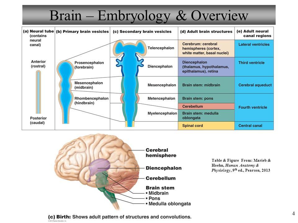
1 Developmental Neuropathology Pathology, Radiology, and Clinical Correlations Reid Heffner MD Distinguished Teaching Professor Department of Pathology and Anatomy
2 I HAVE NO CONFLICTS OF INTEREST OR DISCLOSURES TO DECLARE. I HAVE NO FINANCIAL INTEREST IN, I RECEIVE NO FINANCIAL SUPPORT FROM AND I HAVE NO CONTRACTS WITH OR GRANTS FROM ANY PHARMACEUTICAL COMPANY OR ANY INDUSTRY
3 Contents A. MALFORMATIONS -Microcephaly -Hydrocephalus -Neural tube defects -Midline cerebral defects -Cell migration disorders -Cerebellar anomalies -Spinal cord lesions
4 Contents B. PERINATAL DISORDERS - Periventricular leukomalacia - Germinal matrix hemorrhage - Porencephaly
5 CNS malformations Incidence 1-2% of live births More common in setting of multiple birth defects Different parts of brain develop at different times, so different malformations depend on when injury occurs
6 Causes of CNS malformations Chromosome abnormalities Mutations Maternal factors Infections Medications (valproate), alcohol Diabetes Vitamin deficiencies-ex: folate X-ray exposure
7 Causes of CNS malformations Microcephaly: Viral infections Hydrocephaly: LiCAM gene Neural tube defects: Valproate, folic acid, MTHFR gene Midline defects Holoprosencepaaly: many genes incl. SHH, ZIC2 Agenesis of corpus callosum: Fetal alcohol Cell migration disorders Polymicrogyria: 22q.11 deletions Heteropias Cerebellar abnormalities Arnold-Chiari Dandy-Walker: FoxC1(transcription factor) Spinal cord lesions-hox genes

8 Ultrasound(20 weeks) Chromosome analysis Blood sample-folate Screening
9

10 Four week embryo SHH gene LiCAM gene FoxC1 gene HOX genes
11 Prevalence of CNS malformations Composite of several studies Neural tube disorders 35% Hydrocephalus 20-30% Microcephaly 8-15% Midline disorders 10% Cerebellar disorders 10% Syrinx 1-2% Polymicrogyria 1-2%

 56 hr after infection with ZIKV supernatant, immunostained for ZIKV envelop protein (ZIKVE; green) and DAPI (gray). (D) Production of infectious ZIKV particles by infected hnpcs.")
12 Zika Virus Infects Human Cortical Neural Progenitors and Attenuates Their Growth Hengli Tang, Christy Hammack, Sarah C. Ogden, Zhexing Wen, Xuyu Qian, Yujing Li, Bing Yao, Jaehoon Shin, Feiran Zhang, Emily M. Lee, Kimberly M. Christian, Ruth A. Didier, Peng Jin, Hongjun Song, Guo-li Ming ZIKV Infects hipsc-derived Neural Progenitor Cells with High Efficiency (A and B) Sample confocal images of forebrain-specific hnpcs (A) and immature neurons (B) 56 hr after infection with ZIKV supernatant, immunostained for ZIKV envelop protein (ZIKVE; green) and DAPI (gray). (D) Production of infectious ZIKV particles by infected hnpcs. Supernatant from hnpc cultures 72 hr after ZIKV infection was collected and added to Vero cells for 2 hr. The Vero cells were further cultured for 48 hr. Shown are sample images of ZIKVE immunostaining (green) and DAPI (gray). Scale bars, 20 μm. Copyright 2016 Elsevier Inc. Terms and Conditions
13 Microcephaly 3-18% of CNS malformations Intrauterine infections Zika virus Toxoplasmosis CMV Rubella Medications during pregnancy Alcohol, toxin exposure in utero X-rays (in rats, maybe humans)


14 Brain is also small Destructive CNS lesions Gliosis Calcifications Neuronal loss
15 Macrocephaly
16
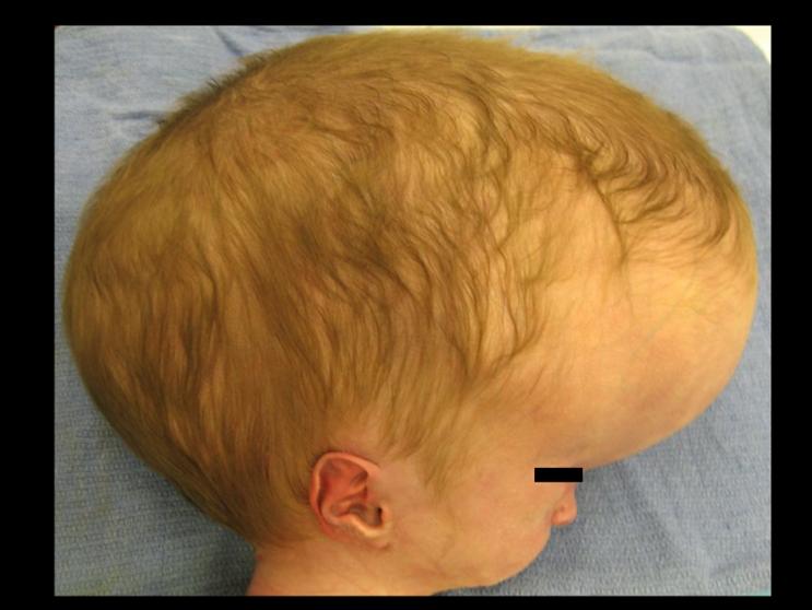
17 Hydrocephalus in newborn Causes Aqueduct stenosis X-linked, L1CAM gene Infections in utero Tumors usually later in life Cognitive and motor deficits Treated by shunting Ventriculoperitoneal catheter
18 HYDROCEPHALUS Acute or chronic High pressure Block in ventricular flow Block in meningeal circulation Block in reabsorption Normal pressure Hydrocephalus Ex vacuo About 500 ml CSF produced each day

19 CSF drainage system Arachnoid villi


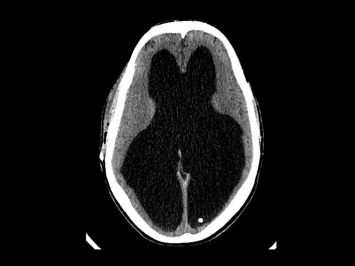
20 Obstructive hydrocephalus Notice the loss of white matter and intact cortex

21 Aqueductal stenosis, atresia, forking

22 Aqueductal stenosis Gliosis of aqueduct, forking (branching into blind alleys)
23 Neural tube defects -Anencephaly -Encephalocele -Spinal dysraphism (Spina bifida or cut in two)

24 Neural tube defects- 8/1000 births Closure by 4 weeks
25 Neural tube defects (dysraphism) Failure of cranial or spinal neural tube closure Teratogens (valproic acid) Genetic factors-200 genes MTHFR gene Increased a-fetoprotein in maternal serum or amniotic fluid Prevention: folic acid (folate deficiency causes disease)


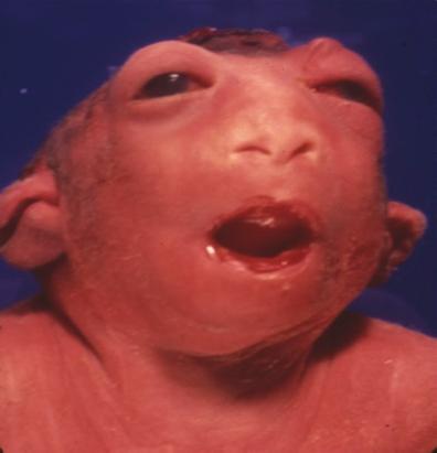
26 Anencephaly Failure of neural tube and anterior neuropore to close Baby has frog-like appearance Begins as exencephaly and neural tissue destroyed by amniotic fluid Remaining tissue is a disorganized mass of neural and vascular tissue This is called the cerebrovasculosa

27 Anencephaly incompatible with life Pituitary insufficiency
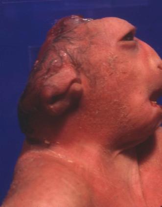
28 Anencephaly


29 Herniation of brain tissue through cranial defect. Occipital is most common 55% survival Encephalocele
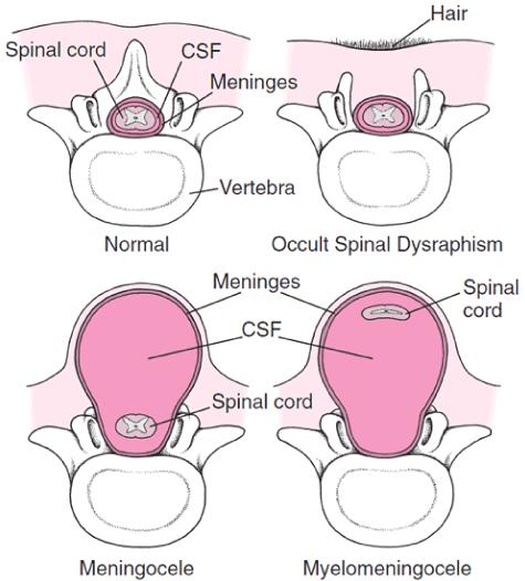
30 Spinal Dysraphism

31 Meningomyelocele Occurs in lumbar sacral region Associated with Arnold-Chiari II malformation Sphincter control problems, paralysis, infection


32 Protrusion of meninges and subarachnoid space through bony defect Meningocele
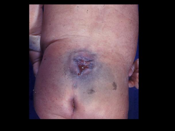
33 Meningocele

34 Midline cerebral defects Holoprosencephaly Arhinencephaly Cyclopia Agenesis of corpus callosum Sonic hedgehog gene Patterning gene Specialization Developing midline structures


35 Holoprosencephaly Failure of formation of the forebrain vesicles Frequently associated with facial anomalies (cyclopia) Associated with mutations of SHH, Zic2, other genes One hemisphere instead of two Abnormal midline structures Single ventricle


36 Holoprosencephaly Posterior fossa structures are normal Anterior Posterior

37 Cyclopia One eye Often have holoprosencpaly Incompatible with life


38 Arhinencephaly can occur alone without HPE Cleft palate and hair lip Agenesis of olfactory nerve, gyrus rectus, olfactory cortex
39 Agenesis of the corpus callosum Causes 8/1000 live births May be recessive or X-linked Infection in utero-12-26th week Fetal alcohol syndrome Clinical features Seizures Feeding problems Psychomotor retardation Associated disorders Polymicrogyria Absence of cingulate sulcus and cingulate gyrus


40 Agenesis of the corpus callosum Normal corpus callosum


41 Agenesis of the corpus callosum Absence of cingulate gyrus
42 Migration disorders Polymicrogyria Heterotopias


43 Normal fetal brain development 17 wk 22 wk 30 wk 28 wk Period of cortical and deep gray matter development
 in germinal matrix")
44 Polymicrogyria is an abnormal migration of neuroblasts to the surface Migrate along radial glia Neuroblasts (stem cells) in germinal matrix

45 Polymicrogyria Usually occurs after fifth month of development Causes are genetic mutations, intrauterine infections and hypoxia May be diffuse or focal Cortical gyri are small; cortical structure abnormal


46 Migration Disorders: Polymicrogyria Diverse etiologies 22q.11 deletion Frequently associated with Epilepsy Other symptoms depend on extent of involvement

47 Polymicrogyria Cortex simplified, consists of 3-4 layers Cobblestone appearance grossly

48 Normal cortical layers polymicrogyria Small convolutions, numerous shallow sulci Three to four layers, not normal six layers


49 Heterotopia Foci of grey matter in white matter or beneath the ependyma Migrating neuroblasts from germinal matrix get stuck and don t proceed to the cortex
50 Cerebellar anomalies Arnold-Chiari malformations Causes are poorly understood Dandy-Walker malformation
51 Arnold-Chiari malformations TYPE I (Chiari malformation) Cerebellar tonsils extend through foramen magnum (chronic tonsillar herniation) Often associated with syringomyelia Not symptomatic until teen years (neck pain, ataxia) TYPE II Vermis, lower hemispheres, medulla extend into spinal canal Brainstem abnormalities-beaking of tectum Meningomyelocele 95% Hydrocephalus common

52 Chiari type I

53 Arnold-Chiari type II Pathogenesis unknown. Cerebellar herniation associated with lumbar myelomeningocele. Beaking of the tectum Elongation of medulla and fourth ventricle 22 wk fetus

54 Chiari II Extension of cerebellar vermis and medulla through foramen magnum into the spinal canal

55 Arnold-Chiari malformation II Extension of vermis and medulla into spinal canal Beaking of midbrain tectum Hydrocephalus
 implicated in some cases Ataxia Hydrocephalus")
56 Dandy-Walker Malformation FoxC1 gene (normal cerebellar development) implicated in some cases Ataxia Hydrocephalus Absence of vermis


57 Dandy-Walker Malformation Cerebellar vermis is absent; cyst is an expanded 4 th ventricle that communicates with the subarachnoid space.
58 Syringomyelia-causes Syrinx is a tube Congenital Arnold-Chiari malformation Neural tube defects Idiopathic-occurring alone May be congenital? Role of the HOX genes Tumors (gliomas), later in like


59 Syringomyelia a fluid-filled cleft within the cord Disease often asymptomatic in childhood Dorsal half of cervical cord commonly involved Cyst expands during life Symptoms result from compression of normal spinal cord structures

60 Tracts of spinal cord Syringomyelia Symptoms in upper extremities Pain or loss of sensation-spinothalamic tract Weakness-pyramidal tract


61 Syrinx often surrounded by gliosis with Rosenthal fibers Cyst has no lining cells Syringomyelia
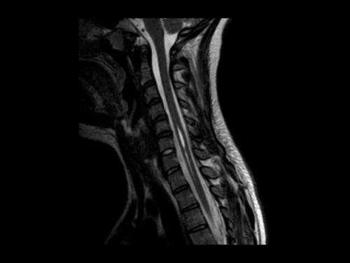
62 Syrinx T2
63 Learning objectives Correlate malformations with embryology of CNS List genes and their respective malformations Describe each malformation pathologically Explain the clinical features of each developmental disease Compare the malformations to each other Discuss how you would diagnose each developmental disease Characterize the outcome of each nalformation


64 The most common malformation
65 Contents B. PERINATAL DISORDERS - Periventricular leukomalacia - Germinal matrix hemorrhage - Porencephaly
66 Perinatal developmental lesions: susceptibility Susceptibility to hypoxia-ischemia, hemorrhage, etc. depends on age

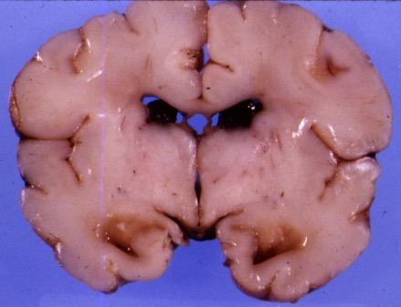
67 Periventricular leukomalacia See bilateral symmetrical white matter lesions in cerebrum PVL is essentially a watershed infarction of subcortical tissue

68 Periventricular leukomalacia Occurs around time of birth Basically an ischemic/hypoxic lesion Affects cerebral white matter, not cortex Looks like there is loss of tissue, discoloration

69 Periventricular leukomalacia Sometimes there is cystic change in the white matter


70 Periventricular leukomalacia Palor of the subcortical structures Gliosis with microglia, astrocytes May be small areas of calcification

71 Germinal matrix Primitive stem cells next to lateral ventricle

72 Germinal Matrix Hemorrhage Premature infants Birth weight<1,500 gm or gestation <35 weeks. Hyaline membrane disease Occurs hrs after birth

73 Germinal matrix hemorrhage Fetal anoxia begins the process Acute venous congestion Dilation of terminal vein Capillary fragility in matrix Matrix substance is also loosely coherent Hemorrhage develops May rupture into ventricle Before ultrasound, it was thought to be uniformly fatal
74 Germinal matrix hemorrhage: Grading Grade 1 Isolated subependymal hemorrhage (SEH) Grade 2 SEH with intraventricular hemorrhage (IVH) but no ventricular enlargement Grade 3 SHE + IVH + enlarged ventricles Grade 4 SHE + IVH + extension of hemorrhage into brain tissue

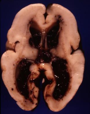
75 Germinal Matrix Hemorrhage

76 WHAT IS SEEN HERE? Cystic structure in the brain, often communicates with ventricle Essentially an infarct occurring in utero Symptoms depend on size and location

77 Porencephaly Unilateral, cystic lesion, partially lined by ependymal or arachnoid
Malformations of the Nervous System November 10, Dr. Peter Ostrow
 Malformations of the Nervous System November 10, 2016 Dr. Peter Ostrow Malformations of the Nervous System 1. Abnormal closure of the neural tube 1. Disorders of forebrain formation 1. Cortical anomalies
intracranial anomalies
 Chapter 5: Fetal Central Nervous System 84 intracranial anomalies Hydrocephaly Dilatation of ventricular system secondary to an increase in the amount of CSF. Effects of hydrocephalus include flattening
Fetal Medicine. Case Presentations. Dr Ermos Nicolaou Fetal Medicine Unit Chris Hani Baragwanath Hospital. October 2003
 Case Presentations Dr Ermos Nicolaou Fetal Medicine Unit Chris Hani Baragwanath Hospital October 2003 Case 1 Ms A M 22year old P0 G1 Referred from Sebokeng Hospital at 36w for polyhydramnios On Ultrasound:
Supplementary Online Content
 Supplementary Online Content Honein MA, Dawson AL, Petersen E, et al; US Zika Pregnancy Registry Collaboration. Birth Defects Among Fetuses and Infants of US Women With Laboratory Evidence of Possible
CNS Embryology 5th Menstrual Week (Dorsal View)
") Imaging of the Fetal Brain; Normal & Abnormal Alfred Abuhamad, M.D. Eastern Virginia Medical School CNS Embryology 5th Menstrual Week (Dorsal View) Day 20 from fertilization Neural plate formed in ectoderm
Imaging of the Fetal Brain; Normal & Abnormal Alfred Abuhamad, M.D. Eastern Virginia Medical School CNS Embryology 5th Menstrual Week (Dorsal View) Day 20 from fertilization Neural plate formed in ectoderm
Prenatal Prediction of The Neurologically Impaired Neonate By Ultrasound
 Prenatal Prediction of The Neurologically Impaired Neonate By Ultrasound Robert H. Debbs, D.O.,F.A.C.O.O.G. Professor of OB-GYN Perelman School of Medicine, University of Pennsylvania Director, Pennsylvania
Central nervous system. Obstetrics Content Outline Obstetrics - Fetal Abnormalities
 Obstetrics Content Outline Obstetrics - Fetal Abnormalities Many congenital malformations of the CNS result from incomplete closure of the neural tube Effective February 2007 10 16% the most common neural
Neuroanatomy. Assistant Professor of Anatomy Faculty of Medicine The University of Jordan Dr Maha ELBeltagy
 Neuroanatomy Dr. Maha ELBeltagy Assistant Professor of Anatomy Faculty of Medicine The University of Jordan 2018 Development of the Central Nervous System Development of the nervous system Development
Symposium: OB/GY US (Room B) CNS Anomalies
 CNS Anomalies") 82 Symposium: OB/GY US (Room B) 11 : 50 1 2 : 10 CNS Anomalies Brain area Midline structure S u p r a t e n t o r i a l ventricular system Cerebral hemisphere Posterior fossa Head size and shape Image
82 Symposium: OB/GY US (Room B) 11 : 50 1 2 : 10 CNS Anomalies Brain area Midline structure S u p r a t e n t o r i a l ventricular system Cerebral hemisphere Posterior fossa Head size and shape Image
Neuropathology Specialty Conference
 Neuropathology Specialty Conference March 22, 2010 Case 2 Rebecca Folkerth, MD Brigham and Women s Hospital Children s Hospital Harvard Medical School Clinical History 18-gestational-week fetus found on
Appendix 3.5 Case Inclusion Guidance for Potentially Zika-related Birth Defects
 Appendix 3.5 Case Inclusion Guidance for Potentially Zika-related Birth Defects Appendix 3.5 A3.5-1 Case Definition Appendix 3.5 Case Inclusion Guidance for Potentially Zika-related Birth Defects Contents
Complex Hydrocephalus
 2012 Hydrocephalus Association Conference Washington, DC - June 27-July1, 2012 Complex Hydrocephalus Marion L. Walker, MD Professor of Neurosurgery & Pediatrics Primary Children s Medical Center University
Neuro. Development. Judy Philbrook, NNP-BC. ! Primary neurulation! Prosencepahlic! Neuronal proliferation. ! 3-4 weeks! 2-3 months!
 Neuro Judy Philbrook, NNP-BC Microsoft clip art Development! Primary neurulation! Prosencepahlic! Neuronal proliferation! Neuronal migration! Organization! Myelination! 3-4 weeks! 2-3 months! 3-4 months!
Characteristic features of CNS pathology. By: Shifaa AlQa qa
 Characteristic features of CNS pathology By: Shifaa AlQa qa Normal brain: - The neocortex (gray matter): six layers: outer plexiform, outer granular, outer pyramidal, inner granular, inner pyramidal, polymorphous
Development of the Nervous System. Leah Militello, class of 2018
 Development of the Nervous System Leah Militello, class of 2018 Learning Objectives 1. Describe the formation and fate of the neural tube and neural crest including timing and germ layer involved. 2. Describe
Central nervous system
 Chapter 2 Central nervous system NORMAL SONOGRAPHIC ANATOMY The fetal brain undergoes major developmental changes throughout pregnancy. At 7 weeks of gestation, a sonolucent area is seen in the cephalic
NEURO IMAGING 2. Dr. Said Huwaijah Chairman of radiology Dep, Damascus Univercity
 NEURO IMAGING 2 Dr. Said Huwaijah Chairman of radiology Dep, Damascus Univercity I. EPIDURAL HEMATOMA (EDH) LOCATION Seventy to seventy-five percent occur in temporoparietal region. CAUSE Most likely caused
Brain Imaging. Bearbeitet von Klaus Sartor, Stefan Hähnel, Bodo Kress
 Brain Imaging Bearbeitet von Klaus Sartor, Stefan Hähnel, Bodo Kress 1. Auflage 2007. Taschenbuch. 312 S. Paperback ISBN 978 3 13 143961 1 Format (B x L): 12,5 x 19 cm Weitere Fachgebiete > Medizin > Sonstige
Developmental Neuropathology
 Developmental Neuropathology EARLY Anterior closure E26 Posterior closure E28 Anencephaly E16-E26 Spina Bifida Holoprosencephaly (anterior midline closure) MID-GESTATION Neuronal migration Gyral formation
Anatomy Lab (1) Theoretical Part. Page (2 A) Page (2B)
 Theoretical Part. Page (2 A) Page (2B)") Anatomy Lab (1) This sheet only includes the extra notes for the lab handout regarding the theoretical part, as for the practical part it includes everything the doctor mentioned. Theoretical Part Page
Anatomy Lab (1) This sheet only includes the extra notes for the lab handout regarding the theoretical part, as for the practical part it includes everything the doctor mentioned. Theoretical Part Page
Department of Cognitive Science UCSD
 Department of Cognitive Science UCSD Verse 1: Neocortex, frontal lobe, Brain stem, brain stem, Hippocampus, neural node, Right hemisphere, Pons and cortex visual, Brain stem, brain stem, Sylvian fissure,
Chiari Malformations. Google. Objectives Seventh Annual NKY TBI Conference 3/22/13. Kerry R. Crone, M.D.
 Chiari Malformations Kerry R. Crone, M.D. Professor of Neurosurgery and Pediatrics University of Cincinnati College of Medicine University of Cincinnati Medical Center Cincinnati Children s Hospital Medical
Central Nervous System Congenital Abnormalities
 Central Nervous System Congenital Abnormalities Eva Brichtova, M.D., Ph.D., Department of Pediatric Sugery, Orthopaedics and Traumatology, University Hospital Brno Neural tube defects Dysraphism uncomplete
Congenital malformation & hydrocephalus
 Congenital malformation & hydrocephalus Objectives: 1- Know the common types of congenital malformations of the CNS and have a basic knowledge of their pathological features. 2- Correlate CNS normal development
The Brain: Prenatal and Postnatal Effects of Congenital Heart Disease. Dianna M. E. Bardo, M D Swedish Cherry Hill Radia, Inc.
 The Brain: Prenatal and Postnatal Effects of Congenital Heart Disease Dianna M. E. Bardo, M D Swedish Cherry Hill Radia, Inc. Seattle, WA embryology We recognize the VACTERL association and frequency of
Prenatal Diagnosis of Central Nervous System (CNS) Pathologies: does Fetal MRI help in their management?
 Pathologies: does Fetal MRI help in their management?") Prenatal Diagnosis of Central Nervous System (CNS) Pathologies: does Fetal MRI help in their management? Daniela Prayer, Division of Neuroradiology and Musculoskeletal Radiology Medical University Vienna/Austria
Prenatal Diagnosis of Central Nervous System (CNS) Pathologies: does Fetal MRI help in their management? Daniela Prayer, Division of Neuroradiology and Musculoskeletal Radiology Medical University Vienna/Austria
Use of MRI in Evaluating Fetal Ventriculomegaly Lisa McLeod, Harvard Medical School Year III Gillian Lieberman, MD
 January 2004 Use of MRI in Evaluating Fetal Ventriculomegaly Lisa McLeod, Harvard Medical School Year III http://bidmc.harvard.edu/content/departments/radiology/files/fetalatlas/default.htm Objectives:
NYEIS Version 4.3 (ICD) ICD - 10 Codes Available in NYEIS at time of version launch (9/23/2015)
 ICD - 10 Codes Available in NYEIS at time of version launch (9/23/2015)") D82.1 Di George's syndrome E63.9 Nutritional deficiency, unspecified E70.21 Tyrosinemia E70.29 Other disorders of tyrosine metabolism E70.30 Albinism, unspecified E70.5 Disorders of tryptophan metabolism
D82.1 Di George's syndrome E63.9 Nutritional deficiency, unspecified E70.21 Tyrosinemia E70.29 Other disorders of tyrosine metabolism E70.30 Albinism, unspecified E70.5 Disorders of tryptophan metabolism
Enhancement of Cranial US: Utility of Supplementary Acoustic Windows and Doppler Harriet J. Paltiel, MD
 Enhancement of Cranial US: Utility of Supplementary Acoustic Windows and Doppler Harriet J. Paltiel, MD Boston Children s Hospital Harvard Medical School None Disclosures Conventional US Anterior fontanelle
Development of Brain Stem, Cerebellum and Cerebrum
 Development of Brain Stem, Cerebellum and Cerebrum The neural tube cranial to the 4th pair of somites develop into the brain. 3 dilatations and 2 flexures form at the cephalic end of the neural tube during
Introduction to Neurosurgical Subspecialties:
 Introduction to Neurosurgical Subspecialties: Pediatric Neurosurgery Brian L. Hoh, MD 1 and Gregory J. Zipfel, MD 2 1 University of Florida, 2 Washington University Pediatric Neurosurgery Pediatric neurosurgeons
Spine and spinal cord
 NEURORADIOLOGY Spine and spinal cord Erika Vörös University of Szeged Department of Radiology SZEGED DISEASES OF SPINE AND SPINAL CORD I. Non-tumourous diseases developmental anomalies vascular disorders
Neurosonography: State of the art
 Neurosonography: State of the art Lisa H Lowe, MD, FAAP Professor and Academic Chair, University MO-Kansas City Pediatric Radiologist, Children s Mercy Hospitals and Clinics Learning objectives After this
Organization of The Nervous System PROF. SAEED ABUEL MAKAREM
 Organization of The Nervous System PROF. SAEED ABUEL MAKAREM Objectives By the end of the lecture, you should be able to: List the parts of the nervous system. List the function of the nervous system.
Central Nervous System (CNS) -> brain and spinal cord. Major Divisions of the nervous system:
 -> brain and spinal cord. Major Divisions of the nervous system:") Central Nervous System (CNS) -> brain and spinal cord Major Divisions of the nervous system: Afferent (sensory input) -> cell bodies outside of the central nervous system (CNS), carry info into the CNS
Central Nervous System (CNS) -> brain and spinal cord Major Divisions of the nervous system: Afferent (sensory input) -> cell bodies outside of the central nervous system (CNS), carry info into the CNS
Chapter 3. Structure and Function of the Nervous System. Copyright (c) Allyn and Bacon 2004
 Allyn and Bacon 2004") Chapter 3 Structure and Function of the Nervous System 1 Basic Features of the Nervous System Neuraxis: An imaginary line drawn through the center of the length of the central nervous system, from the
Chapter 3 Structure and Function of the Nervous System 1 Basic Features of the Nervous System Neuraxis: An imaginary line drawn through the center of the length of the central nervous system, from the
Dissection of the Sheep Brain
 Dissection of the Sheep Brain Laboratory Objectives After completing this lab, you should be able to: 1. Identify the main structures in the sheep brain and to compare them with those of the human brain.
HYDROCEPHALUS OF THE INFANT (ABOUT 86 CASES)
") HYDROCEPHALUS OF THE INFANT (ABOUT 86 CASES) K.EL KHOU;R.ANDALOUSSI;L.OUZIDANE Pediatric radiology department-chu Ibn Rochd Casablanca-Morroco Morroco. Introduction Hydrocephalus of infant is a progressive
HYDROCEPHALUS OF THE INFANT (ABOUT 86 CASES) K.EL KHOU;R.ANDALOUSSI;L.OUZIDANE Pediatric radiology department-chu Ibn Rochd Casablanca-Morroco Morroco. Introduction Hydrocephalus of infant is a progressive
Anatomy and Physiology (Bio 220) The Brain Chapter 14 and select portions of Chapter 16
 The Brain Chapter 14 and select portions of Chapter 16") Anatomy and Physiology (Bio 220) The Brain Chapter 14 and select portions of Chapter 16 I. Introduction A. Appearance 1. physical 2. weight 3. relative weight B. Major parts of the brain 1. cerebrum 2.
Anatomy and Physiology (Bio 220) The Brain Chapter 14 and select portions of Chapter 16 I. Introduction A. Appearance 1. physical 2. weight 3. relative weight B. Major parts of the brain 1. cerebrum 2.
Brain Meninges, Ventricles and CSF
 Brain Meninges, Ventricles and CSF Lecture Objectives Describe the arrangement of the meninges and their relationship to brain and spinal cord. Explain the occurrence of epidural, subdural and subarachnoid
Ultrasound Anomaly Details
 Appendix 2. Association of Copy Number Variants With Specific Ultrasonographically Detected Fetal Anomalies Ultrasound Anomaly Details Abdominal wall Bladder exstrophy Body-stalk anomaly Cloacal exstrophy
Spectrum of Cranio-facial anomalies during 2 Ultrasound. trimester on
 Spectrum of Cranio-facial anomalies during 2 Ultrasound nd trimester on Poster No.: C-0378 Congress: ECR 2015 Type: Scientific Exhibit Authors: K. Dave, S. Solanki; Ahmedabad/IN Keywords: Obstetrics (Pregnancy
Neuroembryology II. Dr. Newton COPH G210
 Neuroembryology II Dr. Newton COPH G210 Anterior and posterior neuropore closure at E25 & E27, respectively, is essential for normal nervous system development. NTDs occur 1/1K births. Incidence can be
CNS pathology Third year medical students. Dr Heyam Awad 2018 Lecture 5: disturbed fluid balance and increased intracranial pressure
 CNS pathology Third year medical students Dr Heyam Awad 2018 Lecture 5: disturbed fluid balance and increased intracranial pressure ILOs Understand causes and symptoms of increased intracranial pressure.
Insults to the Developing Brain & Effect on Neurodevelopmental Outcomes
 Insults to the Developing Brain & Effect on Neurodevelopmental Outcomes Ira Adams-Chapman, MD Assistant Professor of Pediatrics Director, Developmental Progress Clinic Emory University School of Medicine
Central nervous system (CNS): brain and spinal cord Collections of cell body and dendrites (grey matter) are called nuclei/nucleus Nucleus can also
: brain and spinal cord Collections of cell body and dendrites (grey matter) are called nuclei/nucleus Nucleus can also") Chapter 3 Part 1 Orientation Directions in the nervous system are described relatively to the neuraxis An imaginary line drawn through the center of the length of the central nervous system, from the bottom
Chapter 3 Part 1 Orientation Directions in the nervous system are described relatively to the neuraxis An imaginary line drawn through the center of the length of the central nervous system, from the bottom
Slide 1. Slide 2. Slide 3. Tomography vs Topography. Computed Tomography (CT): A simplified Topographical review of the Brain. Learning Objective
: A simplified Topographical review of the Brain. Learning Objective") Slide 1 Computed Tomography (CT): A simplified Topographical review of the Brain Jon Wheiler, ACNP-BC Slide 2 Tomography vs Topography Tomography: A technique for displaying a representation of a cross
Slide 1 Computed Tomography (CT): A simplified Topographical review of the Brain Jon Wheiler, ACNP-BC Slide 2 Tomography vs Topography Tomography: A technique for displaying a representation of a cross
The dura is sensitive to stretching, which produces the sensation of headache.
 Dural Nerve Supply Branches of the trigeminal, vagus, and first three cervical nerves and branches from the sympathetic system pass to the dura. Numerous sensory endings are in the dura. The dura is sensitive
Anatomy, Terminology and Treatment in Pediatric Neurosurgery Part I
 Anatomy, Terminology and Treatment in Pediatric Neurosurgery Part I John Ragheb, MD, FACS, FAAP Professor of Neurosurgery and Pediatrics, Affiliated Faculty of University of Miami, Miller School of Medicine
Arnold Chiari Malformation - A hospital based autopsy study
 Rapotra Megha et al / International Journal of Biomedical Research 2017; 8(05): 250-254. 250 International Journal of Biomedical Research ISSN: 0976-9633 (Online); 2455-0566 (Print) Journal DOI: https://dx.doi.org/10.7439/ijbr
Chapter 5: Fetal Central Nervous System 71
 71 Chapter 5 Fetal Central Nervous System Embryology NEURULATION begins with the formation of the neural plate, the neural folds and their ultimate fusion and closure as the NEURAL TUBE. NEURAL PLATE -
Organization of The Nervous System PROF. MOUSAED ALFAYEZ & DR. SANAA ALSHAARAWY
 Organization of The Nervous System PROF. MOUSAED ALFAYEZ & DR. SANAA ALSHAARAWY Objectives At the end of the lecture, the students should be able to: List the parts of the nervous system. List the function
Brain ميهاربا لض اف دمح ا د The Meninges 1- Dura Mater of the Brain endosteal layer does not extend meningeal layer falx cerebri tentorium cerebelli
 .احمد د فاضل ابراهيم Lecture 15 Brain The Meninges Three protective membranes or meninges surround the brain in the skull: the dura mater, the arachnoid mater, and the pia mater 1- Dura Mater of the Brain
Principles Arteries & Veins of the CNS LO14
 Principles Arteries & Veins of the CNS LO14 14. Identify (on cadaver specimens, models and diagrams) and name the principal arteries and veins of the CNS: Why is it important to understand blood supply
Meninges and Ventricles
 Meninges and Ventricles Irene Yu, class of 2019 LEARNING OBJECTIVES Describe the meningeal layers, the dural infolds, and the spaces they create. Name the contents of the subarachnoid space. Describe the
HEAD AND NECK IMAGING. James Chen (MS IV)
") HEAD AND NECK IMAGING James Chen (MS IV) Anatomy Course Johns Hopkins School of Medicine Sept. 27, 2011 OBJECTIVES Introduce cross sectional imaging of head and neck Computed tomography (CT) Review head
HEAD AND NECK IMAGING James Chen (MS IV) Anatomy Course Johns Hopkins School of Medicine Sept. 27, 2011 OBJECTIVES Introduce cross sectional imaging of head and neck Computed tomography (CT) Review head
BIOL Dissection of the Sheep and Human Brain
 BIOL 2401 Dissection of the Sheep and Human Brain Laboratory Objectives After completing this lab, you should be able to: Identify the main structures in the sheep brain and to compare them with those
A Case of Naso-Ethmoidal Meningoencephalocele
 A Case of Naso-Ethmoidal Meningoencephalocele Divyanshu Dubey, Sonjjay Pande, Pradeep Dubey, Anshudha Sawhney Vol. 3 No. 8 (August 2011) International Journal of Collaborative Research on Internal Medicine
Pediatric Spinal Anomalies
 Department of Radiology University of California San Diego Pediatric Spinal Anomalies John R. Hesselink, M.D. Spine Embryogenesis 1. Primitive streak 2. Proliferation of cells at primitive pit (Hensen's
The neurvous system senses, interprets, and responds to changes in the environment. Two types of cells makes this possible:
 NERVOUS SYSTEM The neurvous system senses, interprets, and responds to changes in the environment. Two types of cells makes this possible: the neuron and the supporting cells ("glial cells"). Neuron Neurons
Ch 13: Central Nervous System Part 1: The Brain p 374
 Ch 13: Central Nervous System Part 1: The Brain p 374 Discuss the organization of the brain, including the major structures and how they relate to one another! Review the meninges of the spinal cord and
Brain and Cranial Nerves (Ch. 15) Human Anatomy lecture. caudal = toward the spinal cord)
 Human Anatomy lecture. caudal = toward the spinal cord)") Insight: Some cranial nerve disorders Brain and Cranial Nerves (Ch. 15) Human Anatomy lecture I. Overview (Directional terms: rostral = toward the forehead caudal = toward the spinal cord) A. 3 Major parts
Insight: Some cranial nerve disorders Brain and Cranial Nerves (Ch. 15) Human Anatomy lecture I. Overview (Directional terms: rostral = toward the forehead caudal = toward the spinal cord) A. 3 Major parts
meninges Outermost layer of the meninge dura mater arachnoid mater pia mater membranes located between bone and soft tissue of the nervous system
 membranes located between bone and soft tissue of the nervous system meninges Outermost layer of the meninge dura mater middle layer of the meninges, contains no blood vessels arachnoid mater Innermost
ACTIVITY 7: NERVOUS SYSTEM HISTOLOGY, BRAIN, CRANIAL NERVES
 ACTIVITY 7: NERVOUS SYSTEM HISTOLOGY, BRAIN, CRANIAL NERVES LABORATORY OBJECTIVES: 1. Histology: Identify structures indicated on three different slides or images of nervous system tissue. These images
Page. Ch 11 A CNS. This set. Major Landmarks: Brain size is proportional to body size only and can be divided into three major portions;
 1 BIO 211: ANATOMY & PHYSIOLOGY I 1 Ch 11 A CNS This set Ch 11 B Notes: PNS Somatic ANS Ch 11 C ANS Dr. Dr. Lawrence G. G. Altman www.lawrencegaltman.com Some illustrations are courtesy of McGraw-Hill.
Development of the Nervous System 1 st month
 Development of the Nervous System 1 st month day 1 - fertilization of egg day 6 - uterine implantation day 18 - trilaminar (3-layered) disc (blastoderm, embryo) ectoderm (dorsal) - nervous system and skin
Introduction to the Central Nervous System: Internal Structure
 Introduction to the Central Nervous System: Internal Structure Objective To understand, in general terms, the internal organization of the brain and spinal cord. To understand the 3-dimensional organization
RESEARCH ARTICLE RELATIVE FREQUENCY OF HYDROCEPHALUS IN RASHT PEDIATRIC PATIENTS
 RESEARCH ARTICLE RELATIVE FREQUENCY OF HYDROCEPHALUS IN RASHT PEDIATRIC PATIENTS Elham BIDABADI MD Assistant Professor of Pediatric Neurology, Guilan University of Medical Sciences,Guilan,Iran Corresponding
Babies First and CaCoon Risk Factors (A Codes and B Codes)
") Babies First and Risk Factors (A Codes and B Codes) (Birth through 4 years of age) Medical Risk Factors A1. Drug exposed infant (See A29) A2. Infant HIV positive A3. Maternal PKU or HIV positive A4. Intracranial
Babies First and Risk Factors (A Codes and B Codes) (Birth through 4 years of age) Medical Risk Factors A1. Drug exposed infant (See A29) A2. Infant HIV positive A3. Maternal PKU or HIV positive A4. Intracranial
Chiari malformations. A fact sheet for patients and carers
 A fact sheet for patients and carers Chiari malformations This fact sheet provides information on Chiari malformations. It focuses on Chiari malformations in adults. Our fact sheets are designed as general
Ventricles, CSF & Meninges. Steven McLoon Department of Neuroscience University of Minnesota
 Ventricles, CSF & Meninges Steven McLoon Department of Neuroscience University of Minnesota 1 Coffee Hour Thursday (Sept 14) 8:30-9:30am Surdyk s Café in Northrop Auditorium Stop by for a minute or an
Biological Bases of Behavior. 3: Structure of the Nervous System
 Biological Bases of Behavior 3: Structure of the Nervous System Neuroanatomy Terms The neuraxis is an imaginary line drawn through the spinal cord up to the front of the brain Anatomical directions are
Spinal Imaging. Bearbeitet von Herwig Imhof. 1. Auflage Taschenbuch. 312 S. Paperback ISBN Format (B x L): 12,5 x 19 cm
: 12,5 x 19 cm") Spinal Imaging Bearbeitet von Herwig Imhof 1. Auflage 2007. Taschenbuch. 312 S. Paperback ISBN 978 3 13 144071 6 Format (B x L): 12,5 x 19 cm Weitere Fachgebiete > Medizin > Sonstige Medizinische Fachgebiete
Spinal Imaging Bearbeitet von Herwig Imhof 1. Auflage 2007. Taschenbuch. 312 S. Paperback ISBN 978 3 13 144071 6 Format (B x L): 12,5 x 19 cm Weitere Fachgebiete > Medizin > Sonstige Medizinische Fachgebiete
b. The groove between the two crests is called 2. The neural folds move toward each other & the fuse to create a
 Chapter 13: Brain and Cranial Nerves I. Development of the CNS A. The CNS begins as a flat plate called the B. The process proceeds as: 1. The lateral sides of the become elevated as waves called a. The
Gross Organization I The Brain. Reading: BCP Chapter 7
 Gross Organization I The Brain Reading: BCP Chapter 7 Layout of the Nervous System Central Nervous System (CNS) Located inside of bone Includes the brain (in the skull) and the spinal cord (in the backbone)
Chapter 8. Pediatric Surgery
 Chapter 8 Pediatric Surgery 8.1 Hydrocephalus Hydrocephalus is a congenital disorder. There may be difficulties during normal vaginal delivery due large size of the head. In 1970s, when these pictures
Brainstem. By Dr. Bhushan R. Kavimandan
 Brainstem By Dr. Bhushan R. Kavimandan Development Ventricles in brainstem Mesencephalon cerebral aqueduct Metencephalon 4 th ventricle Mylencephalon 4 th ventricle Corpus callosum Posterior commissure
The Brain and Cranial Nerves Pg Three Main Regions of the Brain. Forebrain
 The Brain and Cranial Nerves Pg. 129 Three Main Regions of the Brain Forebrain Cerbral hemispheres Diencephalon Midbrain Brain stem Hindbrain Pons Cerebellum Medulla oblongata Interprets sensory inputs
Anatomy & Physiology Central Nervous System Worksheet
 1. What are the two parts of the CNS? 2. What are the four functions of the CNS Anatomy & Physiology Central Nervous System Worksheet 3. What are the four functions of the meninges? (p430) 4. Starting
The Brain and Cranial Nerves Pg. 129
 The Brain and Cranial Nerves Pg. 129 Three Main Regions of the Brain Forebrain Cerbral hemispheres Diencephalon Midbrain Brain stem Hindbrain Pons Cerebellum Medulla oblongata Forebrain Interprets sensory
Chapter 14. The Brain Meninges and Cerebral Spinal Fluid
 Chapter 14 The Brain Meninges and Cerebral Spinal Fluid Meninges of the Brain Skull Brain: Blood vessel Pia mater Gray matter White matter Dura mater: Periosteal layer Meningeal layer Arachnoid villus
Homework Week 2. PreLab 2 HW #2 Synapses (Page 1 in the HW Section)
") Homework Week 2 Due in Lab PreLab 2 HW #2 Synapses (Page 1 in the HW Section) Reminders No class next Monday Quiz 1 is @ 5:30pm on Tuesday, 1/22/13 Study guide posted under Study Aids section of website
Homework Week 2 Due in Lab PreLab 2 HW #2 Synapses (Page 1 in the HW Section) Reminders No class next Monday Quiz 1 is @ 5:30pm on Tuesday, 1/22/13 Study guide posted under Study Aids section of website
SHORT ANSWER. Write the word or phrase that best completes each statement or answers the question.
 Exam Name SHORT ANSWER. Write the word or phrase that best completes each statement or answers the question. Figure 12.3 Using Figure 12.3, match the following: 1) Site of efferent soma. 2) Site of axons
General: Brain tumors are lesions that have mass effect distorting the normal tissue and often result in increased intracranial pressure.
 1 Lecture Objectives Know the histologic features of the most common tumors of the CNS. Know the differences in behavior of the different tumor types. Be aware of the treatment modalities in the various
1. The basic anatomy of the Central Nervous System (CNS)
") Psyc 311A, fall 2008 Conference week 1 Sept 9 th to 11 th TA: Jürgen Germann; e-mail: jurgen.germann@mcgill.ca Overview: 1. The basic anatomy of the Central Nervous System (CNS) 2. Cells of the CNS 3.
Psyc 311A, fall 2008 Conference week 1 Sept 9 th to 11 th TA: Jürgen Germann; e-mail: jurgen.germann@mcgill.ca Overview: 1. The basic anatomy of the Central Nervous System (CNS) 2. Cells of the CNS 3.
The Brain. Brain. Spinal Cord. Cauda Equina
 The Brain Brain Spinal Cord Cauda Equina The Brain Ventricles- cavities in the brain filled with cerebrospinal fluid connected to the subarachnoid space- fluid filled space surrounding the brain Brain
Unit Three. The brain includes: cerebrum, diencephalon, brain stem, & cerebellum. The brain lies within the cranial cavity of the skull.
 Human Anatomy & Physiology 11 Divisions of the Nervous System Karen W. Smith, Instructor Unit Three BRAIN & SPINAL CORD Refer to the following URLs. Be sure to study these along with your book. http://www.sirinet.net/~jgjohnso/nervous.html
By Mr. Danilo Villar Rogayan Jr.
 The Nervous System By Mr. Danilo Villar Rogayan Jr. Instructor I, Department of Natural Sciences College of Agriculture & Veterinary Medicine RMTU San Marcelino Introduction Highly complex system of two
Han-Sung Kwon M.D. Department of Obstetrics and Gynecology Konkuk University School of Medicine Seoul, Korea
 Han-Sung Kwon M.D. Department of Obstetrics and Gynecology Konkuk University School of Medicine Seoul, Korea Embryologic features of the developing hindbrain Embryologic features of the developing hindbrain
Announcement. Danny to schedule a time if you are interested.
 Announcement If you need more experiments to participate in, contact Danny Sanchez (dsanchez@ucsd.edu) make sure to tell him that you are from LIGN171, so he will let me know about your credit (1 point).
PROPERTY OF ELSEVIER SAMPLE CONTENT - NOT FINAL. Gross Anatomy and General Organization of the Central Nervous System
 3 Gross Anatomy and General Organization of the Central Nervous System C h a p t e r O u t l i n e The Long Axis of the CNS Bends at the Cephalic Flexure Hemisecting a Brain Reveals Parts of the Diencephalon,
Posterior fossa malformations
 ANDREA ROSSI, MD Head, Department of Pediatric Neuroradiology G. Gaslini Children s Research Hospital Genoa Italy andrearossi@ospedale-gaslini.ge.it Posterior fossa malformations Cerebellar ataxia Hypotonia
Neurology study of the nervous system. nervous & endocrine systems work together to maintain homeostasis
 Nervous System Neurology study of the nervous system nervous & endocrine systems work together to maintain homeostasis Nervous System works very fast Uses electrical signals called nerve impulses Short-lived
BRAIN PART I (A & B): VENTRICLES & MENINGES
: VENTRICLES & MENINGES") BRAIN PART I (A & B): VENTRICLES & MENINGES Cranial Meninges Cranial meninges are continuous with spinal meninges Dura mater: inner layer (meningeal layer) outer layer (endosteal layer) fused to periosteum
BRAIN PART I (A & B): VENTRICLES & MENINGES Cranial Meninges Cranial meninges are continuous with spinal meninges Dura mater: inner layer (meningeal layer) outer layer (endosteal layer) fused to periosteum
Student Lab #: Date. Lab: Gross Anatomy of Brain Sheep Brain Dissection Organ System: Nervous Subdivision: CNS (Central Nervous System)
") Lab: Gross Anatomy of Brain Sheep Brain Dissection Organ System: Nervous Subdivision: CNS (Central Nervous System) Student Lab #: Date 1 Objectives: 1. Learn the main components making up a motor neuron.
Lab: Gross Anatomy of Brain Sheep Brain Dissection Organ System: Nervous Subdivision: CNS (Central Nervous System) Student Lab #: Date 1 Objectives: 1. Learn the main components making up a motor neuron.
CENTRAL NERVOUS SYSTEM
 Student Name CHAPTER 13 CENTRAL NERVOUS SYSTEM Approximately one hundred billion neurons make up the brain. Everything we are and everything we hope to become are centered in this structure, which is about
Embryology of the Nervous System. Steven McLoon Department of Neuroscience University of Minnesota
 Embryology of the Nervous System Steven McLoon Department of Neuroscience University of Minnesota In the blastula stage embryo, the embryonic disk has two layers. During gastrulation, epiblast cells migrate
CONV. TYPE Unspecified pervasive developmental disorder YES NO Approx. F84.9 Pervasive developmental disorder, unspecified YES YES 307.
 to Conversion Table Billing Code: Billable ICD-9/ -CM code that can be used to indicate a diagnosis for reimbursement purpose Eligibility Code: ICD-9/ associated with diagnosed conditions with a high probability
CNS Developmental. Anke van Eekelen, PhD. Telethon Institute for Child Health Research
 CNS Developmental Anke van Eekelen, PhD Telethon Institute for Child Health Research (Some slides are modified versions of Prof. Alan Harvey s Neuroscience lecture at ANHB and Dr. Joanne Britto s Dev Neuroscience
Fetal CNS MRI. Daniela Prayer. Division of Neuroradiology And Musculoskeletal Radiology. Medical University of Vienna, AUSTRIA
 Fetal CNS MRI Daniela Prayer Division of Neuroradiology And Musculoskeletal Radiology Medical University of Vienna, AUSTRIA Methods Normal development Malformations Acquired pathology MR- methods for assessment