Neuroanatomy. Assistant Professor of Anatomy Faculty of Medicine The University of Jordan Dr Maha ELBeltagy
|
|
|
- Catherine Cox
- 5 years ago
- Views:
Transcription

1 Neuroanatomy Dr. Maha ELBeltagy Assistant Professor of Anatomy Faculty of Medicine The University of Jordan 2018
2 Development of the Central Nervous System

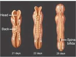
3 Development of the nervous system Development of the neural tube: - At the beginning of the 3 rd week an ectodermal thickening appears in the middle of the trilaminar germ disc known as the neural plate. - The neural plate invaginates to form a neural groove. - The lips of the neural groove approach each other & fuse together transforming the groove into a neural tube with an anterior & posterior neuropores which are obliterated on day 25 & 27 respectively transforming the neural tube into a closed tube.
4 Development of the spinal cord The neural tube is lined by one cell layer called matrix. This epithelium, which extends from the cavity of the tube to the exterior, is referred to as the ventricular zone. Repeated division of the matrix cells results in an increase in length and diameter of the neural tube. cells migrate peripherally to form the intermediate zone (grey matter). The neuroblasts give rise to nerve fibers that grow peripherally and form a layer external to the intermediate zone called the marginal zone (myelinated white matter). neuroblasts give rise to astrocytes and oligodendrocytes. Microglia is derived from surrounding mesenchym

 appears in the lateral wall dividing it into: - Dorsal part (Alar Plate) which expands to form the dorsal (sensory) horn.")
5 the cells in the lateral wall of the neural tube proliferate & are differentiated into 3 layers: - Inner ependymal Layer: forms the ependymal lining of the central canal & ventricles. - Middle Mantle Layer: Cellular layer which forms the grey matter of the spinal cord. - Outer Marginal Layer: forms the white matter of the spinal cord. The thick lateral walls are connected together by thin roof plate (dorsal) & floor plate (ventral). A groove (sulcus limitans) appears in the lateral wall dividing it into: - Dorsal part (Alar Plate) which expands to form the dorsal (sensory) horn. - Ventral part (basal plate) which expands to form the ventral (motor) horn. The cavity of the tube remains narrow & forms the central canal of the spinal cord.

6 Development of sensory and motor roots

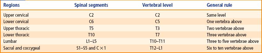
7 Development of the Meninges The pia, arachnoid and dura maters are formed from the mesenchyme. (sclerotome) that surrounds the neural tube. The subarachnoid space develops as a cavity in the mesenchyme, which becomes filled with cerebrospinal fluid. During the first 2 months of intrauterine life, the spinal cord is the same length as the vertebral column. at birth, the coccygeal end of the cord lies at the level of the third lumbar vertebra. In the adult, the lower end of the spinal cord lies at the level of the lower border of the body of the first lumbar vertebra. The oblique spinal nerves below L1 form the cauda equina.
8


9 Congenital Malformations of spinal cord development 1) Spina bifida occulta: Absent vertebral arch with normal spinal cord. It affects the lumbosacral area & is usually covered with hairy skin. 2) Spina bifida cystica: - Meningocele: The meninges herniates through the spina bifida to form subcutaneous sac filled with CSF. - Meningomyelocele: The spinal cord herniates through the meningocele. - Myelocele (Rachischisis): Failure of obliteration of the neural tube.
& the median part develops to form")
: develops to form the midbrain & its cavity forms cerebral aqueduct.")
10 Development of the brain The cranial part of the neural tube forms 3 brain vesicles: - Forebrain vesicle(prosencephalon): forms 2 lateral evaginations which develop to form the 2 cerebral hemispheres (their cavities form the lateral ventricles)& the median part develops to form the diencephalon (its cavity forms the 3 rd ventricle. - Midbrain vesicle (Mesencephalon): develops to form the midbrain & its cavity forms cerebral aqueduct. - Hindbrain vesicle (Rhombencephalon): develops to form pons, medulla & cerebellum. Its cavity forms the 4 th ventricle. Prof Yousry
11 Embryonic (developmental) divisions of the Brain Primary vesicle Secondary vesicle Derivatives Prosencephalon telencephalon Cerebral cortex Cerebral white matter Basal ganglia diencephalon Thalamus Hypothalamus Subthalamus Epithalamus Mesencephalon mesencephalon Midbrain Rhombencephalon metencephalon Cerebellum Pons myelencephalon Medulla oblongata


12 DEVELOPMENT OF THE MEDULLA OBLONGATA As in the development of the spinal cord the medulla will have an alar plate & a basal plate separated by a sulcus limitans & connected by a thin roof plate & a floor plate. The lateral walls move away from each other stretching the roof plate & enlarging its cavity which forms the 4 th ventricle. The alar plate forms the sensory nuclei of the medulla & the basal plate forms the motor nuclei. Between the fourth and fifth months, local resorptions of the roof plate occur, forming lateral foramina of Luschka, and a median foramen of Magendie.

13 Development of the pons & cerebellum The same steps in the development of the medulla occur but the alar plates bend medially to form 2 rhombic lips. The rhombic lips approach each other & fuse together forming a cerebellar plate. The cerebellar plate differentiates into a median part which forms the vermis & 2 lateral masses which form the cerebellar hemispheres. The cavity forms part of the 4 th ventricle.


14 Development of Midbrain As in the development of the spinal cord & the medulla the midbrain will have an alar plate & a basal plate separated by a sulcus limitans & connected by a thin roof plate & a floor plate. The alar plates develop to form the tectum which is divided by a vertical & transverse grooves into 4 colliculi. The basal plate forms the motor nuclei in the tegmentum of midbrain The marginal layer of the basal plate enlarges greatly to form the crus cerebri. It cavity remains narrow & forms the cerebral aqueduct. Prof Yousry

15 Development of the Diencephalon It develops from the median part of the forebrain. It consists of 2 lateral walls connected by a roof plate & a floor plate, its cavity is called the 3 rd ventricle. The roof plate: - Its anterior part forms the choroid plexus of the 3 rd ventricle. - Its posterior part forms the pineal body. A hypothalamic sulcus appears in the lateral wall which separates the thalamus above from the hypothalamus below. The floor plate forms the posterior lobe of the pituitary gland.
 The Infundibulum: Arises from the floor of the diencephalon behind Rathke s pouch.")
16 Development of the pituitary gland It develops from 2 ectodermal strucutres: 1) Rathke s Pouch: It arises from the roof of the stomdeum & ascends upwards to come to lie in front of the infundibulum. It loses its connection to the stomodeum & develops to form the anterior lobe of the pituitary gland. 2) The Infundibulum: Arises from the floor of the diencephalon behind Rathke s pouch. It develops to form the posterior lobe (pars nervosa) of the pituitary gland.

17 Development of the cerebral hemisphere The 2 cerebral hemispheres arise as 2 evaginations from the lateral wall of the forebrain. The cavity of each of them expands to form the lateral ventricle. The wall of the hemisphere consists of 3 layers: ependymal, mantle & marginal. The mantle layer at the base of the hemisphere forms the basal ganglia. The hemispheres enlarge & overlaps the brain stem & cerebellum.

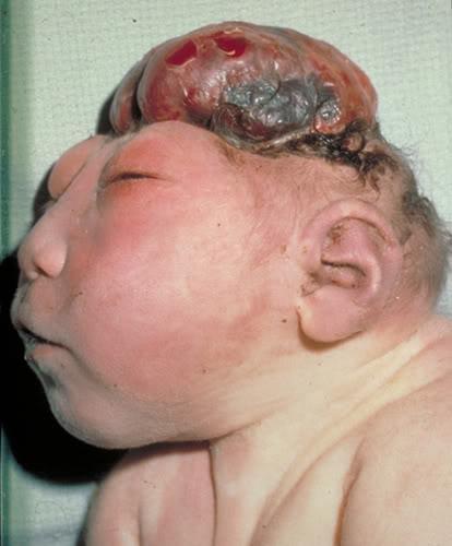
 Exencephaly: It is due to failure of closure of anterior neuropore.")

 Meningoencephalocele: part of the brain herniated through the meningocele.")

18 Congenital Malformations of brain development 1) Hydrocephalus: It is of 2 types - Internal hydrocephalus: Excessive accumulation of CSF within the ventricles of the brain. - External hydrocephalus: Excessive accumulation of the CSF between the brain & arachnoid mater. 2) Exencephaly: It is due to failure of closure of anterior neuropore. The vault of the skull is absent & the brain is exposed. When the brain is degenerated the anaomaly is known as Anencephaly. 3) Menigocele: the meninges herniated through a deficient part of the skull. 4) Meningoencephalocele: part of the brain herniated through the meningocele. 5) Meningo-hydro-enecephalocele: part of the ventricle is found within the brain tissue which herniated through the meningocele. 6) Holoprosencephaly: Results from degeneration of midline structures leading to fusion of lateral ventricles, orbital & nasal cavities.
19 THANK YOU
Development of Brain Stem, Cerebellum and Cerebrum
 Development of Brain Stem, Cerebellum and Cerebrum The neural tube cranial to the 4th pair of somites develop into the brain. 3 dilatations and 2 flexures form at the cephalic end of the neural tube during
Biological Bases of Behavior. 3: Structure of the Nervous System
 Biological Bases of Behavior 3: Structure of the Nervous System Neuroanatomy Terms The neuraxis is an imaginary line drawn through the spinal cord up to the front of the brain Anatomical directions are
Chapter 3. Structure and Function of the Nervous System. Copyright (c) Allyn and Bacon 2004
 Allyn and Bacon 2004") Chapter 3 Structure and Function of the Nervous System 1 Basic Features of the Nervous System Neuraxis: An imaginary line drawn through the center of the length of the central nervous system, from the
Chapter 3 Structure and Function of the Nervous System 1 Basic Features of the Nervous System Neuraxis: An imaginary line drawn through the center of the length of the central nervous system, from the
Review of Nervous System Anatomy
 For the real amazement, if you wish to be amazed, is this process. You start out as a single cell derived from the coupling of a sperm and an egg; this divides in two, then four, then eight, and so on,
Nervous System. Lecture 4
 Nervous System Lecture 4 Neurons Functional unit of the nervous system Also called the nerve cell Soma or body Axon Dendrites Neuroglial cells support cells Schwann cells produce myelin in PNS Oligodendrocytes
Department of Cognitive Science UCSD
 Department of Cognitive Science UCSD Verse 1: Neocortex, frontal lobe, Brain stem, brain stem, Hippocampus, neural node, Right hemisphere, Pons and cortex visual, Brain stem, brain stem, Sylvian fissure,
Development of Spinal Cord & Vertebral Column. Dr. Sanaa Alshaarawi & Prof. Ahmed Fathalla
 Development of Spinal Cord & Vertebral Column Dr. Sanaa Alshaarawi & Prof. Ahmed Fathalla OBJECTIVES At the end of the lecture, students should be able to: q Describe the development of the spinal cord
Fig.9.2. Structure of embryonic brain
 T Chapter 9 Development of Ectodermal Organs he ectoderm gives rise to 3 separate cell populations: neural(plate) ectoderm, neural crest cells, and epiderm (general body ectoderm). A primordium (anlage)
The neurvous system senses, interprets, and responds to changes in the environment. Two types of cells makes this possible:
 NERVOUS SYSTEM The neurvous system senses, interprets, and responds to changes in the environment. Two types of cells makes this possible: the neuron and the supporting cells ("glial cells"). Neuron Neurons
Embryology of the Nervous System. Steven McLoon Department of Neuroscience University of Minnesota
 Embryology of the Nervous System Steven McLoon Department of Neuroscience University of Minnesota In the blastula stage embryo, the embryonic disk has two layers. During gastrulation, epiblast cells migrate
Chapter 5: Fetal Central Nervous System 71
 71 Chapter 5 Fetal Central Nervous System Embryology NEURULATION begins with the formation of the neural plate, the neural folds and their ultimate fusion and closure as the NEURAL TUBE. NEURAL PLATE -
Early Development of Neural Tube Development of Medulla Spinalis and Peripheral Nervous System. Assoc.Prof. E.Elif Güzel, M.D.
 Early Development of Neural Tube Development of Medulla Spinalis and Peripheral Nervous System Assoc.Prof. E.Elif Güzel, M.D. Third week of Embryogenesis Primitive streak/pit appears on the epiblast (day
Neuroanatomy lecture (1)
") Neuroanatomy lecture (1) Introduction: Neuroanatomy has two parts: the central and peripheral nervous system. The central nervous system is composed of brain and spinal cord. The brain has the following
Neuroanatomy lecture (1) Introduction: Neuroanatomy has two parts: the central and peripheral nervous system. The central nervous system is composed of brain and spinal cord. The brain has the following
SOME BASIC TERMINOLOGY CNS: Central Nervous System: Brain + Spinal Cord
 SOME BASIC TERMINOLOGY CNS: Central Nervous System: Brain + Spinal Cord CEREBROSPINAL FLUID (CSF): The fluid filling the ventricles, cerebral aqueduct, central canal, and subarachnoid space. It is a filtrate
Organization of The Nervous System PROF. SAEED ABUEL MAKAREM
 Organization of The Nervous System PROF. SAEED ABUEL MAKAREM Objectives By the end of the lecture, you should be able to: List the parts of the nervous system. List the function of the nervous system.
b. The groove between the two crests is called 2. The neural folds move toward each other & the fuse to create a
 Chapter 13: Brain and Cranial Nerves I. Development of the CNS A. The CNS begins as a flat plate called the B. The process proceeds as: 1. The lateral sides of the become elevated as waves called a. The
Central nervous system (CNS): brain and spinal cord Collections of cell body and dendrites (grey matter) are called nuclei/nucleus Nucleus can also
: brain and spinal cord Collections of cell body and dendrites (grey matter) are called nuclei/nucleus Nucleus can also") Chapter 3 Part 1 Orientation Directions in the nervous system are described relatively to the neuraxis An imaginary line drawn through the center of the length of the central nervous system, from the bottom
Chapter 3 Part 1 Orientation Directions in the nervous system are described relatively to the neuraxis An imaginary line drawn through the center of the length of the central nervous system, from the bottom
1. The basic anatomy of the Central Nervous System (CNS)
") Psyc 311A, fall 2008 Conference week 1 Sept 9 th to 11 th TA: Jürgen Germann; e-mail: jurgen.germann@mcgill.ca Overview: 1. The basic anatomy of the Central Nervous System (CNS) 2. Cells of the CNS 3.
Psyc 311A, fall 2008 Conference week 1 Sept 9 th to 11 th TA: Jürgen Germann; e-mail: jurgen.germann@mcgill.ca Overview: 1. The basic anatomy of the Central Nervous System (CNS) 2. Cells of the CNS 3.
Introduction and Basic structural organization of the nervous system
 Introduction and Basic structural organization of the nervous system **the slides are in bold and the book is in red Done by : razan krishan & marah marahleh INTRODUCTION The nervous system, along with
Anatomy Lab (1) Theoretical Part. Page (2 A) Page (2B)
 Theoretical Part. Page (2 A) Page (2B)") Anatomy Lab (1) This sheet only includes the extra notes for the lab handout regarding the theoretical part, as for the practical part it includes everything the doctor mentioned. Theoretical Part Page
Anatomy Lab (1) This sheet only includes the extra notes for the lab handout regarding the theoretical part, as for the practical part it includes everything the doctor mentioned. Theoretical Part Page
PSY 215 Lecture #5 (01/26/2011) (Anatomy of the Brain) Dr. Achtman PSY 215. Lecture 5 Anatomy of the Brain Chapter 4, pages 86-96
 (Anatomy of the Brain) Dr. Achtman PSY 215. Lecture 5 Anatomy of the Brain Chapter 4, pages 86-96") Corrections: none needed PSY 215 Lecture 5 Anatomy of the Brain Chapter 4, pages 86-96 Announcements: Reminder: The first midterm is in one week! Everyone is encouraged to start studying (recommend 30/night
Corrections: none needed PSY 215 Lecture 5 Anatomy of the Brain Chapter 4, pages 86-96 Announcements: Reminder: The first midterm is in one week! Everyone is encouraged to start studying (recommend 30/night
A&P 1 Brain & Cranial Nerves Guide - Lab Exercises
 A&P 1 Brain & Cranial Nerves Guide - Lab Exercises Please make sure you read the entire set of instructions on Dissection the Sheep Brain before beginning to cut. Also, please do not forget to go over
DEVELOPMENT OF BRAIN
 Ahmed Fathalla OBJECTIVES At the end of the lecture, students should: List the components of brain stem. Describe the site of brain stem. Describe the relations between components of brain stem & their
Ch 13: Central Nervous System Part 1: The Brain p 374
 Ch 13: Central Nervous System Part 1: The Brain p 374 Discuss the organization of the brain, including the major structures and how they relate to one another! Review the meninges of the spinal cord and
Sheep Brain Dissection
 Sheep Brain Dissection Mammalian brains have many features in common. Human brains may not be available, so sheep brains often are dissected as an aid to understanding the mammalian brain since he general
Anatomy of the Nervous System. Brain Components
 Anatomy of the Nervous System Brain Components NERVOUS SYSTEM INTRODUCTION Is the master system of human body, controlling the functions of rest of the body systems Nervous System CLASSIFICATION A. Anatomical
OBJECTIVES (Revised) I. Introduction
 I. Introduction") Neuroembryology: CNS Developmental Anomalies Raymond J. Colello, Ph.D. *Lecture written by Dr. John Povlishock Note: Emphasize Dr. Colello s lecture PPT and developmental anomalies OBJECTIVES (Revised)
Neuroembryology: CNS Developmental Anomalies Raymond J. Colello, Ph.D. *Lecture written by Dr. John Povlishock Note: Emphasize Dr. Colello s lecture PPT and developmental anomalies OBJECTIVES (Revised)
Central Nervous System (CNS) -> brain and spinal cord. Major Divisions of the nervous system:
 -> brain and spinal cord. Major Divisions of the nervous system:") Central Nervous System (CNS) -> brain and spinal cord Major Divisions of the nervous system: Afferent (sensory input) -> cell bodies outside of the central nervous system (CNS), carry info into the CNS
Central Nervous System (CNS) -> brain and spinal cord Major Divisions of the nervous system: Afferent (sensory input) -> cell bodies outside of the central nervous system (CNS), carry info into the CNS
Anatomy and Physiology (Bio 220) The Brain Chapter 14 and select portions of Chapter 16
 The Brain Chapter 14 and select portions of Chapter 16") Anatomy and Physiology (Bio 220) The Brain Chapter 14 and select portions of Chapter 16 I. Introduction A. Appearance 1. physical 2. weight 3. relative weight B. Major parts of the brain 1. cerebrum 2.
Anatomy and Physiology (Bio 220) The Brain Chapter 14 and select portions of Chapter 16 I. Introduction A. Appearance 1. physical 2. weight 3. relative weight B. Major parts of the brain 1. cerebrum 2.
Brain ميهاربا لض اف دمح ا د The Meninges 1- Dura Mater of the Brain endosteal layer does not extend meningeal layer falx cerebri tentorium cerebelli
 .احمد د فاضل ابراهيم Lecture 15 Brain The Meninges Three protective membranes or meninges surround the brain in the skull: the dura mater, the arachnoid mater, and the pia mater 1- Dura Mater of the Brain
Anatomy Lecture Notes Chapter 13
 I. embryonic development of the CNS A. neurulation is the formation of the CNS in the embryo invagination of dorsal ectoderm (outer layer of embryo cells) this process is induced (caused) by the notochord
Histology of the CNS
 Histology of the CNS Lecture Objectives Describe the histology of the cerebral cortex layers. Describe the histological features of the cerebellum; layers and cells of cerebellar cortex. Describe the elements
BRAIN DEVELOPMENT I: ESTABLISHMENT OF BASIC ARCHITECTURE. Thomas Marino, Ph.D.
 BRAIN DEVELOPMENT I: ESTABLISHMENT OF BASIC ARCHITECTURE Thomas Marino, Ph.D. Development of the Brain I. Competencies: Upon completion of this section of the course, the student must be able to: 1. Understand
SHORT ANSWER. Write the word or phrase that best completes each statement or answers the question.
 Exam Name 1) A change in the conditions in the synaptic terminal can influence the soma as a result of axoplasmic transport. 2) The nervous system is composed of the brain and spinal cord. A) efferent
Development of the Nervous System
 Development of the Nervous System Neural tube derivatives Spinal cord (alar vs. basal plate) Brain vesicles Brainstem nuclei Cerebral cortex Neural crest derivatives Puituitary gland development Developmental
Organization of The Nervous System PROF. MOUSAED ALFAYEZ & DR. SANAA ALSHAARAWY
 Organization of The Nervous System PROF. MOUSAED ALFAYEZ & DR. SANAA ALSHAARAWY Objectives At the end of the lecture, the students should be able to: List the parts of the nervous system. List the function
Nervous System The Brain and Spinal Cord Unit 7b
 Nervous System The Brain and Spinal Cord Unit 7b Chetek High School Mrs. Michaelsen 9.12 Meninges A. Meninges 1. The organs of the CNS are covered by membranes a. The meninges are divided into 3 layers:
CNS Embryology 5th Menstrual Week (Dorsal View)
") Imaging of the Fetal Brain; Normal & Abnormal Alfred Abuhamad, M.D. Eastern Virginia Medical School CNS Embryology 5th Menstrual Week (Dorsal View) Day 20 from fertilization Neural plate formed in ectoderm
Imaging of the Fetal Brain; Normal & Abnormal Alfred Abuhamad, M.D. Eastern Virginia Medical School CNS Embryology 5th Menstrual Week (Dorsal View) Day 20 from fertilization Neural plate formed in ectoderm
CNS Developmental. Anke van Eekelen, PhD. Telethon Institute for Child Health Research
 CNS Developmental Anke van Eekelen, PhD Telethon Institute for Child Health Research (Some slides are modified versions of Prof. Alan Harvey s Neuroscience lecture at ANHB and Dr. Joanne Britto s Dev Neuroscience
Organogenesis of Heart, Kidney, Nervous System & Sense Organs. [ GLOSSARY ]
![Organogenesis of Heart, Kidney, Nervous System & Sense Organs. [ GLOSSARY ]](/thumbs/86/94287046.jpg "Organogenesis of Heart, Kidney, Nervous System & Sense Organs. [ GLOSSARY ]") Organogenesis of Heart, Kidney, Nervous System & Sense Organs. [ GLOSSARY ] Subject : Zoology Course : 3rd Year, B.Sc. Undergraduate UGC Syllabus, Model - 1 Paper No. : Z-305B & Title : Developmental Biology
Organogenesis of Heart, Kidney, Nervous System & Sense Organs. [ GLOSSARY ] Subject : Zoology Course : 3rd Year, B.Sc. Undergraduate UGC Syllabus, Model - 1 Paper No. : Z-305B & Title : Developmental Biology
Student Lab #: Date. Lab: Gross Anatomy of Brain Sheep Brain Dissection Organ System: Nervous Subdivision: CNS (Central Nervous System)
") Lab: Gross Anatomy of Brain Sheep Brain Dissection Organ System: Nervous Subdivision: CNS (Central Nervous System) Student Lab #: Date 1 Objectives: 1. Learn the main components making up a motor neuron.
Lab: Gross Anatomy of Brain Sheep Brain Dissection Organ System: Nervous Subdivision: CNS (Central Nervous System) Student Lab #: Date 1 Objectives: 1. Learn the main components making up a motor neuron.
Development of the Nervous System. Leah Militello, class of 2018
 Development of the Nervous System Leah Militello, class of 2018 Learning Objectives 1. Describe the formation and fate of the neural tube and neural crest including timing and germ layer involved. 2. Describe
Nervous System: Part IV The Central Nervous System The Brain
 Nervous System: Part IV The Central Nervous System The Brain Can you survive when part of your brain is destroyed? 2 Essential Knowledge 3.D.2 2. Cells communicate with each other through direct contact
BRAIN PART I (A & B): VENTRICLES & MENINGES
: VENTRICLES & MENINGES") BRAIN PART I (A & B): VENTRICLES & MENINGES Cranial Meninges Cranial meninges are continuous with spinal meninges Dura mater: inner layer (meningeal layer) outer layer (endosteal layer) fused to periosteum
BRAIN PART I (A & B): VENTRICLES & MENINGES Cranial Meninges Cranial meninges are continuous with spinal meninges Dura mater: inner layer (meningeal layer) outer layer (endosteal layer) fused to periosteum
Central Nervous System: Part 2
 Central Nervous System: Part 2 1. Meninges 2. CSF 3. Spinal Cord and Spinal Nerves Explain spinal cord anatomy, including gray and white matter and meninges (give the general functions of this organ).
Ventricles, CSF & Meninges. Steven McLoon Department of Neuroscience University of Minnesota
 Ventricles, CSF & Meninges Steven McLoon Department of Neuroscience University of Minnesota 1 Coffee Hour Thursday (Sept 14) 8:30-9:30am Surdyk s Café in Northrop Auditorium Stop by for a minute or an
Embryonic Brain Development
 Chapter 14 The Brain and Cranial Nerves Largest organ in the body? Brain functions in sensations, memory, emotions, decision making, behavior 19-1 19-2 Embryonic Brain Development Principal Parts of the
Neurology. Dr. Mohd. Zahirul Islam Khan DVM, MS, Ph.D, Postdoc.
 Neurology By, 1. Central Nervous System: a) Brain of different animals. b) Spinal cord. 2. Peripheral Nervous System (PNS): a) Cranial Nerves b) Spinal Nerves and ganglia 3. Autonomic Nervous System (PNS)
Brain and Cranial Nerves (Ch. 15) Human Anatomy lecture. caudal = toward the spinal cord)
 Human Anatomy lecture. caudal = toward the spinal cord)") Insight: Some cranial nerve disorders Brain and Cranial Nerves (Ch. 15) Human Anatomy lecture I. Overview (Directional terms: rostral = toward the forehead caudal = toward the spinal cord) A. 3 Major parts
Insight: Some cranial nerve disorders Brain and Cranial Nerves (Ch. 15) Human Anatomy lecture I. Overview (Directional terms: rostral = toward the forehead caudal = toward the spinal cord) A. 3 Major parts
M555 Medical Neuroscience Lab 1: Gross Anatomy of Brain, Crainal Nerves and Cerebral Blood Vessels
 M555 Medical Neuroscience Lab 1: Gross Anatomy of Brain, Crainal Nerves and Cerebral Blood Vessels Anatomical Directions Terms like dorsal, ventral, and posterior provide a means of locating structures
Huntington s Disease & MARY ET BOYLE, PH.D. DEPARTMENT OF COGNITIVE SCIENCE
 Huntington s Disease & Early Nervous System Development MARY ET BOYLE, PH.D. DEPARTMENT OF COGNITIVE SCIENCE UCSD The cups fell to the floor with a crash. Was this the alarm signal? Or was it forgetting
Development of the Nervous System 1 st month
 Development of the Nervous System 1 st month day 1 - fertilization of egg day 6 - uterine implantation day 18 - trilaminar (3-layered) disc (blastoderm, embryo) ectoderm (dorsal) - nervous system and skin
Lecture 9. General Medicine_3rd semester
 Lecture 9 General Medicine_3rd semester MICROSCOPIC STRUCTURE AND DEVELOPMENT OF THE CENTRAL AND PERIPHERAL NERVOUS SYSTEM Structure of gray matters in the CNS: Iso- and allocortex, cerebellar cortex,
Blood supply to the brain Blood brain barrier isolates neural tissue from general circulation
 The Brain and Cranial Nerves Objectives Name the major regions of the brain and describe their functions. Discuss the formation, circulation, and functions of the CSF. List the main components of the medulla
Unit Three. The brain includes: cerebrum, diencephalon, brain stem, & cerebellum. The brain lies within the cranial cavity of the skull.
 Human Anatomy & Physiology 11 Divisions of the Nervous System Karen W. Smith, Instructor Unit Three BRAIN & SPINAL CORD Refer to the following URLs. Be sure to study these along with your book. http://www.sirinet.net/~jgjohnso/nervous.html
Lecture 1: Neuroembryology Neuroscience 1: Neuroembryology
 The nervous system starts to form at the 3 rd week of life from the ectoderm at the specialized part called the neuroectoderm/neurectoderm. The Notochord forms at the 3 rd week of life o It is the source
Lecture - Chapter 13: Central Nervous System
 Lecture - Chapter 13: Central Nervous System 1. Describe the following structures of the brain, what is the general function of each: a. Cerebrum b. Diencephalon c. Brain Stem d. Cerebellum 2. What structures
action potential afferent neuron Weblike; specifically, the weblike middle layer of the three meninges. arachnoid astrocytes autonomic nervous system
 action potential A large transient depolarization event, including polarity reversal, that is conducted along the membrane of a muscle cell or a nerve fiber. afferent neuron Nerve cell that carries impulses
3 rd week ectoderm thickens to form neural plate, which is later flanked by neural folds This neural groove deepens, forming a neural tube by 4 th
 3 rd week ectoderm thickens to form neural plate, which is later flanked by neural folds This neural groove deepens, forming a neural tube by 4 th week differentiates into the CNS = brain development begins
Chapter 12b. Overview
 Chapter 12b Spinal Cord Overview Spinal cord gross anatomy Spinal meninges Sectional anatomy Sensory pathways Motor pathways Spinal cord pathologies 1 The Adult Spinal Cord About 18 inches (45 cm) long
Chapter 7 Nervous System
 Chapter 7 Nervous System Two message centers: Functions of these systems: 1. * 2. * Overview of the Nervous System Parts: General Functions: Functions Sensory input: Sensation via nerves Integration: interpretation
Unit 3 : Nervous System
 Unit 3 : Nervous System Mind Map Structural Classification The nervous Tissue Disorders of The nervous system Nervous System Central Nervous System Peripheral Nervous System The brain Spinal Cord Sensory
Nervous system. Dr. Rawaa Salim Hameed
 Nervous system Dr. Rawaa Salim Hameed Central nervous system (CNS) CNS consists of the brain (cerebrum, cerebellum, and brainstem) and spinal cord CNS is covered by connective tissue layers, the meninges
Chapter 1 Introduction and overview
 1 Chapter 1 Introduction and overview Components and organisation of the nervous system 1 Neurones and neuroglia 1 Central and peripheral nervous systems 2 Autonomic nervous system 2 Afferent neurones,
BRAIN STEM AND CEREBELLUM..
 Lecture Title: BRAIN STEM AND CEREBELLUM.. (CNS Block, Radiology) Dr. Hamdy Hassan Ass.Prof. Consultant Radiology Department KKHU King Saud University Lecture Objectives.. Students at the end of the lecture
meninges Outermost layer of the meninge dura mater arachnoid mater pia mater membranes located between bone and soft tissue of the nervous system
 membranes located between bone and soft tissue of the nervous system meninges Outermost layer of the meninge dura mater middle layer of the meninges, contains no blood vessels arachnoid mater Innermost
Chapter 14: The Brain and Cranial Nerves. Copyright 2009, John Wiley & Sons, Inc.
 Chapter 14: The Brain and Cranial Nerves Development of the Brain Three to four-week embryo: prosencephalon, mesencephalon and rhombencephalon. Five-week embryo: telencephalon (cerebrum), diencephalon
ACTIVITY 7: NERVOUS SYSTEM HISTOLOGY, BRAIN, CRANIAL NERVES
 ACTIVITY 7: NERVOUS SYSTEM HISTOLOGY, BRAIN, CRANIAL NERVES LABORATORY OBJECTIVES: 1. Histology: Identify structures indicated on three different slides or images of nervous system tissue. These images
Chapter 13 Brain and Cranial Nerves
 Chapter 13 Brain and Cranial Nerves 13-1 Brain and Cranial Nerves Brain Part of CNS contained in cranial cavity Control center for many of body s functions Much like a complex computer but more Parts of
Cerebral hemisphere. Parietal Frontal Occipital Temporal
 Cerebral hemisphere Sulcus / Fissure Central Precental gyrus Postcentral gyrus Lateral (cerebral) Parieto-occipital Cerebral cortex Frontal lobe Parietal lobe Temporal lobe Insula Amygdala Hippocampus
Chapter 2. Central Nervous System; the brain and spinal cord
 Chapter 2 Central Nervous System; the brain and spinal cord CNS 1. Topography; - what are the main components of the brain - how do you recognize them? 2. The location of the major functional areas of
The Nervous System. Chapter 7. Essentials of Human Anatomy & Physiology. Elaine N. Marieb. Seventh Edition
 Essentials of Human Anatomy & Physiology Elaine N. Marieb Seventh Edition Chapter 7 The Nervous System Functions of the Nervous System 1. Sensory input gathering information To monitor changes occurring
Slide 1. Slide 2. Slide 3. Tomography vs Topography. Computed Tomography (CT): A simplified Topographical review of the Brain. Learning Objective
: A simplified Topographical review of the Brain. Learning Objective") Slide 1 Computed Tomography (CT): A simplified Topographical review of the Brain Jon Wheiler, ACNP-BC Slide 2 Tomography vs Topography Tomography: A technique for displaying a representation of a cross
Slide 1 Computed Tomography (CT): A simplified Topographical review of the Brain Jon Wheiler, ACNP-BC Slide 2 Tomography vs Topography Tomography: A technique for displaying a representation of a cross
SHORT ANSWER. Write the word or phrase that best completes each statement or answers the question.
 Exam Name SHORT ANSWER. Write the word or phrase that best completes each statement or answers the question. Figure 12.3 Using Figure 12.3, match the following: 1) Site of efferent soma. 2) Site of axons
Page. Ch 11 A CNS. This set. Major Landmarks: Brain size is proportional to body size only and can be divided into three major portions;
 1 BIO 211: ANATOMY & PHYSIOLOGY I 1 Ch 11 A CNS This set Ch 11 B Notes: PNS Somatic ANS Ch 11 C ANS Dr. Dr. Lawrence G. G. Altman www.lawrencegaltman.com Some illustrations are courtesy of McGraw-Hill.
The Nervous System. Functions of the Nervous System input gathering To monitor occurring inside and outside the body Changes =
 The Nervous System Functions of the Nervous System input gathering To monitor occurring inside and outside the body Changes = To process and sensory input and decide if is needed output A response to integrated
Chapter 13 Lecture Outline *
 Anatomy and Physiology, Seventh Edition Rod R. Seeley Idaho State University Trent D. Stephens Idaho State University Philip Tate Phoenix College Chapter 13 Lecture Outline * *See PowerPoint Image Slides
Dissection of the Sheep Brain
 Dissection of the Sheep Brain Laboratory Objectives After completing this lab, you should be able to: 1. Identify the main structures in the sheep brain and to compare them with those of the human brain.
Meninges and Ventricles
 Meninges and Ventricles Irene Yu, class of 2019 LEARNING OBJECTIVES Describe the meningeal layers, the dural infolds, and the spaces they create. Name the contents of the subarachnoid space. Describe the
4. Development of nervous system. Neural plate. Brain vesicles. Sensory organs.
 4. Development of nervous system. Neural plate. Brain vesicles. Sensory organs. Development of the central nervous system neurulation = ectoderm in front of the primitive node thickens to form the neural
CNS consists of brain and spinal cord Cephalization Evolutionary development of rostral (anterior) portion of CNS Increased number of neurons in head
 portion of CNS Increased number of neurons in head") CNS consists of brain and spinal cord Cephalization Evolutionary development of rostral (anterior) portion of CNS Increased number of neurons in head Highest level reached in human brain 1 Mostly to orient
CNS consists of brain and spinal cord Cephalization Evolutionary development of rostral (anterior) portion of CNS Increased number of neurons in head Highest level reached in human brain 1 Mostly to orient
The Brain Worksheet Sections 5-7
 The Brain Worksheet Sections 5-7 1. neuroglia 2. autonomic nervous system 3. sensory neurons 4. oligodendrocytes 5. ascending tracts 6. descending tracts 7. saltatory propagation 8. continuous propagation
Lecture 4 The BRAINSTEM Medulla Oblongata
 Lecture 4 The BRAINSTEM Medulla Oblongata Introduction to brainstem 1- Medulla oblongata 2- Pons 3- Midbrain - - - occupies the posterior cranial fossa of the skull. connects the narrow spinal cord
Bellringer: The central nervous system is comprised of: What is the name of the outermost layer of the brain? a. Brain. b.
 Bellringer: The central is comprised of: a. Brain b. Spinal cord c. Sensory receptors d. Both a and b What is the name of the outermost layer of the brain? a. Pia mater b. Dura mater c. Arachnoid d. Pons
Chapter 7 The Nervous System
 Chapter 7 The Nervous System Fxns of the Nervous System 1. Sensory input gathering information To monitor Δs occurring inside and outside the body (Δs = stimuli) 2. Integration to process and interpret
The Brain. Brain. Spinal Cord. Cauda Equina
 The Brain Brain Spinal Cord Cauda Equina The Brain Ventricles- cavities in the brain filled with cerebrospinal fluid connected to the subarachnoid space- fluid filled space surrounding the brain Brain
SLIDES 6 AND 10 MM PIG SLIDES; TRANSVERSE SECTIONS AND SAGITAL; FETAL PIGS-1-8INCH; HUMAN SAGITAL DIAGRAMS:DRAWINGS OF THE PIG SECTIONS TO BE
 SLIDES 6 AND 10 MM PIG SLIDES; TRANSVERSE SECTIONS AND SAGITAL; FETAL PIGS-1-8INCH; HUMAN SAGITAL SECTION DIAGRAMS:DRAWINGS OF THE PIG SECTIONS TO BE LABELLED, AND FOUR DRAWINGS TO BE MADE. REFERENCES:PATTEN:
Remember from the first year embryology Trilaminar disc has 3 layers: ectoderm, mesoderm, and endoderm
 Development of face Remember from the first year embryology Trilaminar disc has 3 layers: ectoderm, mesoderm, and endoderm The ectoderm forms the neural groove, then tube The neural tube lies in the mesoderm
The dura is sensitive to stretching, which produces the sensation of headache.
 Dural Nerve Supply Branches of the trigeminal, vagus, and first three cervical nerves and branches from the sympathetic system pass to the dura. Numerous sensory endings are in the dura. The dura is sensitive
Development of the Central Nervous System (CNS) 2. Diencephalon -interbrain ( ) -diencephalons, thalamus, hypothalamus, epithalamus -hollow space =
 2. Diencephalon -interbrain ( ) -diencephalons, thalamus, hypothalamus, epithalamus -hollow space =") Week 2 -neural plate Development of the Central Nervous System (CNS) Week 3 -neural groove Week 4 -neural tube (ice cream cone view) 1. Prosencephalon - 2. Mesencephalon - 3. Rhombencephalon - Week 5 -brain
Week 2 -neural plate Development of the Central Nervous System (CNS) Week 3 -neural groove Week 4 -neural tube (ice cream cone view) 1. Prosencephalon - 2. Mesencephalon - 3. Rhombencephalon - Week 5 -brain
TRANSVERSE SECTION PLANE Scalp 2. Cranium. 13. Superior sagittal sinus
 TRANSVERSE SECTION PLANE 1 1. Scalp 2. Cranium 3. Superior sagittal sinus 4. Dura mater 5. Falx cerebri 6. Frontal lobes of the cerebrum 7. Middle meningeal artery 8. Cortex, grey matter 9. Cerebral vessels
Chapter 9. Nervous System
 Chapter 9 Nervous System Central Nervous System (CNS) vs. Peripheral Nervous System(PNS) CNS Brain Spinal cord PNS Peripheral nerves connecting CNS to the body Cranial nerves Spinal nerves Neurons transmit
NOTES CHAPTER 9 (Brief) The Nervous System LECTURE NOTES
 The Nervous System LECTURE NOTES") NOTES CHAPTER 9 (Brief) The Nervous System LECTURE NOTES I. Divisions of the Nervous System two major divisions A. Central Nervous System (CNS) 1. brain 2. spinal cord B. Peripheral Nervous System (PNS)
NOTES CHAPTER 9 (Brief) The Nervous System LECTURE NOTES I. Divisions of the Nervous System two major divisions A. Central Nervous System (CNS) 1. brain 2. spinal cord B. Peripheral Nervous System (PNS)
13 ANATOMY OF THE NERVOUS SYSTEM
 CHAPTER 13 ANATOMY OF THE NERVOUS SYSTEM 513 13 ANATOMY OF THE NERVOUS SYSTEM Figure 13.1 Human Nervous System The ability to balance like an acrobat combines functions throughout the nervous system. The
Good Morning! Take out your notes and vocab 1-10! Copyright 2003 Pearson Education, Inc. publishing as Benjamin Cummings
 Good Morning! Take out your notes and vocab 1-10! Functions of the Nervous System 1. Sensory input gathering information To monitor changes occurring inside and outside the body (changes = stimuli) 2.
The Nervous System PART B
 7 The Nervous System PART B PowerPoint Lecture Slide Presentation by Jerry L. Cook, Sam Houston University ESSENTIALS OF HUMAN ANATOMY & PHYSIOLOGY EIGHTH EDITION ELAINE N. MARIEB The Reflex Arc Reflex
The Nervous System PART C. PowerPoint Lecture Slide Presentation by Patty Bostwick-Taylor, Florence-Darlington Technical College
 PowerPoint Lecture Slide Presentation by Patty Bostwick-Taylor, Florence-Darlington Technical College The Nervous System 7 PART C Protection of the Central Nervous System Scalp and skin Skull and vertebral
Fondamenti di anatomia e istologia
 Ingegneria delle tecnologie per la salute Fondamenti di anatomia e istologia Sistema Nervoso aa. 2017-18 Nervous System The nervous system can be divided into two major regions: the central nervous system
Chapter 18: The Brain & Cranial Nerves. Origin of the Brain
 Chapter 18: The Brain & Cranial Nerves BIO 218 Fall 2015 Origin of the Brain The brain originates from a structure called the neural tube, which arises during a developmental stage called neurulation.
BIOH111. o Cell Module o Tissue Module o Integumentary system o Skeletal system o Muscle system o Nervous system o Endocrine system
 BIOH111 o Cell Module o Tissue Module o Integumentary system o Skeletal system o Muscle system o Nervous system o Endocrine system Endeavour College of Natural Health endeavour.edu.au 1 Textbook and required/recommended