Bone, soft tissue and skin tumors. By: Shefaa qa qa
|
|
|
- Valentine Bates
- 5 years ago
- Views:
Transcription
1 Bone, soft tissue and skin tumors By: Shefaa qa qa
2 Bone tumors Most bone neoplasms develop during the first several decades of life and have a propensity for the long bones of the extremities. The occurrence in certain age groups and predilection for particular anatomic sites provides important diagnostic clues about specific types of tumors. For example, osteosarcoma peaks during adolescence and most frequently involves the knee whereas chondrosarcoma affects older adults and involves the pelvis and proximal extremities. metastases (adenocarcinoma) and hematopoietic tumors far outnumber primary bone neoplasms. Bone sarcomas are lethal in 50% of cases.
3 Bone tumors may present in a number of ways. The more common benign lesions are often asymptomatic incidental findings. Many tumors, however, produce pain or a slow-growing mass. In some circumstances the first hint of a tumor s presence is a pathologic fracture. Radiographic imaging studies have an important role in diagnosing these lesions. In addition to providing the exact location and extent of the tumor, imaging studies can detect features that narrow the diagnostic possibilities. Ultimately, in almost all instances biopsy is necessary for definitive diagnosis.
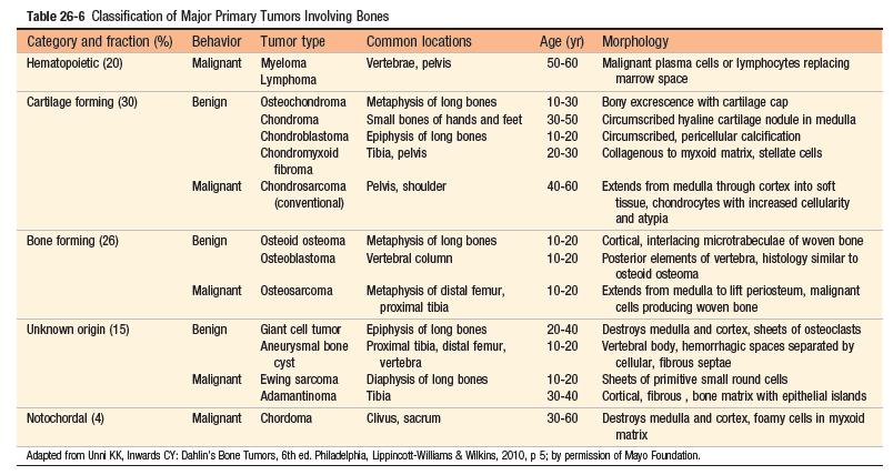
4 When possible, bone tumors are classified according to the normal cell or matrix they produce. Lesions that do not have normal tissue counterparts are grouped according to their clinicopathologic features. Benign tumors greatly outnumber their malignant counterparts and occur with greatest frequency within the first three decades of life, whereas in older adults, a bone tumor is likely to be malignant. Excluding neoplasms originating from hematopoietic cells (myeloma, lymphoma, and leukemia), osteosarcoma is the most common primary cancer of bone, followed by chondrosarcoma and Ewing sarcoma.
5 Insults that induce chronic injury and inflammation, such as bone infarcts, chronic osteomyelitis, Paget disease, radiation, and metal prostheses, increase the risk of bone neoplasia, possibly because proliferation associated with chronic inflammation and repair set the stage for acquisition of oncogenic mutations.
6
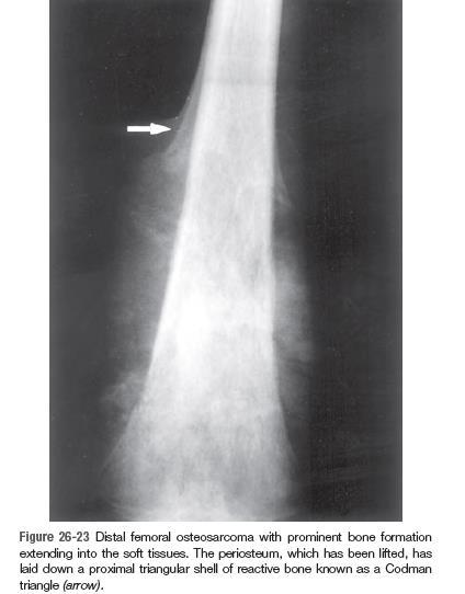
7 Osteosarcoma Osteosarcoma is a malignant tumor in which the cancerous cells produce osteoid matrix or mineralized bone. It is the most common primary malignant tumor of bone, exclusive of myeloma and lymphoma. Osteosarcomas typically present as painful, progressively enlarging masses. Sometimes a sudden fracture of the bone is the first symptom.
8 Osteosarcoma occurs in all age groups but has a bimodal age distribution; 75% occur in persons younger than 20 years of age. The smaller second peak occurs in older adults, who frequently suffer from conditions known to predispose to osteosarcoma Paget disease, bone infarcts, and prior radiation. Overall, men are more commonly affected than women (1.6 : 1). Any bone can be involved. The tumors usually arise in the metaphyseal region of the long bones of the extremities, and almost 50% occur about the knee (i.e., distal femur or proximal tibia).
9
10 Pathogenesis: Approximately 70% of osteosarcomas have acquired genetic abnormalities such as complex structural and numerical chromosomal aberrations. Molecular studies have shown that these tumors usually have mutations in well-known tumor suppressors and oncogenes, including the following: - RB - TP53 - INK4a - MDM2 and CDK4
11 Several subtypes of osteosarcoma are recognized and are grouped according to: - Site of origin (intramedullary, intracortical, or surface) - Histologic grade (low, high) - Primary (underlying bone is unremarkable) or secondary to preexisting disorders (benign tumors, Paget disease, bone infarcts, previous radiation) - Histologic features (osteoblastic, chondroblastic, fibroblastic, telangiectatic, small cell, and giant cell).
12 Clinical Course: Osteosarcoma is treated with a multimodality approach that includes neoadjuvant chemotherapy, which is given under the assumption that all patients have occult metastases at the time of diagnosis, followed by surgery. The prognosis of osteosarcoma has improved substantially since the advent of chemotherapy, with 5-year survival rates reaching 60% to 70% in patients without overt metastases at initial diagnosis. These aggressive neoplasms spread hematogenously to the lungs. Unfortunately, the outcome for patients with metastases, recurrent disease or secondary osteosarcoma is still poor (<20% 5-year survival rate).
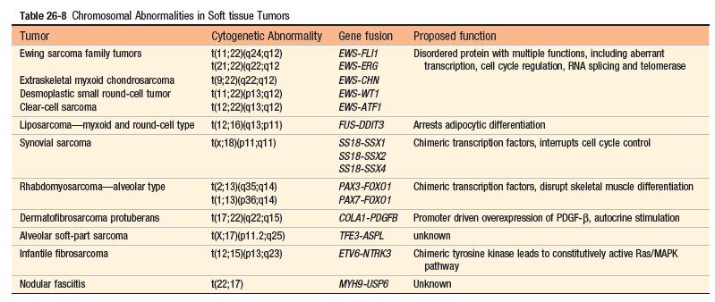
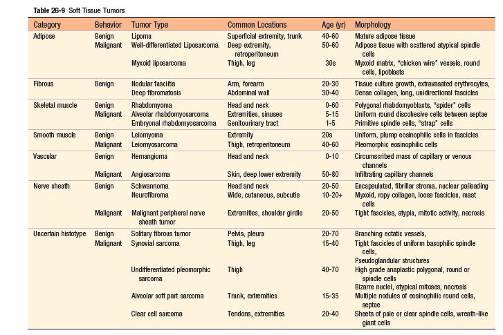
13 Soft tissue tumors soft tissue refers to non-epithelial tissue excluding the skeleton, joints, central nervous system, hematopoietic and lymphoid tissues. With the exception of skeletal muscle neoplasms, benign soft tissue tumors outnumber their malignant counterparts (sarcomas), by 100 fold. Most soft tissue tumors arise in the extremities, especially the thigh.
14 Pathogenesis: The majority of sarcomas are sporadic and have no known predisposing cause. A small minority of soft tissue neoplasms are associated with germline mutations in tumor suppressor genes (neurofibromatosis 1, Gardner syndrome, Li- Fraumeni syndrome, Osler-Weber Rendu syndrome). A few tumors can be linked to known environmental exposures such as radiation, burns or toxins.
15 Although all soft tissue tumors probably arise from pluripotent mesenchymal stem cells, rather than mature cells, tumors can be classified into: - Tumors that recapitulate a mature mesenchymal tissue (e.g., skeletal muscle) can be further subdivided into benign and malignant forms. - Tumors composed of cells for which there is no normal counterpart (e.g., synovial sarcoma, undifferentiated pleomorphic sarcoma).
16 Clinically, soft tissue tumors range from benign, selflimited lesions that require minimal treatment to intermediate grade, locally aggressive tumors with minimal metastatic risk to highly significant metastatic risk and mortality. aggressive malignancies with Pathologic classification integrates morphology (e.g., muscle differentiation), immunohistochemistry and molecular diagnostics.
17
18
19 Skin tumors Epidermal Tumors Tumors of the Dermis Adnexal (Appendage) Tumors
20 Melanoma Melanoma is the most deadly of all skin cancers and is strongly linked to acquired mutations caused by exposure to UV radiation in sunlight. Melanoma is a relatively common neoplasm that can be cured if it is detected and treated when it is in its earliest stages. Today, as a result of increased public awareness of the signs of cutaneous melanoma, most are cured surgically. The great preponderance of melanoma arises in the skin; other sites of origin include the oral and anogenital mucosal surfaces (i.e., oropharynx, gastrointestinal and genitourinary tracts), esophagus, meninges, and the uvea of the eye.
21 Pathogenesis: - The overwhelming majority of melanoma is sporadic and is related to a single predisposing environmental factor: ultraviolet radiation (UVR) damage from sun exposure. UVR is associated strongly with DNA damage. - Melanomas most commonly arise on sun-exposed surfaces, particularly the upper back in men and the back and legs in women. - lightly pigmented individuals are at higher risk than are darkly pigmented individuals (diminishing melanin production in skin thus presumably increasing the amount of damage that sun-exposure wreaks on melanocytes ). - About 10% to 15% of melanomas are inherited as an autosomal dominant trait
22 Since melanomas sometimes occur in dark-skinned individuals and at body sites that are not sunexposed, sunlight is not always an essential predisposing factor, and other environmental factors may also contribute to risk. The more common mutations in melanoma are: CDKN2A p16/ink4a BRAF NRAS
23 Unlike benign nevi, melanomas show striking variations in color, appearing in shades of black, brown, red, dark blue, and gray. On occasion, zones of white or flesh-colored hypopigmentation also appear, sometimes due to focal regression of the tumor. The borders of melanomas are irregular and often notched, unlike the smooth, round, and uniform borders of melanocytic nevi.
24 Clinical Features: The most important warning signs, sometimes called the ABCDEs of melanoma, are: (1) Asymmetry (2) irregular borders (3) Variegated color (4) increasing diameter (5) evolution or change over time, especially if rapid. - Melanoma of the skin is usually asymptomatic, although itching or pain may be early manifestations. - The majority of lesions are greater than 10 mm in diameter at diagnosis. - The most consistent clinical signs are changes in the color, size, or shape of a pigmented lesion. Other features of pigmented lesions that should raise concern are a diameter greater than 6 mm, any change in appearance, and new onset of itching or pain.
25 Squamous Cell Carcinoma Squamous cell carcinoma is the second most common tumor arising on sun-exposed sites in older people, exceeded only by basal cell carcinoma. Invasive squamous cell carcinomas are usually discovered while they are small and resectable. Less than 5% of these tumors metastasize to regional nodes; these lesions are generally deeply invasive and involve the subcutis.
26 Pathogenesis: - The most important cause of cutaneous squamous cell carcinoma is DNA damage induced by exposure to UV light TP53 mutations - A second common association is with immunosuppression, most notably chronic immunosuppression as a result of chemotherapy or organ transplantation. Immunosuppression may contribute to carcinogenesis by reducing host surveillance and increasing the susceptibility of keratinocytes to infection and transformation by oncogenic viruses, particularly human papilloma virus (HPV). - Other risk factors for squamous cell carcinoma include: disorders of DNA repair, such as xeroderma pigmentosum. industrial carcinogens (tars and oils), chronic ulcers and draining osteomyelitis, old burn scars, ingestion of arsenicals, Ionizing radiation, and (in the oral cavity) tobacco and betel nut chewing.
27 Basal Cell Carcinoma Basal cell carcinoma is the most common invasive cancer in humans. These are slow-growing tumors that rarely metastasize. The vast majority is recognized at an early stage and is cured by local excision. However, a small number of tumors (<0.5%) are locally aggressive and potentially disfiguring.
28 They occur at sun-exposed sites in lightly pigmented elderly adults. As with squamous cell carcinoma, the incidence of basal cell carcinoma is increased in the setting of immunosuppression and in disorders of DNA repair, such as xeroderma pigmentosum.
29 Pathogenesis: Most basal cell carcinomas have mutations that lead to unbridled Hedgehog signaling---- Loss of function of PTCH mutations
30 Basal cell carcinomas usually present as pearly papules containing prominent dilated subepidermal blood vessels (telangiectasias). Advanced lesions may ulcerate, and extensive local invasion of bone or facial sinuses may occur after many years of neglect or in unusually aggressive tumors, explaining the archaic designation rodent ulcers.
Dermatopathology: The tumor is composed of keratinocytes which show atypia, increase mitoses and abnormal mitoses.
 Squamous cell carcinoma (SCC): A common malignant tumor of keratinocytes arising in the epidermis, usually from a precancerous condition: 1- UV induced actinic keratosis, usually of low grade malignancy.
MECHANISMS OF HUMAN DISEASE: LABORATORY SESSION PATHOLOGY OF THE SKIN LAB. Friday, February 12, :30 am 11:00 am
 MECHANISMS OF HUMAN DISEASE: LABORATORY SESSION PATHOLOGY OF THE SKIN LAB Friday, February 12, 2012 9:30 am 11:00 am FACULTY COPY GOALS: Describe the basic clinical and morphologic features of various
MECHANISMS OF HUMAN DISEASE: LABORATORY SESSION PATHOLOGY OF THE SKIN LAB. Friday, February 13, :30 am 11:00 am
 MECHANISMS OF HUMAN DISEASE: LABORATORY SESSION PATHOLOGY OF THE SKIN LAB Friday, February 13, 2009 9:30 am 11:00 am FACULTY COPY GOALS: Describe the basic clinical and morphologic features of various
Primary bone tumors > metastases from other sites Primary bone tumors widely range -from benign to malignant. Classified according to the normal cell
 Primary bone tumors > metastases from other sites Primary bone tumors widely range -from benign to malignant. Classified according to the normal cell counterpart and line of differentiation. Among the
Clinical characteristics
 Skin Cancer Fernando Vega, MD Seattle Healing Arts Clinical characteristics Precancerous lesions Common skin cancers ACTINIC KERATOSIS Precancerous skin lesions Actinic keratoses Dysplastic melanocytic
Periocular Malignancies
 Periocular Malignancies Andrew Gurwood, O.D., F.A.A.O., Dipl. Marc Myers, O.D., F.A.A.O. Drs. Myers and Gurwood have no financial interests to disclose. Course Description Discussion of the most common
Cutaneous Malignancies: A Primer COPYRIGHT. Marissa Heller, M.D.
 Cutaneous Malignancies: A Primer Marissa Heller, M.D. Associate Director of Dermatologic Surgery Department of Dermatology Beth Israel Deaconess Medical Center December 10, 2016 Skin Cancer Non-melanoma
Neoplasia part I. Dr. Mohsen Dashti. Clinical Medicine & Pathology nd Lecture
 Neoplasia part I By Dr. Mohsen Dashti Clinical Medicine & Pathology 316 2 nd Lecture Lecture outline Review of structure & function. Basic definitions. Classification of neoplasms. Morphologic features.
Pathology. Skin Tumor. Bayan N. Mohammad 15/10/2015. Mohammad al-orjani. Page 0 of 23
 #7 35 Pathology Skin Tumor Bayan N. Mohammad 15/10/2015 Mohammad al-orjani Page 0 of 23 بسم هللا الرحمن الرحيم GREETINGS This lecture is about skin tumors, all the slides are included and every slide will
Financial disclosures
 Mesenchymal Neoplasms with Melanocytic Differentiation By Konstantinos Linos MD, FCAP, FASDP Bone, Soft Tissue and Dermatopathology Assistant Professor of Pathology Dartmouth-Hitchcock Medical Center Geisel
An Overview of Melanoma. Harriet Kluger, M.D. Associate Professor Section of Medical Oncology Yale Cancer Center
 An Overview of Melanoma Harriet Kluger, M.D. Associate Professor Section of Medical Oncology Yale Cancer Center Melanoma Statistics Median age at presentation 45-55 55 years Incidence: 2003 54,200 cases
* I have no disclosures or any
 Howard Rosenthal, M.D. Associate Professor of Orthopedic Surgery University of Kansas Sarcoma Center I have no disclosures or any conflicts related to the content of this presentation. Objectives 1. Describe
number Done by Corrected by Doctor مها شوماف
 number 15 Done by Ali Yaghi Corrected by Waseem Alhaj Doctor مها شوماف 1 P a g e Epidemiology Epidemiology is the study of the incidence of a disease. It can give us information about the possible causes
Benign versus Cancerous Lesions How to tell the difference FMF 2014 Christie Freeman MD, CCFP, DipPDerm, MSc
 1 Benign versus Cancerous Lesions How to tell the difference FMF 2014 Christie Freeman MD, CCFP, DipPDerm, MSc Benign lesions Seborrheic Keratoses: Warty, stuck-on Genetics and birthdays Can start in late
Malignant Bone Tumours. PathoBasic, Daniel Baumhoer
 Malignant Bone Tumours PathoBasic, 20.03.18 Daniel Baumhoer FNCLCC Grading The differentiation score is defined as the extent to which a tumor resembles adult mesenchymal tissue (score 1), the extent to
Benign and malignant epithelial lesions: Seborrheic keratosis: A common benign pigmented epidermal tumor occur in middle-aged or older persons more
 Benign and malignant epithelial lesions: Seborrheic keratosis: A common benign pigmented epidermal tumor occur in middle-aged or older persons more common on the trunk; but extremities, head and neck are
Dr Rodney Itaki Lecturer Anatomical Pathology Discipline. University of Papua New Guinea School of Medicine & Health Sciences Division of Pathology
 Neoplasia Dr Rodney Itaki Lecturer Anatomical Pathology Discipline University of Papua New Guinea School of Medicine & Health Sciences Division of Pathology General Considerations Overview: Neoplasia uncontrolled,
NEOPLASIA. 3. Which of the following tumour is benign a. Chondrosarcoma b. Osteochondroma c. Chondroblastoma d. Ewing s tumour e.
 NEOPLASIA 1. malignant neoplasms a. are independent of hormonal influence b. are always composed of homogenous cell lines c. arise from differentiated cells by a process of anaplasia d. display abnormal
AllinaHealthSystems 1
 Overview Biology and Introduction to the Genetics of Cancer Denise Jones, MS, CGC Certified Genetic Counselor Virginia Piper Cancer Service Line I. Our understanding of cancer the historical perspective
المركب النموذج--- سبيتز وحمة = Type Spitz's Nevus, Compound SPITZ NEVUS 1 / 7
 SPITZ NEVUS 1 / 7 Epidemiology An annual incidence rate of 1.4 cases of Spitz nevus per 100,000 individuals has been estimated in Australia, compared with 25.4 per 100,000 individuals for cutaneous melanoma
LAC + USC.
 Jeff McDavit,, M.D. LAC + USC mcdavit@usc.edu Clinical History 55 year old male with large, deep, non- tender left thigh mass. Seen at LAC+USC Med Ctr FNA clinic No h/o trauma or radiation Vimentin
Update on Cutaneous Mesenchymal Tumors. Thomas Brenn
 Update on Cutaneous Mesenchymal Tumors Thomas Brenn Cutaneous Mesenchymal Tumours Wide morphological and biological spectrum Myofibroblastic, smooth muscle, neural, vascular, apidocytic, undifferentiated;
Skin and Body Membranes
 4 Skin and Body Membranes PowerPoint Lecture Slide Presentation by Jerry L. Cook, Sam Houston University ESSENTIALS OF HUMAN ANATOMY & PHYSIOLOGY EIGHTH EDITION ELAINE N. MARIEB Skin and Body Membranes
أملس عضلي غرن = Leiomyosarcoma. Leiomyosarcoma 1 / 5
 Leiomyosarcoma 1 / 5 EPIDEMIOLOGY Exact incidence is unknown, but older studies suggest that leiomyosarcomas comprise approximately 3 percent of soft-tissue sarcomas. Superficial leiomyosarcoma occurs
Identifying Skin Cancer. Mary S. Stone MD Professor of Dermatology and Pathology University of Iowa Carver College of Medicine March, 2018
 Identifying Skin Cancer Mary S. Stone MD Professor of Dermatology and Pathology University of Iowa Carver College of Medicine March, 2018 American Cancer Society web site Skin Cancer Melanoma Non-Melanoma
Immunohistochemistry in Bone and Soft Tissue Tumors. Sahar Rassi Zankoul, MD
 Immunohistochemistry in Bone and Soft Tissue Tumors Sahar Rassi Zankoul, MD Introduction Bone tumors represent a wide variety of tumors of various origins and malignant potentials. These different tumor
Multistep nature of cancer development. Cancer genes
 Multistep nature of cancer development Phenotypic progression loss of control over cell growth/death (neoplasm) invasiveness (carcinoma) distal spread (metastatic tumor) Genetic progression multiple genetic
4Ps LUMPS AND BUMPS B.L.&T. BUMPS, LUMPS, AND TATTOOS. Most Common BUMP in the oral cavity Fibroma INTERDENTAL PAPILLAE LESIONS
 B.L.&T. BUMPS, LUMPS, AND TATTOOS LUMPS AND BUMPS DIFFERENTIAL DIAGNOSIS FOR LUMPS AND BUMPS Traumatic Fibroma Papilloma Epulis Fissuratum Inflammatory Papillary Hyperplasia Lesions of Attached Gingiva
Bone Tumors Clues and Cues
 William Herring, M.D. 2002 Bone Tumors Clues and Cues In Slide Show mode, advance the slides by pressing the spacebar All Photos Retain the Copyright of their Authors Clues by Appearance of Lesion Patterns
Integumentary System
 Integumentary System The integumentary system is commonly known as the Skin Largest organ of human body 10% total body weight and would cover over 20 square feet Functions of Skin 1. Protection Barrier
Cancer. Chapter 31 Lesson 2
 Cancer Chapter 31 Lesson 2 Tumors All cancers are tumors- masses of tissue. Not all tumors are cancers. Some tumors are benign- noncancerous. These tumors are surrounded by membranes that prevent them
Pigmented lesions of the Oral cavity
 Oral medicine أ.م.د احسان عبد هللا كميل Pigmented lesions of the Oral cavity Pigmented oral lesions are a large group of disorders in which the dark or brown color is the essential clinical characteristic.
Skin and Body Membranes Body Membranes Function of body membranes Cover body surfaces Line body cavities Form protective sheets around organs
 Skin and Body Membranes Body Membranes Function of body membranes Cover body surfaces Line body cavities Form protective sheets around organs Classification of Body Membranes Epithelial membranes Cutaneous
Glenn D. Goldman, MD. Fletcher Allen Health Care. University of Vermont College of Medicine
 Glenn D. Goldman, MD Fletcher Allen Health Care University of Vermont College of Medicine Recognize and identify the main types of skin cancer Understand how and why Mohs surgery is utilized for the treatment
Skin Malignancies Non - Melanoma & Melanoma Marilyn Ng, MD Dept. of Surgery M&M Conference Downstate Medical Center July 19, 2012
 Skin Malignancies Non - Melanoma & Melanoma Marilyn Ng, MD Dept. of Surgery M&M Conference Downstate Medical Center July 19, 2012 Case Presentation 57 yo man with 3 month hx of a nonhealing < 1 cm right
Skin Cancer. 5 Warning Signs. American Osteopathic College of Occupational and Preventive Medicine OMED 2012, San Diego, Monday, October 8, 2012 C-1
 Skin Cancer AMERICAN OSTEOPATHIC COLLEGE OF OCCUPATIONAL & PREVENTIVE MEDICINE OMED 2012 October 8, 2012 E. Robert Wanat II, D.O., M.P.H. Learning Objectives: Identify the 3 Basic Types of Skin Cancer
IT S FUNDAMENTAL MY DEAR WATSON! A SHERLOCKIAN APPROACH TO DERMATOLOGY
 IT S FUNDAMENTAL MY DEAR WATSON! A SHERLOCKIAN APPROACH TO DERMATOLOGY Skin, Bones, and other Private Parts Symposium Dermatology Lectures by Debra Shelby, PhD, DNP, FNP-BC, FADNP, FAANP Debra Shelby,
Living Beyond Cancer Skin Cancer Detection and Prevention
 Living Beyond Cancer Skin Cancer Detection and Prevention Cutaneous Skin Cancers Identification Diagnosis Treatment options Prevention What is the most common cancer in people? What is the most common
Pathology of the skin. 2nd Department of Pathology, Semmelweis University
 Pathology of the skin 2nd Department of Pathology, Semmelweis University Histology of the skin Epidermis: Stratum corneum Stratum granulosum Stratum spinosum Stratum basale Dermis: papillary and reticular
ONCOLOGY. Csaba Bödör. Department of Pathology and Experimental Cancer Research november 19., ÁOK, III.
 ONCOLOGY Csaba Bödör Department of Pathology and Experimental Cancer Research 2018. november 19., ÁOK, III. bodor.csaba1@med.semmelweis-univ.hu ONCOLOGY Characteristics of Benign and Malignant Neoplasms
performed to help sway the clinician in what the appropriate diagnosis is, which can substantially alter the treatment of management.
 Hello, I am Maura Polansky at the University of Texas MD Anderson Cancer Center. I am a Physician Assistant in the Department of Gastrointestinal Medical Oncology and the Program Director for Physician
Melanoma: The Basics. What is a melanocyte?
 Melanoma: The Basics What is a melanocyte? A melanocyte is a normal cell, found in the skin, which produces melanin. Melanin is a black or dark brown pigment that is seen in the skin, hair, and parts of
Glenn D. Goldman, MD. University of Vermont Medical Center. University of Vermont College of Medicine
 Glenn D. Goldman, MD University of Vermont Medical Center University of Vermont College of Medicine Recognize and identify the main types of skin cancer and their precursors Identify and understand new
Head and Neck Cancer in FA: Risks, Prevention, Screening, & Treatment Options David I. Kutler, M.D., F.A.C.S.
 Head and Neck Cancer in FA: Risks, Prevention, Screening, & Treatment Options David I. Kutler, M.D., F.A.C.S. Associate Professor Division of Head and Neck Surgery Department of Otolaryngology-Head and
Skin and Body Membranes
 Essentials of Human Anatomy & Physiology Elaine N. Marieb Seventh Edition Chapter 4 Skin and Body Membranes Slides 4.1 4.32 Lecture Slides in PowerPoint by Jerry L. Cook Skin and Body Membranes Function
Pathology of Sarcoma ELEANOR CHEN, MD, PHD, ASSISTANT PROFESSOR DEPARTMENT OF PATHOLOGY UNIVERSITY OF WASHINGTON
 Pathology of Sarcoma ELEANOR CHEN, MD, PHD, ASSISTANT PROFESSOR DEPARTMENT OF PATHOLOGY UNIVERSITY OF WASHINGTON Presentation outline Background and epidemiology of sarcomas Sarcoma classification Sarcoma
Cell Death and Cancer. SNC 2D Ms. Papaiconomou
 Cell Death and Cancer SNC 2D Ms. Papaiconomou How do cells die? Necrosis Death due to unexpected and accidental cell damage. This is an unregulated cell death. Causes: toxins, radiation, trauma, lack of
The Radiology Assistant : Bone tumor - ill defined osteolytic tumors and tumor-like lesions
 Bone tumor - ill defined osteolytic tumors and tumor-like lesions Henk Jan van der Woude and Robin Smithuis Radiology department of the Onze Lieve Vrouwe Gasthuis, Amsterdam and the Rijnland hospital,
Chapter 3. Neoplasms. Copyright 2015 Cengage Learning.
 Chapter 3 Neoplasms Terminology Related to Neoplasms and Tumors Neoplasm New growth Tumor Swelling or neoplasm Leukemia Malignant disease of bone marrow Hematoma Bruise or contusion Classification of Neoplasms
NEOPLASIA! Terminology and Classification of Neoplastic cells! Objectives: Asst. Prof. Prasit Suwannalert, Ph.D. Leading Questions
 NEOPLASIA! Asst. Prof. Prasit Suwannalert, Ph.D. (Email: prasit.suw@mahidol.ac.th)! Department of Pathobiology Faculty of Science, Mahidol University! Objectives: After learning, students should be able
Cancer Genetics. What is Cancer? Cancer Classification. Medical Genetics. Uncontrolled growth of cells. Not all tumors are cancerous
 Session8 Medical Genetics Cancer Genetics J avad Jamshidi F a s a U n i v e r s i t y o f M e d i c a l S c i e n c e s, N o v e m b e r 2 0 1 7 What is Cancer? Uncontrolled growth of cells Not all tumors
NEOPLASIA! Terminology and Classification of Neoplastic cells! Asst. Prof. Prasit Suwannalert, Ph.D. Objectives:
 NEOPLASIA! Asst. Prof. Prasit Suwannalert, Ph.D. (SCPA 202: Feb 20, 2018) (Email: prasit.suw@mahidol.ac.th)! Department of Pathobiology Faculty of Science, Mahidol University! 1! Topic: Neoplasia Lecturer
Malignant Melanoma Early Stage. A guide for patients
 This melanoma patient brochure is designed to help educate melanoma patients and their caregivers. It was developed under the guidance of Dr. Michael Smylie, Professor, Department of Oncology, University
Skin Cancer - Non-Melanoma
 Skin Cancer - Non-Melanoma Introduction Each year, millions of people find out that they have skin cancer. Skin cancer is almost 100% curable if found early and treated right away. It is possible to prevent
Diseases of the vulva
 Diseases of the vulva 1. Bartholin Cyst - Infection of the Bartholin gland produces an acute inflammation within the gland (adenitis) and may result in an abscess. Bartholin duct cysts - Are relatively
Vision Health: Conditions, Disorders & Treatments EYELID DISORDERS
 Vision Health: Conditions, Disorders & Treatments EYELID DISORDERS There are a number of disorders that can affect the eyelid. Entropion Entropion is an inward turning of the eyelid and lashes toward the
Dermatology for the PCP Deanna G. Brown, MD, FAAD Susong Dermatology Consulting Staff at CHI Memorial
 Dermatology for the PCP Deanna G. Brown, MD, FAAD Susong Dermatology Consulting Staff at CHI Memorial Cutaneous Oncology for the PCP Deanna G. Brown, MD, FAAD Susong Dermatology Consulting Staff at CHI
Test Bank for Robbins and Cotran Pathologic Basis of Disease 9th Edition by Kumar
 Link full download:https://getbooksolutions.com/download/test-bank-for-robbinsand-cotran-pathologic-basis-of-disease-9th-edition-by-kumar Test Bank for Robbins and Cotran Pathologic Basis of Disease 9th
Cancer statistics (US)
") Disclosure I have no financial relationships to disclose Biology and Introduction to the Genetics of Cancer Vickie Matthias Hagen, MS, CGC Certified Genetic Counselor Virginia Piper Cancer Service Line
Disclosure I have no financial relationships to disclose Biology and Introduction to the Genetics of Cancer Vickie Matthias Hagen, MS, CGC Certified Genetic Counselor Virginia Piper Cancer Service Line
Oncology 101. Cancer Basics
 Oncology 101 Cancer Basics What Will You Learn? What is Cancer and How Does It Develop? Cancer Diagnosis and Staging Cancer Treatment What is Cancer? Cancer is a group of more than 100 different diseases
Skin Cancer 101: Diagnosis and Management of the Most Common Cancer
 Skin Cancer 101: Diagnosis and Management of the Most Common Cancer Sarah Patton, PA-C, MSHS Skin Surgery Center www.skinsurgerycenter.com Seattle/Bellevue, WA Skin cancer Skin cancer is by far the most
MELANOMA IN ADOLESCENTS AND YOUNG ADULTS
 Cancer in Adolescents and Young Adults (AYA) Working Group MELANOMA IN ADOLESCENTS AND YOUNG ADULTS Emmanouil Saloustros MD, DSc General Hospital of Heraklion Venizelio Heraklion, Crete, Greece ESMO Preceptorship
Oral Tumors in Dogs Gingival Enlargement
 Oral Tumors in Dogs Is that lump you re seeing in your dog s mouth normal? Or is it something to be concerned about? The easiest way to know for sure is to have it evaluated by a veterinarian. When you
Evaluation and Management of Head and Neck Cancer in Patients with Fanconi anemia David I. Kutler, M.D., F.A.C.S.
 Evaluation and Management of Head and Neck Cancer in Patients with Fanconi anemia David I. Kutler, M.D., F.A.C.S. Residency Site Director Weill Cornell Medical Center Associate Professor Division of Head
Ocular Neoplasia What s Common? What s New? Richard R Dubielzig
 Ocular Neoplasia What s Common? What s New? Richard R Dubielzig Orbit 288 6% Tumors of the globe make up 3225 out of 6110 total neoplasms = 53%. Tumors of the conjunctiva make up 1192 out of 6110 total
Neoplasia literally means "new growth.
 NEOPLASIA Neoplasia literally means "new growth. A neoplasm, defined as "an abnormal mass of tissue the growth of which exceeds and is uncoordinated with that of the normal tissues and persists in the
PowerPoint Lecture Slide Presentation by Patty Bostwick-Taylor, Florence-Darlington Technical College Skin and Body Membranes
 PowerPoint Lecture Slide Presentation by Patty Bostwick-Taylor, Florence-Darlington Technical College Skin and Body Membranes 4 Body Membranes Function of body membranes Cover body surfaces Line body cavities
Dermoscopy: Recognizing Top Five Common In- Office Diagnoses
 Dermoscopy: Recognizing Top Five Common In- Office Diagnoses Vu A. Ngo, DO Department of Family Medicine and Dermatology Choctaw Nation Health Services Authority Learning Objectives Introduction to dermoscopy
Biology: Cell Division and Cancer
 Cancer Cells [Photo by Dr. Cecil Fox via Wikimedia Commons] Biology: Cell Division and Cancer High School 9-12 Corin Malone Auburn Riverside High School, Auburn School District Materials adapted from Not
ORAL MELANOMA Definition Epidemiology Clinical Presentation
 ORAL MELANOMA Definition Melanoma is a highly malignant neoplasia, arising from melanocytes, the cells that produce the brownish pigment melanin. Melanin is the determinant in skin colour and protects
Unit 4 - The Skin and Body Membranes 1
 Unit 4 - The Skin and Body Membranes 1 I. Unit 4: Skin and Body Membranes A. Body Membranes 1. Function of body membranes a) Cover body surfaces b) Line body cavities c) Form protective sheets around organs
Learning Objectives. Tanning. The Skin. Classic Features. Sun Reactive Skin Type Classification. Skin Cancers: Preventing, Screening and Treating
 Learning Objectives Skin Cancers: Preventing, Screening and Treating Robert A. Baldor, MD, FAAFP Professor, Family Medicine & Community Health University of Massachusetts Medical School Distinguish the
Neoplasia 2018 Lecture 2. Dr Heyam Awad MD, FRCPath
 Neoplasia 2018 Lecture 2 Dr Heyam Awad MD, FRCPath ILOS 1. List the differences between benign and malignant tumors. 2. Recognize the histological features of malignancy. 3. Define dysplasia and understand
BACK TO TABLE OF CONTENTS FOCUS ON MELANOMA Oncology Annual Report BAPTIST HEALTH LEXINGTON ONCOLOGY ANNUAL REPORT
 FOCUS ON MELANOMA 2014 Oncology Annual Report BAPTIST HEALTH LEXINGTON 1 2014 ONCOLOGY ANNUAL REPORT TABLE OF CONTENTS What is melanoma?...3 Who is at risk for melanoma?...3 What causes melanoma?...4 What
Cerebral Parenchymal Lesions: I. Metastatic Neoplasms
 Chapter 4 Cerebral Parenchymal Lesions: I. Metastatic Neoplasms After one has reasonably ruled out the possibility of a nonneoplastic diagnosis (see Chap. 3), one is left with considering a diagnosis of
Cytology of Neoplasms that Occur on the Limbs Rick Alleman, DVM, PhD, DABVP, DACVP
 Cytology of Neoplasms that Occur on the Limbs Rick Alleman, DVM, PhD, DABVP, DACVP I. Introduction The purpose of this material is to provide information that may be useful in the identification of tumors
Neoplasia. (4&5 of 6)
") Neoplasia (4&5 of 6) Carcinogenic agents 3 major classes: -Chemicals -Radiation -Microbes Chemicals Direct-Acting Chimney sweeps and chronic exposure to soot or Indirect-Acting Direct-Acting chemical carcinogens
Neoplasia (4&5 of 6) Carcinogenic agents 3 major classes: -Chemicals -Radiation -Microbes Chemicals Direct-Acting Chimney sweeps and chronic exposure to soot or Indirect-Acting Direct-Acting chemical carcinogens
HPV and Head and Neck Cancer: What it means for you and your patients
 HPV and Head and Neck Cancer: What it means for you and your patients Financial Disclosure: None November 8, 2013 Steven J. Wang, MD Associate Professor Department of Otolaryngology-Head and Neck Surgery
Test Bank for Robbins and Cotran Pathologic Basis of Disease 9th Edition by Kumar
 Link full download: http://testbankair.com/download/test-bank-for-robbins-cotran-pathologic-basis-of-disease-9th-edition-bykumar-abbas-and-aster Test Bank for Robbins and Cotran Pathologic Basis of Disease
Cancer Fundamentals. Julie Randolph-Habecker, Ph.D. Director, Experimental Histopathology Shared Resource
 Cancer Fundamentals Julie Randolph-Habecker, Ph.D. Director, Experimental Histopathology Shared Resource Cancer Overview Leading cause of death in US 1.2 million diagnosed each year More common after age
Chapter 20 Lecture Outline
 Chapter 20 Lecture Outline See separate PowerPoint slides for all figures and tables preinserted into PowerPoint without notes. Copyright 2016 McGraw-Hill Education. Permission required for reproduction
Lecture 2. [Pathophysiology]
![Lecture 2. [Pathophysiology]](/thumbs/83/88432253.jpg "Lecture 2. [Pathophysiology]") II. Rate of Growth Most benign tumors grow slowly, and most cancers (malignant tumors) grow much faster. However, there are some exceptions to this generalization e.g., the rate of growth of leiomyomas
II. Rate of Growth Most benign tumors grow slowly, and most cancers (malignant tumors) grow much faster. However, there are some exceptions to this generalization e.g., the rate of growth of leiomyomas
Unit 4 The Integumentary System
 Unit 4 The Integumentary System I. Classification of Body Membranes A. Epithelial Membranes (3) 1. Cutaneous Membrane > Stratified Squamous > Sits on Dense Connective Tissue > Skin: Epidermis & Dermis
CODING TUMOUR MORPHOLOGY. Otto Visser
 CODING TUMOUR MORPHOLOGY Otto Visser INTRODUCTION The morphology describes the tissue of the tumour closest to normal tissue Well differentiated tumours are closest to normal Undifferentiated tumours show
Overview of Cancer. Mylene Freires Advanced Nurse Practitioner, Haematology
 Overview of Cancer Mylene Freires Advanced Nurse Practitioner, Haematology Aim of the Presentation Review basic concepts of cancer Gain some understanding of the socio-economic impact of cancer Order of
-The cause of testicular neoplasms remains unknown
 - In the 15- to 34-year-old age group, they are the most common tumors of men. - include: I. Germ cell tumors : (95%); all are malignant. II. Sex cord-stromal tumors: from Sertoli or Leydig cells; usually
CNS TUMORS. D r. Ali Eltayb ( U. of Omdurman. I ). M. Path (U. of Alexandria)
. M. Path (U. of Alexandria)") CNS TUMORS D r. Ali Eltayb ( U. of Omdurman. I ). M. Path (U. of Alexandria) CNS TUMORS The annual incidence of intracranial tumors of the CNS ISmore than intraspinal tumors May be Primary or Secondary
CNS TUMORS D r. Ali Eltayb ( U. of Omdurman. I ). M. Path (U. of Alexandria) CNS TUMORS The annual incidence of intracranial tumors of the CNS ISmore than intraspinal tumors May be Primary or Secondary
LENTIGO SIMPLEX. Epidemiology
 LENTIGO SIMPLEX Epidemiology The frequency of lentigo simplex in children and adults has not been determined. There does not appear to be a racial or gender predilection. Lentigo simplex is the most common
Diseases of the breast (2 of 2) Breast cancer
 Breast cancer") Diseases of the breast (2 of 2) Breast cancer Epidemiology & etiology The most common type of cancer & the 2 nd most common cause of cancer death in women 1 of 8 women in USA Affects 7% of women Peak at
Diseases of the breast (2 of 2) Breast cancer Epidemiology & etiology The most common type of cancer & the 2 nd most common cause of cancer death in women 1 of 8 women in USA Affects 7% of women Peak at
All the Rest. What is All the Rest? Integumentary System. First are the FUNCTI0NS. THERMOREGULATION function. PROTECTION function 10/12/16
 What is All the Rest? Integumentary System All the Rest Integumentary System Homeostatic Imbalances Skin Cancer Burns First are the FUNCTI0NS Chemical, Physical/Mechanical, Biological Thermoregulation
PRINCESS MARGARET CANCER CENTRE CLINICAL PRACTICE GUIDELINES GYNECOLOGIC CANCER VULVAR
 PRINCESS MARGARET CANCER CENTRE CLINICAL PRACTICE GUIDELINES GYNECOLOGIC CANCER VULVAR Last Revision Date July 2015 1 Site Group: Gynecologic Cancer Vulvar Author: Dr. Stephane Laframboise 1. INTRODUCTION
Basal cell carcinoma 5/28/2011
 Goal of this Presentation A practical approach to the diagnosis of cutaneous carcinomas and their mimics Thaddeus Mully, MD University of California San Francisco To review common non-melanoma skin cancers
Case 8 Soft tissue swelling
 Case 8 Soft tissue swelling 26-year-old female presented with a swelling on the back of the left knee joint since the last 6 months and chronic pain in the calf and foot since the last 2 months. Pain in
Ch. 4: Skin and Body Membranes
 Ch. 4: Skin and Body Membranes I. Body Membranes A. Function of body membranes 1. Cover body surfaces 2. Line body cavities 3. Form protective sheets around organs II. Classification of Body Membranes
Radiotherapy in feline and canine head and neck cancer
 Bettina Kandel Like surgery radiotherapy is usually a localized type of treatment. Today it is more readily available for the treatment of cancer in companion animals and many clients are well informed
Cutaneous Mesenchymal Neoplasms with EWSR1 Rearrangement
 Cutaneous Mesenchymal Neoplasms with EWSR1 Rearrangement By Konstantinos Linos MD, FCAP, FASDP Bone, Soft Tissue and Dermatopathology Assistant Professor of Pathology Dartmouth-Hitchcock Medical Center
Enterprise Interest Nothing to declare
 Enterprise Interest Nothing to declare Diagnoses one would not like to miss in soft tissue pathology early in your career Marta Sbaraglia, MD Department of Pathology Hospital of Treviso University of Padua
TUMOR,NEOPLASM. Pathology Department, Zhejiang University School of Medicine,
 TUMOR,NEOPLASM Pathology Department, Zhejiang University School of Medicine, 马丽琴,maliqin198@zju.edu.cn The points in this chapter What is a neoplasm (conception) Morphology of neoplasm Macroscopy of Neoplasm
Canine Cutaneous Melanoma
 Canine Cutaneous Melanoma By Elizabeth Downing Clinical Advisor: Dr. Angharad Waite, VMD Basic Science Advisor: Dr. Cheryl Balkman, DVM, DACVIM Senior Seminar Paper Cornell University College of Veterinary