Head&Neck Imaging. ssregypt.com. Parapharyngeal Spaces. Mamdouh mahfouz MD
|
|
|
- Amice Dawson
- 5 years ago
- Views:
Transcription

1 Head&Neck Imaging Parapharyngeal Spaces ssregypt.com Mamdouh mahfouz MD


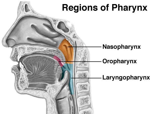
2 Definitio n Fat filled triangular space lateral the pharynx Extends from the skull base to the oropharynx
3 Parapharyngeal Space
![Content s Fat Arteries [ascending pharyngeal, internal maxillary]](/docs-images/85/92777162/images/4-0.jpg "Veins [ pharyngeal veins] Nerves [ branches of the mandibular")
4 Content s Fat Arteries [ascending pharyngeal, internal maxillary] Veins [ pharyngeal veins] Nerves [ branches of the mandibular nerve]

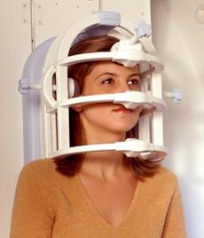
5 Imaging An anatomic landmark for the adjacent spaces Imaging plane is directed for the site pathology CT and MRI 5 mm slices Axial and coronal planes Contrast injection TEST HAYTHAM
6 Scanning techniques AXIAL CORONAL


7 C T AXIAL Calcification Bone erosions Hyperostosis CORONAL


8 Closed magnet M RI Superior contrast resolution Direct multiplanar imaging Vascular imaging without contrast injection No bone artifacts

9 Dynamic magnet

10 Anatomic relations Antrolateral Postrolateral Medial ANTRO- LATERAL ASPECT POSTRO- LATERAL ASPECT


11 Clinical aspects Is difficult to be evaluated clinically Presenting symptoms Sore throat Dysphagia Change of voice Nasal obstruction Cranial nerves IX XII A mass bulges posterior to the angle of mandible
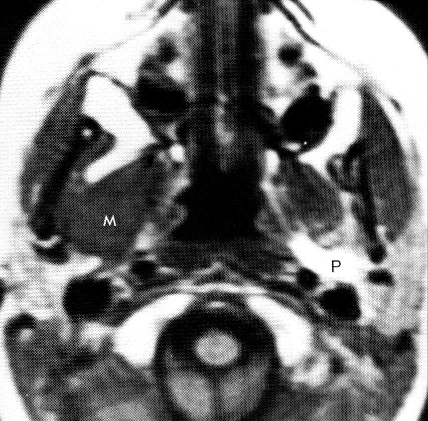
12 Antrolateral aspect [ Infratemporal fossa ] Masticator space Parotid space MASTICATOR SPACE PAROTID SPACE
![Antrolateral aspect [Infratemporal fossa]](/docs-images/85/92777162/images/13-0.jpg "Masticator Space Muscles of masticatio [")
![masseter, temporalis, pterygoid muscles]](/docs-images/85/92777162/images/13-2.jpg "Mandibular ramus Mandibular nerve branches Fat")
13 Antrolateral aspect [Infratemporal fossa] Masticator Space Muscles of masticatio [ masseter, temporalis, pterygoid muscles] Mandibular ramus Mandibular nerve branches Fat behind the antral wall
![Postro -lateral aspect space ] Stylo mandibular tunnel [ Parotid Parotid gland](/docs-images/85/92777162/images/14-0.jpg "(deep lobe) External carotid artery Retromandibular vein Facial nerve Lymph")
14 Postro -lateral aspect space ] Stylo mandibular tunnel [ Parotid Parotid gland (deep lobe) External carotid artery Retromandibular vein Facial nerve Lymph nodes
15 [ extends from the skull base to aortic arch ] Postro -lateral aspect styloid space] Carotid canal Contents Internal carotid artery Internal jugular vein [ Post Cranial nerves 9 to 12, sympathetic plexus Lymph nodes [Internal jugular + lateral retropharyngeal nodes]
![Medial aspect mucosal space] [ Pharyngeal The pharyngeal mucosal space is separated from the PPS by the](/docs-images/85/92777162/images/16-2.jpg "pharyngo- basilar fascia (PBF) The PBF is a tough membrane Maintains patent airway Crossed only by aggressive")
16 Medial aspect mucosal space] [ Pharyngeal The pharyngeal mucosal space is separated from the PPS by the pharyngo- basilar fascia (PBF) The PBF is a tough membrane Maintains patent airway Crossed only by aggressive lesions
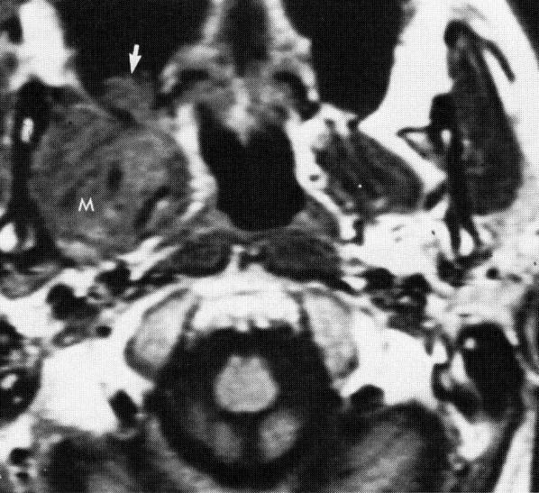
17 Medial Patholo aspect gy mucosal space] Displacement of the PPS fat laterally 98% of masses are carcinomas 80% squamous cell type [ Pharyngeal Other carcinomas [ adenoid cystic & mucoepidermoid] Lymphomas and sarcomas (children) Angiofibroma, plasmacytoma, melanoma (rare)


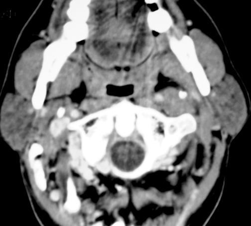
18 Nasopharyngeal anatomy Eustachian tube fossa Fossa of Rosenmuller

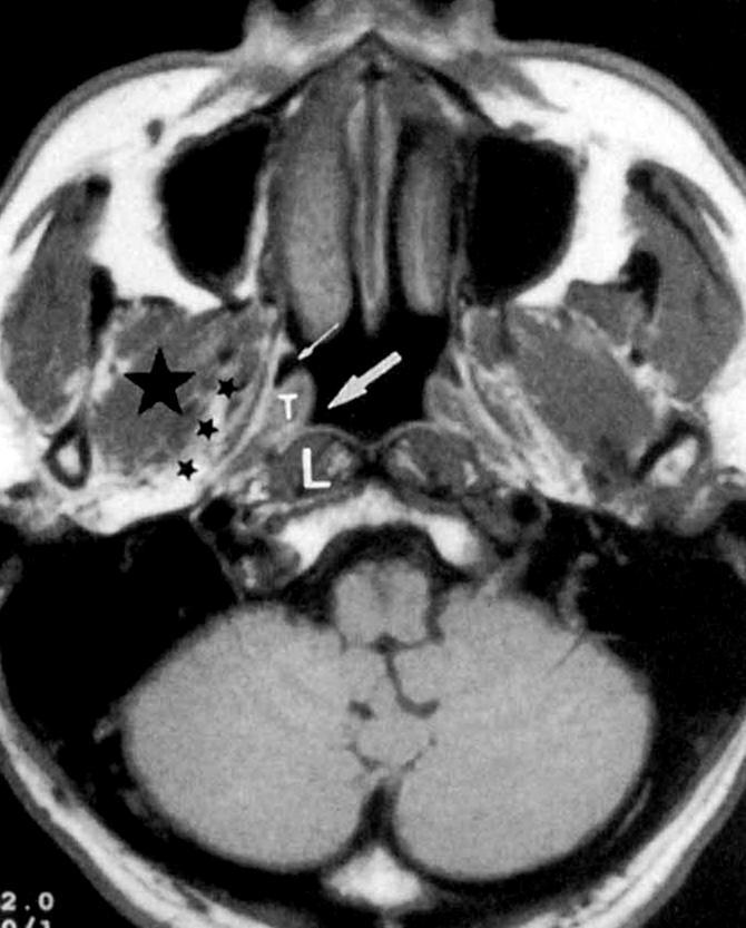
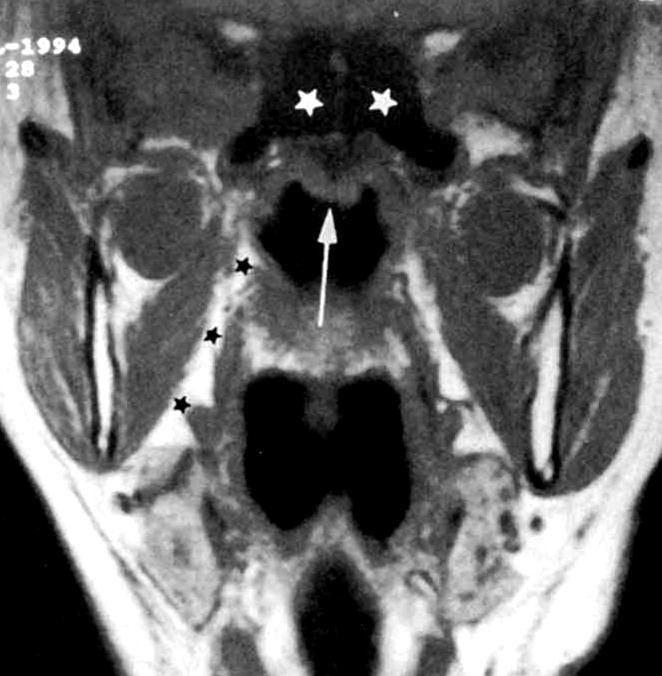
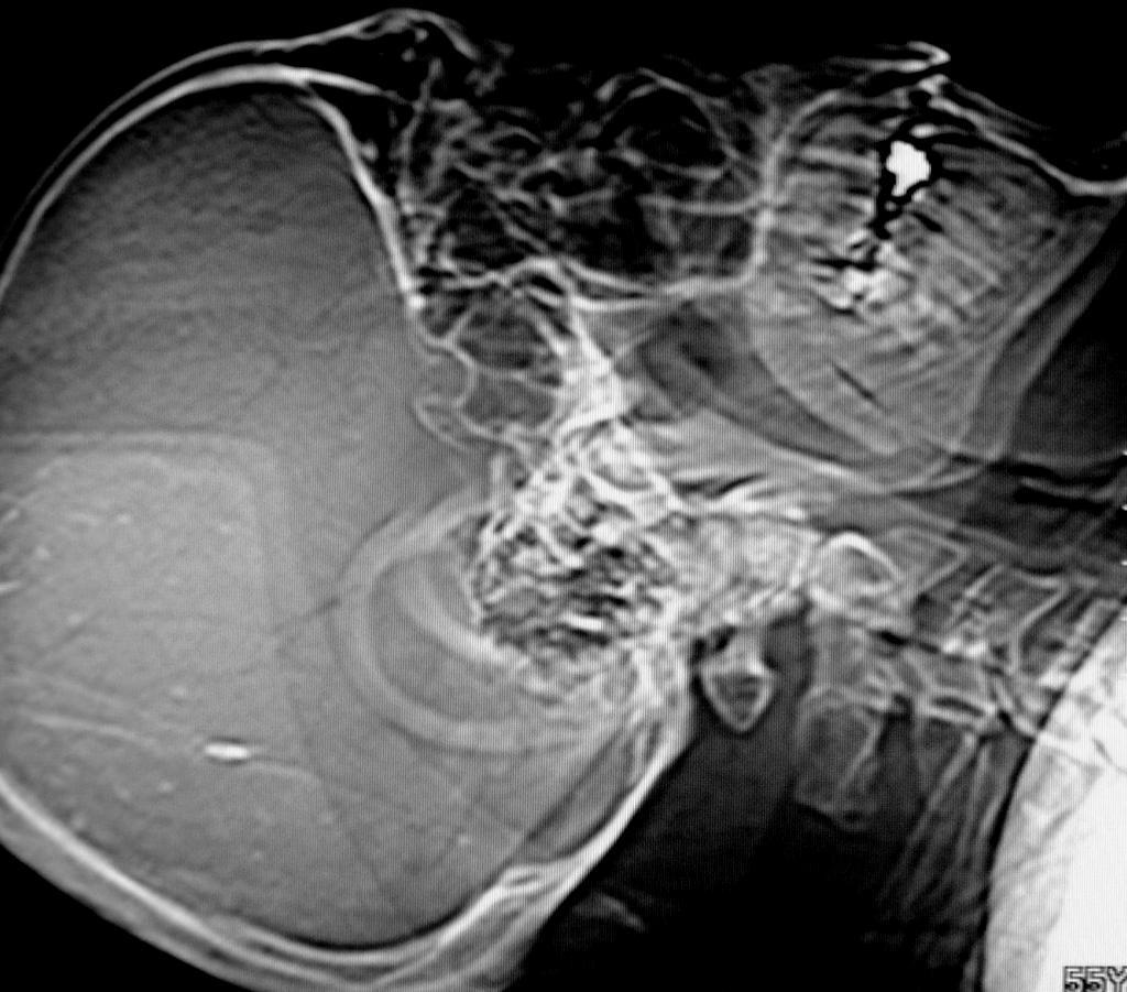
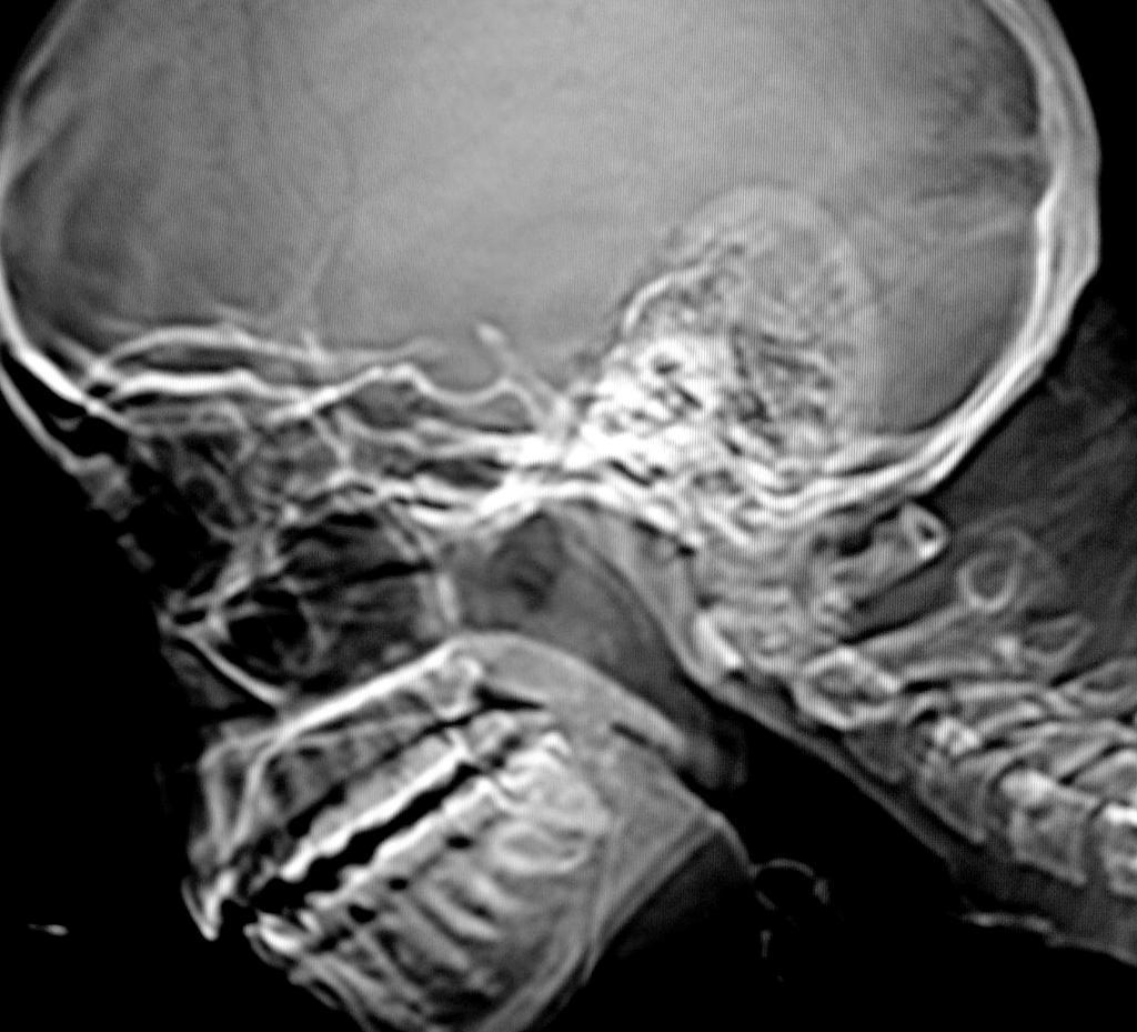
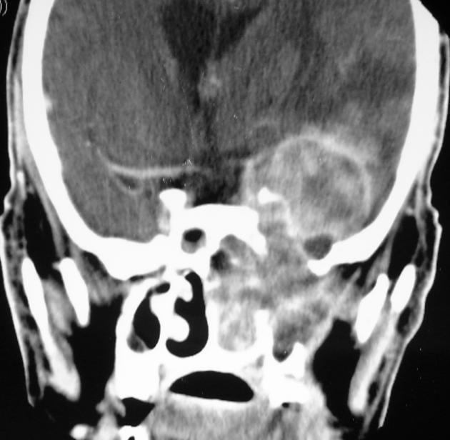
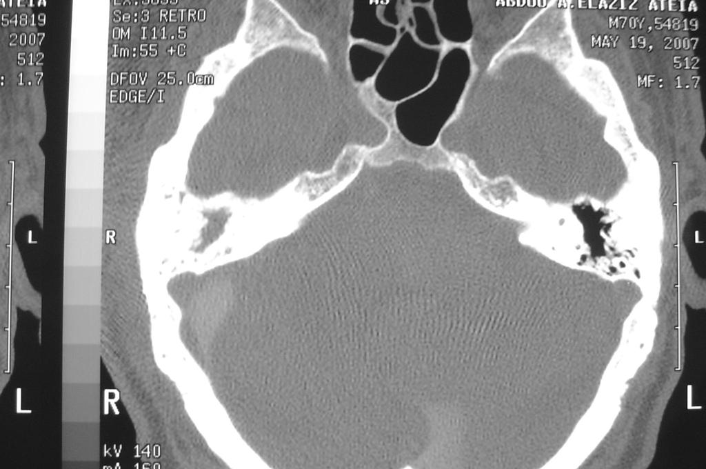
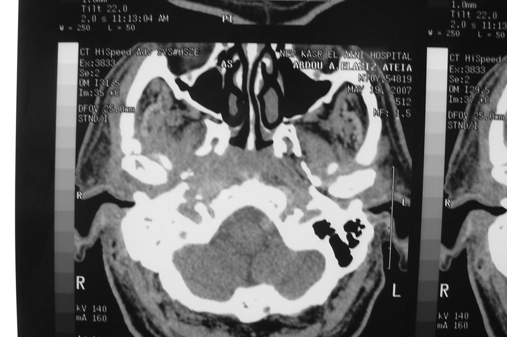
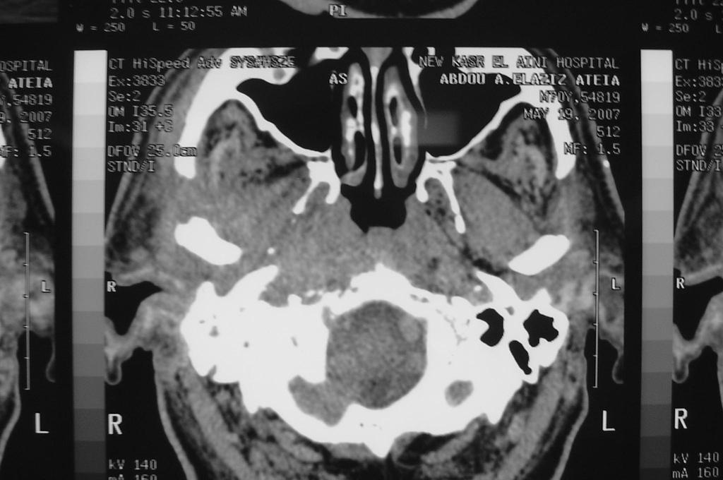
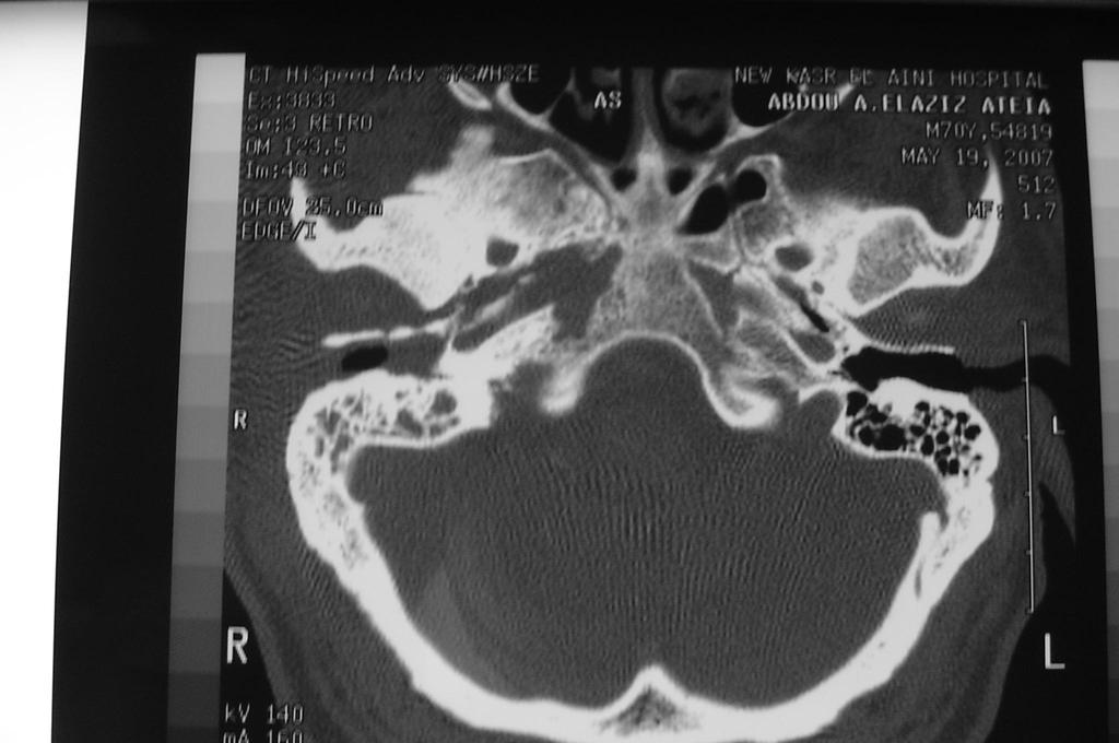
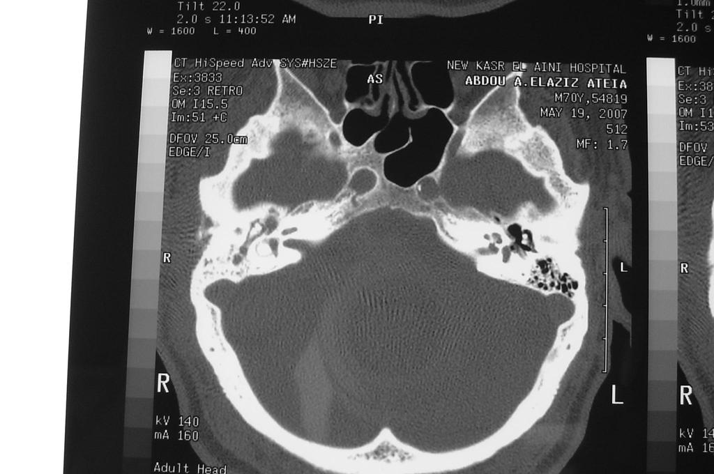
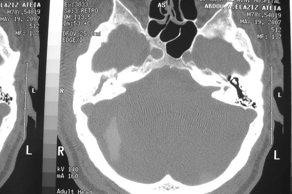
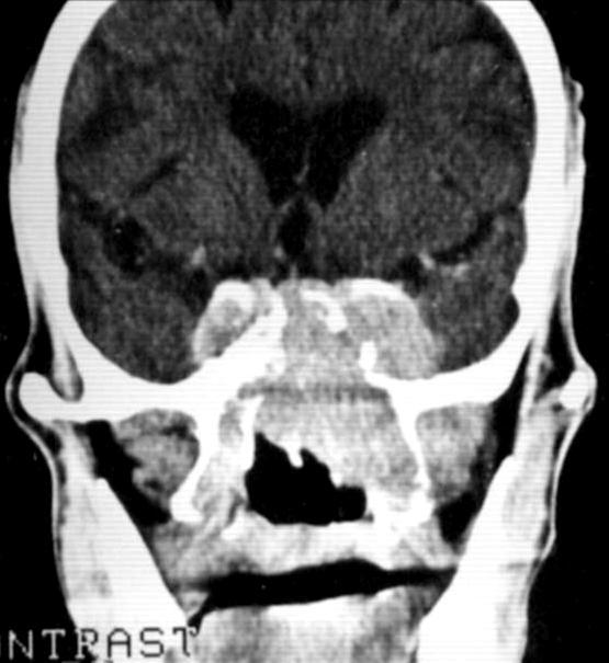
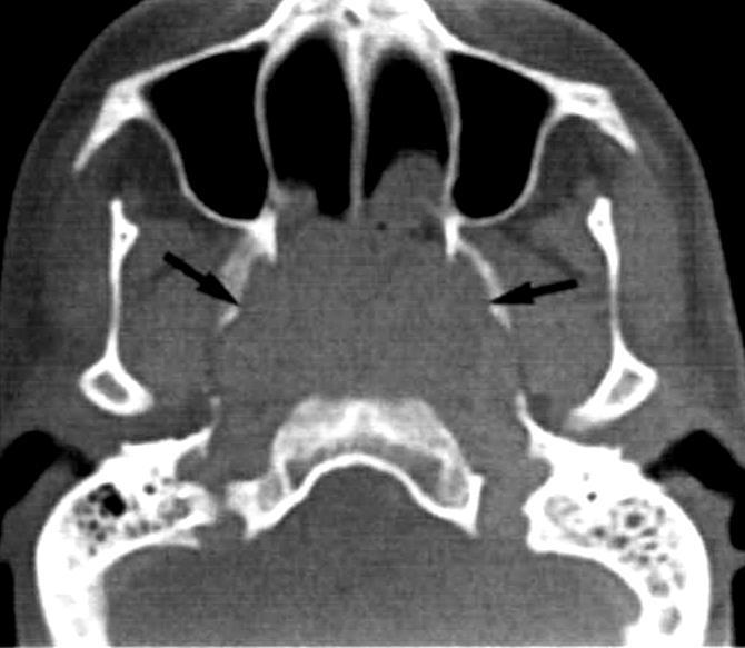
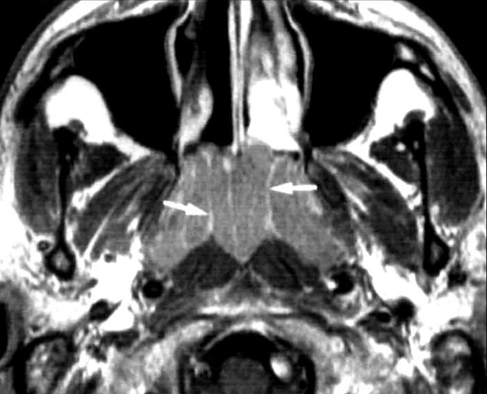
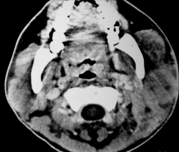
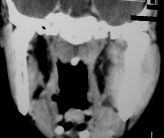
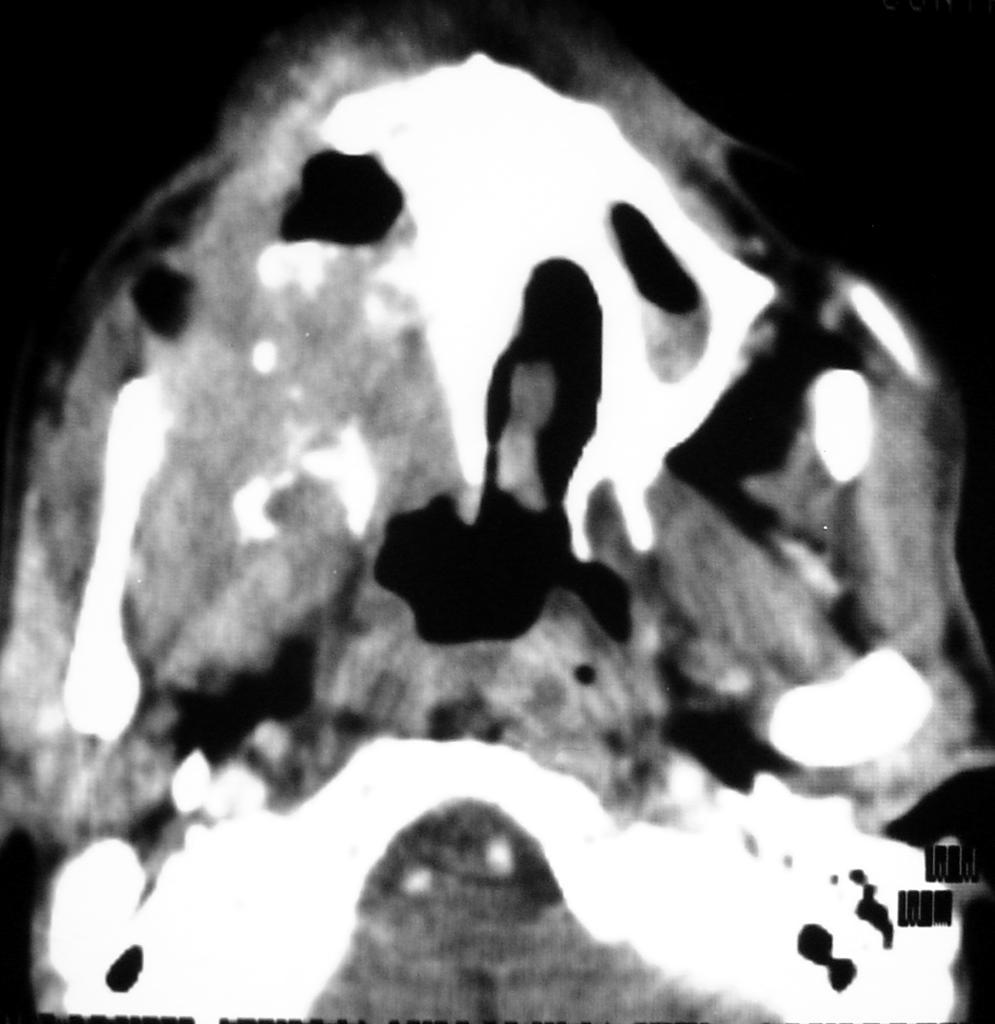
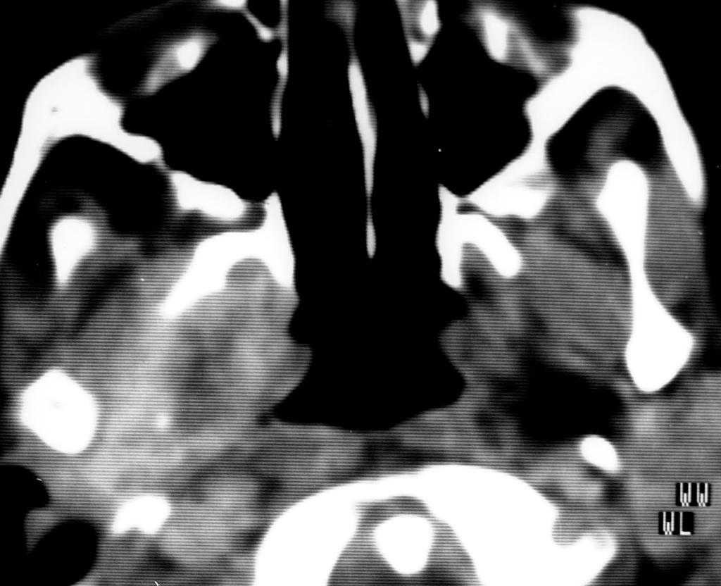
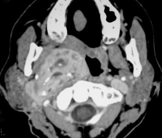
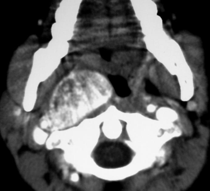
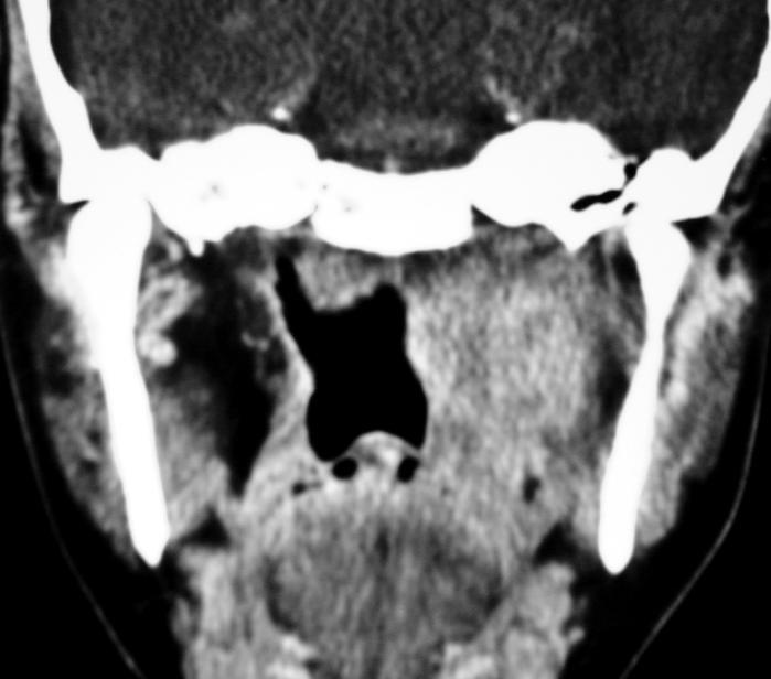
19 Nasopharyngeal carcinoma F 55Y
20 Nasopharyngeal carcinoma

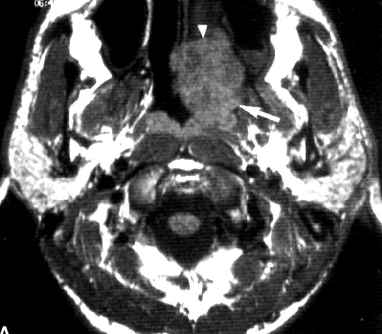
21 Nasopharyngeal Carcinoma Early diagnosis Best on MRI Obliteration of the fat strip between the tensor and levator veli palatini muscles on T1 WIs Extension into the PPS fat Obliteration of the fat plane between the nasopharynx and prevertebral muscles

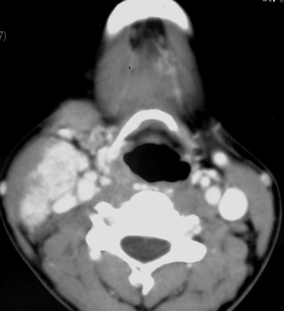
22 Nasopharyngeal carcinoma with nodes
23 Nasopharyngeal carcinoma [ Staging ] T1 T2 T3 T4 Confined to the nasopharynx Extension to Oropharynx or nasal fossa (axial) Invasion of bones or sinuses (axial) Intracranial extension or hypo pharynx or orbit
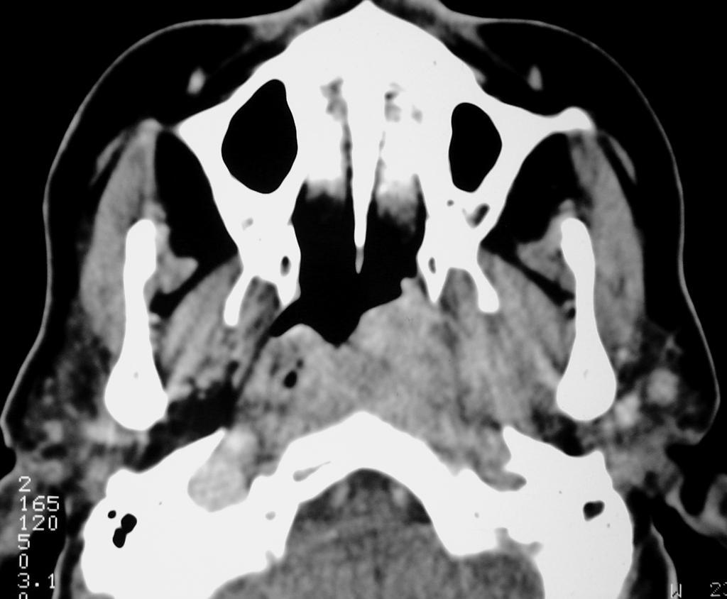
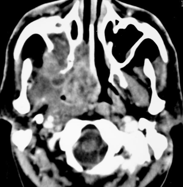
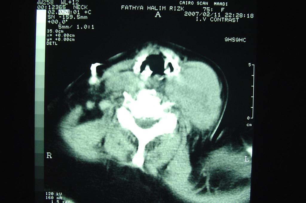
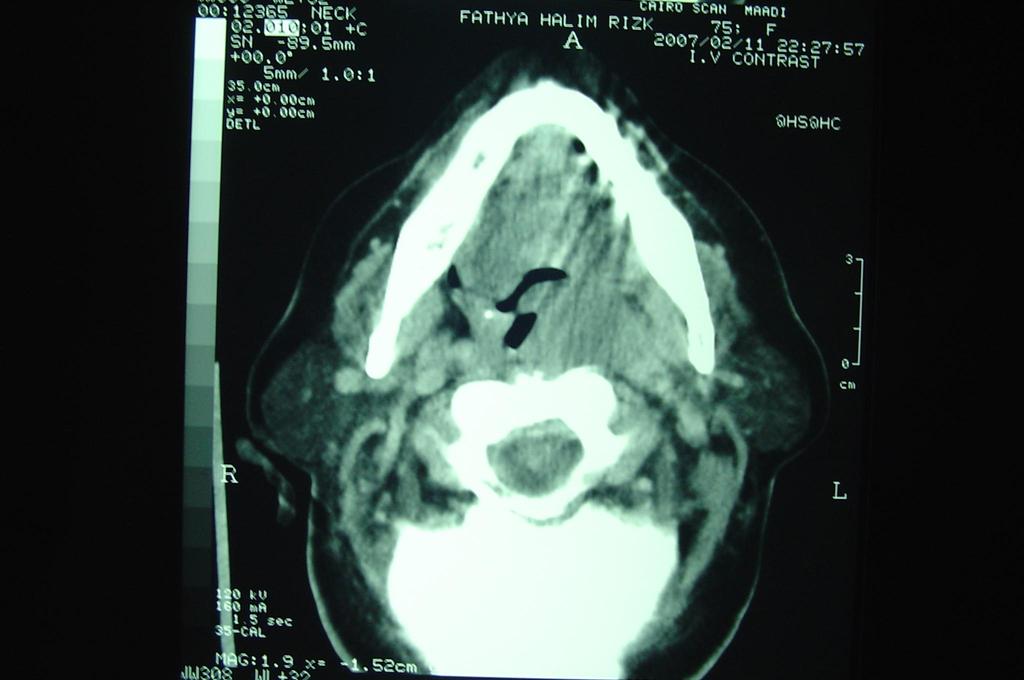
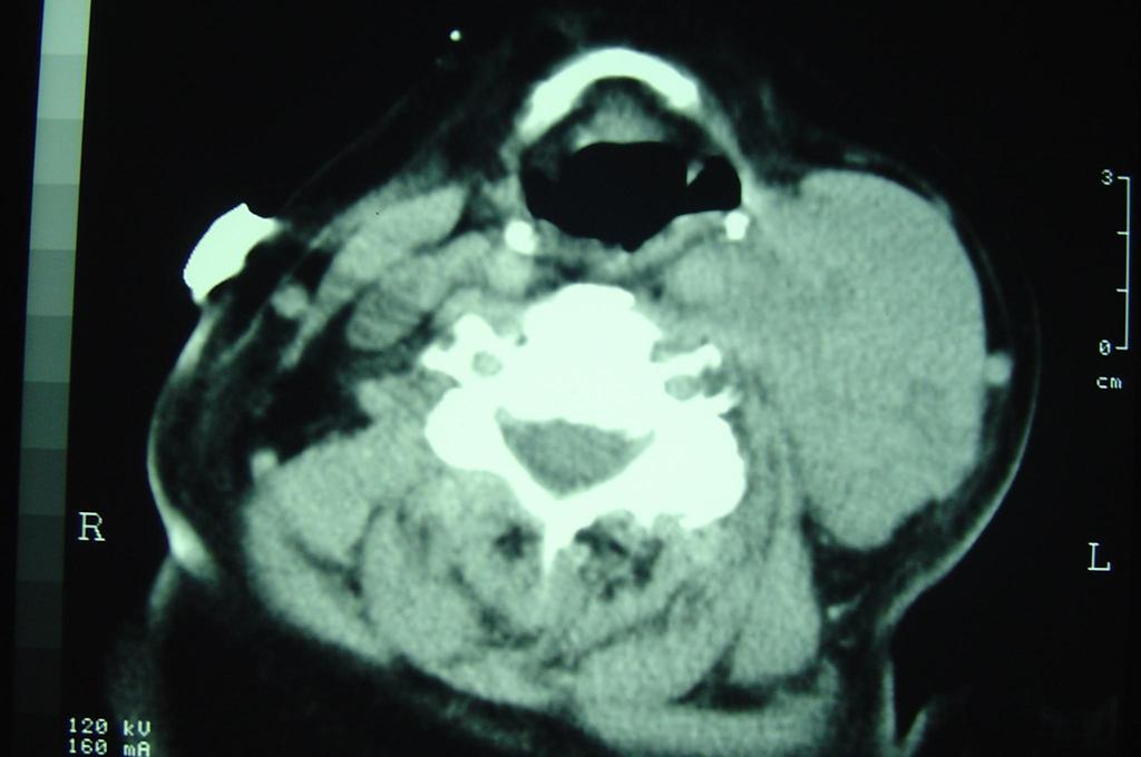
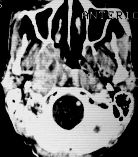
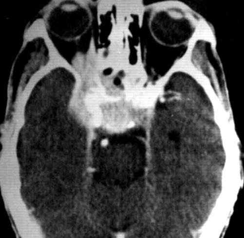
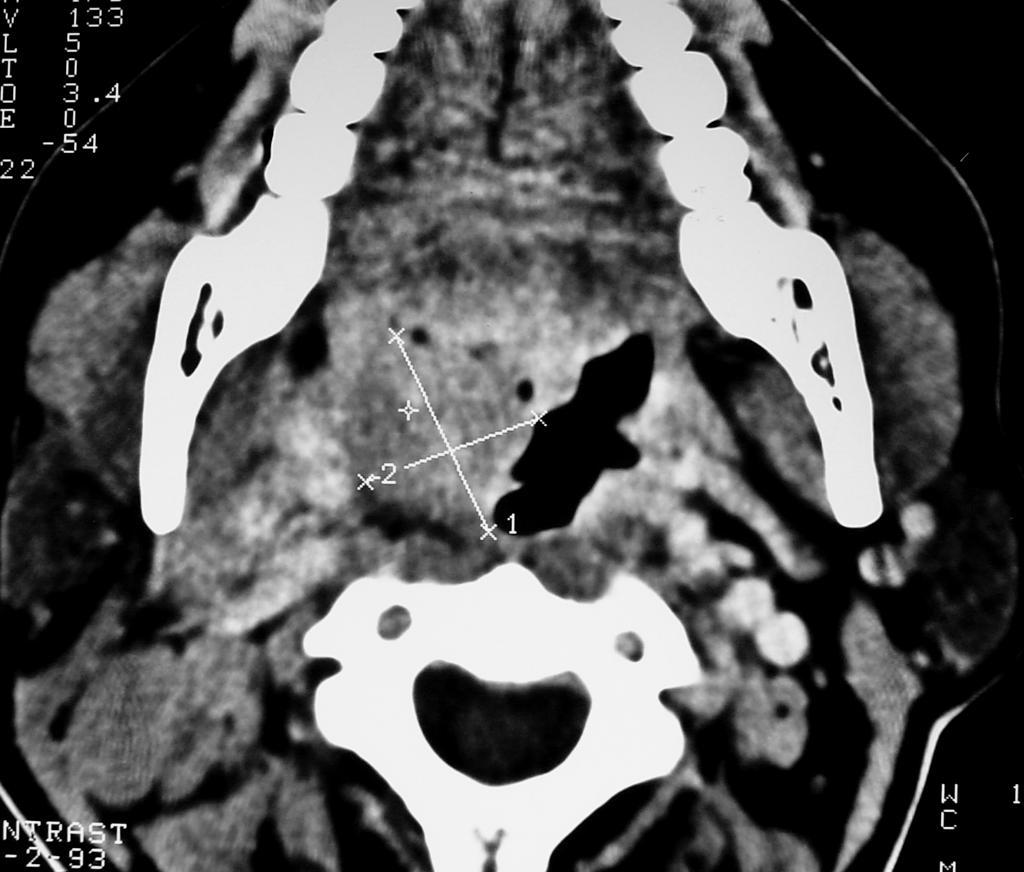
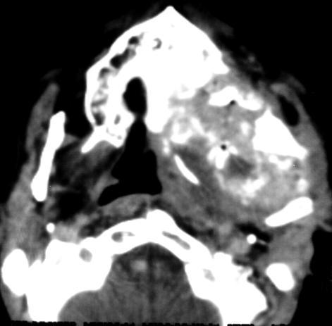
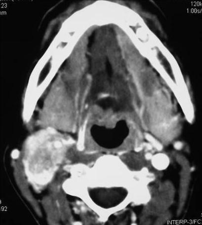
24 Nasopharyngeal Carcinoma Effacement of the FR and ET Heterogeneously enhancing mass in the lateral wall of the nasopharynx Extensions Anteriorly nasal fossa, maxillary sinus, infratemporal fossa Posteriorly prevertebral muscles, carotid sheath Laterally parapharyngeal space, mastecator space

25 Nasopharyngeal Carcinoma Medially nasopharyngeal air space, retropharyngeal to the contra lateral side Inferiorly Oropharynx, tongue Superiorly skull base, intracranial extension
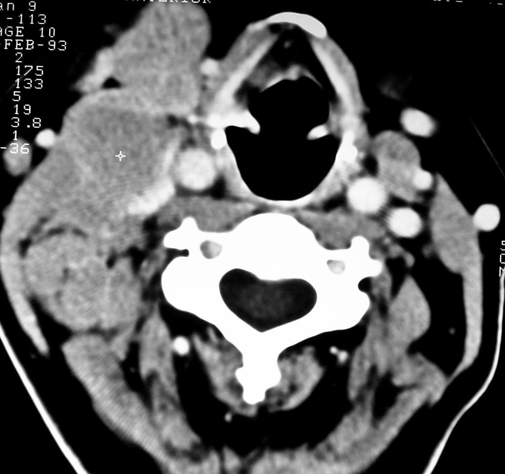
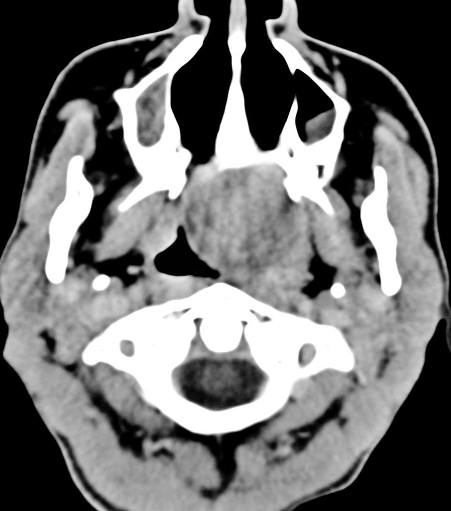
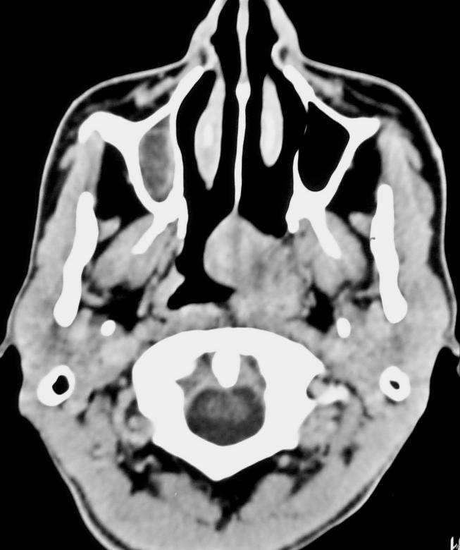
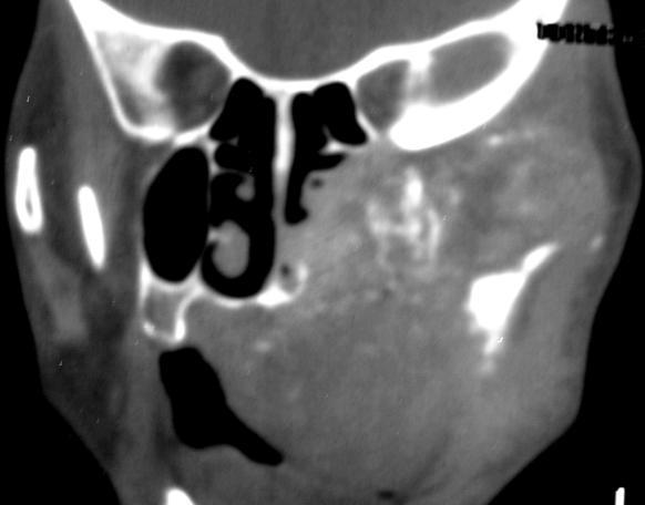
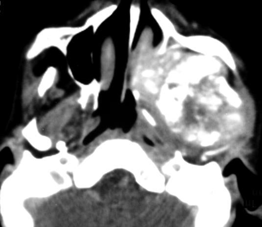
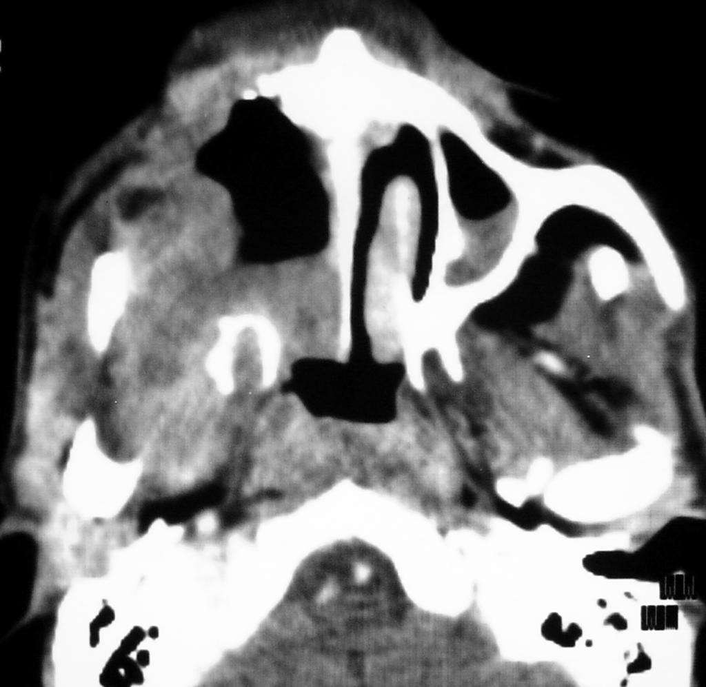
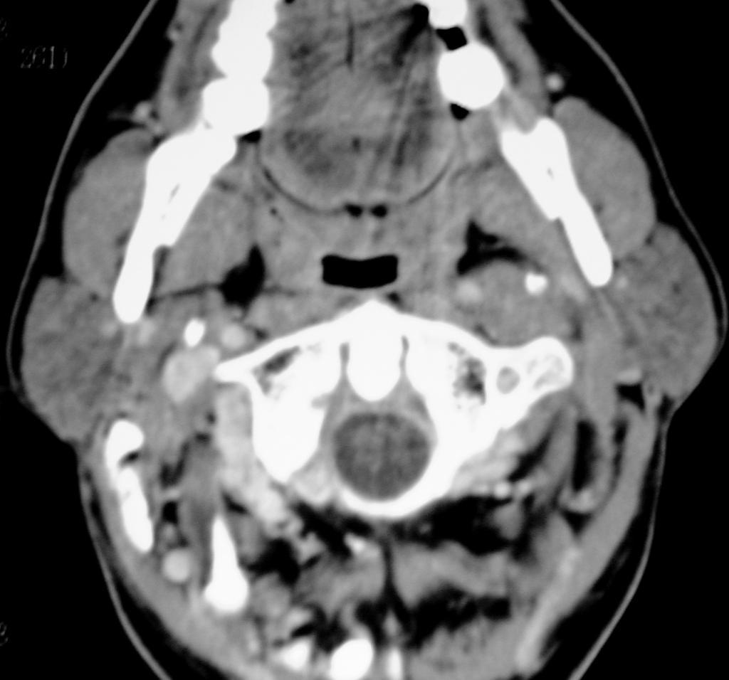
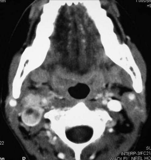
26 Nasopharyngeal Carcinoma F 37Y

27 Nasopharyngeal Carcinoma
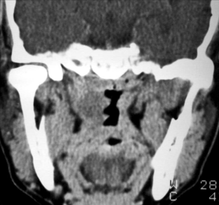
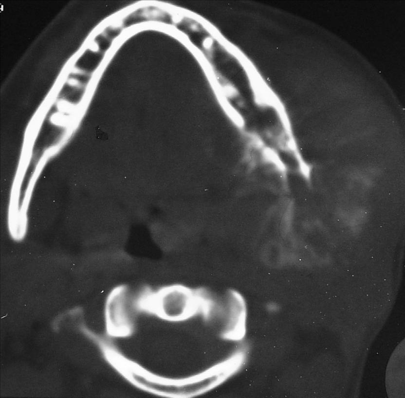
28 Nasopharyngeal Carcinoma, otitis
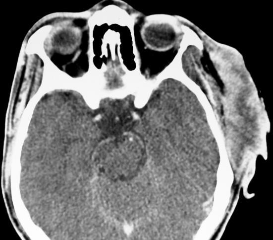
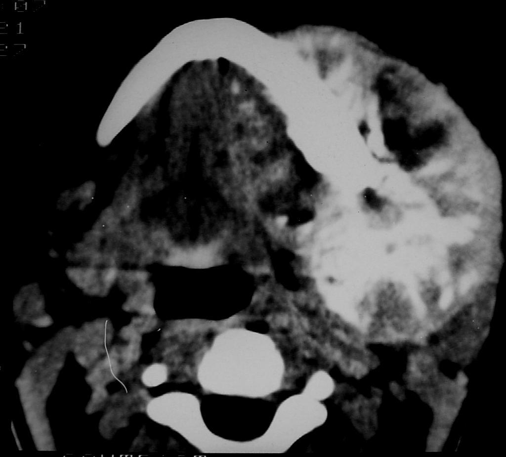
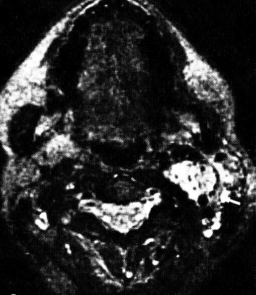
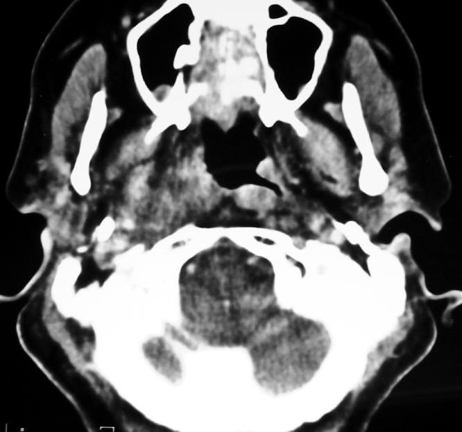
29 M 60Y Nasopharyngeal Carcinoma T4
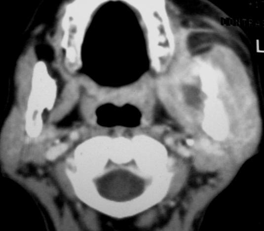
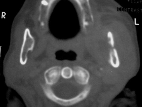
30 Nasopharyngeal Carcinoma T2 55Y M


31 Other malignancies Lymphomas 20% Others 10% Rhabdomyosarcoma Adenoid cystic carcinoma Melanoma, plasmacytoma,.. F 16 Y
32 Nasopharyngeal NHL M 66Y

33 Rhabdomyosarco ma The most common sarcoma of the head and neck Arise from the primitive mesenchymal cells 70% arise before the age of 12 years Orbit > nasopharynx >temporal bone > sinuses > neck Presents by pain and cranial nerve palsies Soft tissue mass with bone destruction Deposits to the lung and bones DD nasopharyngeal carcinoma, angiofibroma,nhl M 11Y
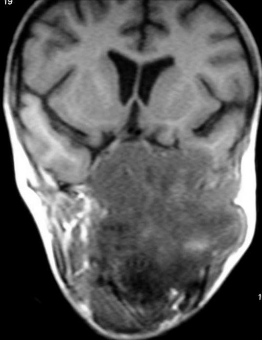
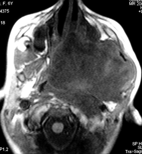
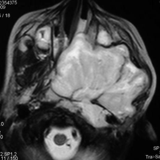
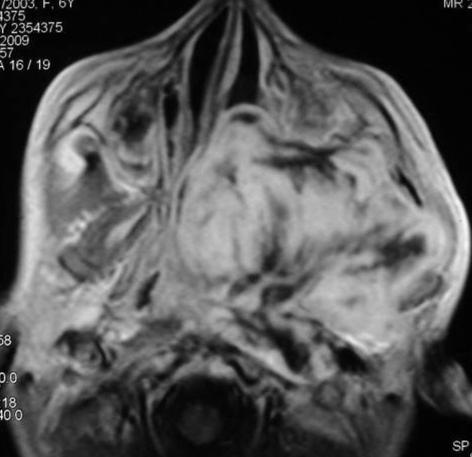
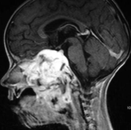
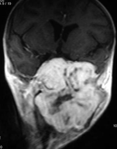
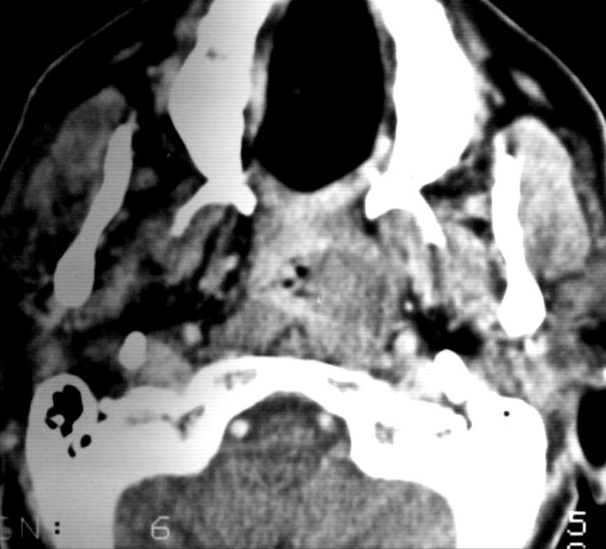
34 Rhabdomyosarcoma F 6Y

35 Adenoid Cystic Carcinoma


36 Nasopharyngeal benign lesions Tornwaldt s cyst A mucous retention cyst Occurs in the midline nasopharynx Low signal in T1 and high signal in T2 WIs


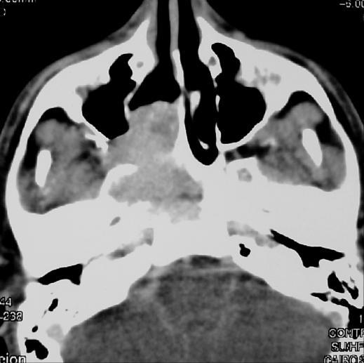
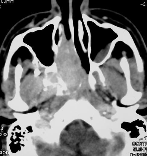
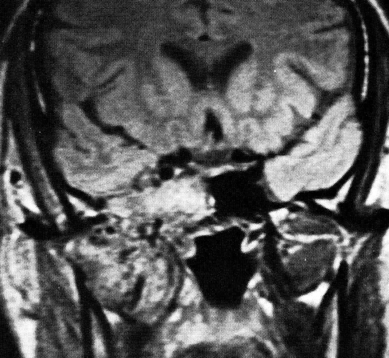
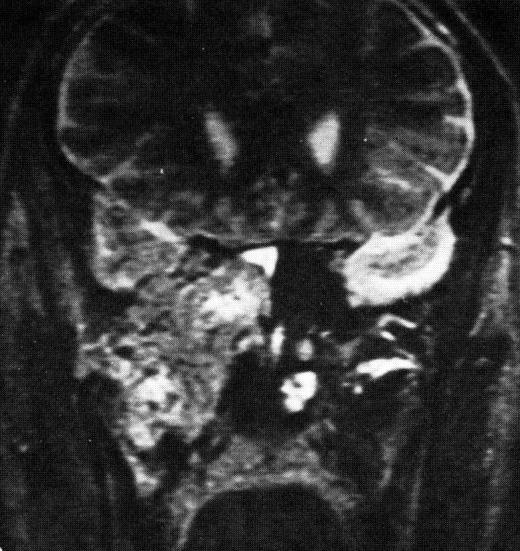
37 Nasopharyngeal angiofibroma Arises near the sphenopalatine foramen Almost exclusively in adolescent boys Epistaxis

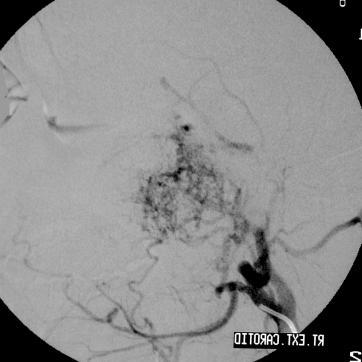
38 Nasopharyngeal angiofibroma Hyper vascular lesion with intense enhancement Supplied by the ascending pharyngeal & ascending palatine branches of the internal maxillary artery M 19Y Forward displacement of the posterior wall of the maxillary sinus [Holman- Miller sign]- classical


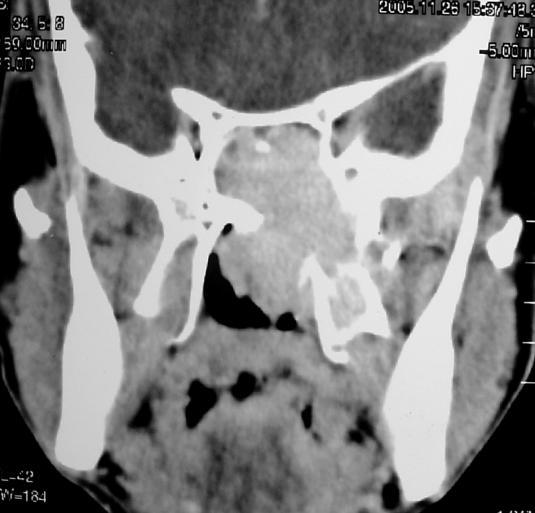
39 Grading of nasopharyngeal Angiofibroma I II III Confined to the nasopharynx Extension into pterygopalatine fossa or Intracranial or intraorbital extension masticator space M 17Y
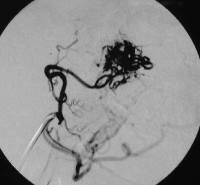
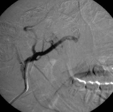
40 Nasopharyngeal angiofibroma M 18Y
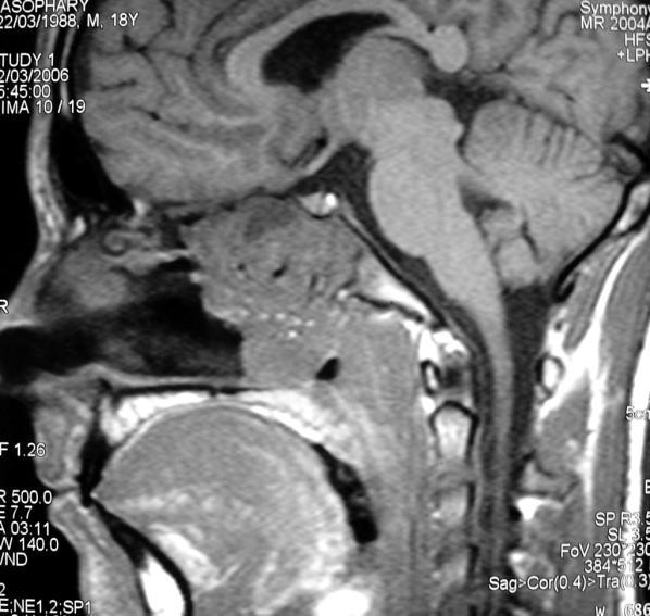
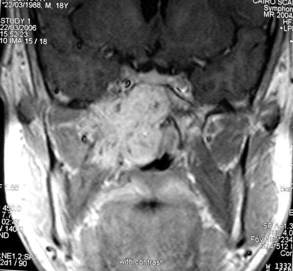
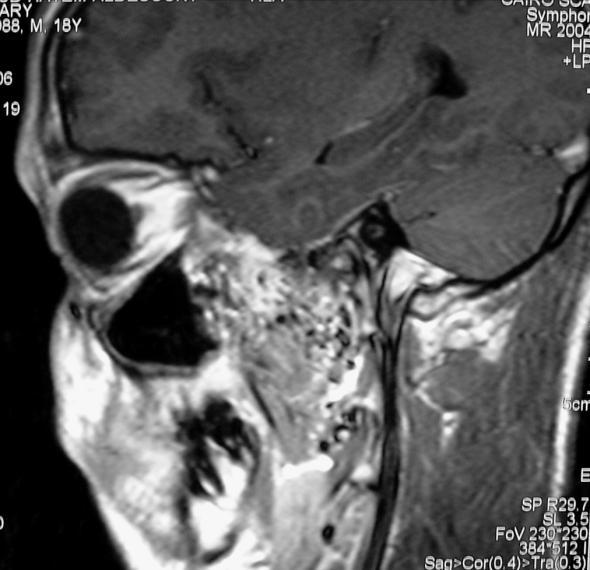
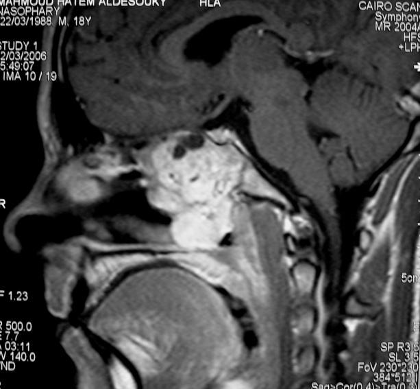
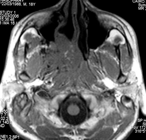
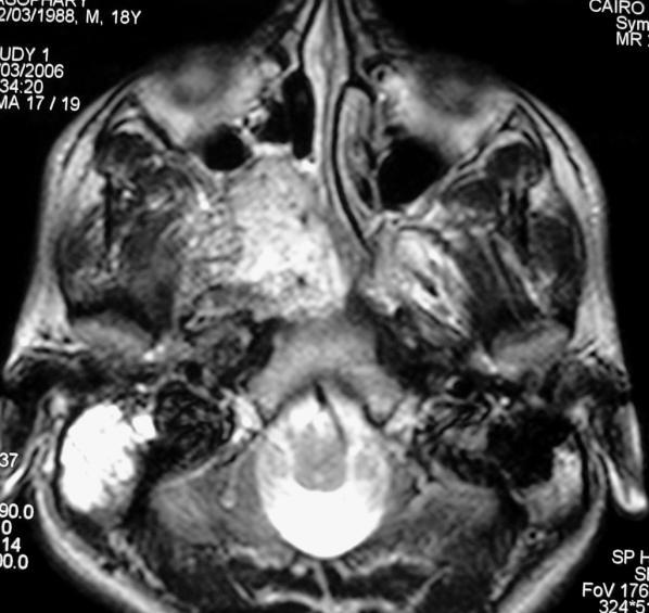
41 Nasopharyngeal angiofibroma M 18Y

42 Oro -pharyngeal anatomy

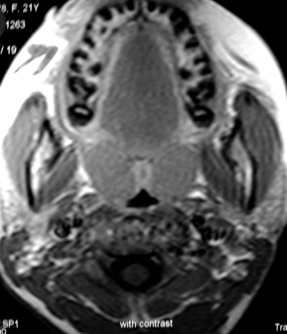
43 Tonsillit is F 21Y
44 Lymphoid hyperplasia
45 Oro -pharyngeal carcinoma

46 NHL tonsils, oropharynx, supraglottic region

47 Oro -pharyngeal carcinoma


48 Lateral Patholo aspect gy [ Parotid space]

49 Adenoid cystic carcinoma

50 Mucoepidermoid carcinoma M 41Y

51 Deep lobe parotid tumor

52 Lymphoma of the nasopharynx and parotid gland M 21Y
53 Antro Patholo -lateral aspect [ gy Masticator space]
54 Adamantinoma of the mandible F 45Y
55 Chondrosarcoma of the hard palate M 45Y
56 Recurrent fibrosarcoma of the maxilla M 50Y
57 Mandibular osteosarcoma M 27Y

58 NHL 10Y M 10Y
59 Rhabdomyosarco ma F 8Y
60 Metastatic renal cell carcinoma in the masticator space

61 Deposit of bronchogenic Carcinoma
![[ extends from the skull base to aortic arch ] Postro -lateral aspect styloid space] Carotid canal Contents Internal carotid artery](/docs-images/85/92777162/images/62-0.jpg "Internal jugular vein [ Post Cranial nerves 9 to 12, sympathetic plexus Lymph nodes [Internal jugular + lateral retropharyngeal")
62 [ extends from the skull base to aortic arch ] Postro -lateral aspect styloid space] Carotid canal Contents Internal carotid artery Internal jugular vein [ Post Cranial nerves 9 to 12, sympathetic plexus Lymph nodes [Internal jugular + lateral retropharyngeal nodes]
63 Glomus Nodes Neurofibrom a
64 LATERAL RETROPHARYNGEAL NODES INTERNAL JUGULAR NODES MEDIAL RETROPHARYNGEAL NODES
65 F 54Y
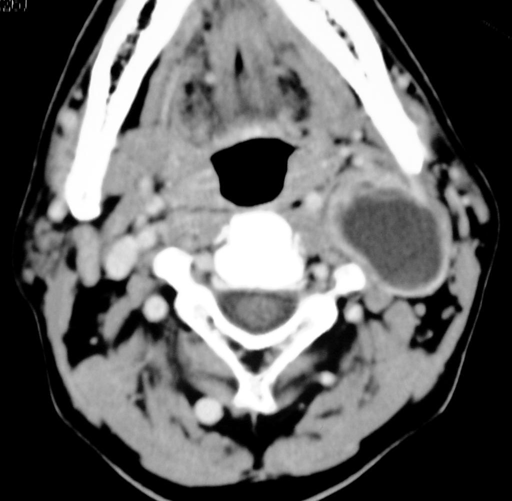
66 Cystic neurofibroma M 44Y
67 Glomus tumor Rare, slowly growing hypervasculer tumor Incidence 1: 1,300,000 Male : female 1: Y Arise from the glomus bodies in and around the jugular bulb Benign hyper vascular lesion supplied by Ascending pharyngeal Carotico -tympanic [ICA] Anterior tympanic [ECA] Stylomastoid [ECA] Meningeal branches [ vertebral]


68 Glomus tumor Mass in the jugular fossa with bone destruction Large at presentation 2-6 cm, intense enhancement Intracranial and extra cranial extension Metastases in 4%, Lung, nodes, liver, bones Salt and pepper appearance on MRI

69 Carotid body tumor


70 Glomus tumor Common presenting symptoms Palsatile tinnitus, cranial nerve palsies,vascular retro-tympanic mass M 41Y


71 Glomus tumor Common jugular fossa masses Meningioma, neurofibroma, deposits, congenitally wide or dehiscent M 55Y

72 Lymphadenopat hy Reactive homogenous,young patient less than 1cc Lymphoma bulky homogenous Direct invasion from near by malignancy Inflammatory septic focus abscess formation
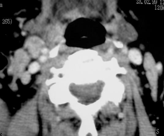
73 Metastatic nodes The most common nodal disease Any malignancy can spread to the retro-pharyngeal nodes Enlarged nodes> 0.8 cm with central necrosis and stranding of the perinodal fat 75% of nasopharyngeal carcinoma,20% of oropharyngeal, 5% of thyroid carcinoma have metastatic nodes at presentation
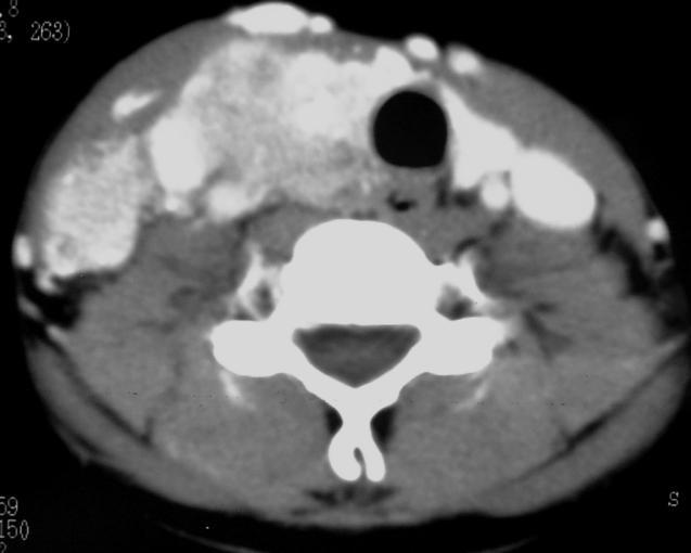
74 Metastatic nodes from thyroid carcinoma F 21 Y

75 PPS LESIONS Antro-lateral aspect Maxilla Mandible Muscles Medial aspect Nasopharynx Oropharynx Tonsil Lateral aspect Deep lobe parotid Posterior aspect Glomus Nodes Neurofibroma

76 Quiz. Tornwaldet cyst
77 Q.. Nasopharyngeal carcinoma

78 Q.. Glomus tumor


79 سبحانك اللهم و بحمدك نشهد ان ال اله اال انت نستغفرك و نتوب اليك ssr-eg.net Thank you Mamdouh Mahfouz MD
80 .. Q
81 .. Q
82
Head and Neck Image 頭頸部放射影像學
 Head and Neck Image 頭頸部放射影像學 陳家媛 台北醫學大學 - 市立萬芳醫院 cychen@wanfang.gov.tw Normal Suprahyoid neck: the old way Nasopharynx Oropharynx Oral cavity Staging of SCC Spaces of Suprahyoid Neck: a New Way Deep
Sinonasal Imaging. Mamdouh Mahfouz MD Professor of Radiology Cairo University. ssregypt.com
 Sinonasal Imaging Mamdouh Mahfouz MD Professor of Radiology Cairo University ssregypt.com Scanning Techniques Routine Study CORONAL Coronal 3-5mm sections from the posterior wall of the sphenoid sinus
Temporal region. temporal & infratemporal fossae. Zhou Hong Ying Dept. of Anatomy
 Temporal region temporal & infratemporal fossae Zhou Hong Ying Dept. of Anatomy Temporal region is divided by zygomatic arch into temporal & infratemporal fossae. Temporal Fossa Infratemporal fossa Temporal
PTERYGOPALATINE FOSSA
 PTERYGOPALATINE FOSSA Outline Anatomical Structure and Boundaries Foramina and Communications with other spaces and cavities Contents Pterygopalatine Ganglion Especial emphasis on certain arteries and
Infratemporal fossa: Tikrit University college of Dentistry Dr.Ban I.S. head & neck Anatomy 2 nd y.
 Infratemporal fossa: This is a space lying beneath the base of the skull between the lateral wall of the pharynx and the ramus of the mandible. It is also referred to as the parapharyngeal or lateral pharyngeal
Temporal fossa Infratemporal fossa Pterygopalatine fossa Terminal branches of external carotid artery Pterygoid venous plexus
 Outline of content Temporal fossa Infratemporal fossa Pterygopalatine fossa Terminal branches of external carotid artery Pterygoid venous plexus Boundary Content Communication Mandibular division of trigeminal
The Pharynx. Dr. Nabil Khouri MD. MSc, Ph.D
 The Pharynx Dr. Nabil Khouri MD. MSc, Ph.D Introduction The pharynx is the Musculo-fascial halfcylinder that links the oral and nasal cavities in the head to the larynx and esophagus in the neck Common
Dr.Ban I.S. head & neck anatomy 2 nd y. جامعة تكريت كلية طب االسنان املرحلة الثانية أ.م.د. بان امساعيل صديق 6102/6102
 جامعة تكريت كلية طب االسنان التشريح مادة املرحلة الثانية أ.م.د. بان امساعيل صديق 6102/6102 Parotid region The part of the face in front of the ear and below the zygomatic arch is the parotid region. The
Clinical evaluation. Imaging Surgical treatment
 Parapharyngeal Space Khalid adhussain AL-Qahtani a MD,MSc,FRCS(c) Assistant Professor Consultant of Otolaryngology Advance Head & Neck Oncology, Thyroid & Parathyroid,Microvascular Reconstruction, ti and
Suprahyoid and Infrahyoid Neck Overview
 10 Imaging Approaches & Indications Neither CT nor MR is a perfect modality for imaging the extracranial H&N. MR is most useful in the suprahyoid neck (SHN) because it is less affected by oral cavity dental
Dr.Ban I.S. head & neck anatomy 2 nd y. جامعة تكريت كلية طب االسنان املرحلة الثانية
 جامعة تكريت كلية طب االسنان التشريح مادة املرحلة الثانية أ.م.د. بان امساعيل صديق 6102-6102 1 The Palate The palate forms the roof of the mouth and the floor of the nasal cavity. It is divided into two
"The Space Between Us:" A Radiographic Review of Common and Uncommon Pathologic Findings within the Deep Spaces of the Neck
 "The Space Between Us:" A Radiographic Review of Common and Uncommon Pathologic Findings within the Deep Spaces of the Neck Poster No.: C-2457 Congress: ECR 2015 Type: Educational Exhibit Authors: A. K.
Prevertebral Region, Pharynx and Soft Palate
 Unit 20: Prevertebral Region, Pharynx and Soft Palate Dissection Instructions: Step1 Step 2 Step 1: Insert your fingers posterior to the sternocleidomastoid muscle, vagus nerve, internal jugular vein,
The Neck the lower margin of the mandible above the suprasternal notch and the upper border of the clavicle
 The Neck is the region of the body that lies between the lower margin of the mandible above and the suprasternal notch and the upper border of the clavicle below Nerves of the neck Cervical Plexus Is formed
Veins of the Face and the Neck
 Veins of the Face and the Neck Facial Vein The facial vein is formed at the medial angle of the eye by the union of the supraorbital and supratrochlear veins. connected through the ophthalmic veins with
Tikrit University collage of dentistry Dr.Ban I.S. head & neck anatomy 2 nd y. Lec [5] / Temporal fossa :
![Tikrit University collage of dentistry Dr.Ban I.S. head & neck anatomy 2 nd y. Lec [5] / Temporal fossa :](/thumbs/88/115294566.jpg "Tikrit University collage of dentistry Dr.Ban I.S. head & neck anatomy 2 nd y. Lec [5] / Temporal fossa :") Lec [5] / Temporal fossa : Borders of the Temporal Fossa: Superior: Superior temporal line. Inferior: gap between zygomatic arch and infratemporal crest of sphenoid bone. Anterior: Frontal process of the
Lec [5] / Temporal fossa : Borders of the Temporal Fossa: Superior: Superior temporal line. Inferior: gap between zygomatic arch and infratemporal crest of sphenoid bone. Anterior: Frontal process of the
Parotid Gland, Temporomandibular Joint and Infratemporal Fossa
 M1 - Anatomy Parotid Gland, Temporomandibular Joint and Infratemporal Fossa Jeff Dupree Sanger 9-057 jldupree@vcu.edu Parotid gland: wraps around the mandible positioned between the mandible and the sphenoid
Structure Location Function
 Frontal Bone Cranium forms the forehead and roof of the orbits Occipital Bone Cranium forms posterior and inferior portions of the cranium Temporal Bone Cranium inferior to the parietal bone forms the
Anatomic Relations Summary. Done by: Sohayyla Yasin Dababseh
 Anatomic Relations Summary Done by: Sohayyla Yasin Dababseh Anatomic Relations Lecture 1 Part-1 - The medial wall of the nose is the septum. - The vestibule lies directly inside the nostrils (Nares). -
Omran Saeed. Luma Taweel. Mohammad Almohtaseb. 1 P a g e
 2 Omran Saeed Luma Taweel Mohammad Almohtaseb 1 P a g e I didn t include all the photos in this sheet in order to keep it as small as possible so if you need more clarification please refer to slides In
Mohammad Hisham Al-Mohtaseb. Lina Mansour. Reyad Jabiri. 0 P a g e
 2 Mohammad Hisham Al-Mohtaseb Lina Mansour Reyad Jabiri 0 P a g e This is only correction for the last year sheet according to our record. If you already studied this sheet just read the yellow notes which
Case Studies in the Skull Base
 Case Studies in the Skull Base Amy C Tsai, MD Neuroradiology Fellow Department of Radiology and Imaging Sciences University of Utah Health Sciences Center Salt Lake City, Utah, USA No disclosures related
Spinal Imaging. ssregypt.com. Mamdouh Mahfouz MD
 Spinal Imaging Degenerative diseases ssregypt.com Mamdouh Mahfouz MD mamdouh.m5@gmail.com MRI Open MRI Closed Extremity MRI Dynamic MRI Dynamic MRI The bed rotates from Upright to Recumbent, stopping at
Brain Imaging. IC calcifications. Mamdouh mahfouz MD
 Brain Imaging IC calcifications www.ssregypt.com Mamdouh mahfouz MD mamdouh.m5@gmail.com CT Hyper dense [ more than100 HU ] MRI Low signal in T1 and T2 WIs [non mobile protons] Exceptions Minute calcifications
Dr. Sami Zaqout, IUG Medical School
 The skull The skull is composed of several separate bones united at immobile joints called sutures. Exceptions? Frontal bone Occipital bone Vault Cranium Sphenoid bone Zygomatic bones Base Ethmoid bone
CT of Nasopharyngeal Carcinoma: Significance of Widening of the Preoccipital Soft Tissue on Axial Scans
 839 CT of Nasopharyngeal Carcinoma: Significance of Widening of the Preoccipital Soft Tissue on Axial Scans John Hoe 1 Axial CT scans of 60 patients with biopsy-proved nasopharyngeal carcinoma were reviewed
Head & Neck Clinical Sub Group. Network Agreed Imaging Guidelines for UAT and Thyroid Cancer. Measure Nos: 11-1C-105i & 11-1C-106i
 Greater Manchester, Lancashire & South Cumbria Strategic Clinical Network & Senate Head & Neck Clinical Sub Group Network Agreed Imaging Guidelines for UAT and Thyroid Cancer Measure Nos: 11-1C-105i &
SCHOOL OF ANATOMICAL SCIENCES Mock Run Questions. 4 May 2012
 SCHOOL OF ANATOMICAL SCIENCES Mock Run Questions 4 May 2012 1. With regard to the muscles of the neck: a. the platysma muscle is supplied by the accessory nerve. b. the stylohyoid muscle is supplied by
Q&A. Fabulous Prizes. Collecting Cancer Data: Pharynx 12/6/12. NAACCR Webinar Series Collecting Cancer Data Pharynx
 Collecting Cancer Data Pharynx NAACCR 2012 2013 Webinar Series Q&A Please submit all questions concerning webinar content through the Q&A panel. Reminder: If you have participants watching this webinar
Maxilla, ORBIT and infratemporal fossa. Neophytos C Demetriades MD, DDS, MSc Associate professor European University of Cyprus School of Medicine
 Maxilla, ORBIT and infratemporal fossa Neophytos C Demetriades MD, DDS, MSc Associate professor European University of Cyprus School of Medicine MAXILLA Superior, middle, and inferior meatus Frontal sinus
AJCC Staging of Head & Neck Cancer (7 th edition, 2010) -LIP & ORAL CAVITY-
 -LIP & ORAL CAVITY-") TX: primary tumor cannot be assessed T0: no evidence of primary tumor Tis: carcinoma in situ. T1: tumor is 2 cm or smaller AJCC Staging of Head & Neck Cancer (7 th edition, 2010) -LIP & ORAL CAVITY- T2:
TX: primary tumor cannot be assessed T0: no evidence of primary tumor Tis: carcinoma in situ. T1: tumor is 2 cm or smaller AJCC Staging of Head & Neck Cancer (7 th edition, 2010) -LIP & ORAL CAVITY- T2:
Downloaded from by on 02/06/18 from IP address Copyright ARRS. For personal use only; all rights reserved
 Downloaded from www.ajronline.org by 46.3.205.55 on 02/06/18 from IP address 46.3.205.55. opyright RRS. For personal use only; all rights reserved Imaging of Parapharyngeal Space Lesions: Focus on the
Skull-2. Norma Basalis Interna Norma Basalis Externa. Dr. Heba Kalbouneh Associate Professor of Anatomy and Histology
 Skull-2 Norma Basalis Interna Norma Basalis Externa Dr. Heba Kalbouneh Associate Professor of Anatomy and Histology Norma basalis interna Base of the skull- superior view The interior of the base of the
C. Douglas Phillips, MD FACR Director of Head and Neck Imaging Weill Cornell Medical Center NewYork Presbyterian Hospital
 C. Douglas Phillips, MD FACR Director of Head and Neck Imaging Weill Cornell Medical Center NewYork Presbyterian Hospital Objectives Review basics of head and neck imaging Discuss our spatial approach
Anatomy: head and Neck (6 questions) 1. Prevertebral Flexor Musculature (lying in front of the vertebrae) include all, EXCEPT: Longus Colli.
 1. Prevertebral Flexor Musculature (lying in front of the vertebrae) include all, EXCEPT: Longus Colli.") Anatomy: head and Neck (6 questions) 1. Prevertebral Flexor Musculature (lying in front of the vertebrae) include all, EXCEPT: Longus Colli. Rectus Capitis Anterior. Rectus Capitis Lateralis. Rectus Capitis
Anatomy: head and Neck (6 questions) 1. Prevertebral Flexor Musculature (lying in front of the vertebrae) include all, EXCEPT: Longus Colli. Rectus Capitis Anterior. Rectus Capitis Lateralis. Rectus Capitis
Parotid Gland. Parotid Gland. Largest of 3 paired salivary glands (submandibular; sublingual) Ramus of Mandible. Medial pterygoid.
 Ramus of Mandible. Medial pterygoid.") Parotid region Parotid Gland Largest of 3 paired salivary glands (submandibular; sublingual) Ramus of Mandible Medial pterygoid Cross section of mandible Masseter D S SCM Parotid Gland Mastoid Process
Parotid region Parotid Gland Largest of 3 paired salivary glands (submandibular; sublingual) Ramus of Mandible Medial pterygoid Cross section of mandible Masseter D S SCM Parotid Gland Mastoid Process
Subdivided into Vestibule & Oral cavity proper
 Extends from the lips to the oropharyngeal isthmus The oropharyngeal isthmus: Is the junction of mouth and pharynx. Is bounded: Above by the soft palate and the palatoglossal folds Below by the dorsum
Trigeminal Nerve Worksheets, Distributions Page 1
 Trigeminal Nerve Worksheet #1 Distribution by Nerve Dr. Darren Hoffmann Dental Gross Anatomy, Spring 2013 We have drawn out each of the branches of CN V in lecture and you have an idea now for their basic
Cranial Nerve VII - Facial Nerve. The facial nerve has 3 main components with distinct functions
 Cranial Nerve VII - Facial Nerve The facial nerve has 3 main components with distinct functions Somatic motor efferent Supplies the muscles of facial expression; posterior belly of digastric muscle; stylohyoid,
Neuroradiology Case of the Day
 Neuroradiology Case of the Day 76 th CAR Annual Meeting, Montreal, Quebec April 27, 2013 Eugene Yu, MD Assistant Professor of Radiology and Otolaryngology-Head and Neck Surgery Head and Neck Imaging Princess
Dr.Ban I.S. head & neck anatomy 2 nd y جامعة تكريت كلية طب االسنان مادة التشريح املرحلة الثانية أ.م.د. بان امساعيل صديق 6102/6102
 جامعة تكريت كلية طب االسنان مادة التشريح املرحلة الثانية أ.م.د. بان امساعيل صديق 6102/6102 Pterygopalatine fossa: The pterygopalatine fossa is a cone-shaped depression, It is located between the maxilla,
Sinonasal Tumors. Objectives. Objectives. Incidence of Paranasal Sinus Tumors. Demographics of Paranasal Sinus Tumors. Paranasal Sinus Tumors
 Sinonasal Tumors Objectives Incidence and demographics of sinonasal tumors Separating tumors from inflammatory changes Common and notable histologic types of sinonasal tumors Staging of sinonasal tumors
Infection of the Pharyngeal Spaces
 Lecture (4) pharynx د.سنمار Infection of the Pharyngeal Spaces Parapharyngeal Abscess Definition: Collection of pus in the parapharyngeal space which is a connective tissue space lies on the lateral side
The Skull and Temporomandibular joint II Prof. Abdulameer Al-Nuaimi. E. mail:
 The Skull and Temporomandibular joint II Prof. Abdulameer Al-Nuaimi E-mail: a.al-nuaimi@sheffield.ac.uk E. mail: abdulameerh@yahoo.com Temporal fossa The temporal fossa is a depression on the temporal
Perineural Tumor Spread. In Head & Neck Cancer
 Head and Neck Imaging Conference University of Perineural Tumor Spread In Head & Neck Cancer Philip Chapman MD University of Alabama, Birmingham OBJECTIVES: 1. Define (PNTS) 2. Distinguish from pathologic
Anatomy of the Trigeminal Nerve
 19 Anatomy of the Trigeminal Nerve.1 Introduction 0. The Central Part of the Trigeminal Nerve 1..1 Origin 1.. Trigeminal Nuclei.3 The Peripheral Part of the Trigeminal Nerve 4.3.1 Ophthalmic Nerve 4.3.
Anatomy and Physiology. Bones, Sutures, Teeth, Processes and Foramina of the Human Skull
 Anatomy and Physiology Chapter 6 DRO Bones, Sutures, Teeth, Processes and Foramina of the Human Skull Name: Period: Bones of the Human Skull Bones of the Cranium: Frontal bone: forms the forehead and the
Neck-2. Dr. Heba Kalbouneh Associate Professor of Anatomy and Histology
 Neck-2 ` Dr. Heba Kalbouneh Associate Professor of Anatomy and Histology Triangles of the neck Side of the neck Midline Lower border of mandible Line between angle of mandible and mastoid Superior nuchal
Basic Anatomy and Physiology of the Lips and Oral Cavity. Dr. Faghih
 Basic Anatomy and Physiology of the Lips and Oral Cavity Dr. Faghih It is divided into seven specific subsites : 1. Lips 2. dentoalveolar ridges 3. oral tongue 4. retromolar trigone 5. floor of mouth 6.
MAXILLA, ORBIT & PTERYGOPALATINE FOSSA. Neophytos C Demetriades MD, DDS, MSc Associate professor European University of Cyprus School of Medicine
 MAXILLA, ORBIT & PTERYGOPALATINE FOSSA Neophytos C Demetriades MD, DDS, MSc Associate professor European University of Cyprus School of Medicine Maxilla MAXILLA Superior, middle, and inferior meatus Frontal
Tumours of Parapharyngeal space
 Tumours of Parapharyngeal space The parapharyngeal spaces (PPS), as the name implies, lie laterally on either side of the pharynx. They are potential spaces, filled with fat and areolar tissue containing
Tympanic Bulla Temporal Bone. Digastric Muscle. Masseter Muscle
 Superior view Hyoid Bone The hyoid bone does not articulate with any other bones. It is held in place by ligaments to the styloid process of the temporal bone and the thyroid cartilage of the larynx. It
Dr. Sami Zaqout Faculty of Medicine IUG
 Auricle External Ear External auditory meatus The Ear Middle Ear (Tympanic Cavity) Auditory ossicles Internal Ear (Labyrinth) Bony labyrinth Membranous labyrinth External Ear Auricle External auditory
Lec [8]: Mandibular nerve:
![Lec [8]: Mandibular nerve:](/thumbs/94/121295776.jpg "Lec [8]: Mandibular nerve:") Lec [8]: Mandibular nerve: The mandibular branch from the trigeminal ganglion lies in the middle cranial fossa lateral to the cavernous sinus. With the motor root of the trigeminal nerve [motor roots lies
Lec [8]: Mandibular nerve: The mandibular branch from the trigeminal ganglion lies in the middle cranial fossa lateral to the cavernous sinus. With the motor root of the trigeminal nerve [motor roots lies
The Ear The ear consists of : 1-THE EXTERNAL EAR 2-THE MIDDLE EAR, OR TYMPANIC CAVITY 3-THE INTERNAL EAR, OR LABYRINTH 1-THE EXTERNAL EAR.
 The Ear The ear consists of : 1-THE EXTERNAL EAR 2-THE MIDDLE EAR, OR TYMPANIC CAVITY 3-THE INTERNAL EAR, OR LABYRINTH 1-THE EXTERNAL EAR Made of A-AURICLE B-EXTERNAL AUDITORY MEATUS A-AURICLE It consists
Gross Anatomy of the. TEMPORAL BONE, EXTERNAL EAR, and MIDDLE EAR
 Gross Anatomy of the TEMPORAL BONE, EXTERNAL EAR, and MIDDLE EAR M1 Gross and Developmental Anatomy 9:00 AM, December 11, 2008 Dr. Milton M. Sholley Professor of Anatomy and Neurobiology Assignment: Head
Learning Outcomes. The Carotid 20/02/2013. Scalp, Face, Parotid. Layers of the Scalp. The Parotid Gland. The Scalp. The Carotid The Facial Artery
 Learning Outcomes The Scalp Layers of the Scalp Bleeding from the Scalp The Carotid The Facial Artery Major Muscles of the Face and Jaw(s) Muscles of Mastication Muscles of Facial Expression The Parotid
Juvenile Angiofibroma
 Juvenile Angiofibroma Disclaimer The pictures used in this presentation have been obtained from a number of sources. Their use is purely for academic and teaching purposes. The contents of this presentation
Trigeminal Nerve Anatomy. Dr. Mohamed Rahil Ali
 Trigeminal Nerve Anatomy Dr. Mohamed Rahil Ali Trigeminal nerve Largest cranial nerve Mixed nerve Small motor root and large sensory root Motor root Nucleus of motor root present in the pons and medulla
Gross Anatomy of the. TEMPORAL BONE, EXTERNAL EAR, and MIDDLE EAR. Assignment: Head to Toe Temporomandibular Joint (TMJ)
") Gross Anatomy the TEMPORAL BONE, EXTERNAL EAR, and MIDDLE EAR M1 Gross and Developmental Anatomy 9:00 AM, December 11, 2008 Dr. Milton M. Sholley Pressor Anatomy and Neurobiology Assignment: Head to Toe
Gross Anatomy the TEMPORAL BONE, EXTERNAL EAR, and MIDDLE EAR M1 Gross and Developmental Anatomy 9:00 AM, December 11, 2008 Dr. Milton M. Sholley Pressor Anatomy and Neurobiology Assignment: Head to Toe
Nasopharyngeal Cancer. Nasopharynx: Anatomy. Disclosures. Objectives. Head and Neck Cancer I. Thank you. Nasopharynx
 Head and Neck Cancer I Nasopharyngeal Cancer Nancy J. Fischbein, MD Neuroradiology Section Stanford Medical Center Disclosures None* Objectives To briefly review the anatomy of the nasopharynx To discuss
*in general the blood supply of the nose comes from branches of the internal and external carotid arteries.
 In the previous lecture we talked about the anatomy of the nasal cavity, today we will talk about its blood supply, venous drainage, innervations, and finally about the paranasal sinuses. When we describe
04 Development of the Face and Neck. Development of the Face Development of the neck
 04 Development of the Face and Neck Development of the Face Development of the neck Development of the face Overview of facial development The fourth week ~ the twelfth week of prenatal development Between
Cranial nerves.
 Cranial nerves eaglezhyxzy@163.com Key Points of Learning Name Components Passing through Peripheral distribution Central connection Function Cranial nerves Ⅰ olfactory Ⅱ optic Ⅲ occulomotor Ⅳ trochlear
Dr.Noor Hashem Mohammad Lecture (5)
") Dr.Noor Hashem Mohammad Lecture (5) 2016-2017 If the mandible is discarded, the anterior part of this aspect of the skull is seen to be formed by the hard palate. The palatal processes of the maxillae
Dr.Noor Hashem Mohammad Lecture (5) 2016-2017 If the mandible is discarded, the anterior part of this aspect of the skull is seen to be formed by the hard palate. The palatal processes of the maxillae
Nasopharyngeal Carcinoma. Rusty Stevens, MD Christopher Rassekh, MD
 Nasopharyngeal Carcinoma Rusty Stevens, MD Christopher Rassekh, MD Introduction Rare in the US, more common in Asia High index of suspicion required for early diagnosis Nasopharyngeal malignancies SCCA
HEPATO-BILIARY IMAGING
 HEPATO-BILIARY IMAGING BY MAMDOUH MAHFOUZ MD PROF.OF RADIOLOGY CAIRO UNIVERSITY mamdouh.m5@gmail.com www.ssregypt.com CT ABDOMEN Indications Patient preparation Patient position Scanogram Fasting 4-6 hours
The following images were all acquired using a CTI Biograph
 Positron Emission Tomography/ Computed Tomography Imaging of Head and Neck Tumors: An Atlas Michael M. Graham, MD, PhD, and Yusuf Menda, MD Department of Radiology, University of Iowa, Iowa City, IA. Address
EXTRACRANIAL MENINGIOMA PRESENTING AS INFRATEMPORAL FOSSA MASS: A CASE SERIES
 Case Series EXTRACRANIAL MENINGIOMA PRESENTING AS INFRATEMPORAL FOSSA MASS: A CASE SERIES Sunil Mathew * 1, Reddy Ravikanth 2, Vijaykishan B 3. ABSTRACT Extradural meningioma occurs as extracranial extension
Lecture 07. Lymphatic's of Head & Neck. By: Dr Farooq Amanullah Khan PMC
 Lecture 07 Lymphatic's of Head & Neck By: Dr Farooq Amanullah Khan PMC Dated: 28.11.2017 Lymphatic Vessels Of the 800 lymph nodes in the human body, 300 are in the Head & neck region. The lymphatic vessels
A CASE OF A Huge Submandibular Pleomorphic Adenoma
 ISPUB.COM The Internet Journal of Head and Neck Surgery Volume 4 Number 2 S VERMA Citation S VERMA.. The Internet Journal of Head and Neck Surgery. 2009 Volume 4 Number 2. Abstract Pleomorphic adenoma
Bisection of Head & Nasal Cavity 頭部對切以及鼻腔. 解剖學科馮琮涵副教授 分機
 Bisection of Head & Nasal Cavity 頭部對切以及鼻腔 解剖學科馮琮涵副教授 分機 3250 E-mail: thfong@tmu.edu.tw Outline: The structure of nose The concha and meatus in nasal cavity The openings of paranasal sinuses Canals, foramens
CLINICAL PRESENTATION AND RADIOLOGY QUIZ QUESTION
 Donald L. Renfrew, MD Radiology Associates of the Fox Valley, 333 N. Commercial Street, Suite 100, Neenah, WI 54956 4/30/2011 Radiology Quiz of the Week # 18 Page 1 CLINICAL PRESENTATION AND RADIOLOGY
Anatomy 2. Parotid bed (V.imp): meaning that gland is sleeping on structures and they are:
: meaning that gland is sleeping on structures and they are:") Anatomy 2 Parotid Gland: "refer to previous sheet for extra details." Its pyramidal in shape, apex is toward pharynx. Its Medial surface is divided into Anterio-medial and posterio-medial and its posterio-medial
Anatomy 2 Parotid Gland: "refer to previous sheet for extra details." Its pyramidal in shape, apex is toward pharynx. Its Medial surface is divided into Anterio-medial and posterio-medial and its posterio-medial
Patterns of perineural spread of head and neck malignancies.
 Patterns of perineural spread of head and neck malignancies. Poster No.: C-1234 Congress: ECR 2014 Type: Educational Exhibit Authors: C. Martins Jarnalo, G. Lycklama à Nijeholt, E. Sanchez-Aliaga, 1 1
Oropharynx and Oral Cavity
 455 17 Oropharynx and Oral Cavity Th. Vogl 17.1 Anatomy, Imaging Techniques and Imaging Findings 456 17.1.1 Normal Anatomy 456 17.1.2 Imaging Techniques 456 17.1.3 Imaging Findings 456 17.2 Congenital
6 th Reprint Handbook Pages AJCC 7 th Edition
 6 th Reprint Handbook Pages AJCC 7 th Edition AJCC 7 th Edition Errata for 6 th Reprint Table 1 Handbook No Significant Staging Clarifications for 6 th Reprint AJCC 7 th Edition Errata for 6 th Reprint
Pharyngeal Apparatus. Pouches Endoderm Grooves Ectoderm Arch Neural Crest Somitomeres Aortic Arch - Vessel
 Pharyngeal Apparatus Pouches Endoderm Grooves Ectoderm Arch Neural Crest Somitomeres Aortic Arch - Vessel Segmental Organization Humans: Arch 1-4 prominent Arch 5 absent Arch 6 - transient First Arch Face
Anterior triangle of neck
 Anterior triangle of neck Dept. of Anatomy Zhou Hong Ying Outline boundary and subdivisions of ant. triangle contents of the triangle Muscles: suprahyoid muscles, infrahyoid muscles Nerves: CNⅩ, CNⅪ, CNⅫ,
APRIL
 APRIL - 2003 OCTOBER - 2003 February 2009 [KU 652] Sub. Code : 4131 FIRST B.D.S DEGREE EXAMINATION (Modified Regulations III) Paper I HUMAN ANATOMY, HISTOLOGY AND EMBRYOLOGY Time : Three hours
For the following questions, indicate the letter that corresponds to the SINGLE MOST APPROPRIATE ANSWER
 GROSS ANATOMY EXAMINATION May 15, 2000 For the following questions, indicate the letter that corresponds to the SINGLE MOST APPROPRIATE ANSWER 1. Pain associated with an infection limited to the middle
Computed Tomography Evaluation of Oral Cavity and Oropharyngeal Cancers
 AIJOC 10.5005/jp-journals-10003-1111 REVIEW ARTICLE Computed Tomography Evaluation of Oral Cavity and Oropharyngeal Cancers Computed Tomography Evaluation of Oral Cavity and Oropharyngeal Cancers Sunita
THYROID & PARATHYROID. By Prof. Saeed Abuel Makarem & Dr. Sanaa Al-Sharawy
 THYROID & PARATHYROID By Prof. Saeed Abuel Makarem & Dr. Sanaa Al-Sharawy 1 OBJECTIVES By the end of the lecture, the student should be able to: Describe the shape, position, relations and structure of
Oral Cancer Risk and Detection
 Oral Cancer Risk and Detection Evan M. Graboyes, MD Assistant Professor Department of Otolaryngology-Head & Neck Surgery Cancer Control Program, Hollings Cancer Center Medical University of South Carolina
University of Palestine. Midterm Exam 2013/2014 Total Grade:
 [ Course No: DNTS2208 Course Title: Head and Neck Anatomy Date: 17/11/1024 No. of Questions: (52) Time: 2hours Using Calculator (No) University of Palestine Midterm Exam 2013/2014 Total Grade: Instructor
University of Palestine. Midterm Exam 2013/2014 Total Grade:
 Course No: DNTS2208 Course Title: Head and Neck Anatomy Date: 09/11/2013 No. of Questions: (50) Time: 1hour Using Calculator (No) University of Palestine Midterm Exam 2013/2014 Total Grade: Instructor
3-Deep fascia: is absent (except over the parotid gland & buccopharngeal fascia covering the buccinator muscle)
") The Face 1-Skin of the Face The skin of the face is: Elastic Vascular (bleed profusely however heal rapidly) Rich in sweat and sebaceous glands (can cause acne in adults) It is connected to the underlying
The Face 1-Skin of the Face The skin of the face is: Elastic Vascular (bleed profusely however heal rapidly) Rich in sweat and sebaceous glands (can cause acne in adults) It is connected to the underlying
Face. Definition: The area between the two ears and from the chin to the eye brows. The muscles of the face
 Face Definition: The area between the two ears and from the chin to the eye brows. The muscles of the face The muscle of facial expression (include the muscle of the face and the scalp). All are derived
THE INTERIOR OF THE PHARYNX. By Dr. Muhammad Imran Qureshi
 THE INTERIOR OF THE PHARYNX By Dr. Muhammad Imran Qureshi The Cavity The cavity of the pharynx is divided into: 1. The Nasal part (called Nasopharynx) 2. The Oral part (called the Oropharynx), 3. And the
Head & Neck Contouring
 Head & Neck Contouring Presented by James Wheeler, MD Center for Cancer Care Goshen, IN 46526 September 12, 2014 Special Thanks to: Spencer Boulter, Director of Operations (AAMD) Adam Moore, RT(T), CMD
A pictorial review of the Benign and Malignant Conditions involving the Salivary Glands and Oral Cavity.
 A pictorial review of the Benign and Malignant Conditions involving the Salivary Glands and Oral Cavity. Poster No.: C-2266 Congress: ECR 2013 Type: Educational Exhibit Authors: L. Lavelle, J. F. Gerstenmaier,
DISORDERS OF THE SALIVARY GLANDS Neoplasms Dr.M.Baskaran Selvapathy S IV
 DISORDERS OF THE SALIVARY GLANDS Neoplasms Dr.M.Baskaran Selvapathy S IV NEOPLASMS A) Epithelial I. Benign Pleomorphic adenoma( Mixed tumour) Adenolymphoma (Warthin s tumour) Oxyphil adenoma (Oncocytoma)
Major Anatomic Components of the Orbit
 Major Anatomic Components of the Orbit 1. Osseous Framework 2. Globe 3. Optic nerve and sheath 4. Extraocular muscles Bony Orbit Seven Bones Frontal bone Zygomatic bone Maxillary bone Ethmoid bone Sphenoid
Congenital Neck Masses C. Stefan Kénel-Pierre, MD
 Congenital Neck Masses C. Stefan Kénel-Pierre, MD SUNY-LICH Medical Center Department of Surgery Case Presentation xx year old male presents with sudden onset left lower neck swelling x 1 week Denies pain,
HBA THE BODY Head & Neck Written Examination October 23, 2014
 HBA 531 - THE BODY Head & Neck Written Examination October 23, 2014 Name: NOTE 2: When asked to trace nerve, artery, or vein pathways, do so by using arrows, e.g., structure a structure b structure c...