The role of PET/CT in the management of Gynaecological Malignancies. Dr Patrick Fielding Consultant Radiologist PETIC UHW 19 th November 2010
|
|
|
- Laurel Murphy
- 5 years ago
- Views:
Transcription
1 The role of PET/CT in the management of Gynaecological Malignancies Dr Patrick Fielding Consultant Radiologist PETIC UHW 19 th November 2010


2 Thank you! ABMU LHB Cheltenham Sept 2010


3 PETIC Partnership Working ALL Wales Centre Expertise
4 Structure The Centre Updates/ pictures A few basics to emphasise PET/CT Gynaecological malignancy (literature based) Cervical Carcinoma Ovarian Carcinoma Current Commissioning policy Suggestions Possible way forward Discussion

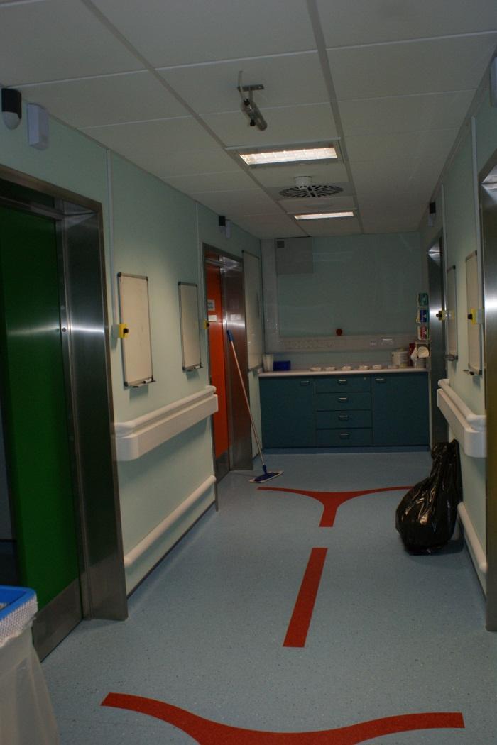
5 The centre
6 Requests Contacts Centre Fax: Reporting Room: Centre Phone: /1


7 The scan process: nuclear physics to patient Before the patient arrives!! (5am)
8 Positron emitting radionuclides Radionuclide Half life 18 F 109 mins 11 C 20.4 mins 13 N 10 mins 15 O 2.1 mins


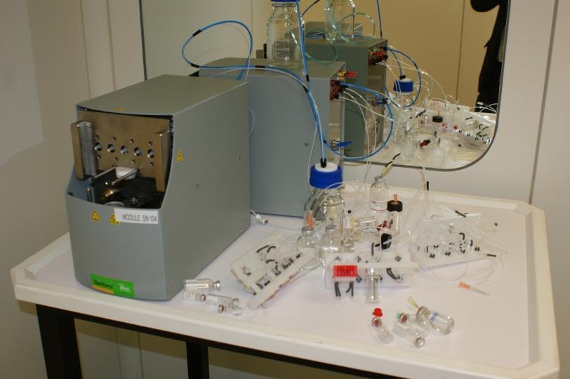
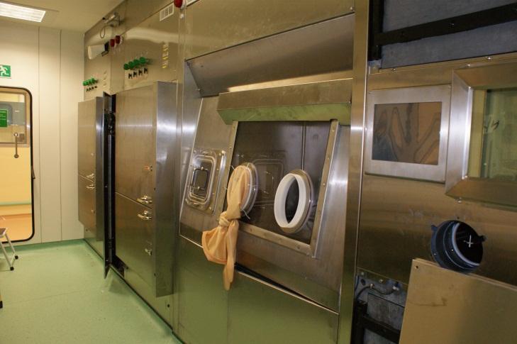

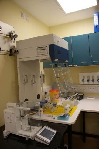
9 Radiochemical Synthesis plant Changing 3 stage cassette radiochemistry QC QC


10 FDG Radioactive sugar


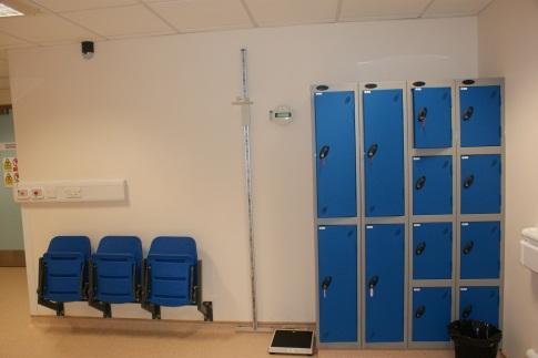


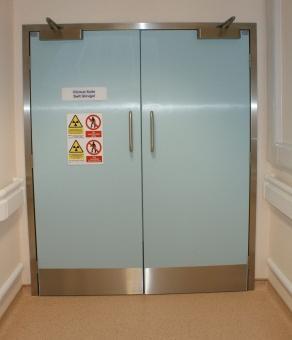
11 Patient Journey Reception Changing/ assessment


12 Injection/uptake rooms Patient Journey


13 Scanner

14 Annihilation coincidence detection


15 Time of flight Time of flight 8hrs 5 mins

16 Time of flight


17 Scanner control room and Reporting area


18 Image presentation


19 What is the SUV? Standardized Uptake Value semiquantitative SUV=measured activity/ patient volume* injected activity Measured activity/expected activity

20 SUV Values Proceed with caution Different protocols Different uptake times Different scanner Different reconstruction algorithms

21 PET/CT in gynaecological Normal appearances malignancies


22 PET/CT in gynaecological malignancies Normal appearances Nucl Med Feb;45(2): Normal and abnormal 18F-FDG endometrial and ovarian uptake in preand postmenopausal patients: assessment by PET/CT. Lerman H, Metser U, Grisaru D, Fishman A, Lievshitz G, Even-Sapir E. Ann Nucl Med : Benign ovarian and endometrial uptake on FDG PET-CT patterns and pitfalls Yiyan Liu
23 Study Design Single centre, Tel-Aviv April to July 2003 All consecutive female patients attending for PET/CT over 3 month period All straightforward menstrual questionnaire Gynae history, contraceptive, HRT 360 total, 75 previous hysterectomy 285 study group Mean age 50.2+/-16.9 Range Nucl Med Feb;45(2): Normal and abnormal 18F-FDG endometrial and ovarian uptake in preand postmenopausal patients: assessment by PET/CT. Lerman H, Metser U, Grisaru D, Fishman A, Lievshitz G, Even-Sapir E.
24 Study Design

25 Endometrial uptake On non contrast CT images, if could see a hypoattenuating stripe then this used otherwise centre of uterus
26 Endometrial uptake: Premenopausal patients
27 Endometrial uptake Premenopausal patients: 4 had IUD 4 COC No sig differences in FDG uptake (few other studies suggest some decrease in uptake with COC and increase with IUD) Amenorrhoea 8 patients 1.9+/- 1.2 similar to post menopausal pts Oligomenorrhoea 19 patients 3.4 +/- 1.2 similar to ovulatory phase pts
28 Endometrial uptake Postmenopausal patients: no gynae malignancy Characteristic No hormonal therapy Hormonal therapy Benign uterine pathology No. of patients SUV SD Range

29 Cervical or endometrial Carcinoma 21 cervical /- 7.3 SUV 5 endometrial /- 9 SUV
30 Ovarian uptake (Increased uptake in ovaries noted in 7 patients with known ovarian malignancy) Seen in 21 premenopausal patients with no gynae malignancy. Of these 15 ovulatory phase 3 oligomenorhoea Ovarian uptake around time ovulation and from corpus luteum cysts well described Of note: No increased FDG uptake was seen in any of the 134 postmenopausal patients with no history of gynaecological malignancy This finding confirmed in subsequent studies significant ovarian uptake in postmenopausal patients considered pathological

31 Ovarian Uptake

32 Benign Uterine tumours


33 Leiomyoma Typical findings Low grade uptake SUV max 2.0 Atypical findings Higher grade uptake SUV 8.5 Premenopausal Luteal phase Degeneration Degenerating fibroid Low grade uptake SUV max 3.9
34 Normal appearances: summary Cyclical uptake of FDG is normal in premenopausal patients in both ovaries and uterus Significant uptake in ovaries in postmenopausal women is considered pathological Benign leiomyomas may show sometimes marked FDG uptake We don t yet have a normal database
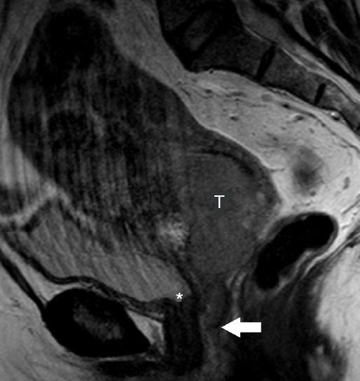
35 Carcinoma Cervix


36 Primary staging: Cervical carcinoma

37 Carcinoma Cervix: staging


38 Carcinoma Cervix: staging: MRI 1B1 1B2 11B IIIA IVA
39 Nodal staging: carcinoma cervix Nodes not incorporated to FIGO staging: but significant prognostic and treatment consequences Using standard size only : MRI limited sensitivity (29-86%) DWI MR shows promise in increasing sensitivity
40 Carcinoma of cervix FDG: general Most Cervical carcinomas are avid for FDG Adenocarcinomas are often non FDG avid General view is PET/CT may add to staging in stages IIB to IVB but of little value in stages I to IIA General view is that PET/CT is of value not in the local but in nodal and distant staging


41 Case 1 :Nodal staging Ca cervix DB 40 woman advanced cervical tumour : staging? Remote disease? Nodal status


42 Case 2 Carcinoma cervix: pelvic nodes

43 Nodal staging of Ca Cervix: a metanalysis
44 Nodal staging of Ca Cervix: a Published March 2010 Searches At least 20 patients Histological confirmation 768 studies 41 included 20 data on CT 31 data on MRI 20 data on PET or PET/CT metanalysis
45 ROC Curves: receiver operator characteristics 1 Sensitivity Perfect Q* Any node above 1mm Area under curve Useless 0 1 Any node above Specificity 0 4cm
46
47 Ca cervix Modality Sensitivity Specificity CT 50% 92% MRI 56% 91% PET or PET/CT 82% 95%
48 Ca cervix: distant metastases Prospective study 120 patients All FIGO 1B or greater (study also looked at nodal status) Distant Metastases in true positive, 9 false positive 100 considered true negative PPV 63% NPV 100% Sensitivity 100% Specificity 94% Gynecol Oncol Jul;106(1): Epub 2007 May 7. The diagnostic value of PET/CT scanning in patients with cervical cancer: a prospective study. Loft A, Berthelsen AK, Roed H, Ottosen C, Lundvall L, Knudsen J, Nedergaard L, Højgaard L, Engelholm SA.
49 Carcinoma cervix ACR guidelines


50 Prognosis carcinoma cervix
51 Ca cervix prognosis
52 Cervical Carcinoma: recurrence Chemoradiotherapy Exenteration May be options: Careful Staging appropriate prior to major surgery/ chemotherapy etc 55 patients with suspected or confirmed recurrence PET provided additional information and altered management in in 36/55 (65%) J Nucl Med Oct;45(10): Defining the priority of using 18F-FDG PET for recurrent cervical cancer. Yen TC, See LC, Chang TC, Huang KG, Ng KK, Tang SG, Chang YC, Hsueh S, Tsai CS, Hong JH, Lin CT, Chao A, Ma SY, Lin WJ, Fu YK, Fan CC, Lai CH.


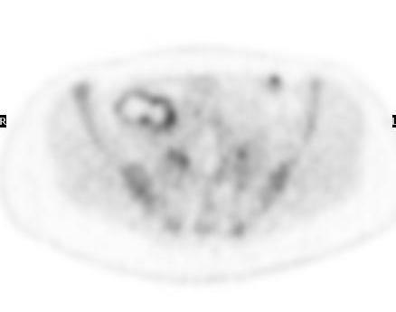

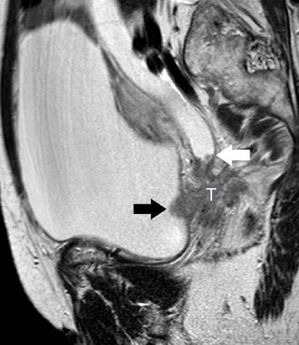
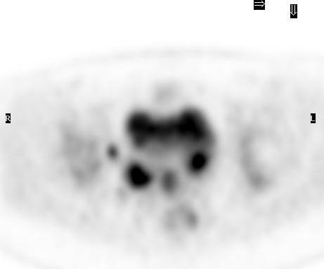
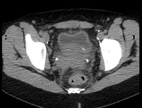
53 Carcinoma cervix:? recurrence


54 Carcinoma cervix? recurrence Cystic lesion No increased FDG uptake? Cause: No FDG avid abnormality seen No definite evidence recurrence

55 Recommendations: possible recurrence
56 Carcinoma cervix: summary No obvious value in local staging Evidence of value in assessment of nodal and distant disease (ACR) Prognostic value in monitoring response to radiotherapy Of value in assessment of possible recurrence especially when radical therapy contemplated (SIGN)

57 Carcinoma Ovary

58 Case 4: Ovarian pathology characterisation PS 64 woman. Incidental findings of 9mm lung nodule and indeterminate ovarian/ adenexal mass
59 Carcinoma Ovary: assessment of primary lesions Prospective study 55 patients all scheduled for surgery All PET/CT contrast enhanced CT and TVUS SUV max 3 or more considered malignant 32 ovarian malignancy/ 18 benign at histology Sens Spec NPV PPV Accuracy PET/CT 87% 100% 81% 100% 92% TVUS 90% 61% 78% 80% 80% Nucl Med Commun 2007; 28: Diagnostic accuracy of 18F-FDG PET/CT in characterizing ovarian lesions and staging ovarian cancer: correlation with transvaginal ultrasonography, computed tomography, and histology. Castellucci P, Perrone AM, Picchio M.
60 Carcinoma ovary: staging PET/CT concordance with pathological staging 22/32 patients (69%) CT concordance 17/32 (53%) Of note 4 of the 6 stage IV patients were missed by CT and correctly characterised by PET/CT Liver Pleura Mediastinum Supraclavicular (also correctly identified a patient with ductal breast carcinoma) Nucl Med Commun 2007; 28: Diagnostic accuracy of 18F-FDG PET/CT in characterizing ovarian lesions and staging ovarian cancer: correlation with transvaginal ultrasonography, computed tomography, and histology. Castellucci P, Perrone AM, Picchio M.

61 Carcinoma Ovary staging Notes: lesion size less than 1 cm much less reliable Mucinous tumours tend to have much lower FDG uptake

62 Carcinoma Ovary: restaging Prospective study 70 patients referred to tertiary centre with suspected recurrent ovarian carcinoma NPV 83.3%, PPV 76.9% Specificity 55% sensitivity 93% Provided complementary information to laparoscopy to inform therapy Fagotti a F. Fanfani Treatment Selection Protocol for Recurrent Ovarian Cancer Patients: The Role of FDG-PET/CT and Staging Laparoscopy A. Oncology 2008;75: A
63 Carcinoma ovary: summary Some literature on characterisation of ovarian/ adenexal lesions Evidence of value in staging especially extrapelvic Evidence of value in assessment of possible recurrence
64 Current funding guidelines/ criteria: WHSCC Lung cancer: Colorectal cancer Lymphoma Oesophageal cancer Head and Neck cancer Other Cancer Sites Other cancer sites (including thyroid, melanoma, sarcoma, testicular, brain, spinal cord and unknown primary) where there is difficulty in staging, restaging or assessment of possible recurrence.
65 Suggestions for a way forward Need to continue to work closely Clinical collaboration Research collaboration Establish normal ranges/ database for SUV value in pre and post menopausal women
66 Clinical recommendations Staging scenario: all 3 tumours For problem solving/ equivocal cases where radical treatment is contemplated Restaging/ recurrence For problem solving/ equivocal cases where radical treatment is contemplated Building the case for future agreed indications: case recording

67
68 Endometrial Carcinoma
69 Ca endometrium: staging Retrospective study: 32 patients histological diagnosis primary high risk endometrial carcinoma Primary carcinoma detection 29 of 32 patients Sens Spec PPV NPV Accuracy Nodes 57% 100% 100% 86% 89% Distant Mets 100% 96% 88% 100% 97% Conventional imaging suspicious for M1 in 2 patients, PET correctly identified M1 in 7 patients (22%) Nucl Med Commun Jun;31(6): High-grade endometrial cancer: value of [(18)F]FDG PET/CT in preoperative staging. Picchio M, Mangili G, Samanes Gajate AM, De Marzi P, Spinapolice EG, Mapelli P, Giovacchini G, Sigismondi C, Viganò R, Sironi S, Messa C. Department of Nuclear Medicine, San Raffaele Scientific Institute, Milan, Italy. picchio.maria@hsr.it
70 Case 3 Endometrial carcinoma JE 74 woman IIa cervical carcinoma. Pelvic recurrence feb Lung nodules felt to be stable? Co incidendal benign lung nodules
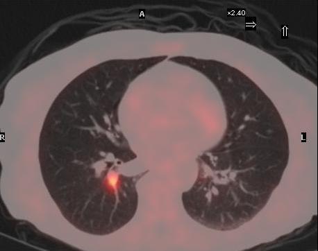


71 Endometrial carcinoma Likely metastatic nodules

72 Spare picts

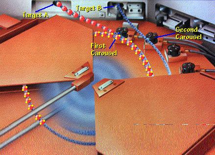
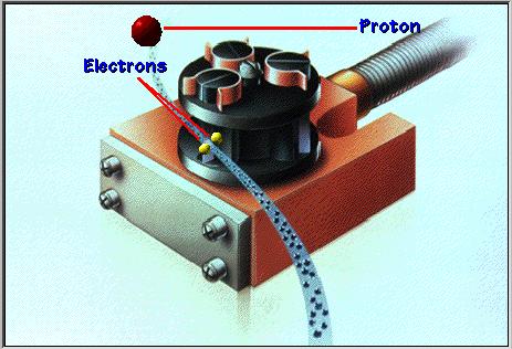

73 Cyclotron
74 Target reaction 17 8 O +1 1 P 18 9 F
75 FDG and the cancer cell Extracellular compartment Cancer cell GLUT 1 GLUT 1 Hexokinase FDG FDG ( ) FDG-6-PO 4 G-6-PO 4 ase GLUT 1 GLUT 1

76 Case 3: Carcinoma cervix? recurrence SA 56 woman ca cervix Hysterectomy and post op RT. Recurrence vaginal vault Treated with chemotherapy. Recent left adenexal mass, new hydroureteronephrosis
FDG-PET/CT in Gynaecologic Cancers
 Friday, August 31, 2012 Session 6, 9:00-9:30 FDG-PET/CT in Gynaecologic Cancers (Uterine) cervical cancer Endometrial cancer & Uterine sarcomas Ovarian cancer Little mermaid (Edvard Eriksen 1913) honoring
FDG-PET Findings in an Ovarian Endometrioma: A Case Report
 FDG-PET Findings in an Ovarian Endometrioma: A Case Report Jia-Huei Lin 1, Victor Chit-kheng Kok 2, Jian-Chiou Su 3 1 Department of Nuclear medicine, Kuang Tien General Hospital, Sha-Lu, Taichung, Taiwan
Spectrum of FDG PET/CT Findings of Uterine Tumors
 Nuclear Medicine and Molecular Imaging Pictorial Essay Kitajima et al. FDG PET/CT of Uterine Tumors Nuclear Medicine and Molecular Imaging Pictorial Essay Downloaded from www.ajronline.org by 37.44.205.17
PET-CT findings in surgically transposed ovaries
 The British Journal of Radiology, 79 (2006), 110 115 1,2,3 R ZISSIN, MD, 1 U METSER, MD, 1 H LERMAN, MD, 1 G LIEVSHITZ, MD, 4 T SAFRA, MD and 1,3 E EVEN-SAPIR, MD, PhD Department of 1 Nuclear Medicine
CT PET SCANNING for GIT Malignancies A clinician s perspective
 CT PET SCANNING for GIT Malignancies A clinician s perspective Damon Bizos Head, Surgical Gastroenterology Charlotte Maxeke Johannesburg Academic Hospital Case presentation 54 year old with recent onset
Dr Sneha Shah Tata Memorial Hospital, Mumbai.
 Dr Sneha Shah Tata Memorial Hospital, Mumbai. Topics covered Lymphomas including Burkitts Pediatric solid tumors (non CNS) Musculoskeletal Ewings & osteosarcoma. Neuroblastomas Nasopharyngeal carcinomas
Index. Surg Oncol Clin N Am 16 (2007) Note: Page numbers of article titles are in boldface type.
 Note: Page numbers of article titles are in boldface type.") Surg Oncol Clin N Am 16 (2007) 465 469 Index Note: Page numbers of article titles are in boldface type. A Adjuvant therapy, preoperative for gastric cancer, staging and, 339 B Breast cancer, metabolic
Surg Oncol Clin N Am 16 (2007) 465 469 Index Note: Page numbers of article titles are in boldface type. A Adjuvant therapy, preoperative for gastric cancer, staging and, 339 B Breast cancer, metabolic
Pet Scan And Gynaecological Malignancies: Hospital Sultanah Bahiyah Experience
 ORIGINAL ARTICLE Pet Scan And Gynaecological Malignancies: Hospital Sultanah Bahiyah Experience T Shahila, MMED (O&G), M N Rushdan, MMED (O&G) Gynaecological Oncology Unit, Department of Obstetrics & Gynaecology,
Page: 1 of 29. For this policy, PET scanning is discussed for the following 4 applications in oncology:
 Emission Tomography Scanning Page: 1 of 29 Last Review Status/Date: June 2015 Description Positron emission tomography (PET) scans are based on the use of positron-emitting radionuclide tracers coupled
Role of PET/CT in Ovarian Cancer
 Residents Section Structured Review rticle Prakash et al. PET/T in Ovarian ancer Residents Section Structured Review rticle Downloaded from www.ajronline.org by 148.251.232.83 on 04/26/18 from IP address
FDG-PET value in deep endometriosis
 Gynecol Surg (2011) 8:305 309 DOI 10.1007/s10397-010-0652-6 ORIGINAL ARTICLE FDG-PET value in deep endometriosis A. Setubal & S. Maia & C. Lowenthal & Z. Sidiropoulou Received: 3 December 2010 / Accepted:
Staging. Carcinoma confined to the corpus. Carcinoma confined to the endometrium. Less than ½ myometrial invasion. Greater than ½ myometrial invasion
 5 th of June 2009 Background Most common gynaecological carcinoma in developed countries Most cases are post-menopausal Increasing incidence in certain age groups Increasing death rates in the USA 5-year
Medical Policy An independent licensee of the Blue Cross Blue Shield Association
 PET Scanning: Oncologic Applications Page 1 of 42 Medical Policy An independent licensee of the Blue Cross Blue Shield Association Title: See also: Positron Emission Tomography (PET) Scanning: Oncologic
Cervical Cancer: 2018 FIGO Staging
 Cervical Cancer: 2018 FIGO Staging Jonathan S. Berek, MD, MMS Laurie Kraus Lacob Professor Stanford University School of Medicine Director, Stanford Women s Cancer Center Senior Scientific Advisor, Stanford
PET/CT Frequently Asked Questions
 PET/CT Frequently Asked Questions General Q: Is FDG PET specific for cancer? A: No, it is a marker of metabolism. In general, any disease that causes increased metabolism can result in increased FDG uptake
performed to help sway the clinician in what the appropriate diagnosis is, which can substantially alter the treatment of management.
 Hello, I am Maura Polansky at the University of Texas MD Anderson Cancer Center. I am a Physician Assistant in the Department of Gastrointestinal Medical Oncology and the Program Director for Physician
PET/CT in Gynaecological Cancers. Stroobants Sigrid, MD, PhD Departement of Nuclear Medicine University Hospital,Antwerp
 PET/CT in Gynaecological Cancers Stroobants Sigrid, MD, PhD Departement of Nuclear Medicine University Hospital,Antwerp Cervix cancer Outline of this talk Initial staging Treatment monitoring/guidance
Colorectal Cancer and FDG PET/CT
 Hybrid imaging in colorectal & esophageal cancer Emmanuel Deshayes IAEA WorkShop, November 2017 Colorectal Cancer and FDG PET/CT 1 Clinical background Cancer of the colon and rectum is one of the most
Los Angeles Radiological Society 62 nd Annual Midwinter Radiology Conference January 31, 2010
 Los Angeles Radiological Society 62 nd Annual Midwinter Radiology Conference January 31, 2010 Self Assessment Module on Nuclear Medicine and PET/CT Case Review FDG PET/CT IN LYMPHOMA AND MELANOMA Submitted
PET/CT in oncology. Positron emission tomography
 Clinical Medicine 2012, Vol 12, No 4: 368 72 PET/CT in oncology Fahim-Ul-Hassan, SpR Nuclear Medicine, Guy s Hospital, London; Gary J Cook, professor of Clinical PET, KCL Division of Imaging Sciences &
Clinical indications for positron emission tomography
 Clinical indications for positron emission tomography Oncology applications Brain and spinal cord Parotid Suspected tumour recurrence when anatomical imaging is difficult or equivocal and management will
North of Scotland Cancer Network Clinical Management Guideline for Carcinoma of the Uterine Cervix
 THIS DOCUMENT North of Scotland Cancer Network Carcinoma of the Uterine Cervix UNCONTROLLED WHEN PRINTED DOCUMENT CONTROL Prepared by A Kennedy/AG Macdonald/Others Approved by NOT APPROVED Issue date April
3 Summary of clinical applications and limitations of measurements
 CA125 (serum) 1 Name and description of analyte 1.1 Name of analyte Cancer Antigen 125 (CA125) 1.2 Alternative names Mucin-16 1.3 NLMC code To follow 1.4 Description of analyte CA125 is an antigenic determinant
PET CT for Staging Lung Cancer
 PET CT for Staging Lung Cancer Rohit Kochhar Consultant Radiologist Disclosures Neither I nor my immediate family members have financial relationships with commercial organizations that may have a direct
WHAT DOES PET IMAGING ADD TO CONVENTIONAL STAGING OF HEAD AND NECK CANCER PATIENTS?
 doi:10.1016/j.ijrobp.2006.12.044 Int. J. Radiation Oncology Biol. Phys., Vol. 68, No. 2, pp. 383 387, 2007 Copyright 2007 Elsevier Inc. Printed in the USA. All rights reserved 0360-3016/07/$ see front
Index. B Bilateral salpingo-oophorectomy (BSO), 69
, 69") A Advanced stage endometrial cancer diagnosis, 92 lymph node metastasis, 92 multivariate analysis, 92 myometrial invasion, 92 prognostic factors FIGO stage, 94 histological grade, 94, 95 histologic cell
A Advanced stage endometrial cancer diagnosis, 92 lymph node metastasis, 92 multivariate analysis, 92 myometrial invasion, 92 prognostic factors FIGO stage, 94 histological grade, 94, 95 histologic cell
The Use of PET Scanning in Urologic Oncology
 The Use of PET Scanning in Urologic Oncology Dr Nicholas C. Buchan Uro-oncology Fellow 1 2 Aims To understand the basic concepts underlying PET scanning. Understand the emerging role of PET Scanning for
3/25/2019. Rare uterine cancers ~3% Leiomyosarcoma Carcinosarcoma (MMMT) Endometrial Stromal Sarcomas Aggressive tumors High Mortality Rates
 Endometrial Stromal Sarcomas Aggressive tumors High Mortality Rates") J. Anthony Rakowski D.O., F.A.C.O.O.G. MSU SCS Board Review Coarse Rare uterine cancers ~3% Leiomyosarcoma Carcinosarcoma (MMMT) Endometrial Stromal Sarcomas Aggressive tumors High Mortality Rates Signs
J. Anthony Rakowski D.O., F.A.C.O.O.G. MSU SCS Board Review Coarse Rare uterine cancers ~3% Leiomyosarcoma Carcinosarcoma (MMMT) Endometrial Stromal Sarcomas Aggressive tumors High Mortality Rates Signs
Triage of Ovarian Masses. Andreas Obermair Brisbane
 Triage of Ovarian Masses Andreas Obermair Brisbane Why Triage? In ovarian cancer, best outcomes for patients can be achieved when patients are treated in tertiary centres by a multidisciplinary team led
PET/CT in breast cancer staging
 PET/CT in breast cancer staging Anni Morsing Consultant, PhD, DMSc Rigshospitalet 1 18F- FDG PET/CT for breastcancer staging Where is the clinical impact? To which women should 18F- FDG PET/CT be offered?
Oncologic Applications of PET Scanning
 6.01.26 Oncologic Applications of PET Scanning Section 6.0 Radiology Subsection Effective Date February 15, 2015 Original Policy Date January 26, 2009 Next Review Date December 2015 Description Positron
1 Introduction. 2 Materials and methods. LI Na 1 LI Yaming 1,* YANG Chunming 2 LI Xuena 1 YIN Yafu 1 ZHOU Jiumao 1
 Nuclear Science and Techniques 20 (2009) 354 358 18 F-FDG PET/CT in diagnosis of skeletal metastases LI Na 1 LI Yaming 1,* YANG Chunming 2 LI Xuena 1 YIN Yafu 1 ZHOU Jiumao 1 1 Department of Nuclear Medicine,
MRI in Cervix and Endometrial Cancer
 28th Congress of the Hungarian Society of Radiologists RCR Session Budapest June 2016 MRI in Cervix and Endometrial Cancer DrSarah Swift St James s University Hospital Leeds, UK Objectives Cervix and endometrial
Unusual Osteoblastic Secondary Lesion as Predominant Metastatic Disease Spread in Two Cases of Uterine Leiomyosarcoma
 49 Unusual Osteoblastic Secondary Lesion as Predominant Metastatic Disease Spread in Two Cases of Uterine Leiomyosarcoma Loredana Miglietta a Maria Angela Parodi b Luciano Canobbio b Luca Anselmi c a Medical
Medical Policy An independent licensee of the Blue Cross Blue Shield Association
 PET Scanning: Oncologic Applications Page 1 of 88 Medical Policy An independent licensee of the Blue Cross Blue Shield Association Title: Positron Emission Tomography (PET) Scanning: Oncologic Applications
An Introduction to PET Imaging in Oncology
 January 2002 An Introduction to PET Imaging in Oncology Janet McLaren, Harvard Medical School Year III Basics of PET Principle of Physiologic Imaging: Allows in vivo visualization of structures by their
Cervical cancer presentation
 Carcinoma of the cervix: Carcinoma of the cervix is the second commonest cancer among women worldwide, with only breast cancer occurring more commonly. Worldwide, cervical cancer accounts for about 500,000
New Cancer Cases By Site Breast 28% Lung 14% Colo-Rectal 10% Uterus 6% Thyroid 5% Lymphoma 4% Ovary 3%
 Uterine Malignancy New Cancer Cases By Site 2010 Breast 28% Lung 14% Colo-Rectal 10% Uterus 6% Thyroid 5% Lymphoma 4% Ovary 3% Cancer Deaths By Site 2010 Lung 26% Breast 15% Colo-Rectal 9% Pancreas 7%
Appendix 1: Regional Lymph Node Stations for Staging Esophageal Cancer
 Appendix 1: Regional Lymph Node Stations for Staging Esophageal Cancer Locoregional (N stage) disease was redefined in the seventh edition of the AJCC Cancer Staging Manual as any periesophageal lymph
Management of Endometrial Hyperplasia
 Management of Endometrial Hyperplasia I have nothing to disclose. Stefanie M. Ueda, M.D. Assistant Clinical Professor UCSF Division of Gynecologic Oncology Female Malignancies in the United States New
Pathway Gynaecology Cancer & Diagnostic Protocol for Inter Trust transfer
 NICaN Pathway Gynaecology Cancer & Diagnostic Protocol for Inter Trust transfer Timed schedules to enable the proactive management of the patient from point of receipt of referral to first definitive treatment
Sarah Burton. Lead Gynae Oncology Nurse Specialist Cancer Care Cymru
 Sarah Burton Lead Gynae Oncology Nurse Specialist Cancer Care Cymru Gynaecological Cancers Cervical Cancers Risk factors Presentation Early sexual activity Multiple sexual partners Smoking Human Papiloma
receive adjuvant chemotherapy
 Women with high h risk early stage endometrial cancer should receive adjuvant chemotherapy Michael Friedlander The Prince of Wales Cancer Centre and Royal Hospital for Women The Prince of Wales Cancer
The solitary pulmonary nodule: Assessing the success of predicting malignancy
 The solitary pulmonary nodule: Assessing the success of predicting malignancy Poster No.: C-0829 Congress: ECR 2010 Type: Scientific Exhibit Topic: Chest Authors: R. W. K. Lindsay, J. Foster, K. McManus;
One of the commonest gynecological cancers,especially in white Americans.
 Gynaecology Dr. Rozhan Lecture 6 CARCINOMA OF THE ENDOMETRIUM One of the commonest gynecological cancers,especially in white Americans. It is a disease of postmenopausal women with a peak incidence in
objectives Pitfalls and Pearls in PET/CT imaging Kevin Robinson, DO Assistant Professor Department of Radiology Michigan State University
 objectives Pitfalls and Pearls in PET/CT imaging Kevin Robinson, DO Assistant Professor Department of Radiology Michigan State University To determine the regions of physiologic activity To understand
PET/CT for the evaluation of gynecological
 PET/CT for the evaluation of gynecological cancer Gynecological cancer Barcelona, September 16 th 2018 Dr. Pilar Paredes, MD, PhD Nuclear Medicine Department Hospital Clínic Barcelona (Spain) PET/CT in
Adjuvant Therapies in Endometrial Cancer. Emma Hudson
 Adjuvant Therapies in Endometrial Cancer Emma Hudson Endometrial Cancer Most common gynaecological cancer Incidence increasing in Western world 1-2% cancer deaths 75% patients postmenopausal 97% epithelial
ROLE OF PET-CT IN BREAST CANCER, GUIDELINES AND BEYOND. Prof Jamshed B. Bomanji Institute of Nuclear Medicine UCL Hospitals London
 ROLE OF PET-CT IN BREAST CANCER, GUIDELINES AND BEYOND Prof Jamshed B. Bomanji Institute of Nuclear Medicine UCL Hospitals London CANCER Key facts Estimated 15.2 million new cases per year in 2015 worldwide
Bone and CT Scans Are Complementary for Diagnoses of Bone Metastases in Breast Cancer When PET Scans Findings Are Equivocal: A Case Report
 Bone and CT Scans Are Complementary for Diagnoses of Bone Metastases in Breast Cancer When Scans Findings Are Equivocal: A Case Report Yuk-Wah Tsang 1, Jyh-Gang Leu 2, Yen-Kung Chen 3, Kwan-Hwa Chi 1,4
Deppen S, et al. Annals of Thoracic Surgery 2011;92:
 Deppen S, et al. Annals of Thoracic Surgery 2011;92:428-33. http://www.nationmaster.com/graph/ mor_his-mortality-histoplasmosis http://www.humirarems.com/brochure.aspx Baddley, John W., et al. Emerging
Staging and Treatment Update for Gynecologic Malignancies
 Staging and Treatment Update for Gynecologic Malignancies Bunja Rungruang, MD Medical College of Georgia No disclosures 4 th most common new cases of cancer in women 5 th and 6 th leading cancer deaths
New Visions in PET: Surgical Decision Making and PET/CT
 New Visions in PET: Surgical Decision Making and PET/CT Stanley J. Goldsmith, MD Director, Nuclear Medicine Professor, Radiology & Medicine New York Presbyterian Hospital- Weill Cornell Medical Center
A phase II study of weekly paclitaxel and cisplatin followed by radical hysterectomy in stages IB2 and IIA2 cervical cancer AGOG14-001/TGOG1008
 A phase II study of weekly paclitaxel and cisplatin followed by radical hysterectomy in stages IB2 and IIA2 cervical cancer AGOG14-001/TGOG1008 NCT02432365 Chyong-Huey Lai, MD On behalf of Principal investigator
Appendix C: Evidence Tables Studies of PET for Oncology Indications NR = not reported ND = not done
 Retrospective case series with medical records review of patients with melanoma for treatment plans before and after FDG PET in three university medical centers (1992 2004) Change in disease management
Positron Emission Tomography in Lung Cancer
 May 19, 2003 Positron Emission Tomography in Lung Cancer Andrew Wang, HMS III Patient DD 53 y/o gentleman presented with worsening dyspnea on exertion for the past two months 30 pack-year smoking Hx and
PET-imaging: when can it be used to direct lymphoma treatment?
 PET-imaging: when can it be used to direct lymphoma treatment? Luca Ceriani Nuclear Medicine and PET-CT centre Oncology Institute of Southern Switzerland Bellinzona Disclosure slide I declare no conflict
What is endometrial cancer?
 Uterine cancer What is endometrial cancer? Endometrial cancer is the growth of abnormal cells in the lining of the uterus. The lining is called the endometrium. Endometrial cancer usually occurs in women
Chapter II.8 Gynecological Tumors
 Chapter II.8 Gynecological Tumors Farrokh Dehdashti and Barry A. Siegel Introduction Gynecological cancers as a group comprise approximately 11% of female cancer. 1 In the United States, it is estimated
Using PET/CT in Prostate Cancer
 Using PET/CT in Prostate Cancer Legal Disclaimer These materials were prepared in good faith by MITA as a service to the profession and are believed to be reliable based on current scientific literature.
Cervical Cancer - Suspected
 Cervical Cancer - Suspected Presentation for patients Asymptomatic presentation Symptomatic presentation History and examination Consider differential diagnoses RED FLAG! Cervix appears normal after examination
Esophageal Cancer. What is the value of performing PET scan routinely for staging of esophageal cancers
 Esophageal Cancer What is the value of performing PET scan routinely for staging of esophageal cancers What is the sensitivity and specificity of PET scan for metastatic lesions When should PET scan be
Hybrid Imaging SPECT/CT PET/CT PET/MRI. SNMMI Southwest Chapter Aaron C. Jessop, MD
 Hybrid Imaging SPECT/CT PET/CT PET/MRI SNMMI Southwest Chapter 2014 Aaron C. Jessop, MD Assistant Professor, Department of Nuclear Medicine UT MD Anderson Cancer Center, Houston, Texas Complimentary role
Staging of Prostatic Carcinoma - The evolving use of SPECT-CT and Positron Emission Tomography (PET)
") Staging of Prostatic Carcinoma - The evolving use of SPECT-CT and Positron Emission Tomography (PET) Poster No.: C-2477 Congress: ECR 2015 Type: Educational Exhibit Authors: B. Rawal, N. Vasdev, R. P.
Staging of Prostatic Carcinoma - The evolving use of SPECT-CT and Positron Emission Tomography (PET) Poster No.: C-2477 Congress: ECR 2015 Type: Educational Exhibit Authors: B. Rawal, N. Vasdev, R. P.
Proposed All Wales Vulval Cancer Guidelines. Dr Amanda Tristram
 Proposed All Wales Vulval Cancer Guidelines Dr Amanda Tristram Previous FIGO staging FIGO Stage Features TNM Ia Lesion confined to vulva with
GENERAL DATA. Sex : female Age : 40 years old Marriage status : married
 GENERAL DATA Sex : female Age : 40 years old Marriage status : married CHIEF COMPLAINT Bilateral ovarian tumors discovered by sonography accidentally PRESENT ILLNESS 2003-06-26 :bilateral ovarian tumors
ESGO-ESTRO-ESP Cervical Cancer Clinical Practice Guidelines Management of early stages: algorithms focusing on the histological data
 ESGO-ESTRO-ESP Cervical Cancer Clinical Practice Guidelines Management of early stages: algorithms focusing on the histological data David Cibula Gynecologic Oncology Centre General University Hospital
When do you need PET/CT or MRI in early breast cancer?
 When do you need PET/CT or MRI in early breast cancer? Elizabeth A. Morris MD FACR Chief, Breast Imaging Service Memorial Sloan-Kettering Cancer Center NY, NY Objectives What is the role of MRI in initial
The Role of PET / CT in Lung Cancer Staging
 July 2004 The Role of PET / CT in Lung Cancer Staging Vlad Vinarsky, Harvard Medical School Year IV Patient AM HPI: 81 yo F p/w hemoptysis x 1 month LLL lesion on CXR, not responsive to Abx 35 pack-year
Enterprise Interest None
 Enterprise Interest None Cervical Cancer -Management of late stages ESP meeting Bilbao Spain 2018 Dr Mary McCormack PhD FRCR Consultant Clinical Oncologist University College Hospital London On behalf
Molecular Imaging and Cancer
 Molecular Imaging and Cancer Cancer causes one in every four deaths in the United States, second only to heart disease. According to the U.S. Department of Health and Human Services, more than 512,000
Molecular Imaging in the Development of Cancer Therapeutics. Johannes Czernin
 Molecular Imaging in the Development of Cancer Therapeutics Johannes Czernin Ahmanson Biological Imaging Division University of California Los Angeles Cancer Statistics Cancer Type 5-year Survival Rate
PET/CT F-18 FDG. Objectives. Basics of PET/CT Imaging. Objectives. Basic PET imaging
 Basics of PET/CT Imaging Kevin Robinson, DO Department of Radiology Michigan State University Objectives Basic PET imaging Evaluating the therapeutic response Evaluating the big 5 Lymphoma Breast Lung
Squamous cell carcinoma arising in a dermoid cyst of the ovary: a case series
 DOI: 10.1111/j.1471-0528.2007.01478.x www.blackwellpublishing.com/bjog Gynaecological oncology Squamous cell carcinoma arising in a dermoid cyst of the ovary: a case series JL Hurwitz, a A Fenton, a WG
Contents. 3 Pneumology Introduction Positron Emission Tomography: Past and Present 1. 2 Fundamentals. xxx
 xxx IX Contents 1 Introduction Positron Emission Tomography: Past and Present 1 1.1 Survey.......................... 1 Physical and Biochemical Fundamentals.... 2 PET in National and International Medical
MRI for cervical and endometrial cancers. Dr Robert Bleehen Consultant Radiologist Cardiff & Vale UHB
 MRI for cervical and endometrial cancers Dr Robert Bleehen Consultant Radiologist Cardiff & Vale UHB RCR 06(1) RCR 06(1) Technique Pelvic multiphased-array coil Fasting? Buscopan? ABDOMEN!!! Cx:+/- HR
Evaluation and Management of Thyroid Nodules. Nick Vernetti, MD, FACE Palm Medical Group Las Vegas, Nevada
 Evaluation and Management of Thyroid Nodules Nick Vernetti, MD, FACE Palm Medical Group Las Vegas, Nevada Disclosure Consulting Amgen Speaking Amgen Objectives Understand the significance of incidental
Gynecologic Cancer Surveillance and Survivorship: Informing Practice and Policy
 Gynecologic Cancer Surveillance and Survivorship: Informing Practice and Policy Stephanie Yap, M.D. University Gynecologic Oncology Northside Cancer Institute Our Learning Objectives Review survival rates,
Original Policy Date
 MP 6.01.17 Oncologic Applications of PET Scanning Medical Policy Section Radiology Issue 12:2013 Original Policy Date 12:2013 Last Review Status/Date Reviewed with literature/12:2013 Return to Medical
FDG PET/CT in Lung Cancer Read with the experts. Homer A. Macapinlac, M.D.
 FDG PET/CT in Lung Cancer Read with the experts Homer A. Macapinlac, M.D. Patient with suspected lung cancer presents with left sided chest pain T3 What is the T stage of this patient? A) T2a B) T2b C)
Prostate Case Scenario 1
 Prostate Case Scenario 1 H&P 5/12/16: A 57-year-old Hispanic male presents with frequency of micturition, urinary urgency, and hesitancy associated with a weak stream. Over the past several weeks, he has
Ryan Niederkohr, M.D. Slides are not to be reproduced without permission of author
 Ryan Niederkohr, M.D. CMS: PET/CT CPT CODES 78814 Limited Area (e.g., head/neck only; chest only) 78815 78816 Regional (skull base to mid-thighs) True Whole Body (skull vertex to feet) SELECTING FIELD
Upper GI Malignancies Imaging Guidelines for the Management of Gastric, Oesophageal & Pancreatic Cancers 2012
 Upper GI Malignancies Imaging Guidelines for the Management of Gastric, Oesophageal & Pancreatic Cancers 2012 Version Control This is a controlled document please destroy all previous versions on receipt
Assessing the lung and mediastinum in cancer-is tissue the issue? George Santis
 1 Assessing the lung and mediastinum in cancer-is tissue the issue? George Santis Optimal management of Cancer Histological diagnosis & accurate staging at presentation Molecular analysis of primary tumour
Clinical summary. Male 30 year-old with past history of non-seminomous germ cell tumour. Presents with retroperitoneal lymphadenopathy on CT.
 Clinical summary Male 30 year-old with past history of non-seminomous germ cell tumour. Presents with retroperitoneal lymphadenopathy on CT. For restaging PET/CT. PET/CT findings No significant FDG uptake
Case Scenario 1. History
 History Case Scenario 1 A 53 year old white female presented to her primary care physician with post-menopausal vaginal bleeding. The patient is not a smoker and does not use alcohol. She has no family
Staging and restaging for distant metastatic disease in breast cancer: Has anything changed?
 Staging and restaging for distant metastatic disease in breast cancer: Has anything changed? Sarah J Vinnicombe Clinical Senior Lecturer in Cancer Imaging Dundee Cancer Centre s.vinnicombe@dundee.ac.uk
Positron emission tomography scanning is coming to a hospital near you soon!
 PROFESSIONAL ISSUES Positron emission tomography scanning is coming to a hospital near you soon! Humayun Bashir, Gregory Shabo and TO Nunan Humayun Bashir MB BS FCPS, Specialist Registrar in Nuclear Medicine
PRINCESS MARGARET CANCER CENTRE CLINICAL PRACTICE GUIDELINES GYNECOLOGIC CANCER CERVIX
 PRINCESS MARGARET CANCER CENTRE CLINICAL PRACTICE GUIDELINES GYNECOLOGIC CANCER CERVIX Site Group: Gynecology Cervix Author: Dr. Stephane Laframboise 1. INTRODUCTION 3 2. PREVENTION 3 3. SCREENING AND
Value of true whole-body FDG- PET/CT scanning protocol in oncology and optimization of its use based on primary malignancy
 Value of true whole-body FDG- PET/CT scanning protocol in oncology and optimization of its use based on primary malignancy Ronnie Sebro MD, Ph.D Carina Mari Aparici MD, Miguel Hernandez Pampaloni MD, PhD
THE ROLE OF CONTEMPORARY IMAGING AND HYBRID METHODS IN THE DIAGNOSIS OF CUTANEOUS MALIGNANT MELANOMA(CMM) AND MERKEL CELL CARCINOMA (MCC)
 AND MERKEL CELL CARCINOMA (MCC)") THE ROLE OF CONTEMPORARY IMAGING AND HYBRID METHODS IN THE DIAGNOSIS OF CUTANEOUS MALIGNANT MELANOMA(CMM) AND MERKEL CELL CARCINOMA (MCC) I.Kostadinova, Sofia, Bulgaria CMM some clinical facts The incidence
THE ROLE OF CONTEMPORARY IMAGING AND HYBRID METHODS IN THE DIAGNOSIS OF CUTANEOUS MALIGNANT MELANOMA(CMM) AND MERKEL CELL CARCINOMA (MCC) I.Kostadinova, Sofia, Bulgaria CMM some clinical facts The incidence
PET IMAGING (POSITRON EMISSION TOMOGRAPY) FACT SHEET
 FACT SHEET") Positron Emission Tomography (PET) When calling Anthem (1-800-533-1120) or using the Point of Care authorization system for a Health Service Review, the following clinical information may be needed to
Positron Emission Tomography (PET) When calling Anthem (1-800-533-1120) or using the Point of Care authorization system for a Health Service Review, the following clinical information may be needed to
North of Scotland Cancer Network Clinical Management Guideline for Endometrial Cancer
 THIS DOCUMENT North of Scotland Cancer Network Clinical Management Guideline for Endometrial Cancer Based on WOSCAN CMG with further extensive consultation within NOSCAN UNCONTROLLED WHEN PRINTED DOCUMENT
FDG PET/CT STAGING OF LUNG CANCER. Dr Shakher Ramdave
 FDG PET/CT STAGING OF LUNG CANCER Dr Shakher Ramdave FDG PET/CT STAGING OF LUNG CANCER FDG PET/CT is used in all patients with lung cancer who are considered for curative treatment to exclude occult disease.
Endometrial Cancer. Incidence. Types 3/25/2019
 Endometrial Cancer J. Anthony Rakowski DO, FACOOG MSU SCS Board Review Coarse Incidence 53,630 new cases yearly 8,590 deaths yearly 4 th most common malignancy in women worldwide Most common GYN malignancy
Staging recurrent ovarian cancer with 18 FDG PET/CT
 ONCOLOGY LETTERS 5: 593-597, 2013 Staging recurrent ovarian cancer with FDG PET/CT SANJA DRAGOSAVAC 1, SOPHIE DERCHAIN 2, NELSON M.G. CASERTA 3 and GUSTAVO DE SOUZA 2 1 DIMEN Medicina Nuclear and PET/CT
Research Article Prevalence of Clinically Significant Extraosseous Findings on Unenhanced CT Portions of 18 F-Fluoride PET/CT Bone Scans
 The Scientific World Journal Volume 2012, Article ID 979867, 5 pages doi:10.1100/2012/979867 The cientificworldjournal Research Article Prevalence of Clinically Significant Extraosseous Findings on Unenhanced
Does PET/CT Have an Additional Value in Detection of Osteolytic Bone Metastases.
 Egyptian J. Nucl. Med., Vol 2, No. 2, Dec. 2009 65 ONCOLOGY, Original Article Does PET/CT Have an Additional Value in Detection of Osteolytic Bone Metastases. R. Riad, M.D.*, M. Awad, M.D. **, E. Eldebawy,
Para-aortic laparoscopic lymph-node dissection for advanced cervical cancers
 Para-aortic laparoscopic lymph-node dissection for advanced cervical cancers P. Mathevet, Hôpital Femme-Mère-Enfant, Bron Lymph-node involvement Is one of the major prognostic factor in gynecologic cancers.
GCIG Rare Tumour Brainstorming Day
 GCIG Rare Tumour Brainstorming Day Relatively (Not So) Rare Tumours Adenocarcinoma of Cervix Keiichi Fujiwara, Ros Glasspool Benedicte Votan, Jim Paul Aim of the Day To develop at least one clinical trial