THE ROLE OF CONTEMPORARY IMAGING AND HYBRID METHODS IN THE DIAGNOSIS OF CUTANEOUS MALIGNANT MELANOMA(CMM) AND MERKEL CELL CARCINOMA (MCC)
|
|
|
- Merry Powell
- 5 years ago
- Views:
Transcription
 AND MERKEL CELL CARCINOMA (MCC) I.")
1 THE ROLE OF CONTEMPORARY IMAGING AND HYBRID METHODS IN THE DIAGNOSIS OF CUTANEOUS MALIGNANT MELANOMA(CMM) AND MERKEL CELL CARCINOMA (MCC) I.Kostadinova, Sofia, Bulgaria

2 CMM some clinical facts The incidence of cutaneous melanoma has doubled over the last two decades in the United States and around the world. Melanoma is the least common but the most deadly skin cancer, accounting for only about 5% of all cases, but the vast majority of skin cancer-related death*. 10% of all patients with melanoma have a family history. Incidence rates are higher in women than in men below the age of 50, but by age 65, rates in men double those in women, and by age 80 they are triple. ** Disease stage on initial evaluation is the most important predictor of outcome and determinant of the appropriate management and a disease free survival. *SEER (Surveillance, Epidemiology and End Results) data. seer.cancer.gov. ** American Cancer Society. Cancer Facts and Figures Atlanta: American Cancer Society; 2016.
* TX Primary tumor cannot be assessed T0No evidence of primary Tis Melanoma in")

3 * NCCN Clinical Practice Guidelines in Oncology: Melanoma. V TNM Classification for Malignant Melanoma Primary tumor (T)* TX Primary tumor cannot be assessed T0No evidence of primary Tis Melanoma in situ T1:Melanoma 1.0 mm in thickness T1a: Without ulceration and mitoses < 1/mm 2 T1b: With ulceration or mitoses 1/mm 2 T2: Melanomas mm in thickness T2a: Without ulceration T2b: With ulceration T3: Melanomas mm in thickness T3a: Without ulceration T3b: With ulceration T4: Melanomas > 4.0 mm in thickness T4a: Without ulceration T4b: With ulceration
4 TNM Classification for Malignant Melanoma-N,M NX: Patients in whom the regional nodes cannot be assessed N0: No regional metastases detected N1-3: Regional metastases based upon number of metastatic nodes and presence or absence of intralymphatic metastases (in transit or satellite metastases) N1: 1 lymph node N1a: Micrometastases N1b: Macrometastases N2: 2 or 3 lymph nodes N2a: Micrometastases N2b: Macrometastases N2c: In-transit met(s)/satellite(s) without metastatic lymph nodes N3: 4 metastatic lymph nodes, or matted lymph nodes, or in-transit met(s)/satellite(s) with metastatic lymph node(s) M0: No detectable evidence of distant metastases M1a: Metastases to skin, subcutaneous, or distant lymph nodes, normal LDH level M1b: Lung metastases, normal LDH level M1cMetastases to any site combined with an elevated serum LDH level Clinical staging Stage I ( T1 )and Stage II ( T2-4 )-no lymph node involvement Stage III nodal involvement Stage IV distant mets


5 Lymphatic and hematogeneous dissemination of CMM and location of the primary tumor Tarik Z. Belhocine et al. J Nucl Med 2006;47:957, 967
,")

6 Choice of imaging modality for staging of CMM Imaging methods have a primary role in the staging of CMM, surgical planning and therapy approach, prognosis and long term follow-up. Basic imaging modalities such as US, CT, MRI as well as PET/CT, are used mainly for detection of local and distant metastases, but have no role in the staging of the primary process (T stage), especially in patients with disease stage I or II. Meta-analysis of 74 studies on patients show that ultrasound is superior for regional lymph nodes and PET/CT for distant metastases* CT of the abdomen, pelvis and thorax is the modality of choice for restaging of patients with stage III or IV. Patients at risk of developing cerebral or liver metastases need to have MRI examination or CT with contrast. *Xing Y. et al., J.Natl.Cancer, 2011, 103,

7 Choice of imaging modality for staging of CMM Currently, whole body MRI with diffusion weighted imaging /WB-MRI with DWI/, is established as one of the most promising modern functional imaging modalities. It is based on detection of the free movement of water molecules, which is limited when cell membrane is ruptured or in the cases of the changed tissue cellularity in the tumor*. *Schmidt G. et al.,topics in MRI, 18 /3/, 2007,

8 Choice of imaging modality for staging of CMM Additional functional information obtained from detailed anatomic WB-MRI-DWI, through the use of T1, T2 and STIR (short tau inversion recovery) sequences, gives a more detailed and accurate staging of tumours. Benefits of a whole body MRI are the absence of radiation and high sensitivity for lesions located in the bone marrow, brain and liver. Still, CT remains the modality of choice and gold standard for lesions located in the lungs, bones and thoracic cavity. Prospective studies show that WB-MRI-DWI has a lower sensitivity for detecting distant metastases, compared to PET/CT, respectively 78.8% and 86.7%, but MRI has a higher accuracy for lesions located in the bone marrow and liver*. *Pfannenberg C. et al.. Eur J Cancer 2007;43: 55764
.")
9 Application of hybrid imaging methods SPECT/CT The most important factors determining the prognosis of CMM are: invasion depth of the tumor, presence of ulceration, number of mitosis and histologic/oncogenic status of the first lymph node responsible for drainage of the tumor (SLN). The aim of SLN visualization and histological examination in patients without palpable lymph node enlargement, is the staging of disease, since lymphatic spread in CMM is often unpredictable. Therefore, its histopathology suggests the status of the rest of the lymph nodes in the lymphatic region and gives stage and prognosis. The metastatic invasion of the SLN requires a total lymphadenectomy for therapeutic benefit.
10 Application of hybrid imaging methods SPECT/CT Because of its high sensitivity up to 100% SLNscintigraphy is the modality of choice for visualization of SLN and detection of occult metastases, especially when CMM is located in the head, neck or torso and has Clark level <4 and Breslow thickness mm* Compared to palpation, SLN scintigraphy detects more lymphatic drainage basins in 50% of the patients with CMM in the torso and in 33% of the patients with CMM located in the head and neck region **. *SNM Guidance, 2002 **Belhocine T., A.Scott, E.Sapir et al., JNM, 6 / 47/, 2006,


11 Advantages of SLN scintigraphy, combined with SPECT/CT Improved image quality, compared to a planar imaging and improved SLN detestability with up to 43%, especially when combined with intraoperative gamma probe detection.* Allows exact localization of SLN** Gives additional preoperative staging and determination of prognosis of disease* *Mar M.et al., JNM,2005,46,50 ** Kostadinova I, M.Garcheva, BCNM, Belgrade,2013

12 SPECT/CT in a patient with CMM on the back - there are SLNs in the right axilla and right scalula

13 SPECT/CT of the same patient used for staging a single lung nodule - 9mm

14 Application the hybrid imaging method PET/CT 1. Staging PET/CT has no role in the diagnosis and local tumor spread (T stage). In the early stages of disease PET/CT has a very low sensitivity of up to 17%, as micrometastases are usually missed. In this situation SLN scintigraphy, combined with surgical excision, is a choice. PET/CT examination is recommended in patients with higher risk of tumor invasion, with thickness of 2-4mm or more, where 50-70% of the patients have already invasion of lymph nodes and 10% have distant metastases* If there is no possibility for PET/CT scanning, alternatives are CT or WB- MRI-DWI** *Acland K. et al., JClin.Oncology, 19 /10/,2001, **Pfannenberg C., Radiologie Feb;55(2):

15 Staging with PET/CT
16 Application of the hybrid imaging method-pet/ct 2. Role of PET/CT in restaging of the patients In patients with disease stage III or IV, MRI of the brain and liver, CT of the abdomen and thorax as well as ultrasonography have a significant role in the visualization of recurrences and have sensitivity ranging from 57% to 81% and specificity ranging from 45% to 87%*. PET/CT has a higher sensitivity and specificity, compared to CIM for the detection of distant metastases with accuracy reaching up to 97%. In high-risk melanoma patients 18 F-FDG PET/CT is more accurate (97%) for the detection of tumor recurrence than the tumor marker S100 (65%)**. PET/CT alters the therapeutic approach in 22-29% of the patients***. *Friedman K., R.Wahl, Sem.NM, 34, 2004, **Buchbender Chr.et al.jnm,53,2012. ***Schwimmer J. et al. Q J Nucl Med. 2000;44:
17 Role of PET/CT in restaging of the patients PET/CT allows for detection of metastases up to 6 months earlier, compared to clinical investigation or CT alone*, especially in localizations, which are difficult. PET/CT influences decision making for surgical intervention in patients with disease stage III or IV. High accumulation of F18- FDG (measured by SUV) in involved lymph nodes has an important negative prognostic value for PFS. Lesions in the lung parenchyma up to 5-7mm, small lesions in the liver or in the brain can lead to false negative results on PET/CT. They require further investigation using CT or MRI**. Summarized data show that in high risk patients (disease stage III or IV) the optimal approach is to combine PET/CT with organ-specific MRI of suspicious lesions**. Very promising is the new hybrid technique PET/MRI, which is even preferred over the latter two modalities***. * Fuster D. et al., J Nucl Med. 2004;45: ** Walker R. et al., Melanoma, in Hybrid PET-CT imaging, 2010, ***Pfannenberg C, Schwenzer N. Radiologe Feb;55(2):120-6.


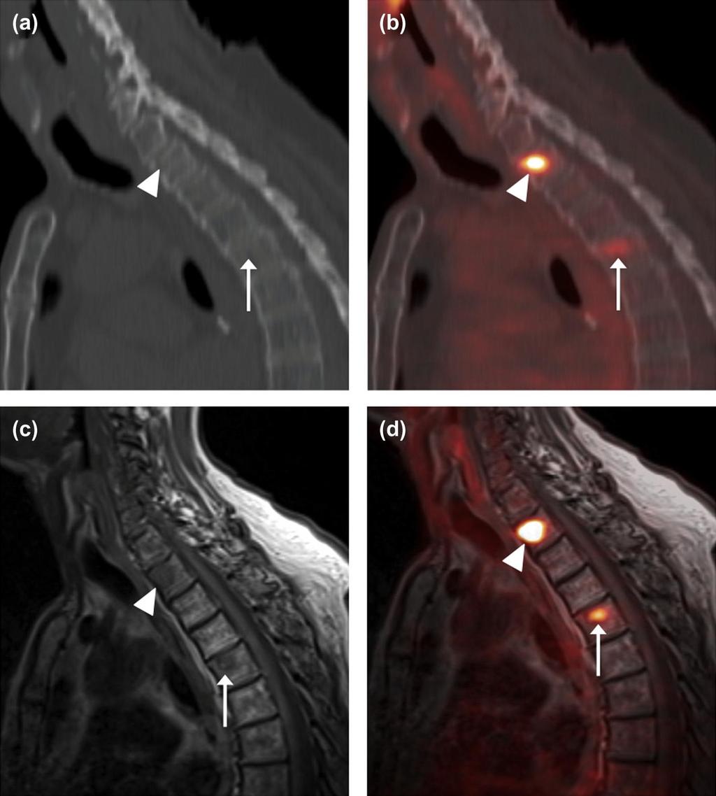
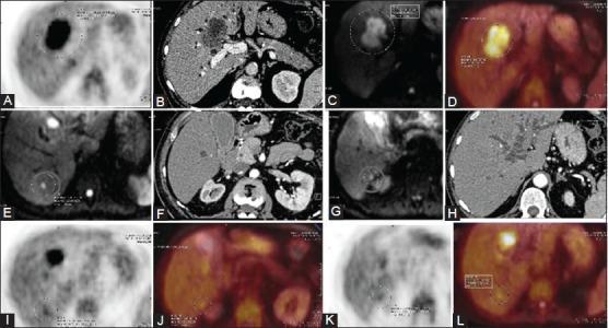
18 Restaging with PET/CT in a patient with CMM and multiple metastatic lesions

19 Restaging with PET/CT in a patient with CMM and multiple metastatic lesions
20 Application of the hybrid imaging method-pet/ct 3. PET/CT in the follow-up of therapy effect PET/CT may play an important role in the follow up of the therapy effect in patients with local and distant metastases, who are treated with chemo- or immunotherapy. Responders to chemotherapy identified by 18 F-FDG PET/CT have been proven to have a longer progression-free and overall survival than nonresponders * Follow-up using serial scanning PET/CT can contribute to patient-tailored therapy. PET/CT can be implemented in clinical trials involving new pharmacological substances. *Strobel K.et al., Eur J Nucl Med Mol Imaging. 2008;35:


")
21 PET/CT in the follow up after chemotherapy of a patient with progressive disease with CMM located in the right thigh ( thickness 3mm ) and initial metastases in the inguinal lymph nodes (not shown). PET-CT at 6 months (on the left) and at 11 months (on the right) after diagnosis of CMM.

22 Application of PET/CT in Merkel Cell Carcinoma (MCC) MCC is a rare cutaneous neoplasm, which occurs with an increasing frequency in individuals over the age of 50, grows very quickly and has a poor prognosis. It is characterized by an increased expression of somatostatine receptors, which can be utilized both for radionuclide diagnostics with 68 Ga DOTATOC/DOTATATE, reaching accuracy of up to %, as well as for specific targeted radionuclide therapy (theranostics) with radioactive labelled 90Y/177Lu - radiopharmaceuticals* According to other sources, detection rate of MCC with F18-FDG is even higher, reaching sensitivity of up to 94-97% and specificity of up to 87-96%, thus changing therapy approach in 33% of the patients ** The simultaneous visualization of the tumor using both radiopharmaceuticals is offering the possibility of individualization the of the therapy to the patient. *Beylergil V., et al.,cancers 2014, **George R. et al., Nucl.Med.Comm., 35 /3/, 2014,


23 PET/CT of a patient with MCC in the left eyelid without metastases / left /. Four months later multiple metastatic lesions were observed / right /.
24 New hybrid imaging modality - PET/MRI in the diagnostics of MCC and CMM Advantages - It is used primarily for restaging and for the detection of cerebral, subcutaneous, muscle, hepatic and bone marrow metastases. - It gives lower radiation exposure - Lesions in the soft tissues have a better contrast, compared to the CT(PET/CT) examinations, which may lead to a higher accuracy and a change in the therapeutic approach*. Disadavantages - duration of the study, high costs, contraindication due to metallic implants or cardiac devices, patients prone to claustrophobia and low sensitivity in detecting pulmonary lesions** * Buchbender Ch.et al.,jnm,53/8/, 2012, **Pace L, Clin Transl Imaging (2013) 1:31 44.
25 PET-MRI

26 PET/CT and PET/MRI-cutaneous melanoma of the right leg-t4b
27 Conclusion SLN scintigraphy, combined with surgical excision of the node is the modality of choice in stages I and II of CMM. PET/CT is the modality of choice in the primary staging and restaging of CMM in disease stage III and IV If occurrence of metastatic lesions in the brain, liver or the lungs is suspected, MRI, CT or PET/MRI play a primary role.
28 Conclusion Future role of WB/MRI and hybrid imaging modalities, such as 18F-FDG-PET/CT and PET/MRI will continue to grow and eventually replace multimodality imaging approach, due to the possibility of acquiring exact anatomical and functional data about tumor spread in a one stop shop diagnostic procedure. 18F-and 68Ga-labelled PET/CT radiopharmaceuticals can be applied simultaneously in the staging and restaging of MCC, in order to customize and individualize therapy of the patient.


29 Sofia- the host of 6-th Balkan congress of nuclear medicine
Radionuclide detection of sentinel lymph node
 Radionuclide detection of sentinel lymph node Sophia I. Koukouraki Assoc. Professor Department of Nuclear Medicine Medicine School, University of Crete 1 BACKGROUND The prognosis of malignant disease is
Dr Sneha Shah Tata Memorial Hospital, Mumbai.
 Dr Sneha Shah Tata Memorial Hospital, Mumbai. Topics covered Lymphomas including Burkitts Pediatric solid tumors (non CNS) Musculoskeletal Ewings & osteosarcoma. Neuroblastomas Nasopharyngeal carcinomas
Case Scenario 1 Worksheet. Primary Site C44.4 Morphology 8743/3 Laterality 0 Stage/ Prognostic Factors
 CASE SCENARIO 1 9/10/13 HISTORY: Patient is a 67-year-old white male and presents with lesion located 4-5cm above his right ear. The lesion has been present for years. No lymphadenopathy. 9/10/13 anterior
Index. Surg Oncol Clin N Am 16 (2007) Note: Page numbers of article titles are in boldface type.
 Note: Page numbers of article titles are in boldface type.") Surg Oncol Clin N Am 16 (2007) 465 469 Index Note: Page numbers of article titles are in boldface type. A Adjuvant therapy, preoperative for gastric cancer, staging and, 339 B Breast cancer, metabolic
Surg Oncol Clin N Am 16 (2007) 465 469 Index Note: Page numbers of article titles are in boldface type. A Adjuvant therapy, preoperative for gastric cancer, staging and, 339 B Breast cancer, metabolic
Surgery for Melanoma and What s on the Horizon
 and What s on the Horizon Giorgos C. Karakousis, M.D. Assistant Professor of Surgery Perelman School of Medicine at the University of Pennsylvania Background/Overview 76,870 cases of melanoma estimated
Melanoma Quality Reporting
 Melanoma Quality Reporting September 1, 2013 December 31, 2016 Laurence McCahill, MD Surgical Oncologist Metro Health Surgical Oncology Metro Health Professional Building 2122 Health Drive SW Wyoming,
performed to help sway the clinician in what the appropriate diagnosis is, which can substantially alter the treatment of management.
 Hello, I am Maura Polansky at the University of Texas MD Anderson Cancer Center. I am a Physician Assistant in the Department of Gastrointestinal Medical Oncology and the Program Director for Physician
FDG PET/CT STAGING OF LUNG CANCER. Dr Shakher Ramdave
 FDG PET/CT STAGING OF LUNG CANCER Dr Shakher Ramdave FDG PET/CT STAGING OF LUNG CANCER FDG PET/CT is used in all patients with lung cancer who are considered for curative treatment to exclude occult disease.
NAACCR Webinar Series 1
 Collecting Cancer Data: Melanoma 2013 2014 NAACCR Webinar Series April 3, 2014 Q&A Please submit all questions concerning webinar content through the Q&A panel. Reminder: If you have participants watching
Primary Cutaneous Melanoma Pathology Reporting Proforma DD MM YYYY. *Tumour site. *Specimen laterality. *Specimen type
 Primary Cutaneous Melanoma Pathology Reporting Proforma Includes the International Collaboration on Cancer reporting dataset denoted by * Family name Given name(s) Date of birth DD MM YYYY Sex Male Female
PET CT for Staging Lung Cancer
 PET CT for Staging Lung Cancer Rohit Kochhar Consultant Radiologist Disclosures Neither I nor my immediate family members have financial relationships with commercial organizations that may have a direct
PET/CT in lung cancer
 PET/CT in lung cancer Andrei Šamarin North Estonia Medical Centre 3 rd Baltic Congress of Radiology 08.10.2010 Imaging in lung cancer Why do we need PET/CT? CT is routine imaging modality for staging of
The GOSTT concept. (radio)guided intraoperative Scintigraphic Tumor Targeting. Emmanuel Deshayes. GOSTT = Radioguided Surgery
guided intraoperative Scintigraphic Tumor Targeting. Emmanuel Deshayes. GOSTT = Radioguided Surgery") IAEA WorkShop, November 2017 Emmanuel Deshayes With the kind help of Pr Francesco Giammarile The GOSTT concept GOSTT = Radioguided Surgery (radio)guided intraoperative Scintigraphic Tumor Targeting 1 Radioguided
IAEA WorkShop, November 2017 Emmanuel Deshayes With the kind help of Pr Francesco Giammarile The GOSTT concept GOSTT = Radioguided Surgery (radio)guided intraoperative Scintigraphic Tumor Targeting 1 Radioguided
North of Scotland Cancer Network Clinical Management Guideline for Malignant Melanoma
 Nth of Scotland Cancer Netwk Clinical Management Guideline f Malignant Melanoma Based on WOSCAN CMG with further consultation within NOSCAN UNCONTROLLED WHEN PRINTED Prepared by Approved by Issue date
1. Written information to patient /GP: fax ASAP to GP & offer copy of consultation letter.
 Skin Cancer follow up guidelines If NEW serious diagnosis given: 1. Written information to patient /GP: fax ASAP to GP & offer copy of consultation letter. 2. Free prescription information details. 3.
Hybrid Imaging SPECT/CT PET/CT PET/MRI. SNMMI Southwest Chapter Aaron C. Jessop, MD
 Hybrid Imaging SPECT/CT PET/CT PET/MRI SNMMI Southwest Chapter 2014 Aaron C. Jessop, MD Assistant Professor, Department of Nuclear Medicine UT MD Anderson Cancer Center, Houston, Texas Complimentary role
Precision Surgery for Melanoma
 Precision Surgery for Melanoma Giorgos C. Karakousis, M.D. Assistant Professor of Surgery Perelman School of Medicine at the University of Pennsylvania Background 87,110 cases of melanoma estimated in
CT PET SCANNING for GIT Malignancies A clinician s perspective
 CT PET SCANNING for GIT Malignancies A clinician s perspective Damon Bizos Head, Surgical Gastroenterology Charlotte Maxeke Johannesburg Academic Hospital Case presentation 54 year old with recent onset
47. Melanoma of the Skin
 1 Terms of Use The cancer staging form is a specific document in the patient record; it is not a substitute for documentation of history, physical examination, and staging evaluation, or for documenting
STAGE CATEGORY DEFINITIONS
 CLINICAL Extent of disease before any treatment y clinical staging completed after neoadjuvant therapy but before subsequent surgery TX Tis Tis (DCIS) Tis (LCIS) Tis (Paget s) T1 T1mi T1a T1b T1c a b c
The Concept of GOSTT
 IAEA Regional Training Course on Sentinel Lymph Node Mapping and Radioguided Surgery The Concept of GOSTT Giuliano Mariani Regional Center of Nuclear Medicine, University of Pisa Medical School, Pisa,
AJCC 8 Implementation January 1, 2018 Melanoma of the Skin. Suraj Venna
 AJCC 8 Implementation January 1, 2018 Melanoma of the Skin Suraj Venna Personalized Medicine AJCC 8 th Edition This Time It s Personal Traditional AJCC (TNM) population-based analyses of large databases
Melanoma Case Scenario 1
 Melanoma Case Scenario 1 History and physical 11/5/16 Patient is a single, 48-year-old male in good health who presented to his primary physician for a yearly physical exam during which a 3.4 x 2.8 x 1.5
PET-MRI in malignant bone tumours. Lars Stegger Department of Nuclear Medicine University Hospital Münster, Germany
 PET-MRI in malignant bone tumours Lars Stegger Department of Nuclear Medicine University Hospital Münster, Germany Content From PET to PET/MRI General considerations Bone metastases Primary bone tumours
Cutaneous Melanoma: Epidemiology (USA) The Sentinel Node in Head and Neck Melanoma. Cutaneous Melanoma: Epidemiology (USA)
 The Sentinel Node in Head and Neck Melanoma. Cutaneous Melanoma: Epidemiology (USA)") The Sentinel Node in Head and Neck Melanoma Cutaneous Melanoma: Epidemiology (USA) 6 th leading cause of cancer among men and women 68,720 new cases of invasive melanoma in 2009 8,650 deaths from melanoma
The Sentinel Node in Head and Neck Melanoma Cutaneous Melanoma: Epidemiology (USA) 6 th leading cause of cancer among men and women 68,720 new cases of invasive melanoma in 2009 8,650 deaths from melanoma
Melanoma Case Scenario 1
 Melanoma Case Scenario 1 History and physical 11/5/16 Patient is a single, 48-year-old male in good health who presented to his primary physician for a yearly physical exam during which a 3.4 x 2.8 x 1.5
SPECT/CT Imaging of the Sentinel Lymph Node
 IAEA Regional Training Course on Hybrid Imaging SPECT/CT Imaging of the Sentinel Lymph Node Giuliano Mariani Regional Center of Nuclear Medicine, University of Pisa Medical School, Pisa, Italy Vilnius,
Work-up/Follow-up: Baseline and Surveillance Studies for Cutaneous Melanoma Patients
 2018 AAD Annual Meeting, San Diego, CA Work-up/Follow-up: Baseline and Surveillance Studies for Cutaneous Melanoma Patients Susan M. Swetter, MD, FAAD Professor of Dermatology Director, Pigmented Lesion
FieldStrength. Leuven research is finetuning. whole body staging
 FieldStrength Publication for the Philips MRI Community Issue 40 May 2010 Leuven research is finetuning 3.0T DWIBS for whole body staging The University Hospital of Leuven is researching 3.0T whole body
Bone PET/MRI : Diagnostic yield in bone metastases and malignant primitive bone tumors
 Bone PET/MRI : Diagnostic yield in bone metastases and malignant primitive bone tumors Lars Stegger, Benjamin Noto Department of Nuclear Medicine University Hospital Münster, Germany Content From PET to
LYMPHATIC DRAINAGE IN THE HEAD & NECK
 LYMPHATIC DRAINAGE IN THE HEAD & NECK Like other parts of the body, the head and neck contains lymph nodes (commonly called glands). Which form part of the overall Lymphatic Drainage system of the body.
STAGING AND FOLLOW-UP STRATEGIES
 ATHENS 4-6 October 2018 European Society of Urogenital Radiology STAGING AND FOLLOW-UP STRATEGIES Ahmet Tuncay Turgut, MD Professor of Radiology Hacettepe University, Faculty of Medicine Ankara 2nd ESUR
Los Angeles Radiological Society 62 nd Annual Midwinter Radiology Conference January 31, 2010
 Los Angeles Radiological Society 62 nd Annual Midwinter Radiology Conference January 31, 2010 Self Assessment Module on Nuclear Medicine and PET/CT Case Review FDG PET/CT IN LYMPHOMA AND MELANOMA Submitted
PET/CT in breast cancer staging
 PET/CT in breast cancer staging Anni Morsing Consultant, PhD, DMSc Rigshospitalet 1 18F- FDG PET/CT for breastcancer staging Where is the clinical impact? To which women should 18F- FDG PET/CT be offered?
PET/CT Frequently Asked Questions
 PET/CT Frequently Asked Questions General Q: Is FDG PET specific for cancer? A: No, it is a marker of metabolism. In general, any disease that causes increased metabolism can result in increased FDG uptake
Completing the Puzzle AJCC TNM Staging Breast. Nicole Catlett, CTR 2017 Kentucky Cancer Registry Fall Conference, September 21 & 22, 2017
 Completing the Puzzle AJCC TNM Staging Breast Nicole Catlett, CTR 2017 Kentucky Cancer Registry Fall Conference, September 21 & 22, 2017 OBJECTIVES Understanding of Breast TNM staging Identify clinical
ARTICLE. Imaging of metastatic melanoma
 Cancer Imaging (2006) 6, 204 208 DOI: 10.1102/1470-7330.2006.0033 CI ARTICLE Imaging of metastatic melanoma D Michael King Royal Marsden Foundation Trust, London, UK Corresponding address: D Michael King,
ROLE OF PET-CT IN BREAST CANCER, GUIDELINES AND BEYOND. Prof Jamshed B. Bomanji Institute of Nuclear Medicine UCL Hospitals London
 ROLE OF PET-CT IN BREAST CANCER, GUIDELINES AND BEYOND Prof Jamshed B. Bomanji Institute of Nuclear Medicine UCL Hospitals London CANCER Key facts Estimated 15.2 million new cases per year in 2015 worldwide
Positron emission tomography/computer tomography in the evaluation of head and neck cancer treatment
 Positron emission tomography/computer tomography in the evaluation of head and neck cancer treatment Severina Šedienė 1, Ilona Kulakienė 1, Viktoras Rudžianskas 2 1 Lithuanian University of Health Sciences,
Subject Index. Dry desquamation, see Skin reactions, radiotherapy
 Subject Index Actinic keratosis disseminated disease 42 surgical excision 42 AIDS, see Kaposi s sarcoma Amifostine, skin reaction prophylaxis 111 Basal cell carcinoma, superficial X-ray therapy Bowen s
Colorectal Cancer and FDG PET/CT
 Hybrid imaging in colorectal & esophageal cancer Emmanuel Deshayes IAEA WorkShop, November 2017 Colorectal Cancer and FDG PET/CT 1 Clinical background Cancer of the colon and rectum is one of the most
Prof. Dr. NAGUI M. ABDELWAHAB,M.D.; MARYSE Y. AWADALLAH, M.D. AYA M. BASSAM, Ms.C.
 Role of Whole-body Diffusion MR in Detection of Metastatic lesions Prof. Dr. NAGUI M. ABDELWAHAB,M.D.; MARYSE Y. AWADALLAH, M.D. AYA M. BASSAM, Ms.C. Cancer is a potentially life-threatening disease,
Index. Note: Page numbers of article titles are in boldface type. A Age as factor in melanoma, Anorectal melanoma RT for, 1035
 Index Note: Page numbers of article titles are in boldface type. A Age as factor in melanoma, 947 948 Anorectal melanoma RT for, 1035 B Bacille Calmette-Guerin (BCG) in melanoma, 1008 BCG. See Bacille
POSITRON EMISSION TOMOGRAPHY (PET)
") Status Active Medical and Behavioral Health Policy Section: Radiology Policy Number: V-27 Effective Date: 08/27/2014 Blue Cross and Blue Shield of Minnesota medical policies do not imply that members should
Status Active Medical and Behavioral Health Policy Section: Radiology Policy Number: V-27 Effective Date: 08/27/2014 Blue Cross and Blue Shield of Minnesota medical policies do not imply that members should
Chapter 2 Staging of Breast Cancer
 Chapter 2 Staging of Breast Cancer Zeynep Ozsaran and Senem Demirci Alanyalı 2.1 Introduction Five decades ago, Denoix et al. proposed classification system (tumor node metastasis [TNM]) based on the dissemination
MUSCLE - INVASIVE AND METASTATIC BLADDER CANCER
 10 MUSCLE - INVASIVE AND METASTATIC BLADDER CANCER Recommendations from the EAU Working Party on Muscle Invasive and Metastatic Bladder Cancer G. Jakse (chairman), F. Algaba, S. Fossa, A. Stenzl, C. Sternberg
SEER Summary Stage Still Here!
 SEER Summary Stage Still Here! CCRA NORTHERN REGION STAGING SYMPOSIUM SEPTEMBER 20, 2017 SEER Summary Stage Timeframe: includes all information available through completion of surgery(ies) in the first
Radiological assessment of neoadjuvent chemotherapy for breast cancer
 XV th Balkan Congress of Radiology Budapest, Hungary, October 12 15, 2017 Radiological assessment of neoadjuvent chemotherapy for breast cancer V. Bešlagić C l i n i c o f R a d i o l o g y, U n i v e
Whole Body MRI. Dr. Nina Tunariu. Prostate Cancer recurrence, progression and restaging
 Whole Body MRI Prostate Cancer recurrence, progression and restaging Dr. Nina Tunariu Consultant Radiology Drug Development Unit and Prostate Targeted Therapies Group 12-13 Janeiro 2018 Evolving Treatment
Esophageal Cancer. What is the value of performing PET scan routinely for staging of esophageal cancers
 Esophageal Cancer What is the value of performing PET scan routinely for staging of esophageal cancers What is the sensitivity and specificity of PET scan for metastatic lesions When should PET scan be
An Introduction to PET Imaging in Oncology
 January 2002 An Introduction to PET Imaging in Oncology Janet McLaren, Harvard Medical School Year III Basics of PET Principle of Physiologic Imaging: Allows in vivo visualization of structures by their
Collaborative Stage for TNM 7 - Revised 12/02/2009 [ Schema ]
![Collaborative Stage for TNM 7 - Revised 12/02/2009 [ Schema ]](/thumbs/82/86783199.jpg "Collaborative Stage for TNM 7 - Revised 12/02/2009 [ Schema ]") CS Tumor Size Collaborative Stage for TNM 7 - Revised 12/02/2009 [ Schema ] Note: the specific tumor size as documented in the medical record. If the ONLY information regarding tumor size is the physician's
CS Tumor Size Collaborative Stage for TNM 7 - Revised 12/02/2009 [ Schema ] Note: the specific tumor size as documented in the medical record. If the ONLY information regarding tumor size is the physician's
WHAT DOES THE PATHOLOGY REPORT MEAN?
 Melanoma WHAT IS MELANOMA? Melanoma is a type of cancer that affects cells called melanocytes. These cells are found mainly in skin but also in the lining of other areas such as nose and rectum, and also
FDG-PET/CT in Gynaecologic Cancers
 Friday, August 31, 2012 Session 6, 9:00-9:30 FDG-PET/CT in Gynaecologic Cancers (Uterine) cervical cancer Endometrial cancer & Uterine sarcomas Ovarian cancer Little mermaid (Edvard Eriksen 1913) honoring
Clinical indications for positron emission tomography
 Clinical indications for positron emission tomography Oncology applications Brain and spinal cord Parotid Suspected tumour recurrence when anatomical imaging is difficult or equivocal and management will
Staging Colorectal Cancer
 Staging Colorectal Cancer CT is recommended as the initial staging scan for colorectal cancer to assess local extent of the disease and to look for metastases to the liver and/or lung Further imaging for
GOSTT General concept
 GOSTT General concept Francesco GIAMMARILE «Aut tace aut loquere meliora silentio» Presentation Outline Introduction: GOSTT and radioguided surgery The Sentinel Node Concept Latest technological knowledge
Clinical summary. Male 30 year-old with past history of non-seminomous germ cell tumour. Presents with retroperitoneal lymphadenopathy on CT.
 Clinical summary Male 30 year-old with past history of non-seminomous germ cell tumour. Presents with retroperitoneal lymphadenopathy on CT. For restaging PET/CT. PET/CT findings No significant FDG uptake
MRI and metastases of PCa
 MRI and metastases of PCa François CORNUD Céline COUVIDAT David EISS Arnaud LEFEVRE IRM Paris 16, France, Paris, France Université Paris Descartes, Paris, France When imaging should be considered for detection
COLORECTAL CANCER STAGING in 2010
 COLORECTAL CANCER STAGING in 2010 Robert A. Halvorsen, MD, FACR MCV Hospitals / VCU Medical Center Richmond, Virginia I do not have any relevant financial relationships with any commercial interests COLON
MRI-PET: Oncologic Applications
 MRI-PET: Oncologic Applications Pablo R. Ros, MD University Hospitals Case Medical Center Case Western Reserve University SCBT-MR Boston, MA October, 2012 Pablo.Ros@UHhospitals.org Acknowledgement Osman
Disclosures. SLNB for Melanoma 25/02/2014 SENTINEL LYMPH NODE BIOPSY FOR MELANOMA: CURRENT GUIDELINES AND THEIR CLINICAL APPLICATION
 8 th Canadian Melanoma Conference February 22, 2014 Rimrock Resort Hotel, Banff, Alberta SENTINEL LYMPH NODE BIOPSY FOR MELANOMA: CURRENT GUIDELINES AND THEIR CLINICAL APPLICATION Christopher Bichakjian,
Imaging in gastric cancer
 Imaging in gastric cancer Gastric cancer remains a deadly disease because of late diagnosis. Adenocarcinoma represents 90% of malignant tumors. Diagnosis is based on endoscopic examination with biopsies.
Breast Imaging: Multidisciplinary Approach. Madelene Lewis, MD Assistant Professor Associate Program Director Medical University of South Carolina
 Breast Imaging: Multidisciplinary Approach Madelene Lewis, MD Assistant Professor Associate Program Director Medical University of South Carolina No Disclosures Objectives Discuss a multidisciplinary breast
Collaborative Stage for TNM 7 - Revised 07/14/2009 [ Schema ]
![Collaborative Stage for TNM 7 - Revised 07/14/2009 [ Schema ]](/thumbs/71/65936251.jpg "Collaborative Stage for TNM 7 - Revised 07/14/2009 [ Schema ]") MelanomaSkin CS Tumor Size Collaborative Stage for TNM 7 - Revised 07/14/2009 [ Schema ] Code 000 No mass/tumor found Description 001-988 001-988 millimeters (code exact size in millimeters) 989 989 millimeters
MelanomaSkin CS Tumor Size Collaborative Stage for TNM 7 - Revised 07/14/2009 [ Schema ] Code 000 No mass/tumor found Description 001-988 001-988 millimeters (code exact size in millimeters) 989 989 millimeters
The Use of PET Scanning in Urologic Oncology
 The Use of PET Scanning in Urologic Oncology Dr Nicholas C. Buchan Uro-oncology Fellow 1 2 Aims To understand the basic concepts underlying PET scanning. Understand the emerging role of PET Scanning for
Melanoma. Kaushik Mukherjee MD A. Scott Pearson MD
 Melanoma Kaushik Mukherjee MD A. Scott Pearson MD Disclosures You still have to study Not all inclusive No Western blots Extensive use of Google Image Search and Sabiston Melanoma Basics 8 th most common
Citation for published version (APA): Francken, A. B. (2007). Primary and metastatic melanoma: aspects of follow-up and staging s.n.
: Francken, A. B. (2007). Primary and metastatic melanoma: aspects of follow-up and staging s.n.") University of Groningen Primary and metastatic melanoma Francken, Anne Brecht IMPORTANT NOTE: You are advised to consult the publisher's version (publisher's PDF) if you wish to cite from it. Please check
University of Groningen Primary and metastatic melanoma Francken, Anne Brecht IMPORTANT NOTE: You are advised to consult the publisher's version (publisher's PDF) if you wish to cite from it. Please check
Springer Healthcare. Staging and Diagnosing Cutaneous Melanoma. Concise Reference. Dirk Schadendorf, Corinna Kochs, Elisabeth Livingstone
 Concise Reference Staging and Diagnosing Cutaneous Melanoma Dirk Schadendorf, Corinna Kochs, Elisabeth Livingstone Extracted from Handbook of Cutaneous Melanoma: A Guide to Diagnosis and Treatment Published
Imaging Work-Up of a Neck Mass - Adults & Children
 Disclosures Imaging Work-Up of a Neck Mass - Adults & Children I have nothing to disclose Christine M Glastonbury MBBS Professor of Radiology & Biomedical Imaging Otolaryngology-Head & Neck Surgery and
4/22/2010. Hakan Korkmaz, MD Assoc. Prof. of Otolaryngology Ankara Dıșkapı Training Hospital-Turkey.
 Management of Differentiated Thyroid Cancer: Head Neck Surgeon Perspective Hakan Korkmaz, MD Assoc. Prof. of Otolaryngology Ankara Dıșkapı Training Hospital-Turkey Thyroid gland Small endocrine gland:
Value of true whole-body FDG- PET/CT scanning protocol in oncology and optimization of its use based on primary malignancy
 Value of true whole-body FDG- PET/CT scanning protocol in oncology and optimization of its use based on primary malignancy Ronnie Sebro MD, Ph.D Carina Mari Aparici MD, Miguel Hernandez Pampaloni MD, PhD
Case Scenario 1 History and Physical 3/15/13 Imaging Pathology
 Case Scenario 1 History and Physical 3/15/13 The patient is an 84 year old white female who presented with an abnormal mammogram. The patient has a five year history of refractory anemia with ringed sideroblasts
MUSCLE-INVASIVE AND METASTATIC BLADDER CANCER
 MUSCLE-INVASIVE AND METASTATIC BLADDER CANCER (Text update March 2008) A. Stenzl (chairman), N.C. Cowan, M. De Santis, G. Jakse, M. Kuczyk, A.S. Merseburger, M.J. Ribal, A. Sherif, J.A. Witjes Introduction
GUIDELINES ON RENAL CELL CANCER
 20 G. Mickisch (chairman), J. Carballido, S. Hellsten, H. Schulze, H. Mensink Eur Urol 2001;40(3):252-255 Introduction is characterised by a constant rise in incidence over the last 50 years, with a predominance
When do you need PET/CT or MRI in early breast cancer?
 When do you need PET/CT or MRI in early breast cancer? Elizabeth A. Morris MD FACR Chief, Breast Imaging Service Memorial Sloan-Kettering Cancer Center NY, NY Objectives What is the role of MRI in initial
Collaborative Staging Manual and Coding Instructions Part II: Primary Site Schema
 C44.0-C44.9, C51.0-C51.2, C51.8-C51.9, C60.0-C60.2, C60.8-C60.9, C63.2 (M-8720-8790) C44.0 Skin of lip, NOS C44.1 Eyelid C44.2 External ear C44.3 Skin of ear and unspecified parts of face C44.4 Skin of
Talk to Your Doctor. Fact Sheet
 Talk to Your Doctor Hearing the words you have skin cancer is overwhelming and would leave anyone with a lot of questions. If you have been diagnosed with Stage I or II cutaneous melanoma with no apparent
PEDIATRIC Ariel Katz MD
 PEDIATRIC Ariel Katz MD Dept. Otolaryngology Head &Neck Surgery Wolfson Medical Center Holon, Israel OBJECTIVES Overview/Background Epidemiology/Etiology Intro to Guidelines Workup Treatment Follow-Up
INTRODUCTION TO CANCER STAGING
 INTRODUCTION TO CANCER STAGING Patravoot Vatanasapt, MD Dept. Otorhinolaryngology Khon Kaen Cancer Registry Faculty of Medicine Khon Kaen University THAILAND Staging is the attempt to assess the size
You Are Going to Cut How Much Skin? Locoregional Surgical Treatment. Justin Rivard MD, MSc, FRCSC September 21, 2018
 You Are Going to Cut How Much Skin? Locoregional Surgical Treatment Justin Rivard MD, MSc, FRCSC September 21, 2018 Presenter Disclosure Faculty/Speaker: Justin Rivard Relationships with financial sponsors:
Radiological staging of lung cancer. Shukri Loutfi,MD,FRCR Consultant Thoracic Radiologist KAMC-Riyadh
 Radiological staging of lung cancer Shukri Loutfi,MD,FRCR Consultant Thoracic Radiologist KAMC-Riyadh Bronchogenic Carcinoma Accounts for 14% of new cancer diagnoses in 2012. Estimated to kill ~150,000
Ryan Niederkohr, M.D. Slides are not to be reproduced without permission of author
 Ryan Niederkohr, M.D. CMS: PET/CT CPT CODES 78814 Limited Area (e.g., head/neck only; chest only) 78815 78816 Regional (skull base to mid-thighs) True Whole Body (skull vertex to feet) SELECTING FIELD
Protocol applies to melanoma of cutaneous surfaces only.
 Melanoma of the Skin Protocol applies to melanoma of cutaneous surfaces only. Procedures Biopsy (No Accompanying Checklist) Excision Re-excision Protocol revision date: January 2005 Based on AJCC/UICC
Case Reports: Tumor Detection by Diffusion-Weighted MRI and ADC-Mapping with Correlation to PET/CT Results
 Case Reports: Tumor Detection by Diffusion-Weighted MRI and ADC-Mapping with Correlation to PET/CT Results Matthias Philipp Lichy, M.D.; Philip Aschoff, M.D.; Christina Pfannenberg, M.D.; Schlemmer Heinz-Peter,
Appendix 1: Regional Lymph Node Stations for Staging Esophageal Cancer
 Appendix 1: Regional Lymph Node Stations for Staging Esophageal Cancer Locoregional (N stage) disease was redefined in the seventh edition of the AJCC Cancer Staging Manual as any periesophageal lymph
Prostate Case Scenario 1
 Prostate Case Scenario 1 H&P 5/12/16: A 57-year-old Hispanic male presents with frequency of micturition, urinary urgency, and hesitancy associated with a weak stream. Over the past several weeks, he has
Cervical Cancer: 2018 FIGO Staging
 Cervical Cancer: 2018 FIGO Staging Jonathan S. Berek, MD, MMS Laurie Kraus Lacob Professor Stanford University School of Medicine Director, Stanford Women s Cancer Center Senior Scientific Advisor, Stanford
PET/CT in Breast Cancer
 PET/CT in Breast Cancer Rodolfo Núñez Miller, M.D. Nuclear Medicine and Diagnostic Imaging Section Division of Human Health International Atomic Energy Agency Vienna, Austria Overview Introduction Locorregional
Sentinel Lymph Node Biopsies in Cutaneous Melanoma: A systematic review of the literature. Sasha Jenkins
 Sentinel Lymph Node Biopsies in Cutaneous Melanoma: A systematic review of the literature By Sasha Jenkins A Master s Paper submitted to the faculty of the University of North Carolina at Chapel Hill in
Seventh Edition Staging 2017 Melanoma. Overview. This webinar is sponsored by. the Centers for Disease Control and Prevention.
 Seventh Edition Staging 2017 Melanoma Donna M. Gress, RHIT, CTR Validating science. Improving patient care. No materials in this presentation may be repurposed in print or online without the express written
Dr Rosalie Stephens. Mr Richard Martin. Medical Oncologist Auckland City Hospital Auckland
 Dr Rosalie Stephens Medical Oncologist Auckland City Hospital Auckland Mr Richard Martin General Surgeon Melanoma Unit Team Waitemata District Health Board Auckland 8:30-9:25 WS #99: Interactive Case Studies
PET IMAGING (POSITRON EMISSION TOMOGRAPY) FACT SHEET
 FACT SHEET") Positron Emission Tomography (PET) When calling Anthem (1-800-533-1120) or using the Point of Care authorization system for a Health Service Review, the following clinical information may be needed to
Positron Emission Tomography (PET) When calling Anthem (1-800-533-1120) or using the Point of Care authorization system for a Health Service Review, the following clinical information may be needed to
Radiology- Pathology Conference 4/29/2012. Lymph Nodes. John McGrath
 Radiology- Pathology Conference 4/29/2012 Lymph Nodes John McGrath 1 Presentation material is for education purposes only. All rights reserved. 2012 URMC Radiology Page 1 of 24 Case 1: 51 year-old male
GUIDELINES ON PROSTATE CANCER
 10 G. Aus (chairman), C. Abbou, M. Bolla, A. Heidenreich, H-P. Schmid, H. van Poppel, J. Wolff, F. Zattoni Eur Urol 2001;40:97-101 Introduction Cancer of the prostate is now recognized as one of the principal
SELF-ASSESSMENT MODULE REFERENCE SPR 2018 Oncologic Imaging Course Adrenal Tumors November 10, :00 12:10 p.m.
 SELF-ASSESSMENT MODULE REFERENCE SPR 2018 Oncologic Imaging Course Adrenal Tumors November 10, 2018 10:00 12:10 p.m. Staging Susan E. Sharp, MD 1. In the International Neuroblastoma Risk Group Staging
GUIDELINES FOR CANCER IMAGING Lung Cancer
 GUIDELINES FOR CANCER IMAGING Lung Cancer Greater Manchester and Cheshire Cancer Network Cancer Imaging Cross-Cutting Group April 2010 1 INTRODUCTION This document is intended as a ready reference for
Translating Evidence into Practice: Primary Cutaneous Melanoma Guidelines. Sentinel Lymph Node Biopsy
 American Academy of Dermatology 2018 Annual Meeting San Diego, CA, February 17, 2018 Translating Evidence into Practice: Primary Cutaneous Melanoma Guidelines. Sentinel Lymph Node Biopsy Christopher Bichakjian,
Nuclear medicine in oncology. 1. Diagnosis 2. Therapy
 Nuclear medicine in oncology 1. Diagnosis 2. Therapy Diagnosis - Conventional methods - Nonspecific radiopharmaceuticals cumulating in tumours - Specific radiopharmaceuticals (receptor- and immunoscintigraphy)