Results of bevacizumab as the primary treatment for retinal vein occlusions.
|
|
|
- Jessie Clark
- 5 years ago
- Views:
Transcription


1 Results of bevacizumab as the primary treatment for retinal vein occlusions. Marta S Figueroa, Inés Contreras, Susana Noval, Carolina Arruabarrena To cite this version: Marta S Figueroa, Inés Contreras, Susana Noval, Carolina Arruabarrena. Results of bevacizumab as the primary treatment for retinal vein occlusions.. British Journal of Ophthalmology, BMJ Publishing Group, 2010, 94 (8), pp < /bjo >. <hal > HAL Id: hal Submitted on 2 Feb 2011 HAL is a multi-disciplinary open access archive for the deposit and dissemination of scientific research documents, whether they are published or not. The documents may come from teaching and research institutions in France or abroad, or from public or private research centers. L archive ouverte pluridisciplinaire HAL, est destinée au dépôt et à la diffusion de documents scientifiques de niveau recherche, publiés ou non, émanant des établissements d enseignement et de recherche français ou étrangers, des laboratoires publics ou privés.
2 1 TITLE PAGE Title: Results of bevacizumab as the primary treatment for retinal vein occlusions. Authors: Marta S. Figueroa, MD, PhD; 1, 2 Inés Contreras, MD, PhD; 1 Susana Noval, MD, PhD; 2, 3 Carolina Arruabarrena, MD. 2, 4 1. Hospital Universitario Ramón y Cajal. Madrid, Spain. 2. Vissum Corporación Oftalmológica. Madrid, Spain. 3. Hospital Universitario La Paz. Madrid, Spain. 4. Hospital Príncipe de Asturias. Alcalá de Henares. Spain. Corresponding author: Inés Contreras Address: c/ Ferraz nº35 2ºizquierda Madrid, Spain. inescon3@yahoo.com Phone: Fax: No competing interests. The Corresponding Author has the right to grant on behalf of all authors and does grant on behalf of all authors, an exclusive licence (or non-exclusive for government employees) on a worldwide basis to the BMJ Publishing Group Ltd and its Licensees to permit this article (if accepted) to be published in British Journal of Ophthalmology and
3 2 any other BMJPGL products to exploit all subsidiary rights, as set out in our licence (
4 3 ABSTRACT Background: Our purpose was to evaluate the efficacy of intravitreal bevacizumab as the primary treatment of macular edema due to retinal vein occlusions. Methods: Patients diagnosed with central retinal vein occlusion (CRVO) or branch retinal vein occlusion (BRVO) with visual acuity (VA) of less than 20/40 and macular edema with more than 300 μm central retinal thickness were recruited. Patients that had received any prior treatment were excluded. After an initial intravitreal injection of bevacizumab, re-treatment was performed if intra- or subretinal fluid with distortion of the foveal depression was found in optical coherence tomography. Results: 18 eyes with CRVO and 28 eyes with BRVO were included. During a 6-month period, the mean number of injections per patient was 3.7 (BRVO group) and 4.6 (CRVO group). In the BRVO group, mean baseline logmar VA was 0.80 (SD 0.38) and macular thickness was μm (SD μm). After six months, mean logmar VA improved significantly to 0.44 (SD 0.34), p< Mean macular thickness decreased significantly to μm (SD 62.5 μm), p< In the CRVO group, mean baseline logmar VA was 1.13 (SD 0.21) and macular thickness was μm (SD μm). Mean final logmar VA improved significantly to 0.83 (SD 0.45), p< Mean macular thickness decreased significantly to μm (SD μm), p< Conclusions: Intravitreal bevacizumb seems to be an effective primary treatment option for macular edema due to retinal occlusions. Its main drawback is that multiple injections are necessary to maintain visual and anatomic improvements. KEYWORDS: retinal vein occlusions, bevacizumab, macular edema, treatment, branch retinal vein occlusion, central retinal vein occlusion.
5 4 Retinal vein occlusions are a frequent cause of visual impairment. Central retinal vein occlusion (CRVO) is less common than branch retinal vein occlusion (BRVO), but in both cases the development of macular edema (ME) is the main cause of visual loss. Treatment remains controversial. The Central Retinal Vein Occlusion Study Group found that laser treatment had a beneficial effect on neovasculatization but it failed to produce visual improvement in ME.[1] The Branch Vein Occlusion Study provided evidence that grid laser photocoagulation of the edematous macular area leads to a statistically significant benefit in terms of both visual acuity (VA) and persistence of ME as compared to the natural course of the disease.[2] However, mean improvement in VA was only 1.3 lines, and laser therapy leads to the development of scotomas.[2] Triamcinolone acetonide represents an alternative to laser treatment for retinal vein occlusions. Although results are positive in terms of reduction of ME and improvement in VA, its effects are only transient, requiring repeated injections.[3,4] Furthermore, its use has been associated with the development of posterior subcapsular cataracts and elevation of the intraocular pressure.[3-5] Initial treatment outcomes of a long-acting intravitreal fluocinolone acetonide sustained drug delivery implant have been reported in eyes with CRVO and chronic refractory ME.[6] Although VA improved in a significant proportion of eyes, with a reduction in ME, cataracts developed in all phakic patients in the study and 13 of 14 eyes required medical or surgical intraocular pressure lowering interventions.[6] Two clinical trials are ongoing to evaluate the treatment of ME in retinal vein occlusions with intravitreal corticosteroids. First, the multicenter randomized study SCORE, which is comparing the effectiveness and safety of standard care versus triamcinolone acetonide injection in patients with CRVO and BRVO. There is also a Phase 3 trial, evaluating the effect of an intravitreal sustained dexamethasone drug delivery system (Posurdex ) versus observation for ME secondary to retinal vein
6 5 occlusions.[7] Different surgical approaches have also been reported for the treatment of both CRVO (vitrectomy with radial optic neurotomy) and BRVO (vitrectomy with or without sheathotomy at the arteriovenous crossing).[8] Vascular endothelial growth factor (VEGF) is a cytokine produced by the hypoxic retina that increases vascular permeability, leading to ME. VEGF also stimulates endothelial cell hypertrophy, which reduces the capillary lumen and causes more ischemia and thus tends to perpetuate the edema. Anti-VEGF therapy could break this cycle and facilitate resolution of ME. Bevacizumab (Avastin; Genentech Inc., San Francisco, Calif.) is a monoclonal antibody that inhibits all isoforms of VEGF. It has been used off-label to treat several ischemic and edematous diseases. The purpose of this study is to evaluate the efficacy and safety of intravitreal bevacizumab as the sole treatment of retinal vein occlusions presenting with decreased VA due to ME. Patients and methods Prospective non-randomized interventional case series of patients diagnosed with CRVO or BRVO. Ethics committee approval was obtained. The experimental, offlabel use of bevacizumab was explained in detail to all patients before inclusion in the study and all patients granted informed consent. At the visit prior to inclusion in the study, all patients underwent a complete ophthalmological examination, including best-corrected VA, biomicroscopy of the anterior and posterior segments, intraocular pressure measurement (Goldman applanation tonometry) and macular evaluation with optical coherence tomography, Fast Macular Thickness Map protocol, StratusOCT (Carl Zeiss Meditec Inc., Dublin, CA, USA). Central retinal thickness was measured with the Retinal Map analysis
7 6 protocol in a circle of 1 mm in diameter centered on the fovea. OCT was repeated on each follow-up visit. Inclusion criteria were as follows: VA of less than 20/40 and ME of more than 300 μm of central retinal thickness in OCT. Patients previously treated for retinal vein occlusions or with any other ocular diseases were not included in the study. Bevacizumab injections of 1.25 mg/0.05 ml were administered under sterile conditions in the operating theatre. Antibiotic drops were prescribed for one week, starting three days before the injection. Patient follow-up was performed one day, one week and one month after each injection and monthly thereafter. Eyes were retreated if either intra- or subretinal fluid with distortion of the normal foveal depression was found in the OCT examination. Statistical analysis was performed with SPSS 13.0 software. VA was converted to logmar prior to analysis. Quantitative variables are described with mean, median and range qualitative variables as percentages. The Shapiro-Wilk test was used to evaluate whether the variables analysed followed a normal distribution. Since variables did not follow a normal distribution, non-parametric tests were performed to evaluate changes in VA and retinal thickness. RESULTS Forty-six patients were included in the study, 18 patients with CRVO and 28 patients with BRVO. Twenty-one patients were women (46%) and 25 men (54%), with a mean age of years (range between 50 and 78 years). No significant complications developed in either group after treatment with bevacizumab. BRVO group
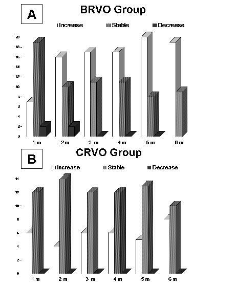
8 7 In the BRVO group, mean baseline logmar VA was 0.80 (SD 0.38; range 0.30 to 1.30); Snellen equivalents 0.22 (SD 0.16; range 0.05 to 0.5). Mean baseline macular thickness was μm (SD μm; range 302 to 753 μm). The mean time elapsed between the diagnosis of BRVO and the first bevacizumab injection was 4 months (range from 5 days to 2 years). Every patient was followed for at least 6 months after the first injection. During these 6 months, the mean number of injections per patient was 3.7 (range 1 to 6). Only one case resolved after a single injection. Visual acuity improved progressively throughout the follow-up period (Figure 1, top), although the main improvement occurred during the first two months. This improvement was statistically significant as compared to baseline VA at all visits (Figure 1, top). At six months of follow-up, mean logmar VA was 0.44 (SD 0.34; range 0 to 1.30); Snellen equivalent 0.46 (SD 0.28; range 0.05 to 1). Figure 2.A, shows the percentage of patients that gained 2 or more Snellen lines, that remained stable and that lost 2 or more lines. Only two patients lost 2 or more lines as compared to baseline during the first two months of treatment; however, after 3 months, vision improved or remained stable in all cases. The median improvement at 6 months was 2 Snellen lines. Final visual acuity was 0.5 or better (Snellen) in only 14% of patients with a baseline visual acuity equal to or lower than 0.2, as compared with 100% of patients with a baseline VA higher than 0.2. Mean macular thickness decreased significantly after the injection of bevacizumab (p< 0.001; Wilcoxon test). The reduction persisted throughout the whole follow-up period (Figure 1, bottom). Final mean macular thickness was μm (SD 62.5 μm). Pre-injection VA was significantly correlated with macular thickness before treatment (p<0.001, R=0.686) and with final VA (p<0.001, R=0.772). Similarly, final VA was
9 8 significantly correlated with pre-injection macular thickness (p=0.001, R=0.597) and with macular thickness at 6 months (p=0.001, R=0.578). CRVO group In the CRVO group, mean baseline logmar VA was 1.13 (SD 0.21, range 0.7 to 1.30); Snellen equivalent 0.08 (SD 0.05; range 0.05 to 0.2). Mean baseline macular thickness was μm (SD μm; range 400 to 757 μm). The mean time elapsed between the diagnosis of CRVO and the first bevacizumab injection was 6 months (range 1 week to 2 years). During the 6-month follow-up, the mean number of injections per patient was 4.6 (range 3 to 6). Mean final logmar VA was 0.83 (SD 0.45; range 0.15 to 1.30); Snellen equivalent 0.23 (SD 0.21; range 0.05 to 0.7). Visual acuity improved progressively, although the main improvement occurred in the first month. This improvement was statistically significant compared to baseline VA at all visits (Figure 3, top). Median improvement at 6 months was 1 Snellen line. Mean macular thickness decreased significantly after injections of bevacizumab (p< 0.001; Wilcoxon test). This reduction persisted throughout the follow-up period (Figure 3, bottom). Final mean macular thickness was μm (SD μm). Pre-injection VA was significantly correlated with macular thickness before treatment (p=0.002, R=0.679) and with final VA (p<0.001, R=0.781). Similarly, final VA was significantly correlated with pre-injection macular thickness (p=0.07, R=0.613) and with macular thickness at 6 months (p=0.044, R=0.479). A correlation was also found between macular thickness reduction at one month and final VA: for each 100 microns of ME reduction, there was an improvement of 1 line in VA.
10 9 DISCUSSION Retinal vein occlusions cause visual loss due to initial hypoxia and delayed ME. The edema may cause an additional reduction in VA that often exceeds the primary ischemic damage; it is the most frequent complication of vein occlusions.[9] It has been shown that intravitreal levels of VEGF are significantly increased after retinal vein occlusions and that the degree of ME is correlated with VEGF levels in aqueous humor.[10] Therefore, several authors have studied the usefulness of intravitreal injections of bevacizumab in both types of retinal vein occlusions.[9] Previous reports suggest that visual results are better in BRVO as compared with CRVO. The mean VA improvement was about 0.3 logmar units in most series of BRVO treated with bevacizumab.[11-14] An important feature of our study was that prior treatment was an exclusion criteria: patients were treated exclusively with bevacizumab. Few reports have prospectively studied patients with BRVO treated with bevacizumab as the sole treatment. Russo et al evaluated bevacizumab versus macular grid laser photocoagulation for ME in 30 patients with BRVO: after treatment, eyes receiving bevacizumab had better VA than those receiving photocoagulation at all times.[13] VA improved by 0.3 logmar units 6 months after treatment with bevacizumab. Rensch et al evaluated the results of early treatment with bevacizumab after BRVO: mean VA improved by 0.21 logmar units 6 months after treatment. In this study, mean logmar VA improved by 0.36 logmar units after treatment. Thus, most studies to date report similarly positive VA results after the injection of bevacizumab.
11 10 Most series have also shown a significant decrease in macular thickness after treatment with bevacizumab, though it is not always correlated with VA.[15,16] Rensch et al found that the improvement in VA correlated significantly with the decrease in macular thickness. We have found that both initial and final macular thicknesses are correlated with final VA. There may be several explanations for these results. Chung et al found that in the group of BRVO with no visual improvement, there was a higher proportion of angiographically documented macular ischemia. In this group, the decrease in central macular thickness was not accompanied by a visual acuity improvement. Although baseline visual acuity was not statistically different between groups, patients with no visual improvement had lower visual acuities than those that did improve. They also found that those patients that responded early to the first injections were more likely to benefit from the bevacizumab treatment.[17] Better initial visual acuities may reflect a preservation of macular function that may not be directly correlated with macular thickness; this would explain why patients with poor initial VA often have poor final VA in spite of the reduction of ME. Visual results are much more variable in patients with CRVO, since no visual benefits, minimal improvements of 9 letters or significant VA improvements have been published.[11,12,17-20] In our study, mean VA improvement was 0.30 logmar units, which is similar to that described by Rensch et al in a series of patients with ME caused by non-ischemic CRVO, with a mean duration prior to the first injection of 4.2 days (SD 3.6).[11] In our study, mean macular thickness in patients with CRVO decreased significantly after treatment. As in the patients with BRVO, macular thickness was correlated with VA. Rensch et al also found that the improvement in VA correlated significantly with the decrease in macular thickness.[11,21] Again, not all studies have found a relationship between retinal thickness and visual acuity in patients with CRVO.
12 11 Beutel et al published a retrospective study with intravitreal bevacizumab for nonischemic CRVO in which they obtained a significant decrease in central retinal thickness without significant improvement of VA after 12 months of follow-up.[20] In a prospective study of ranibizumab for ME due to CRVO, VA and central retinal thickness were not correlated.[16] Sakamoto et al found that the preservation of the foveal inner/outer segment photoreceptor line after resolution of ME was significantly correlated with good visual function.[22] As in BRVO, initial VA may reflect macular function and thus bevacizumab may lead to poorer visual improvement in patients with CRVO as compared with patients with BRVO because the macular function may be more seriously damaged due to the greater size of the initial ischemia in CRVO. No significant complications developed in our series after treatment with bevacizumab. The main drawback of bevacizumab for the treatment of ME after retinal vein occlusions is the need for repeated injections.[11,13,15-18,21-24] No retreatment schedule has been defined and new injections are performed at monthly or longer intervals at the discretion of the treating physician. However, Jaissle et al observed that the number of re-injections necessary to maintain the effect in BRVO declined over time.[9] Even though an optimal treatment regimen is as yet unclear, our data suggest that treatment should be initiated even if BRVO or CRVO have been present for some time: in our patients the temporal delay (range from 5 days to 2 years) from diagnosis to treatment initiation did not influence visual outcome, as reported in other series. An additional benefit that bevacizumab injections may provide as compared with other treatment options is that it may prevent the development of neovascularization or lead to its regression if it is already present. In summary, this case series supports previous reports that suggest that bevacizumab is a valid, effective treatment for ME due to retinal vein occlusions. However, randomized
13 12 multicenter studies are necessary to determine the optimal time after the vascular event for initiation of treatment and the schedule of injections.
14 13 FIGURE LEGENDS Figure 1. Branch retinal vein occlusion group. Top: Snellen visual acuity (VA) at baseline and at each visit. Bottom: Central macular thickness at baseline and at each visit. (p values provided are those of the comparison between each visit and baseline values, Wilcoxon test). Figure 2. Number of patients in which visual acuity remained stable, increased by two or more Snellen lines or decreased by two or more Snellen lines at each visit. A(top): Branch retinal vein occlusion (BRVO) group. B(bottom): Central retinal vein occlusion group (CRVO). m months Figure 3. Central retinal vein occlusion group. Top: Snellen visual acuity at baseline and at each visit. Bottom: Central macular thickness at baseline and at each visit. (p values provided are those of the comparison between each visit and baseline values, Wilcoxon test).
15 14 REFERENCES 1. Natural history and clinical management of central retinal vein occlusion. The Central Vein Occlusion Study Group. Arch Ophthalmol 1997;115: Argon laser photocoagulation for macular edema in branch vein occlusion. The Branch Vein Occlusion Study Group. Am J Ophthalmol 1984;98: Jonas JB, Akkoyun I, Kamppeter B, et al. Intravitreal triamcinolone acetonide for treatment of central retinal vein occlusion. Eur J Ophthalmol 2005;15: Jonas JB, Akkoyun I, Kamppeter B, et al. Branch retinal vein occlusion treated by intravitreal triamcinolone acetonide. Eye 2005;19: Suarez-Figueroa M, Contreras I, Noval S. [Side-effects of triamcinolone in young patients]. Arch Soc Esp Oftalmol 2006;81: Ramchandran RS, Fekrat S, Stinnett SS, et al. Fluocinolone acetonide sustained drug delivery device for chronic central retinal vein occlusion: 12-month results. Am J Ophthalmol 2008;146: Kuppermann BD, Blumenkranz MS, Haller JA, et al. Randomized controlled study of an intravitreous dexamethasone drug delivery system in patients with persistent macular edema. Arch Ophthalmol 2007;125: Rehak J, Rehak M. Branch retinal vein occlusion: pathogenesis, visual prognosis, and treatment modalities. Curr Eye Res 2008;33: Abegg M, Tappeiner C, Wolf-Schnurrbusch U, et al. Treatment of branch retinal vein occlusion induced macular edema with bevacizumab. BMC Ophthalmol 2008;8:18.
16 Funk M, Kriechbaum K, Prager F, et al. Intraocular concentrations of growth factors and cytokines in retinal vein occlusion and the effect of therapy with bevacizumab. Invest Ophthalmol Vis Sci 2009;50: Rensch F, Jonas JB, Spandau UH. Early intravitreal bevacizumab for nonischaemic branch retinal vein occlusion. Ophthalmologica 2009;223: Jaissle GB, Leitritz M, Gelisken F, et al. One-year results after intravitreal bevacizumab therapy for macular edema secondary to branch retinal vein occlusion. Graefes Arch Clin Exp Ophthalmol 2009;247: Russo V, Barone A, Conte E, et al. Bevacizumab compared with macular laser grid photocoagulation for cystoid macular edema in branch retinal vein occlusion. Retina 2009;29: Kreutzer TC, Alge CS, Wolf AH, et al. Intravitreal bevacizumab for the treatment of macular oedema secondary to branch retinal vein occlusion. Br J Ophthalmol 2008;92: Chung EJ, Hong YT, Lee SC, et al. Prognostic factors for visual outcome after intravitreal bevacizumab for macular edema due to branch retinal vein occlusion. Graefes Arch Clin Exp Ophthalmol 2008;246: Spaide RF, Chang LK, Klancnik JM, et al. Prospective study of intravitreal ranibizumab as a treatment for decreased visual acuity secondary to central retinal vein occlusion. Am J Ophthalmol 2009;147: Beutel J, Ziemssen F, Luke M, et al. Intravitreal bevacizumab treatment of macular edema in central retinal vein occlusion: one-year results. Int Ophthalmol 2008 Dec 20. [Epub ahead of print]
17 Prager F, Michels S, Kriechbaum K, et al. Intravitreal bevacizumab (Avastin) for macular oedema secondary to retinal vein occlusion: 12-month results of a prospective clinical trial. Br J Ophthalmol 2009;93: Iturralde D, Spaide RF, Meyerle CB, et al. Intravitreal bevacizumab (Avastin) treatment of macular edema in central retinal vein occlusion: a short-term study. Retina 2006;26: Ferrara DC, Koizumi H, Spaide RF. Early bevacizumab treatment of central retinal vein occlusion. Am J Ophthalmol 2007;144: Rensch F, Jonas JB, Spandau UH. Early intravitreal bevacizumab for nonischaemic central retinal vein occlusion. Acta Ophthalmol 2009;87: Sakamoto A, Tsujikawa A, Ota M, et al. Evaluation of potential visual acuity in eyes with macular oedema secondary to retinal vein occlusion. Clin Experiment Ophthalmol 2009;37: Gregori NZ, Gaitan J, Rosenfeld PJ, et al. Long-term safety and efficacy of intravitreal bevacizumab (Avastin) for the management of central retinal vein occlusion. Retina 2008;28: Wu L, Arevalo JF, Roca JA, et al. Comparison of two doses of intravitreal bevacizumab (Avastin) for treatment of macular edema secondary to branch retinal vein occlusion: results from the Pan-American Collaborative Retina Study Group at 6 months of follow-up. Retina 2008;28:
18
19
20
Intravitreal bevacizumab (Avastin) in the treatment of macular edema secondary to retinal vein occlusion
 in the treatment of macular edema secondary to retinal vein occlusion") ORIGINAL RESEARCH Intravitreal bevacizumab (Avastin) in the treatment of macular edema secondary to retinal vein occlusion Juan Carlos Mesa Gutiérrez Luis Arias Barquet Josep Maria Caminal Mitjana Sergi
ORIGINAL RESEARCH Intravitreal bevacizumab (Avastin) in the treatment of macular edema secondary to retinal vein occlusion Juan Carlos Mesa Gutiérrez Luis Arias Barquet Josep Maria Caminal Mitjana Sergi
Bevacizumab for Macular Edema in Branch and Central Retinal Vein Occlusion
 RESEARCH ARTICLE Acta Medica Marisiensis 2012;58(5):329-333 Bevacizumab for Macular Edema in Branch and Central Retinal Vein Occlusion Rusu Monica-Blanka 1, Egyed ZsI 2, Maki Cristina 1 1 Ophthalmology
Efficacy of combined intravitreal bevacizumab and triamcinolone for branch retinal vein occlusion
 AOP*** 1 Original Article Efficacy of combined intravitreal bevacizumab and triamcinolone for branch retinal vein occlusion Rasha I Ali1, Kapil G Kapoor 1,2, Adeel N Khan 1, Syed K Gibran 1 Purpose: To
Macular edema (ME) is the most common
 is the most common") MANAGEMENT OF RETINAL VEIN OCCLUSIONS * Peter A. Campochiaro, MD ABSTRACT Macular edema (ME) is the most common cause of reduced vision in patients with retinal vein occlusions (RVOs). The primary cause
MANAGEMENT OF RETINAL VEIN OCCLUSIONS * Peter A. Campochiaro, MD ABSTRACT Macular edema (ME) is the most common cause of reduced vision in patients with retinal vein occlusions (RVOs). The primary cause
Clinical Case Presentation. Branch Retinal Vein Occlusion. Sarita M. Registered Nurse Whangarei Base Hospital
 Clinical Case Presentation on Branch Retinal Vein Occlusion Sarita M. Registered Nurse Whangarei Base Hospital Introduction Case Study Pathogenesis Clinical Features Investigations Treatment Follow-up
Comparison of Ranibizumab and Bevacizumab for Macular Edema Associated with Branch Retinal Vein Occlusion
 pissn: 1011-8942 eissn: 2092-9382 Korean J Ophthalmol 2017;31(3):209-216 https://doi.org/10.3341/kjo.2015.0158 Original Article Comparison of Ranibizumab and Bevacizumab for Macular Edema Associated with
Clinical Outcomes After Intravitreal Bevacizumab Injection for Diabetic Macular Edema
 Original Article Clinical Outcomes After Intravitreal Bevacizumab Injection for Diabetic Macular Edema Karen Joyce G. Castro, MD, Marie Joan V. Loy, MD International Eye Institute St. Luke s Medical Center
Yoshiro Minami 1*, Taiji Nagaoka 2, Akihiro Ishibazawa 1,2 and Akitoshi Yoshida 2
 Minami et al. BMC Ophthalmology (2017) 17:90 DOI 10.1186/s12886-017-0485-4 RESEARCH ARTICLE Open Access Correlation between short- and long-term effects of intravitreal ranibizumab therapy on macular edema
Bevacizumab versus Dexamethasone Implant Followed by Bevacizumab for the Treatment of Macula Edema Associated with Branch Retinal Vein Occlusion
 pissn: 1011-8942 eissn: 2092-9382 Korean J Ophthalmol 2018;32(1):29-37 https://doi.org/10.3341/kjo.2016.0134 Original Article Bevacizumab versus Dexamethasone Implant Followed by Bevacizumab for the Treatment
Early Avastin management in acute retinal vein occlusion
 Saudi Journal of Ophthalmology (2010) 24, 87 94 King Saud University Saudi Journal of Ophthalmology www.ksu.edu.sa www.sciencedirect.com ORIGINAL ARTICLE Early Avastin management in acute retinal vein
Vascular Disease Ocular Manifestations of Systemic Hypertension
 Vascular Disease Ocular Manifestations of Systemic Hypertension Maynard L. Pohl, OD, FAAO Pacific Cataract & Laser Institute 10500 NE 8 th Street, Suite 1650 Bellevue, WA 98004 USA 425-462-7664 Cerebrovascular
International Journal of Health Sciences and Research ISSN:
 International Journal of Health Sciences and Research www.ijhsr.org ISSN: 2249-9571 Original Research Article A Multivariate Analysis of Intravitreal Injection of Anti-VEGF Bevacizumab in the Treatment
Retinal Vein Occlusion (RVO) Treatment pathway- Northeast England. Retinal Vein Occlusion (RVO) with Macular oedema (MO)
 Treatment pathway- Northeast England. Retinal Vein Occlusion (RVO) with Macular oedema (MO)") Retinal Vein Occlusion (RVO) Treatment pathway- Northeast England (Royal Victoria Infirmary, Sunderland Eye Infirmary, James Cook University Hospital, Darlington Memorial Hospital, University Hospital
Retinal Vein Occlusion (RVO) Treatment pathway- Northeast England (Royal Victoria Infirmary, Sunderland Eye Infirmary, James Cook University Hospital, Darlington Memorial Hospital, University Hospital
COMPARISON OF INTRAVITREAL TRIAMCINOLONE INJECTION VS LASER PHOTOCOAGULATION IN ANGIOGRAPHIC MACULAR EDEMA IN DIABETIC RETINOPATHY
 Original Article COMPARISON OF INTRAVITREAL TRIAMCINOLONE INJECTION VS LASER PHOTOCOAGULATION IN ANGIOGRAPHIC MACULAR EDEMA IN DIABETIC RETINOPATHY Aggarwal Somesh V 1, Shah Sonali N 2, Bharwada Rekha
Dexamethasone Intravitreal Implant Rescue Treatment for Bevacizumab Refractory Macular Edema Secondary to Branch Retinal Vein Occlusion
 pissn: 1011-8942 eissn: 2092-9382 Korean J Ophthalmol 2017;31(2):108-114 https://doi.org/10.3341/kjo.2017.31.2.108 Original Article Dexamethasone Intravitreal Implant Rescue Treatment for Bevacizumab Refractory
EFFICACY OF INTRAVITREAL TRIAMCINOLONE ACETONIDE FOR THE TREATMENT OF DIABETIC MACULAR EDEMA
 Basrah Journal Of Surgery EFFICACY OF INTRAVITREAL TRIAMCINOLONE ACETONIDE FOR THE TREATMENT OF DIABETIC MACULAR EDEMA Salah Zuhair Abed Al-Asadi MB,ChB, FICMS, Lecturer, Department of Surgery, College
Original Article Vascular Retinopathies Pak Armed Forces Med J 2014; 64 (1): 24-28
: 24-28") Original Article Vascular Retinopathies Pak Armed Forces Med J 2014; 64 (1): 24-28 CHANGES IN VISUAL ACUITY AND MACULAR THICKNESS AFTER INTRAVITREAL BEVACIZUMAB IN VASCULAR RETINOPATHIES Asad Jamal Dar,
Original Article Vascular Retinopathies Pak Armed Forces Med J 2014; 64 (1): 24-28 CHANGES IN VISUAL ACUITY AND MACULAR THICKNESS AFTER INTRAVITREAL BEVACIZUMAB IN VASCULAR RETINOPATHIES Asad Jamal Dar,
Bilateral anterior uveitis secondary to erlotinib
 Bilateral anterior uveitis secondary to erlotinib Lik Thai Lim, Robert Alexander Blum, Chee Peng Cheng, Abdul Hanifudin To cite this version: Lik Thai Lim, Robert Alexander Blum, Chee Peng Cheng, Abdul
An Update on Branch Retinal Vein Occlusion Treatment Studies. Amiee Ho, O.D. Pacific University College of Optometry
 An Update on Branch Retinal Vein Occlusion Treatment Studies Amiee Ho, O.D. Pacific University College of Optometry Course Description This course focuses on current treatment options available for macular
Prevalence and Management of Non-albicans Vaginal Candidiasis
 Prevalence and Management of Non-albicans Vaginal Candidiasis Nalin Hetticarachchi, Ruth Ashbee, Janet D Wilson To cite this version: Nalin Hetticarachchi, Ruth Ashbee, Janet D Wilson. Prevalence and Management
Christina L. Ryu, 1 Adrian Elfersy, 1 Uday Desai, 1 Thomas Hessburg, 1 Paul Edwards, 1 and Hua Gao 1,2. 1. Introduction
 Ophthalmology, Article ID 317694, 6 pages http://dx.doi.org/10.1155/2014/317694 Research Article The Effect of Antivascular Endothelial Growth Factor Therapy on the Development of Neovascular Glaucoma
황반부부종을동반한분지망막정맥폐쇄의치료에있어서유리체강내베바시주맙의반응을예측하는인자
 Journal of Retina 2018;3(1):20-25 ORIGINAL ARTICLE pissn 2508-1926 eissn 2508-3589 황반부부종을동반한분지망막정맥폐쇄의치료에있어서유리체강내베바시주맙의반응을예측하는인자 Predictive Factors for a Favorable Response to Intravitreal Bevacizumab for
Research Article Differentiation between Good and Low-Responders to Intravitreal Ranibizumab for Macular Edema Secondary to Retinal Vein Occlusion
 Hindawi Publishing Corporation Journal of Ophthalmology Volume 2016, Article ID 9875741, 6 pages http://dx.doi.org/10.1155/2016/9875741 Research Article Differentiation between Good and Low-Responders
Bevacizumab for Macular Edema in Central Retinal Vein Occlusion: A Prospective, Randomized, Double-Masked Clinical Study
 Bevacizumab for Macular Edema in Central Retinal Vein Occlusion: A Prospective, Randomized, Double-Masked Clinical Study David L.J. Epstein, MD, Peep V. Algvere, MD, PhD, Gunvor von Wendt, MD, PhD, Stefan
Research Article Long-Term Outcome after Vitrectomy for Macular Edema with Retinal Vein Occlusion Dividing into the Occlusion Site
 Hindawi Publishing Corporation Journal of Ophthalmology Volume 2014, Article ID 198782, 6 pages http://dx.doi.org/10.1155/2014/198782 Research Article Long-Term Outcome after Vitrectomy for Macular Edema
Recalcitrant Diabetic Macular Oedema: Therapeutic Options
 December 2007 A. Giridhar et al. - Recalcitrant DME 451 CONSULTATION S E C T I O N Recalcitrant Diabetic Macular Oedema: Therapeutic Options Dr. Cyrus M Shroff 1, Dr. N S Muralidhar 2, Dr. R Narayanan
Study of clinical significance of optical coherence tomography in diagnosis & management of diabetic macular edema
 Original Research Article Study of clinical significance of optical coherence tomography in diagnosis & management of diabetic macular edema Neha Kantilal Desai 1,*, Somesh Vedprakash Aggarwal 2, Sonali
Volume measurement by using super-resolution MRI: application to prostate volumetry
 Volume measurement by using super-resolution MRI: application to prostate volumetry Estanislao Oubel, Hubert Beaumont, Antoine Iannessi To cite this version: Estanislao Oubel, Hubert Beaumont, Antoine
Quantitative Reduction in Central Foveal Thickness After First Anti-VEGF Injection as a Predictor of Final Outcome in BRVO Patients
 Original clinical study Quantitative Reduction in Central Foveal Thickness After First Anti-VEGF Injection as a Predictor of Final Outcome in BRVO Patients Rupak Roy, MS, Kumar Saurabh, MS, Avirupa Ghose,
Effectiveness of Ranibizumab for Neovascular Age-related Macular Degeneration using Clinician Determined Retreatment Strategy
 Effectiveness of Ranibizumab for Neovascular Age-related Macular Degeneration using Clinician Determined Retreatment Strategy Anupma Kumar, Jayashree N Sahni, Alexandros N Stangos, Claudio Campa, Simon
ILUVIEN IN DIABETIC MACULAR ODEMA
 1 ILUVIEN IN DIABETIC MACULAR ODEMA Marie Tsaloumas Consultant Ophthalmic Surgeon Queen Elizabeth Hospital, Birmingham bars conference 2104 1 2 Declaration of interest I have sat on Advisory boards for
A retrospective nonrandomized study was conducted at 3
 Department of Ophthalmology, Kangbuk Samsung Hospital, Sungkyunkwan University College of Medicine 1, Seoul, Korea Hangil Eye Hospital 2, Incheon, Korea Seoul National University Bundang Hospital 3, Seongnam,
Diabetic maculopathy 11/ An update on. Miss Vasuki Sivagnanavel
 Miss Vasuki Sivagnanavel Consultant Ophthalmologist An update on Diabetic maculopathy Despite advances in the management of diabetes, diabetic retinopathy is already the commonest cause of blindness among
ROLE OF LASER PHOTOCOAGULATION VERSUS INTRAVITREAL TRIAMCINOLONE ACETONIDE IN ANGIOGRAPHIC MACULAR EDEMA IN DIABETES MELLITUS
 ORIGINAL ARTICLE ROLE OF LASER PHOTOCOAGULATION VERSUS INTRAVITREAL TRIAMCINOLONE ACETONIDE IN ANGIOGRAPHIC MACULAR EDEMA IN DIABETES MELLITUS Aggarwal Somesh VP 1, Shah Sonali N 2, Bharwada Rekha M 3,
 Research Article http://www.alliedacademies.org/clinical-ophthalmology-and-vision-science/ A 2-year retrospective study of the treatment of retinal vein occlusion with dexamethasone 0.7 mg intravitreal
EU Regulatory workshop Ophthalmology clinical development and scientific advice. Industry view on DME and macular edema secondary to RVO
 EU Regulatory workshop Ophthalmology clinical development and scientific advice. Industry view on DME and macular edema secondary to RVO Yehia Hashad, M.D. Vice President and Global Therapeutic Area Head
Intravitreal versus Posterior Subtenon Injection of Triamcinolone Acetonide for Diabetic Macular Edema
 Intravitreal versus Posterior Subtenon Injection of Triamcinolone Acetonide for Diabetic Macular Edema Young Jae Choi, MD, In Kyung Oh, MD, Jae Ryung Oh, MD, PhD, Kuhl Huh, MD, PhD Department of Ophthalmology,
HOW COST-EFFECTIVE IS NO SMOKING DAY?
 HOW COST-EFFECTIVE IS NO SMOKING DAY? Daniel Kotz, John A. Stapleton, Lesley Owen, Robert West To cite this version: Daniel Kotz, John A. Stapleton, Lesley Owen, Robert West. HOW COST-EFFECTIVE IS NO SMOKING
Retinal vein occlusion (RVO) is a vascular disease
 is a vascular disease") INTRAVITREAL RANIBIZUMAB FOR RETINAL VEIN OCCLUSION THROUGH 1 YEAR IN CLINICAL PRACTICE TROELS BRYNSKOV, MD,* HENRIK KEMP, MD,* TORBEN L. SØRENSEN, MD, DMSC* Purpose: To evaluate the efficacy and safety
INTRAVITREAL RANIBIZUMAB FOR RETINAL VEIN OCCLUSION THROUGH 1 YEAR IN CLINICAL PRACTICE TROELS BRYNSKOV, MD,* HENRIK KEMP, MD,* TORBEN L. SØRENSEN, MD, DMSC* Purpose: To evaluate the efficacy and safety
Off-label use of intravitreal bevacizumab in non-ischemic macular edema secondary to retinal vein obstructions
 Romanian Journal of Ophthalmology, Volume 60, Issue 2, April-June 2016. pp:90-95 GENERAL ARTICLE Off-label use of intravitreal bevacizumab in non-ischemic macular edema secondary to retinal vein obstructions
Natural Short-term Course of Recurrent Macular Edema Following Intravitreal Bevacizumab Therapy in Branch Retinal Vein Occlusion
 pissn: 1011-8942 eissn: 2092-9382 Korean J Ophthalmol 2017;31(2):95-101 https://doi.org/10.3341/kjo.2017.31.2.95 Original Article Natural Short-term Course of Recurrent Macular Edema Following Intravitreal
A model for calculation of growth and feed intake in broiler chickens on the basis of feed composition and genetic features of broilers
 A model for calculation of growth and feed intake in broiler chickens on the basis of feed composition and genetic features of broilers Bernard Carré To cite this version: Bernard Carré. A model for calculation
NATIONAL INSTITUTE FOR HEALTH AND CARE EXCELLENCE. Health Technology Appraisal. Aflibercept for treating diabetic macular oedema.
 NATIONAL INSTITUTE FOR HEALTH AND CARE EXCELLENCE Health Technology Appraisal Aflibercept for treating diabetic macular oedema Final scope Final remit/appraisal objective To appraise the clinical and cost
Daily alternating deferasirox and deferiprone therapy for hard-to-chelate β-thalassemia major patients
 Daily alternating deferasirox and deferiprone therapy for hard-to-chelate β-thalassemia major patients Manuela Balocco, Paola Carrara, Valeria Pinto, Gian Luca Forni To cite this version: Manuela Balocco,
Efficacy of intravitreal bevacizumab (Avastin TM ) for shortterm treatment of diabetic macular edema
 for shortterm treatment of diabetic macular edema") 111 ORIGINAL Efficacy of intravitreal bevacizumab (Avastin TM ) for shortterm treatment of diabetic macular edema Toshihiko Nagasawa, Takeshi Naito, Shingo Matsushita, Hiroyuki Sato, Takashi Katome, and
111 ORIGINAL Efficacy of intravitreal bevacizumab (Avastin TM ) for shortterm treatment of diabetic macular edema Toshihiko Nagasawa, Takeshi Naito, Shingo Matsushita, Hiroyuki Sato, Takashi Katome, and
CSME BCVA. OCT Bevacizumab CMT CMT BCVA. BCVA LogMAR. p CMT NPDR PDR PDR. Bevacizumab PDR FDA. Ranibizumab. Bevacizumab.
 email: rezaj76@yahoo.com Bevacizumab FDA mg Ranibizumab - Bevacizumab NPDR PDR PDR PDR CSME FAZFA BCVA CMT OCT Bevacizumab BCVA CMT BCVA LogMAR p CMT p CSME CRVO CSME BRVO CRVOBRVO (fovea avascular zone)
RETINAL VENOUS OCCLUSIVE DISEASE IS PROBABLY
 Clinical, Anatomic, and Electrophysiologic Evaluation Following Intravitreal Bevacizumab for Macular Edema in Retinal Vein Occlusion SIVAKAMI A. PAI, ROHIT SHETTY, PRIYA B. VIJAYAN, VENKATASUBRAMANIAM
Jay M. Haynie, O.D.; F.A.A.O. Olympia Tacoma Renton Kennewick Washington
 Jay M. Haynie, O.D.; F.A.A.O. Olympia Tacoma Renton Kennewick Washington I Jay M. Haynie, OD, FAAO have received honoraria from the following companies: Reichert Technologies Notal Vision Carl Zeiss Meditec
Andrew J. Barkmeier, MD; Benjamin P. Nicholson, MA; Levent Akduman, MD
 c l i n i c a l s c i e n c e Effectiveness of Laser Photocoagulation in Clinically Significant Macular Edema With Focal Versus Diffuse Parafoveal Thickening on Optical Coherence Tomography Andrew J. Barkmeier,
Case Report Macular Oedema Related to Idiopathic Macular Telangiectasia Type 1 Treated with Dexamethasone Intravitreal Implant (Ozurdex)
") Case Reports in Ophthalmological Medicine, Article ID 231913, 5 pages http://dx.doi.org/10.1155/2014/231913 Case Report Macular Oedema Related to Idiopathic Macular Telangiectasia Type 1 Treated with Dexamethasone
Case Reports in Ophthalmological Medicine, Article ID 231913, 5 pages http://dx.doi.org/10.1155/2014/231913 Case Report Macular Oedema Related to Idiopathic Macular Telangiectasia Type 1 Treated with Dexamethasone
Abbreviated Drug Evaluation: Fluocinolone acetonide intravitreal implant (Retisert )
") Copyright 2012 Oregon State University. All Rights Reserved Drug Use Research & Management Program Oregon State University, 500 Summer Street NE, E35, Salem, Oregon 97301-1079 Phone 503-947-5220 Fax 503-947-1119
Copyright 2012 Oregon State University. All Rights Reserved Drug Use Research & Management Program Oregon State University, 500 Summer Street NE, E35, Salem, Oregon 97301-1079 Phone 503-947-5220 Fax 503-947-1119
Update on the Use of Anti-VEGF Intravitreal Therapies for Retinal Vein Occlusion. Yi Jiang, MD, and William F. Mieler, MD
 REVIEW ARTICLe Update on the Use of Anti-VEGF Intravitreal Therapies for Retinal Vein Occlusion Yi Jiang, MD, and William F. Mieler, MD Abstract: The use of anti vascular endothelial growth factor (VEGF)
Efficacy of Anti-VEGF Agents in the Treatment of Age-Related Macular Degeneration
 Efficacy of Anti-VEGF Agents in the Treatment of Age-Related Macular Degeneration Marilita M. Moschos Abstract- Purpose: To evaluate by OCT and mf-erg the macular function in eyes with CNV due to ARMD
Posterior Segment Macular Edema
 Posterior Segment Macular Edema Treatment of Macular Edema following Branch Retinal Vein Occlusion Raafay Sophie, MD 1 and Peter A Campochiaro, MD 2 1. Post-doctoral Fellow; 2. Eccles Professor of Ophthalmology
Efficacy and safety of Pro Re Nata regimen without loading dose ranibizumab injections in retinal vein occlusion
 Open Access Original Article Efficacy and safety of Pro Re Nata regimen without loading dose ranibizumab injections in retinal vein occlusion Erkan Unsal 1, Kadir Eltutar 2, Pınar Sultan 3, Hulya Gungel
Applying New Data to Improve the Standard of Care in Retinal Diseases Managing Macular Edema Associated With Retinal Venous Occlusions
 Supplement to November/December 2013 CME Activity Applying New Data to Improve the Standard of Care in Retinal Diseases Managing Macular Edema Associated With Retinal Venous Occlusions By Michael Singer,
Persistent Macular Thickening After Ranibizumab Treatment for Diabetic Macular Edema With Vision Impairment
 9:30 AM Persistent Macular Thickening After Ranibizumab Treatment for Diabetic Macular Edema With Vision Impairment Lee Jampol, MD OBJECTIVE To assess subsequent visual and anatomic outcomes of eyes with
Ranibizumab for Macular Edema Due to Retinal Vein Occlusions: Implication of VEGF as a Critical Stimulator
 original article Ranibizumab for Macular Edema Due to Retinal Vein Occlusions: Implication of VEGF as a Critical Stimulator Peter A Campochiaro 1, Gulnar Hafiz 1, Syed Mahmood Shah 1, Quan Dong Nguyen
Diabetic Macular Edema Treatment in the 21st Century
 Transcript Details This is a transcript of a continuing medical education (CME) activity accessible on the ReachMD network. Additional media formats for the activity and full activity details (including
Spectral domain optical coherence tomography detects
 Spectral domain optical coherence tomography detects early stages of chloroquine retinopathy similar to multifocal electroretinography, fundus autofluorescence and near-infrared autofluorescence Simone
From universal postoperative pain recommendations to procedure-specific pain management
 From universal postoperative pain recommendations to procedure-specific pain management Hélène Beloeil, Francis Bonnet To cite this version: Hélène Beloeil, Francis Bonnet. From universal postoperative
 Research Article http://www.alliedacademies.org/clinical-ophthalmology-and-vision-science/ The trial: A pilot study to assess the efficacy, durability, and safety of combination ranibizumab + peripheral
The Diabetic Retinopathy Clinical Research Network. Management of DME in Eyes with PDR
 The Diabetic Retinopathy Clinical Research Network Management of DME in Eyes with PDR 1 What Has Been Learned? Diabetic Retinopathy Treatment Protocol F: Results suggest that clinically meaningful differences
Efficacy and safety of anti-vegf therapy with intravitreal ranibizumab (LUCENTIS ) for naïve retinal vein occlusion: one-year follow-up
 for naïve retinal vein occlusion: one-year follow-up") Efficacy and safety of anti-vegf therapy with intravitreal ranibizumab (LUCENTIS ) for naïve retinal vein occlusion: one-year follow-up Alfredo Pece, Vincenzo Isola, Stefano Piermarocchi, Giliola Calori
Efficacy and safety of anti-vegf therapy with intravitreal ranibizumab (LUCENTIS ) for naïve retinal vein occlusion: one-year follow-up Alfredo Pece, Vincenzo Isola, Stefano Piermarocchi, Giliola Calori
The Era of anti- - - VEGF Kirk L. Halvorson, OD
 The Era of anti- - - VEGF Kirk L. Halvorson, OD Introduction: Anti- - - Vascular Endothelial Growth Factor (Anti- - - VEGF) medication is a relatively a new line of medications used in treating a variety
OCCLUSIVE VASCULAR DISORDERS OF THE RETINA
 OCCLUSIVE VASCULAR DISORDERS OF THE RETINA Learning outcomes By the end of this lecture the students would be able to Classify occlusive vascular disorders (OVD) of the retina. Correlate the clinical features
Diabetic retinopathy (DR) progressively
 progressively") FOCUS ON DIABETIC MACULAR EDEMA * Frederick L. Ferris III, MD ABSTRACT The current state of treatment for diabetic macular edema (DME) is focused on slowing the rate of vision loss through assessment of
FOCUS ON DIABETIC MACULAR EDEMA * Frederick L. Ferris III, MD ABSTRACT The current state of treatment for diabetic macular edema (DME) is focused on slowing the rate of vision loss through assessment of
Multi-template approaches for segmenting the hippocampus: the case of the SACHA software
 Multi-template approaches for segmenting the hippocampus: the case of the SACHA software Ludovic Fillon, Olivier Colliot, Dominique Hasboun, Bruno Dubois, Didier Dormont, Louis Lemieux, Marie Chupin To
An Alternate, Egg-Free Radiolabeled Meal Formulation for Gastric-Emptying Scintigraphy
 An Alternate, Egg-Free Radiolabeled Meal Formulation for Gastric-Emptying Scintigraphy Philippe Garrigue, Aurore Bodin-Hullin, Sandra Gonzalez, Quentin Sala, Benjamin Guillet To cite this version: Philippe
Applying New Data to Improve the Standard of Care in Retinal Disease. With articles by Gaurav K. Shah, MD Carl D. Regillo, MD.
 Supplement to July/August 2012 CME Activity Applying New Data to Improve the Standard of Care in Retinal Disease With articles by Gaurav K. Shah, MD Carl D. Regillo, MD Release date: August 2012. Expiration
Clinical Trials in Diabetic Retinopathy. Harry W. Flynn Jr., M.D. Nidhi Relhan Batra, M.D.
 1 Clinical Trials in Diabetic Retinopathy 2018 Harry W. Flynn Jr., M.D. Nidhi Relhan Batra, M.D. Bascom Palmer Eye Institute 900 N.W. 17th Street Miami, FL 33136 Phone: (305) 326-6118 Fax: (305) 326-6417
Facts About Diabetic Eye Disease
 Facts About Diabetic Eye Disease Points to Remember 1. Diabetic eye disease comprises a group of eye conditions that affect people with diabetes. These conditions include diabetic retinopathy, diabetic
Virtual imaging for teaching cardiac embryology.
 Virtual imaging for teaching cardiac embryology. Jean-Marc Schleich, Jean-Louis Dillenseger To cite this version: Jean-Marc Schleich, Jean-Louis Dillenseger. Virtual imaging for teaching cardiac embryology..
Y O R K. Health Economics C O N S O R T I U M NOVARTIS. Bevacizumab (Avastin) for Macular Edema Secondary to RVO
 for Macular Edema Secondary to RVO") Y O R K Health Economics C O N S O R T I U M NOVARTIS Bevacizumab (Avastin) for Macular Edema Secondary to RVO Review of Evidence on Clinical Effectiveness and Safety from Non-Randomised Controlled Trials
Y O R K Health Economics C O N S O R T I U M NOVARTIS Bevacizumab (Avastin) for Macular Edema Secondary to RVO Review of Evidence on Clinical Effectiveness and Safety from Non-Randomised Controlled Trials
ORIGINAL ARTICLE. Introduction. June 2009 Kerala Journal of Ophthalmology 139
 June 2009 Kerala Journal of Ophthalmology 139 ORIGINAL ARTICLE Intravitreal Monotherapy With Bevacizumab (IVB) and Triamcinolone Acetonide (IVTA) Versus Combination Therapy (IVB and IVTA) for Recalcitiant
VISUAL OUTCOME IN DIABETIC MACULAR EDEMA AFTER GRID LASER TREATMENT
 The Professional Medical Journal DOI: 10.17957/TPMJ/16.2856 ORIGINAL PROF-2856 VISUAL OUTCOME IN DIABETIC MACULAR EDEMA AFTER GRID LASER TREATMENT 1. MBBS.FCPS Assistant Professor Ophthalmology Independent
Diabetic eye disease. Diabetic retinopathy. Sam S. Dahr, M.D. Retina Center of Oklahoma.
 Diabetic eye disease Sam S. Dahr, M.D. Retina Center of Oklahoma www.rcoklahoma.com Downloaded from: The Retina (on 28 May 2007 12:48 AM) 2007 Elsevier Diabetic retinopathy Downloaded from: The Retina
Optimal electrode diameter in relation to volume of the cochlea
 Optimal electrode diameter in relation to volume of the cochlea Dan Gnansia, Thomas Demarcy, Clair Vandersteen, Charles Raffaelli, Nicolas Guevara, Hervé Delingette, Nicholas Ayache To cite this version:
Ophthalmology Macular Pathways
 Ophthalmology Macular Pathways Age related Macular Degeneration Diabetic Macular Oedema Macular Oedema secondary to Central Retinal Macular Oedema secondary to Branch Retinal CNV associated with pathological
Clinical Study Switch to Aflibercept in Diabetic Macular Edema Patients Unresponsive to Previous Anti-VEGF Therapy
 Hindawi Ophthalmology Volume 2017, Article ID 5632634, 4 pages https://doi.org/10.1155/2017/5632634 Clinical Study Switch to Aflibercept in Diabetic Macular Edema Patients Unresponsive to Previous Anti-VEGF
Foveal Sensitivity and Morphology in Major and Macular Branch Retinal Vein Occlusion
 Send Orders of Reprints at bspsaif@emirates.net.ae 104 The Open Ophthalmology Journal, 2012, 6, 104-109 Open Access Foveal Sensitivity and Morphology in Major and Macular Branch Retinal Vein Occlusion
Improving HIV management in Sub-Saharan Africa: how much palliative care is needed?
 Improving HIV management in Sub-Saharan Africa: how much palliative care is needed? Karilyn Collins, Richard Harding To cite this version: Karilyn Collins, Richard Harding. Improving HIV management in
LYMPHOGRANULOMA VENEREUM PRESENTING AS PERIANAL ULCERATION: AN EMERGING CLINICAL PRESENTATION?
 LYMPHOGRANULOMA VENEREUM PRESENTING AS PERIANAL ULCERATION: AN EMERGING CLINICAL PRESENTATION? Tajinder K Singhrao, Elizabeth Higham, Patrick French To cite this version: Tajinder K Singhrao, Elizabeth
Clinical Study Evaluation of Peripapillary Nerve Fiber Layer after Dexamethasone Implantation (Ozurdex) in Branch Retinal Vein Occlusions
 in Branch Retinal Vein Occlusions") Ophthalmology Volume 2016, Article ID 2050796, 4 pages http://dx.doi.org/10.1155/2016/2050796 Clinical Study Evaluation of Peripapillary Nerve Fiber Layer after Dexamethasone Implantation (Ozurdex) in
Ophthalmology Volume 2016, Article ID 2050796, 4 pages http://dx.doi.org/10.1155/2016/2050796 Clinical Study Evaluation of Peripapillary Nerve Fiber Layer after Dexamethasone Implantation (Ozurdex) in
Long term visual outcome after arteriolar constriction in patients with branch retinal vein occlusion
 Long term visual outcome after arteriolar constriction in patients with branch retinal vein occlusion Jiri Rehak a, Ladislav Dusek b, Martin Sin a, Barbora Babkova a, Zuzana Pracharova a, Matus Rehak a,c
Clinical Trials Related to Age Related Macular Degeneration
 Clinical Trials Related to Age Related Macular Degeneration Kirti Singh MD, DNB, FRCS Kirti Singh MD, DNB, FRCS, Pooja Jain MBBS, Nitasha Ahir MBBS, Divya Jain MD, DNB Guru Nanak Eye Centre, Maulana Azad
Mathieu Hatt, Dimitris Visvikis. To cite this version: HAL Id: inserm
 Defining radiotherapy target volumes using 18F-fluoro-deoxy-glucose positron emission tomography/computed tomography: still a Pandora s box?: in regard to Devic et al. (Int J Radiat Oncol Biol Phys 2010).
Clinically Significant Macular Edema (CSME)
") Clinically Significant Macular Edema (CSME) 1 Clinically Significant Macular Edema (CSME) Sadrina T. Shaw OMT I Student July 26, 2014 Advisor: Dr. Uwaydat Clinically Significant Macular Edema (CSME) 2
Clinically Significant Macular Edema (CSME) 1 Clinically Significant Macular Edema (CSME) Sadrina T. Shaw OMT I Student July 26, 2014 Advisor: Dr. Uwaydat Clinically Significant Macular Edema (CSME) 2
Intravitreal Monotherapy with Bevacizumab and Triamcinolone Versus Combination Therapy in Recalcitiant Diabetic Macular Edema
 Intravitreal Monotherapy with Bevacizumab and Triamcinolone Versus Combination Therapy in Recalcitiant Diabetic Macular Edema Meena Chakrabarti MS, DO, DNB Meena Chakrabarti MS, DO, DNB, Sonia Rani John
Anti VEGF Agents in Retinal Disorders Current Scenario
 Retina Anti VEGF Agents in Retinal Disorders Current Scenario Charu Gupta MS Charu Gupta MS, Cyrus M. Shroff MD Shroff Eye Centre, New Delhi T is a group of proteins involved in the regulation of angiogenesis,
OCT Assessment of the Vitreoretinal Relationship in CSME
 December 2007 Sonia Rani John et al. - IFIS 375 ORIGINAL ARTICLE OCT Assessment of the Vitreoretinal Relationship in CSME Dr. Manoj S. DNB FRCS, Dr. Unnikrishnan Nair MS DO FRCS, Dr. Gargi Sathish MS Introduction
Iodide mumps: Sonographic appearance
 Iodide mumps: Sonographic appearance Salvatore Greco, Riccardo Centenaro, Giuseppe Lavecchia, Francesco Rossi To cite this version: Salvatore Greco, Riccardo Centenaro, Giuseppe Lavecchia, Francesco Rossi.
Pharmacokinetics of caspofungin in a critically ill patient with liver cirrhosis
 Pharmacokinetics of caspofungin in a critically ill patient with liver cirrhosis Isabel Spriet, Wouter Meersseman, Pieter Annaert, Jan Hoon, Ludo Willems To cite this version: Isabel Spriet, Wouter Meersseman,
Comparison of BRVO and CRVO management
 Comparison of BRVO and CRVO management Francesco Bandello, MD, FEBO Department of Ophthalmology University Vita-Salute Scientific Institute San Raffaele Milan, Italy 1 Financial Disclosure Advisory Board
Optical Coherence Tomography (OCT) in Uveitis Piergiorgio Neri, BMedSc, MD, PhD Head Ocular Immunology Unit
 in Uveitis Piergiorgio Neri, BMedSc, MD, PhD Head Ocular Immunology Unit") The Eye Clinic Polytechnic University of Marche Head: Prof Alfonso Giovannini November, 1991 Optical Coherence Tomography (OCT) in Uveitis Piergiorgio Neri, BMedSc, MD, PhD Head Ocular Immunology Unit
The Eye Clinic Polytechnic University of Marche Head: Prof Alfonso Giovannini November, 1991 Optical Coherence Tomography (OCT) in Uveitis Piergiorgio Neri, BMedSc, MD, PhD Head Ocular Immunology Unit
AGE-RELATED MACULAR DEGENERATION (AMD) IS A
 IS A") Intravitreal Bevacizumab for the Management of Choroidal Neovascularization in Age-related Macular Degeneration ZIAD F. BASHSHUR, MD, ALI BAZARBACHI, MD, ALEXANDRE SCHAKAL, MD, ZEINA A. HADDAD, MD, CHRISTELLE
Intravitreal Bevacizumab for the Management of Choroidal Neovascularization in Age-related Macular Degeneration ZIAD F. BASHSHUR, MD, ALI BAZARBACHI, MD, ALEXANDRE SCHAKAL, MD, ZEINA A. HADDAD, MD, CHRISTELLE
Original Article. Authors
 Original Article APMC-303 Efficacy of Intra-Vitreal Bevacizumab Combined with Phaco- Emulsification in the Prophylaxis of Macular Edema in Patients with Non-Proliferative Diabetic Retinopathy Muhammad
Bevacizumab (Avastin) Therapy for Macular Oedema in Central Retinal Vein Occlusion Long Term Results
 Therapy for Macular Oedema in Central Retinal Vein Occlusion Long Term Results") December 2008 G.J. Manayath - Avastin in CRVO 355 ORIGINAL ARTICLE Bevacizumab (Avastin) Therapy for Macular Oedema in Central Retinal Vein Occlusion Long Term Results Dr. George J Manayath MS Abstract
December 2008 G.J. Manayath - Avastin in CRVO 355 ORIGINAL ARTICLE Bevacizumab (Avastin) Therapy for Macular Oedema in Central Retinal Vein Occlusion Long Term Results Dr. George J Manayath MS Abstract
Optical Coherence Tomography: Pearls for the Anterior Segment Surgeon Basic Science Michael Stewart, M.D.
 Optical Coherence Tomography: Pearls for the Anterior Segment Surgeon Basic Science Michael Stewart, M.D. Disclosure OCT Optical Coherence Tomography No relevant financial relationships I will refer to
HHS Public Access Author manuscript J Vitreoretin Dis. Author manuscript; available in PMC 2017 November 03.
 Prediction of Anti-VEGF Response in Diabetic Macular Edema After 1 Injection Ankoor R. Shah, M.D. 1,2, Yoshihiro Yonekawa, M.D. 3,7, Bozho Todorich, M.D. Ph.D. 3,7, Lily Van Laere, M.D. 3, Rehan Hussain,