Optical Coherence Tomography: Pearls for the Anterior Segment Surgeon Basic Science Michael Stewart, M.D.
|
|
|
- Donald Terry
- 5 years ago
- Views:
Transcription


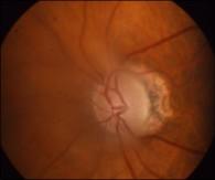
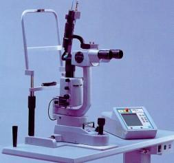
1 Optical Coherence Tomography: Pearls for the Anterior Segment Surgeon Basic Science Michael Stewart, M.D. Disclosure OCT Optical Coherence Tomography No relevant financial relationships I will refer to off-label use of medications Laser Cross section What happens when light hits the retina? What happens when light hits the retina? What happens when light hits the retina? Only 10-5 to 10-9 of incident light is reflected back. Transmission Absorption Reflection OCT images are created by What pigments are found within the eye? Ocular Pigments: melanin, xanthophylls, hemoglobin 1. Ocular pigments 2. Interfaces with differing refractive indices 1. Xanthophylls 2. Hemoglobin 3. Melanin 1


/tan(ө")

/2 Modified from:")


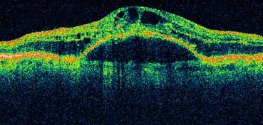
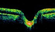
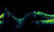
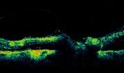
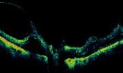
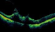
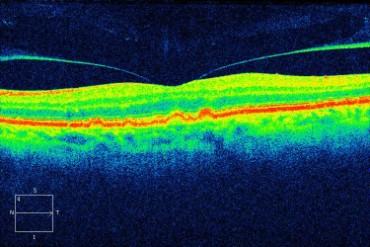
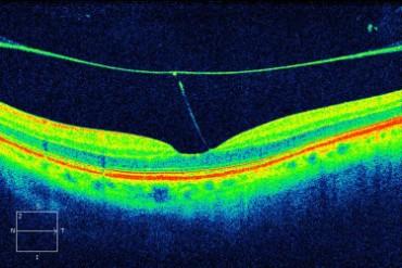
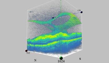
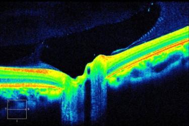
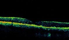
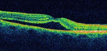
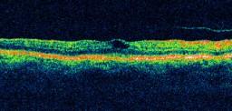
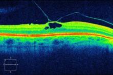
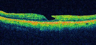
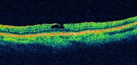
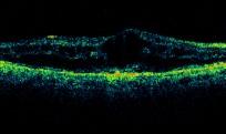
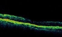
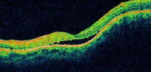
2 OCT Imaging = Optical Biopsy Normal Macular Anatomy Xanthophyll Highest concentration in macula What color is the retina? Melanin OCT B-Scan Normal Macular Anatomy R + T = 1 Fresnel Equations Pre-operative macular hole NFL GCL IPL OPL ONL ELM IS/OS RPE Choroid For light polarized in Ө the plane of the diagram: i R s = (sin(ө i - Ө t)/sin (Ө i + Ө t)) 2 For light polarized perpendicular to the plane of the diagram: R p = (tan (Ө i - Ө t)/tan(ө i + Ө t)) 2 Ө r Ө t Post-operative For non-polarized light: R = (R s + R p)/2 Modified from: Retina 5 th Ed, Chapter 3, Optical Coherence Tomography Within retinal tissue this is: 10-5 to 10-9 Previous vitrectomy with silicone oil 20 minutes later Aqueous 2


3 OCT tomogram of cervical epithelium Optical coherence tomography showing prostatic capsule and cancer cells underneath the thin layer Resolution 150 µ TD-OCT: Resolution 10 µ SD-OCT Resolution 3 to 6 µ 3.5 or 6 mm 3


 maps (Cirrus, software 6.")
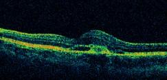
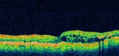
: SDOCT Imaging of Wet AMD Vitreomacular interface abnormalities RPE RPE floor RPE RPE floor Vol: 0.")
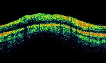
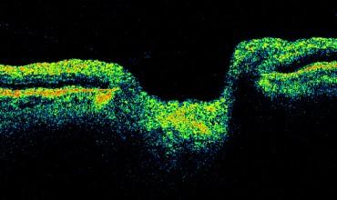
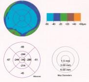
4 Spectral Domain OCT (SDOCT) Imaging Faster image acquisition A-scans/sec: currently available instruments. 200, ,000 A-scans/sec: research instruments. Higher density scans over larger areas, image averaging to improve image quality, fewer eye motion artifacts. SDOCT Imaging of Macula Qualitative assessment of retinal anatomy: Subretinal Fluid PED Subretinal Fluid SDOCT Imaging of Wet AMD Segmentation of retinal layers necessary for quantitative assessments: ILM segmentation SDOCT Imaging of Wet AMD Segmentation algorithms result in reliable retinal thickness maps: Retinal thickness VA: 20/100 SDOCT Imaging of Wet AMD: Anti-VEGF Therapy Retinal thickness map Retinal thickness RPE segmentation ILM segmentation RPE segmentation RPE segmentation VA: 20/40 Anti-VEGF Therapy: 1 month post-injection SDOCT Imaging of Dry AMD Segmentation algorithms result in reliable RPE elevation (drusen) maps (Cirrus, software 6.0): SDOCT Imaging of Wet AMD Segmentation algorithms result in reliable RPE elevation (PED) maps (Cirrus, software 6.0): SDOCT Imaging of Wet AMD Vitreomacular interface abnormalities RPE RPE floor RPE RPE floor Vol: 0.58mm 3 Epiretinal membrane RPE segmentation RPE Elevation map Gregori G, Wang F, Rosenfeld PJ, et al. Spectral domain optical coherence tomography imaging of drusen in nonexudative age-related macular degeneration. Ophthalmology. 2011;118(7): RPE segmentation Retinal thickness RPE Elevation map 4

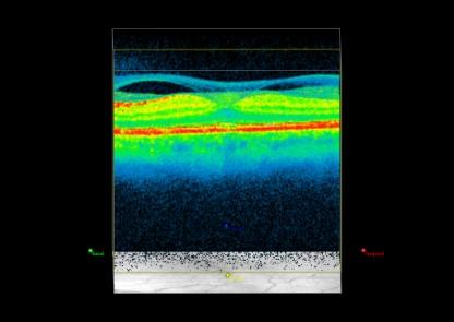
5 Macular edema RPED 5

6 Optical Coherence Tomography: Pearls for the Anterior Segment Surgeon Retina Michael Stewart, M.D. 6


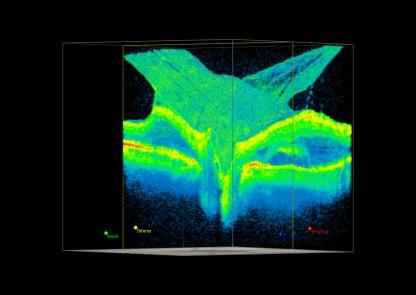
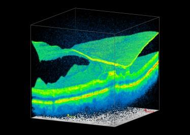
7 Why this shadow? Why this shadow? Gas bubble 7


8 1945µ 2819µ 8


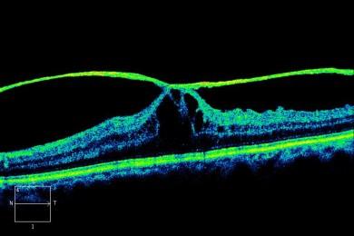
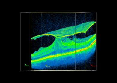
9 2 weeks later OS 20/40 Stage 2 OD 20/25 Stage 1 Lamellar hole Cyst? Lamellar hole 9

10 Epiretinal Membrane 20/40 Pre-Op 20/30 Post-Op 10
11 9/29/ /31/ /30/


 Wet AMD")


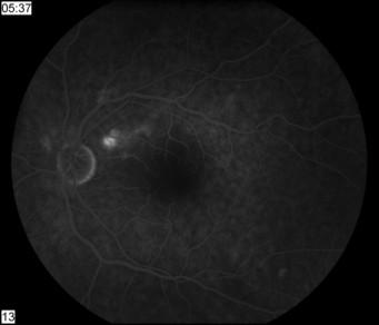
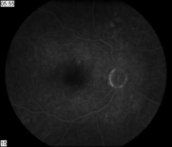
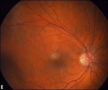
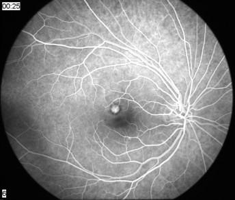
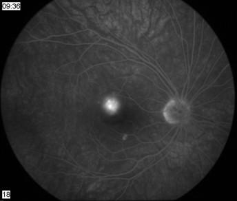
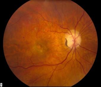
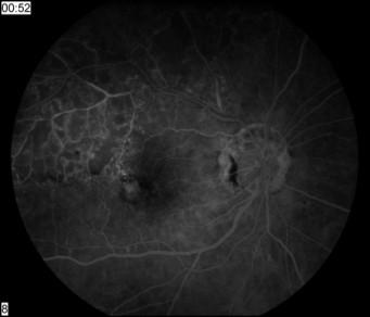
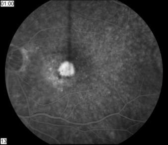
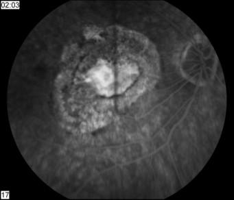
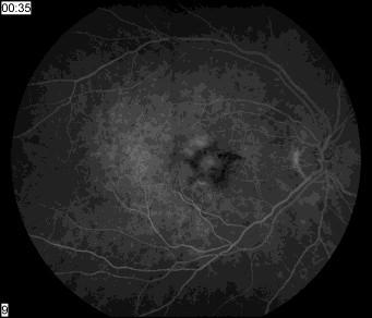
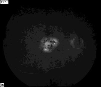
12 1/11/2006 Subretinal fluid RPED Case 71 yo man with wet AMD OS Multiple injections for the last 12 months Complains of worsening in VA after treatment Case 74 y.o. man decreased vision OD (20/200) Wet AMD Dry AMD 20/200 Ranibizumab #1 Case After 1 month Case Monthly Injections 73 y.o. woman with neovascular AMD VA: 20/40 Ranibizumab #1 Vol: 0.73 mm 3 VA: 20/20 Ranibizumab #2 Case 3 65 y/o woman with a vascularized, hemorrhagic PED Vol: 0.63 mm 3 20/80 Ranibizumab #2 VA: 20/20 Ranibizumab #3 Vol: 0.56 mm 3 Color photo Early FA Late FA 12


13 Baseline Anti-VEGF #1, Anti-VEGF #2 4w post Inj. #2 Observed 6w post Inj. #2 Observed Volume: 6.9 mm 3 Volume: 2.6 mm 3 Case 4: Dosing Every 2 Weeks 66 year old woman 10 injections: Bev X6, Ran X4 (~q5wks) July 2007 FA-early 20/50 FA-late OCT Baseline 20/200 Ran Inj. #1 2 w post Inj. #1 20/40 Bev Inj. #2 8w post Inj. #2 Anti-VEGF #3 Volume: 3.2 mm 3 1 year later 20/200 2 w post Inj. #2 20/30 Ran Inj. #3 4w post Inj. #3 Observed Volume: 3.9 mm 3 2 w post Inj.#3 20/30 Bev Inj. #4 Volume: 1.6 mm 3 Modified from: Stewart MW, et al. Pharmacokinetic Rationale For Dosing Every 2 Weeks Versus 4 Weeks With Intravitre Ranibizumab, Bevacizumab, And Aflibercept (Vascular Endothelial Growth Factor Trap-eye). Retina Dec


14 Diagnosis? IOP = 4 OD BDR OS Pre-op Baseline 1 day Post-op Following steroid injection OD 1 month later OS 6 months VA: 20/20 c/o superior field defect 14


15 Acute retinal artery occlusion Inner retina thickening Retinal thinning Treatment options: 1. External beam irradiation 2. Systemic chemotherapy Treatment 1.Observation 2.Laser photocoagulation 15


16 NFL Lines in nasal periphery 65 y.o. s/p scleral buckle for RD Now with VA of 20/100 for 6 months. 2 nd opinion: 68 y.o with iritis and VA of 20/40 OU 16
Technicians & Nurses Program
 ASCRS ASOA Symposium & Congress Technicians & Nurses Program April 17-21, 2015 San Diego, California Optical Coherence Tomography: Essentials in Anterior and Posterior Segment Imaging Michael Stewart,
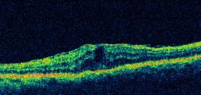
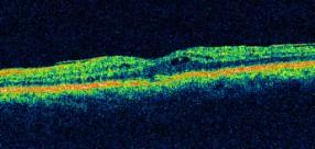
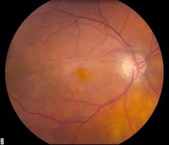
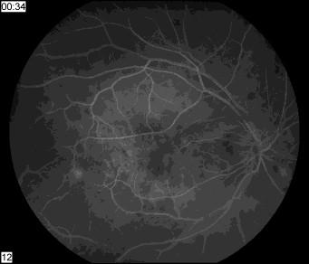
Optical Coherence Tomography in Diabetic Retinopathy. Mrs Samantha Mann Consultant Ophthalmologist Clinical Lead of SEL-DESP
 Optical Coherence Tomography in Diabetic Retinopathy Mrs Samantha Mann Consultant Ophthalmologist Clinical Lead of SEL-DESP Content OCT imaging Retinal layers OCT features in Diabetes Some NON DR features
OCT Interpretation in Retinal Disease
 OCT Interpretation in Retinal Disease Jay M. Haynie, OD, FAAO Financial Disclosure I have received honoraria or am on the advisory board for the following companies: Carl Zeiss Meditec Advanced Ocular
Ganglion cell analysis by optical coherence tomography (OCT) Jonathan A. Micieli, MD Valérie Biousse, MD
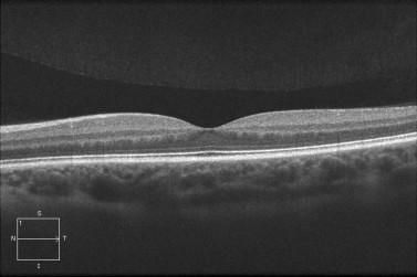
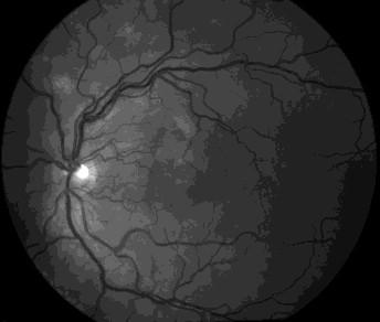
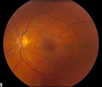
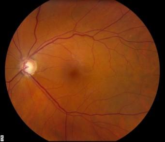
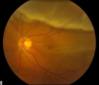
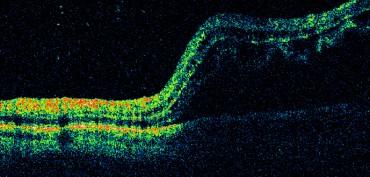
 Jonathan A. Micieli, MD Valérie Biousse, MD") Ganglion cell analysis by optical coherence tomography (OCT) Jonathan A. Micieli, MD Valérie Biousse, MD Figure 1. Normal OCT of the macula (cross section through the line indicated on the fundus photo)
Ganglion cell analysis by optical coherence tomography (OCT) Jonathan A. Micieli, MD Valérie Biousse, MD Figure 1. Normal OCT of the macula (cross section through the line indicated on the fundus photo)
OCT Interpretation. Financial Disclosure. Jay M. Haynie, OD, FAAO. OCT Image Layers 7/21/2014
 OCT Interpretation Jay M. Haynie, OD, FAAO Financial Disclosure I have received honoraria or am on the advisory board for the following companies: Olympia Tacoma Renton Kennewick - Washington Carl Zeiss
OCT Angiography The Next Frontier
 Choroid Retina avascular 5/13/2017 OCT Angiography The Next Frontier Pierce Kenworthy OD, FAAO June 9, 2017 OCT Angiography (OCTA) 2016 Non-invasive, motion contrast imaging Represents erythrocyte movement
OCT Image Analysis System for Grading and Diagnosis of Retinal Diseases and its Integration in i-hospital
 Progress Report for1 st Quarter, May-July 2017 OCT Image Analysis System for Grading and Diagnosis of Retinal Diseases and its Integration in i-hospital Milestone 1: Designing Annotation tool extraction
The Quick Guide to OCT Mastery 50 Real Cases with Expert Analysis
 OPTICAL COHERENCE TOMOGRAPHY The Quick Guide to OCT Mastery 50 Real Cases with Expert Analysis VOL 1 Sanjay Sharma, MD, FRCS, MSc (Epid), MBA Ophthalmologist, Epidemiologist Queen s University, Canada
Mark Dunbar: Disclosure
 Important Things to Understand About OCT Mark T. Dunbar, O.D., F.A.A.O. Bascom Palmer Eye Institute University of Miami, School of Medicine Mark Dunbar: Disclosure Optometry Advisory Board for: Allergan
Past ocular history. DME Case 1. Patient presents blurred VA. Hemoglobin A1c 11.5% -- patient states sugars have not been in good control
 Past ocular history Patient presents blurred VA DME Case 1 Hemoglobin A1c 11.5% -- patient states sugars have not been in good control PDR with macular edema OU Rishi Singh MD Cleveland Clinic OD OS 1
Course # Getting to Know Your OCT
 Course # 140 Getting to Know Your OCT Course Title: Lecturer: Getting to Know Your OCT Brad Sutton, OD, FAAO IU School of Optometry Financial Disclosures No financial disclosures Optical Coherence Tomography-OCT
Diagnosis in AMD. Managing your AMD Patients
 Managing your AMD Patients Robert W. Dunphy, O.D., F.A.A.O. Diagnosis in AMD Have suspicion Identify relative risk Conduct surveillance Biometry Utilize technology to facilitate detection of change / stability
Clinical Case Presentation. Branch Retinal Vein Occlusion. Sarita M. Registered Nurse Whangarei Base Hospital
 Clinical Case Presentation on Branch Retinal Vein Occlusion Sarita M. Registered Nurse Whangarei Base Hospital Introduction Case Study Pathogenesis Clinical Features Investigations Treatment Follow-up
Retinal Complications of Obstructive Sleep Apnea A Growing Concern!
 Retinal Complications of Obstructive Sleep Apnea A Growing Concern! Jay M. Haynie, OD, FAAO Financial Disclosure I have received honoraria or am on the advisory board for the following companies: Carl
Fundus Autofluorescence. Jonathan A. Micieli, MD Valérie Biousse, MD
 Fundus Autofluorescence Jonathan A. Micieli, MD Valérie Biousse, MD The retinal pigment epithelium (RPE) has many important functions including phagocytosis of the photoreceptor outer segments Cone Rod
When optical coherence tomography (OCT)
") Macular Imaging: SD-OCT in nterior Segment Surgical Practice Many pathologic processes of the macula can be visualized or quantified only with this modality. y Steven G. Safran, MD When optical coherence
Macular Imaging: SD-OCT in nterior Segment Surgical Practice Many pathologic processes of the macula can be visualized or quantified only with this modality. y Steven G. Safran, MD When optical coherence
Retina Conference. Janelle Fassbender, MD, PhD University of Louisville Department of Ophthalmology and Visual Sciences 09/04/2014
 Retina Conference Janelle Fassbender, MD, PhD University of Louisville Department of Ophthalmology and Visual Sciences 09/04/2014 Subjective CC/HPI: 64 year old Caucasian female referred by outside ophthalmologist
OPTIC DISC PIT Pathogenesis and Management OPTIC DISC PIT
 OPTIC DISC PIT Pathogenesis and Management Abdel-Latif Siam Ain Shams University Cairo Egypt OPTIC DISC PIT Congenital pit is an atypical coloboma usually located on the temporal edge of the disc, associated
Diagnosis and treatment of diabetic retinopathy. Blake Cooper MD Ophthalmologist Vitreoretinal Surgeon Retina Associates Kansas City
 Diagnosis and treatment of diabetic retinopathy Blake Cooper MD Ophthalmologist Vitreoretinal Surgeon Retina Associates Kansas City Disclosures Consulted for Novo Nordisk 2017,2018. Will be discussing
International Journal of Health Sciences and Research ISSN:
 International Journal of Health Sciences and Research www.ijhsr.org ISSN: 2249-9571 Original Research Article A Multivariate Analysis of Intravitreal Injection of Anti-VEGF Bevacizumab in the Treatment
Management of Neovascular AMD
 Kapusta AMD Part 1 Management of Neovascular AMD Dr. Michael A. Kapusta, MD, FRCSC Ophthalmologist in Chief Jewish General Hospital Vitreoretinal Surgeon 1 FINANCIAL DISCLOSURES Consulting honoraria Bayer,
EFFICACY OF ANTI-VASCULAR ENDOTHELIAL GROWTH FACTOR AGENTS IN RETINAL DISORDER FOR BETTER VISUAL ACUITY
 EFFICACY OF ANTI-VASCULAR ENDOTHELIAL GROWTH FACTOR AGENTS IN RETINAL DISORDER FOR BETTER VISUAL ACUITY Diwakar chaudhary *1, 2, Hu shuqiong, Long Yuan and Xiong kun 1 Yangtze University, 1 Nanhuan Road
Diabetic Macular Edema Treatment in the 21st Century
 Transcript Details This is a transcript of a continuing medical education (CME) activity accessible on the ReachMD network. Additional media formats for the activity and full activity details (including
ZEISS AngioPlex OCT Angiography. Clinical Case Reports
 Clinical Case Reports Proliferative Diabetic Retinopathy (PDR) Case Report 969 PROLIFERATIVE DIABETIC RETINOPATHY 1 1-year-old diabetic female presents for follow-up of proliferative diabetic retinopathy
Cirrus TM HD-OCT. Details defi ne your decisions
 Cirrus TM HD-OCT Details defi ne your decisions 2 With high-defi nition OCT Carl Zeiss Meditec takes you beyond standard spectral domain Built on 10 years experience at the vanguard of innovation, Carl
11/29/2016 MACULAR MALADIES: TYPICAL & ATYPICAL CASES
 MACULAR MALADIES: TYPICAL & ATYPICAL CASES Dawn Pewitt, OD, FAAO Triad Eye Institute, Grove, OK Dpewitt@triadeye.com Disclosure Statement: No financial disclosures COPE 51218-PS Please silence all mobile
Instudies of vascular endothelial growth factor
 MONITORING THERAPEUTIC EFFICACY IN THE ANTI-VEGF ERA* SriniVas R. Sadda, MD ABSTRACT One of the most important questions with vascular endothelial growth factor (VEGF) inhibitors for age-related macular
RETINAL CONDITIONS RETINAL CONDITIONS
 GENERAL INFORMATION RETINAL CONDITIONS RETINAL CONDITIONS WHAT ARE RETINAL CONDITIONS? Retinal conditions affect the light-sensitive tissue at the back of eye known as the retina. They include diseases
ZEISS AngioPlex OCT Angiography Making the revolutionary, routine.
 ZEISS AngioPlex OCT Angiography Making the revolutionary, routine. The moment that revolutionary insight becomes routine. // OCT ANGIOGRAPHY MADE BY ZEISS CIRRUS with AngioPlex creates a new era in both
Michael P. Blair, MD Retina Consultants, Ltd Libertyville/Des Plaines, Illinois Clinical Associate University of Chicago 17 October 2015
 Michael P. Blair, MD Retina Consultants, Ltd Libertyville/Des Plaines, Illinois Clinical Associate University of Chicago 17 October 2015 So What Parts of the Eye Retina are Affected by VHL Neural tissue
The College of Optometrists - Learning outcomes for the Professional Certificate in Medical Retina
 Learning outcomes for the Professional Certificate in Medical Retina, incorporating diabetic retinopathy screening and age related macular degeneration The professional certificate is a prerequisite to
evaluation of vitreoretinal adhesions in exudative AMD using optical coherence tomography
 evaluation of vitreoretinal adhesions in exudative AMD using optical coherence tomography Dr. Mahmoud Alaa Abouhusssein, FRCO Lecturer of ophthalmology, Alexandria university Dr. Amir Ramadan Gomaa, MD
Clinically Significant Macular Edema (CSME)
") Clinically Significant Macular Edema (CSME) 1 Clinically Significant Macular Edema (CSME) Sadrina T. Shaw OMT I Student July 26, 2014 Advisor: Dr. Uwaydat Clinically Significant Macular Edema (CSME) 2
Clinically Significant Macular Edema (CSME) 1 Clinically Significant Macular Edema (CSME) Sadrina T. Shaw OMT I Student July 26, 2014 Advisor: Dr. Uwaydat Clinically Significant Macular Edema (CSME) 2
Non-arteritic anterior ischemic optic neuropathy (NAION) with segmental optic disc edema. Jonathan A. Micieli, MD Valérie Biousse, MD
 with segmental optic disc edema. Jonathan A. Micieli, MD Valérie Biousse, MD") Non-arteritic anterior ischemic optic neuropathy (NAION) with segmental optic disc edema Jonathan A. Micieli, MD Valérie Biousse, MD A 75 year old white woman lost vision in the inferior part of her visual
Non-arteritic anterior ischemic optic neuropathy (NAION) with segmental optic disc edema Jonathan A. Micieli, MD Valérie Biousse, MD A 75 year old white woman lost vision in the inferior part of her visual
Intraocular Radiation Therapy for Age-Related Macular Degeneration
 Medical Policy Manual Medicine, Policy No. 134 Intraocular Radiation Therapy for Age-Related Macular Degeneration Next Review: April 2019 Last Review: June 2018 Effective: August 1, 2018 IMPORTANT REMINDER
Why Is Imaging Critical in My Uveitis Practice?
 Why Is Imaging Critical in My Uveitis Practice? Dilraj S. Grewal, MD Developed in collaboration Imaging Is the Backbone of Uveitis Workup and Monitoring Treatment Response FP FAF B- scan Multimodal Imaging
OCT Angiography. Financial Disclosures: Pre-Test: Which one is Correct?
 OCT Angiography Brandon Lujan, MD Medical Director, Casey Reading Center Assistant Professor of Ophthalmology Financial Disclosures: Genentech (Consultant, Grant support, Educational training) UC Berkeley
Optical Coherence Tomograpic Features in Idiopathic Retinitis, Vasculitis, Aneurysms and Neuroretinitis (IRVAN)
") Columbia International Publishing Journal of Ophthalmic Research (2014) Research Article Optical Coherence Tomograpic Features in Idiopathic Retinitis, Vasculitis, Aneurysms and Neuroretinitis (IRVAN)
Columbia International Publishing Journal of Ophthalmic Research (2014) Research Article Optical Coherence Tomograpic Features in Idiopathic Retinitis, Vasculitis, Aneurysms and Neuroretinitis (IRVAN)
Diabetic Retinopatathy
 Diabetic Retinopatathy Jay M. Haynie, OD, FAAO Financial Disclosure I have received honoraria or am on the advisory board for the following companies: Carl Zeiss Meditec Arctic DX Macula Risk Advanced
8/6/17. Disclosures Aerie Pharmaceuticals Alcon BioTissue Diopsys Optovue Shire
 Nathan Lighthizer, O.D., F.A.A.O. Associate Professor Assistant Dean for Clinical Care Director of Continuing Education Chief of Specialty Care Clinics Oklahoma College of Optometry Tahlequah, OK lighthiz@nsuok.edu
OCT Angiography in Primary Eye Care
 OCT Angiography in Primary Eye Care An Image Interpretation Primer Julie Rodman, OD, MS, FAAO and Nadia Waheed, MD, MPH Table of Contents Diabetic Retinopathy 3-6 Choroidal Neovascularization 7-9 Central
ACTIVATED OR NOT? RETINAL CASE PRESENTATION Shorye Payne, MD Medical Retinal Specialist Robley Rex VA Eye Clinic
 ACTIVATED OR NOT? RETINAL CASE PRESENTATION Shorye Payne, MD Medical Retinal Specialist Robley Rex VA Eye Clinic C We anticipate that the future management of posterior uveal melanoma (PUM) will focus
Measuring of the fovea and foveola using line scans and 3D Macular scans obtained with spectral domain optical coherent tomography.
 Measuring of the fovea and foveola using line scans and 3D Macular scans obtained with spectral domain optical coherent tomography. Vakhrameeva O.A. 1, Moiseenko G.A. 1, Maltsev D.S. 2, Sukhinin M.V. 2,
From Outdated to Updated: A Review of Important Clinical Trials in Ocular Disease from 2014
 From Outdated to Updated: A Review of Important Clinical Trials in Ocular Disease from 2014 1. This course is designed to review the important ophthalmic literature that was released between October 2013
Advances in OCT Murray Fingeret, OD
 Disclosures Advances in OCT Murray Fingeret, OD Consultant Alcon, Allergan, Bausch & Lomb, Carl Zeiss Meditec, Diopsys, Heidelberg Engineering, Reichert, Topcon Currently Approved OCT Devices OCT Devices
P. Stavrakas MD PhD. Consultant Ophthalmologist and vitreoretinal surgeon 2 nd Department of Ophthalmology University of Athens
 P. Stavrakas MD PhD Consultant Ophthalmologist and vitreoretinal surgeon 2 nd Department of Ophthalmology University of Athens Primary diagnose of exudative AMD Anti-VEGF monotherapy treatment Loss of
OCT Angiography: The Next Step in Retinal Imaging Jonathan Zelenak D.O.
 OCT Angiography: The Next Step in Retinal Imaging Jonathan Zelenak D.O. Hillsdale Hospital Michigan State University Overview Evolution of OCT How does OCT angiography work? Clinical examples Potential
R&M Solutions
 Mohamed Hosny El-Bradey, MD., Assistant Professor of Ophthalmology, Tanta University. Wael El Haig, MD., Professor of Ophthalmology. Zagazeeg University. 1 Myopic CNV is considered the most common vision
VMA at the macula resulting in VMT
 Ocriplasmina for pharmacologic treatment in VMT Teresio Avitabile 1 Introduction PVD is a normal, physiologic process that occurs with aging; however, in some cases, PVD is incomplete Incomplete PVD localized
Practical Care of the Cataract Patient with Retinal Disease
 Practical Care of the Cataract Patient with Retinal Disease Brooks R. Alldredge, OD, FAAO Kelly L. Cyr, OD, FAAO The Retina Center Eye Associates of New Mexico 4411 The 25 Way NE, Suite 325 Albuquerque,
3/6/2014. Hoda MH Mostafa MD Associate Professor of Ophthalmology Cairo University. The author has no proprietary interest. Today s Objectives
 Hoda MH Mostafa MD Associate Professor of Ophthalmology Cairo University The author has no proprietary interest Today s Objectives Identify the CLINICAL SCENARIOS IN MACULAR EDEMA where OCT plays a MAJOR
NIH Public Access Author Manuscript JAMA Ophthalmol. Author manuscript; available in PMC 2013 September 10.
 NIH Public Access Author Manuscript Published in final edited form as: JAMA Ophthalmol. 2013 May ; 131(5): 693 694. doi:10.1001/jamaophthalmol.2013.692. Effect of Intravitreous Anti Vascular Endothelial
Ophthalmic Imager Role
 MASTER OCT Ophthalmic Photographers Society October 18, 2014 Chicago, IL James B Soque, CRA COA Pamela A Weber, MD Island Retina Shirley, New York Commack, New York Financial Disclosure Genentech Ophthotech
Choroidal Mapping; a Novel Approach for Evaluating Choroidal Thickness and Volume
 Imaging Technique Choroidal Mapping; a Novel Approach for Evaluating Choroidal Thickness and Volume Jila Noori 1, MD; Mohammad Riazi Esfahani 1,2, MD Fedra Hajizadeh 2, MD; Mohammad-Mehdi Zaferani 1, MD
Incorporating OCT Angiography Into Patient Care
 Incorporating OCT Angiography Into Patient Care Beth A. Steele, OD, FAAO OCT A: Introduction Isolates microvascular circulation from OCT image data Axial resolution = 5 microns (i.e. fine capillaries visible)
Persistent Macular Thickening After Ranibizumab Treatment for Diabetic Macular Edema With Vision Impairment
 9:30 AM Persistent Macular Thickening After Ranibizumab Treatment for Diabetic Macular Edema With Vision Impairment Lee Jampol, MD OBJECTIVE To assess subsequent visual and anatomic outcomes of eyes with
Cirrus TM HD-OCT. Details define your decisions
 Cirrus TM HD-OCT Details define your decisions 2 With high-definition OCT Carl Zeiss Meditec takes you beyond standard spectral domain Built on 10 years experience at the vanguard of innovation, Carl Zeiss
The Foundation WHAT IS THE RETINA? continued next page. RETINA HEALTH SERIES Facts from the ASRS
 The Foundation American Society of Retina Specialists Committed to improving the quality of life of all people with retinal disease. Vitreomacular Traction Syndrome The vitreous humor is a transparent,
DOME SHAPED MACULOPATHY. Ιωάννης Ν. Βαγγελόπουλος Χειρ. Οφθαλμίατρος - Βόλος
 DOME SHAPED MACULOPATHY Ιωάννης Ν. Βαγγελόπουλος Χειρ. Οφθαλμίατρος - Βόλος DOME SHAPED MACULOPATHY-DEFINITIONS The entity Dome Shaped Macula ( DSM ) was first described by Gaucher and associates in 2008
Learn Connect Succeed. JCAHPO Regional Meetings 2017
 Learn Connect Succeed JCAHPO Regional Meetings 2017 How Retinal Imaging Guides Treatment Odette Margit Houghton MD Question 1 Which OCT has the highest resolution? A: Swept source OCT B: Spectral domain
Diabetic Retinopathy: Managing the Extremes. J. Michael Jumper, MD West Coast Retina
 Diabetic Retinopathy: Managing the Extremes J. Michael Jumper, MD West Coast Retina Case 1: EC 65 y.o. HM No vision complaints Meds: Glyburide Metformin Pioglitazone Va: 20/20 OU 20/20 Case 2: HS 68 y.o.
Clinical results of OPT-302 (VEGF-C/D Trap ) Combination Treatment in namd and DME
 Combination Treatment in namd and DME") Clinical results of OPT-302 (VEGF-C/D Trap ) Combination Treatment in namd and DME Ophthalmology Innovation Summit @ AAO, October 25 2018 Megan Baldwin PhD, CEO & Managing Director Disclaimer Investment
Clinical results of OPT-302 (VEGF-C/D Trap ) Combination Treatment in namd and DME Ophthalmology Innovation Summit @ AAO, October 25 2018 Megan Baldwin PhD, CEO & Managing Director Disclaimer Investment
HHS Public Access Author manuscript Ophthalmic Surg Lasers Imaging Retina. Author manuscript; available in PMC 2016 January 14.
 High-Speed Ultrahigh-Resolution OCT of Bruch s Membrane in Membranoproliferative Glomerulonephritis Type 2 Mehreen Adhi, MD, Sarah P. Read, MD, PhD, Jonathan J. Liu, PhD, James G. Fujimoto, PhD, and Jay
A Comparative Study of Age Related Macular Degeneration In Relation To SD-OCTand Fundus Photography.
 IOSR Journal of Dental and Medical Sciences (IOSR-JDMS) e-issn: 2279-0853, p-issn: 2279-0861.Volume 14, Issue 11 Ver. III (Nov. 2015), PP 33-37 www.iosrjournals.org A Comparative Study of Age Related Macular
Citation BioMed Research International, 2015, v. 2015, article no Creative Commons: Attribution 3.0 Hong Kong License
 Title Relationship between Outer Retinal Layers Thickness and Visual Acuity in Diabetic Macular Edema Author(s) Wong, RLM; Lee, JWY; Yau, GSK; Wong, IYH Citation BioMed Research International, 2015, v.
Clinical results of OPT-302 (VEGF-C/D Trap ) Combination Treatment in namd and DME
 Combination Treatment in namd and DME") Clinical results of OPT-302 (VEGF-C/D Trap ) Combination Treatment in namd and DME Ophthalmology Innovation Summit @ AAO, October 25 2018 Megan Baldwin PhD, CEO & Managing Director Disclaimer Investment
Clinical results of OPT-302 (VEGF-C/D Trap ) Combination Treatment in namd and DME Ophthalmology Innovation Summit @ AAO, October 25 2018 Megan Baldwin PhD, CEO & Managing Director Disclaimer Investment
The Foundation WHAT IS THE RETINA?
 Age-Related Macular Degeneration (AMD) is a deterioration of the retina and choroid that leads to a substantial loss in visual acuity (sharpness of vision). AMD is the leading cause of significant visual
Diabetic Retinopathy
 Diabetic Retinopathy Diabetes can be classified into type 1 diabetes mellitus and type 2 diabetes mellitus, formerly known as insulin-dependent diabetes mellitus, and non-insulin diabetes mellitus, respectively.
You can C-ME after Uveitis
 You can C-ME after Uveitis Abstract: Approximately 50% of uveitis patients will present with vision loss secondary to cystoid macular edema[1]. Two patients with uveitis present with a constant decrease
Case Report Increase in Central Retinal Edema after Subthreshold Diode Micropulse Laser Treatment of Chronic Central Serous Chorioretinopathy
 Case Reports in Ophthalmological Medicine Volume 2015, Article ID 813414, 4 pages http://dx.doi.org/10.1155/2015/813414 Case Report Increase in Central Retinal Edema after Subthreshold Diode Micropulse
Overview. Macular OCT Artifact Study
 Imaging Artifacts Sarah Moyer, CRA, OCT-C Director, Ophthalmic Imaging Kittner Eye Center University of North Carolina Chapel Hill, NC Disclose financial interest now Overview Sarah s Thoughts on Artifacts
Diabetic Retinopathy Clinical Research Network
 Diabetic Retinopathy Clinical Research Network Comparison of Time Domain OCT and Spectral Domain OCT Retinal Thickness Measurement in Diabetic Macular Edema Version 1.0 June 16, 2009 comparison of td vs
University Hospital Basel. Optical Coherence Tomography Emerging Role in the Assessment of MS PD Dr. Konstantin Gugleta
 University Hospital Basel Optical Coherence Tomography Emerging Role in the Assessment of MS PD Dr. Konstantin Gugleta 15th State of the Art SMSS, Lucerne January 2013 Retinal Nerve Fiber Layer 1.200.000
Ophthalmology Macular Pathways
 Ophthalmology Macular Pathways Age related Macular Degeneration Diabetic Macular Oedema Macular Oedema secondary to Central Retinal Macular Oedema secondary to Branch Retinal CNV associated with pathological
Moving forward with a different perspective
 Moving forward with a different perspective The Leader In Vision Diagnostics Offers A New Perspective Marco has served the eyecare community by offering exceptional lane products and automated high tech
Citation. As Published Publisher. Version
 Effect of Intravitreous Anti Vascular Endothelial Growth Factor Therapy on Choroidal Thickness in Neovascular Age-Related Macular Degeneration Using Spectral-Domain The MIT Faculty has made this article
Macular Morphology and Visual Acuity in the Comparison of Age-related Macular Degeneration Treatments Trials
 Macular Morphology and Visual Acuity in the Comparison of Age-related Macular Degeneration Treatments Trials Glenn J. Jaffe, MD, 1 Daniel F. Martin, MD, 2 Cynthia A. Toth, MD, 1 Ebenezer Daniel, MPH, PhD,
References 1. Melberg NS, Thomas MA AJO 120: , Welch JC AJO 124: 698, Hirata A, Yonemura N, et al. AJO 130:611, 2000.
 Central or Paracentral Scotoma Associated with Nasal Placement of Chandelier Infusion During Vitrectomy with Fluid-Air Exchange J. Michael Jumper MD, Sara J. Haug MD PhD, Arthur D. Fu MD, Robert N. Johnson
Spectral-domain Optical Coherence Tomography Imaging of Age-related Macular Degeneration
 Imaging Spectral-domain Optical Coherence Tomography Imaging of Age-related Macular egeneration Carlos Alexandre de Amorim Garcia Filho, 1 Philip J Rosenfeld, 2 Zohar Yehoshua 3 and Giovanni Gregori 3
Retina Grandrounds. Retinal Grand Rounds Exhibition of Common Entities
 Retina Grandrounds Retinal Grand Rounds Exhibition of Common Entities Mohammad R Rafieetary, O.D. mrafieetary@charlesrteina.com Exhibitions 1) Choroidal Neovascular Membrane 2) Vitreomacular Interface
NATIONAL INSTITUTE FOR HEALTH AND CARE EXCELLENCE. Health Technology Appraisal. Aflibercept for treating diabetic macular oedema.
 NATIONAL INSTITUTE FOR HEALTH AND CARE EXCELLENCE Health Technology Appraisal Aflibercept for treating diabetic macular oedema Final scope Final remit/appraisal objective To appraise the clinical and cost
Anti VEGF Agents in Retinal Disorders Current Scenario
 Retina Anti VEGF Agents in Retinal Disorders Current Scenario Charu Gupta MS Charu Gupta MS, Cyrus M. Shroff MD Shroff Eye Centre, New Delhi T is a group of proteins involved in the regulation of angiogenesis,
The Human Eye. Cornea Iris. Pupil. Lens. Retina
 The Retina Thin layer of light-sensitive tissue at the back of the eye (the film of the camera). Light rays are focused on the retina then transmitted to the brain. The macula is the very small area in
What Is O.C.T. and Why Should I Give A Rip? OCT & Me How Optical Coherence Tomography Changed the Life of a Small Town Optometrist 5/19/2014
 OCT & Me How Optical Coherence Tomography Changed the Life of a Small Town Optometrist Email: myoder@wcoil.com Mark A. Yoder, O.D. 107 N. Main Street PO Box 123 Bluffton, OH 45817 @yoderod 115.02 Histoplasma
Posterior Segment Update
 Posterior Segment Update Featured Speaker: Dr. Kyle Cheatham, FAAO, DIP ABO DISCLOSURE STATEMENT We have no direct financial or proprietary interest in any companies, products or services mentioned in
Is OCT-A Needed As An Investigative Tool During The Management Of Diabetic Macular Edema
 Is OCT-A Needed As An Investigative Tool During The Management Of Diabetic Macular Edema Ayman M Khattab MD, FRCS Professor of Ophthalmology Cairo University Diabetic Macular Edema (DME) Diabetic macular
History/principles of the OCT What does the normal retinal OCT look like Vitreal disorders Retinal/RPE disorders Choroidal disorders
 Nathan Lighthizer, O.D., F.A.A.O. Assistant Professor Assistant Dean for Clinical Care Director of Continuing Education Chief of Specialty Care Clinics Chief of Electrodiagnostics Clinic Oklahoma College
Opthea Initiates OPT-302 Diabetic Macular Edema Clinical Trial
 ASX and Media Release 3 January 2018 Opthea Initiates OPT-302 Diabetic Macular Edema Clinical Trial Melbourne, Australia; January 3 2018 Opthea Limited (ASX:OPT), a late stage biopharmaceutical company
Retinal Diseases. Age-Related Macular Degeneration. What Is AMD? Risk Factors for AMD
 Santosh C. Patel, M.D. Retina Specialists The Latest And Greatest in the Management of Retinal Diseases February 13, 2007 Retinal Diseases Majority of Blindness in Civilized World AMD Diabetic Retinopathy
Do You See What I See!!! Shane R. Kannarr, OD
 Do You See What I See!!! Shane R. Kannarr, OD skannarr@kannarreyecare.com Define Specialty Testing Additional Test to: Prove/Disprove Diagnosis To monitor progression of a condition To document a condition
Disclosures. Definitions. Goals. Imaging and glaucoma 3/22/2016
 Pinakin Davey OD, PhD, FAAO Professor and Director of Research Disclosures Principal investigator for ivue OCT trial Principal investigator Topcon FDA trials for Maestro and OCT 2000 Consultant for Topcon
Anti-VEGF drugs & CATT Results. in AMD. Apollo Hospitals, Hyderabad. Mallika Goyal, MD
 Anti-VEGF drugs & CATT Results in AMD Mallika Goyal, MD Apollo Hospitals, Hyderabad Vascular Endothelial Growth Factor Napoleone Ferrara at Genentech 1989 identified & cloned the gene Protein that causes
Treatment for Central-Involved Diabetic Macular Edema in Eyes with Very Good Visual Acuity
 Diabetic Retinopathy Clinical Research Network Treatment for Central-Involved Diabetic Macular Edema in Eyes with Very Good Visual Acuity Version 3.0 April 18, 2014 Treatment of CIDME in Eyes with Good
Mariam Raouf Fadel M.B., B.Ch. M.Sc., Cairo University. A thesis. Submitted by. For partial fulfillment of. MD Degree in Ophthalmology
 Correlation of fundus autofluorescence and spectral domain OCT findings of the macula with visual outcome after successful repair of rhegmatogenous retinal detachment A thesis Submitted by Mariam Raouf
The Wilmer Eye Institute s 34 th Annual Current Concepts in Ophthalmology March 13-17, 2017 Vail Marriott * Vail, Colorado
 The Wilmer Eye Institute s 34 th Annual Current Concepts in Ophthalmology March 13-17, 2017 Vail Marriott * Vail, Colorado Tentative 1/23/17 PROGRAM MONDAY, MARCH 13, 2017 Morning Session 6:00-7:00 Registration
The Use of Intravitreal Aflibercept in the Treatment of Wet Type of Age Related Macular Degeneration
 BMH Medical Journal 2015;2(2):31-36 Review Article The Use of Intravitreal Aflibercept in the Treatment of Wet Type of Age Related Macular Degeneration Rejith Rag, MBBS, FRCS, DO, FICO Department of Ophthalmology,
The MP-1 Microperimeter Clinical Applications in Retinal Pathologies
 The MP-1 Microperimeter Clinical Applications in Retinal Pathologies Nelson R. Sabates, MD Director, Retina/Vitreous Service Vice-Chairman Department of Ophthalmology University of Missouri Kansas City
Use of Aflibercept in cases of recurrent or refractory neovascular AMD previously treated with other antiangiogenic drug: anatomo-functional outcomes
 Use of Aflibercept in cases of recurrent or refractory neovascular AMD previously treated with other antiangiogenic drug: anatomo-functional outcomes Daniel E. Charles, MD, Martin Charles, MD, Gisela Jelusich,
Vitreomacular interface disorders. Ghanbari MD 1393:10:25
 Vitreomacular interface disorders Ghanbari MD 1393:10:25 Human vitreous after dissection of the sclera, choroid, and retina. Lamellar structure of the posterior vitreous cortex (PVC) in the monkey. V =
Retinal Vein Occlusion (RVO) Treatment pathway- Northeast England. Retinal Vein Occlusion (RVO) with Macular oedema (MO)
 Treatment pathway- Northeast England. Retinal Vein Occlusion (RVO) with Macular oedema (MO)") Retinal Vein Occlusion (RVO) Treatment pathway- Northeast England (Royal Victoria Infirmary, Sunderland Eye Infirmary, James Cook University Hospital, Darlington Memorial Hospital, University Hospital
Retinal Vein Occlusion (RVO) Treatment pathway- Northeast England (Royal Victoria Infirmary, Sunderland Eye Infirmary, James Cook University Hospital, Darlington Memorial Hospital, University Hospital