Author Manuscript Faculty of Biology and Medicine Publication
|
|
|
- Beryl Houston
- 5 years ago
- Views:
Transcription

1 Serveur Académique Lausannois SERVAL serval.unil.ch Author Manuscript Faculty of Biology and Medicine Publication This paper has been peer-reviewed but dos not include the final publisher proof-corrections or journal pagination. Published in final edited form as: Title: Ectodysplasin A (EDA) - EDA receptor signalling and its pharmacological modulation. Authors: Kowalczyk-Quintas C 1, Schneider P 2.Journal: Cytokine & growth factor reviews Year: 2014 Volume: 25(2) Pages: DOI: /j.cytogfr In the absence of a copyright statement, users should assume that standard copyright protection applies, unless the article contains an explicit statement to the contrary. In case of doubt, contact the journal publisher to verify the copyright status of an article.
2 Kowalczyk and Schneider Modulation of EDA-EDAR signalling Page 1 ECTODYSPLASIN A (EDA) EDA RECEPTOR SIGNALLING AND ITS PHARMACOLOGICAL MODULATION. Christine Kowalczyk-Quintas 1 and Pascal Schneider 1 1 Department of Biochemistry, University of Lausanne, CH-1066 Epalinges, Switzerland. Running title: Modulation of EDA-EDAR signalling. Address correspondence to: Pascal Schneider, Department of Biochemistry, University of Lausanne, Boveresses 155, CH-1066 Epalinges, Switzerland; Phone: ; Fax: ; pascal.schneider@unil.ch. Keywords: ectodysplasin, ectodermal dysplasia, embryo, hair, tooth, TNF family Abstract: The TNF family ligand Ectodysplasin A (EDA) regulates the induction, morphogenesis and/or maintenance of skin-derived structures such as teeth, hair, sweat glands and several other glands. Deficiencies in the EDA - EDA receptor (EDAR) signalling pathway cause hypohidrotic ectodermal dysplasia (HED). This syndrome is characterized by the absence or malformation of several skin-derived appendages resulting in hypotrychosis, hypodontia, heat-intolerance, dry skin and dry eyes, susceptibility to airways infections and crusting of various secretions. The EDA-EDAR system is an important effector of canonical Wnt signalling in developing skin appendages. It functions by stimulating NF-κB-mediated transcription of effectors or inhibitors of the Wnt, Sonic hedgehog (SHH), Fibroblast growth factor (FGF) and Transforming growth factor beta (TGFβ) pathways that regulate interactions within or between epithelial and mesenchymal cells and tissues. In animal models of Eda-deficiency, soluble EDAR agonists can precisely correct clinically relevant symptoms with low side effects even at high agonist doses, indicating that efficient negative feedback signals occur in treated tissues. Hijacking of the placental antibody transport system can help deliver active molecules to developing foetuses in a timely manner. EDAR agonists may serve to treat certain forms of ectodermal dysplasia.
3 Kowalczyk and Schneider Modulation of EDA-EDAR signalling Page 2 Ectodysplasin A and ectodermal dysplasias Ectodermal dysplasias comprise more than 180 different heritable syndromes affecting at least two ectodermal structures such as hair, teeth, nails or exocrine glands (Visinoni et al., 2009). The most common ectodermal dysplasia, X-linked hypohidrotic ectodermal dysplasia (XLHED), is characterized by missing or sparse hair (hypotrychosis), abnormal or missing teeth (hypodontia or anodontia), reduced sweating ability (hypohidrosis), and defects of various lipid- or mucussecreting glands. XLHED is caused by mutations of the ectodysplasin A (EDA) gene (Kere et al., 1996). EDA mutations are also associated with selective tooth agenesis (STA), an inherited condition in which teeth only are affected, with no pathologic involvement of other ectodermal appendages (Mues et al., 2010). EDA is a TNF family ligand that binds to EDA receptor (EDAR). EDAR signals via an adaptor protein, EDAR-associated protein with a death domain (EDARADD). Mutations in the EDAR or EDARADD genes cause hypohidrotic ectodermal dysplasias identical to XLHED, except for their autosomal dominant or recessive modes of transmission (Headon et al., 2001; Headon and Overbeek, 1999). Ectodysplasin A The human EDA gene was first identified by positional cloning in XLHED patients and is located on the long arm of the X chromosome (Kere et al., 1996). EDA is a 391 amino acid residues-long membrane protein with a short intracellular domain, a transmembrane domain, a stalk region of uncharacterized function, a consensus furin cleavage sequence responsible for proteolytic processing of EDA, a short positively-charged sequence required for interactions with heparan-sulfate proteoglycans, a bi-partite collagen-like domain and a 150 amino acid residueslong C-terminal TNF homology domain (THD) responsible for receptor binding (Fig. 1A). EDA transcripts undergo complicated splicing events giving rise to numerous EDA isoforms (up to nine in mouse keratinocytes), of which only the longest two, EDA1 and EDA2, contain the THD, interact with receptors and are known to be biologically active (Bayes et al., 1998; Hashimoto et al., 2006). EDA1 and EDA2 differ by two amino acid residues in the THD (Glu308 and Val309) as a result of differential usage of a splice donor site at the end of exon 7 (Fig. 1A) (Yan et al., 2000). EDA1 binds to EDAR, while the 2 amino acid residues shorter EDA2 specifically interacts with another TNF receptor family member, XEDAR (Yan et al., 2000). EDA1, EDA2 and their receptor binding specificity are conserved in mammals and birds (Kowalczyk-Quintas et al., 2014). XEDAR is a p53-induced gene with no obvious implications in ectodermal appendage development (Brosh et al., 2010; Newton et al., 2004). EDA mutations identified in XLHED patients touch several distinct regions of the protein that are therefore believed to be of functional importance (Schneider et al., 2001) (Fig. 1A). The first one lies at the beginning of the stalk region, but why this region is important for EDA expression or function is unknown. Interestingly, fishes that have evolved in fresh water display a dramatic reduction in their bony armour compared to ocean fishes of the same species, which is due to the selection of a lowabundance allele of Eda that includes, among other polymorphisms, a Leu79Pro transition at the beginning of the stalk region (Colosimo et al., 2005). The second region is the furin consensus cleavage site, indicating that EDA1 must be released to a soluble form to display activity (Fig. 1A, B). The third region is the THD. The THD forms homotrimers that can bind three individual receptors at each monomer-monomer interface (Hymowitz et al., 2003). XLHED- and STAcausing mutations in this domain interfere either with trimer formation, or with receptor binding (Mues et al., 2010; Schneider et al., 2001). The fourth region is the collagen domain that serves to keep two or more EDA1 trimers in close proximity within the same molecule and therefore
4 Kowalczyk and Schneider Modulation of EDA-EDAR signalling Page 3 potentiate its ability to stimulate EDAR signalling (Schneider et al., 2001; Swee et al., 2009) (Fig. 1A,B). Finally, the proteoglycan-binding domain of EDA, which is mostly coded by the very short exon 3, may restrict EDA diffusion in tissues once it is released in a soluble form (Swee et al., 2009) (Fig. 1B). Mutations in the proteoglycan-binding region have not been reported in XLHED patients, either because this interaction is not essential for the function of EDA, or because disruption of the interaction can be achieved neither with a single point mutation nor with exon 3 deletion, which would induce a frame shift. EDAR EDAR is a typical TNF receptor family member with a signal peptide, three cystein-rich domains (CRDs), a transmembrane domain and an intracellular region comprising a so-called death domain (Headon and Overbeek, 1999) (Fig. 1A). Most mutations in patients with autosomal HED are located within CRD2, which is involved in ligand binding, or within the death domain, which is involved in signal transmission (Fig. 1A). A function activating polymorphism (Val370Ala) found in the death domain of EDAR in East Asian and Native American populations is associated with a thicker hair phenotype (Mou et al., 2008; Sabeti et al., 2007). In the death receptors Fas and TRAILR2, the death domain recruits Fas-associated protein with a death domain (FADD), that itself recruits and activates cystein proteases of the caspase family to execute apoptotic cell death. EDAR does not interact with FADD, but instead recruits EDARADD, another adaptor protein with a death domain (Headon et al., 2001) (Fig. 1A). EDAR activates canonical NF-kB Deficiencies for EDA, EDAR or for the adaptor protein EDARADD all induce HED with similar if not identical manifestations. EDARADD, unlike FADD, does not recruit caspases, but possesses consensus binding sites for TNF receptor associated factors (TRAFs), and in particular TRAF6 (Fujikawa et al., 2013; Headon et al., 2001). TRAF6 mediates the activation of the transcription factor NF-κB downstream of several TNF receptor family members (e.g. EDAR, RANK and CD40) or other receptors (IL1R, TLRs). Thus, Traf6-deficient mice do not only develop ectodermal dysplasia similar to Edar- or Edaradd-deficient mice, but also osteopetrosis, alymphoplasia -RANK signalling is essential for the generation of osteoclasts and lymph nodes-, B cell abnormalities -CD40 is important for B cell activation- and blunted signalling by IL1 receptor and Toll-like receptors (Naito et al., 2002). TRAF6 is an E3 ubiquitin ligase that generates K63-linked polyubiquitin chains to which binds NEMO, the regulatory component of the inhibitor of NF-κB kinase (IKK) complex (Chen, 2012). NEMO is on the X chromosome. Although complete NEMO-deficiency causes a severe and lethal inflammatory skin phenotype in human males, hypomorphic NEMO mutations are viable and cause HED with immunodeficiency, sometimes complicated by osteopetrosis for reasons similar to those explained above for Traf6-deficiency (Smahi et al., 2002; Zonana et al., 2000). The crucial implication of NF-κB downstream of EDAR is demonstrated by the phenotype of IκBαΔN transgenic mice that express a non-phosphorylable, and therefore non-degradable form of a NFκB inhibitor. These mice suffer from HED, osteopetrosis and immunodeficiency (Schmidt- Ullrich et al., 2001). In embryonic skin of wild type mice, NF-κB is activated in primary hair placodes (Schmidt-Ullrich et al., 2006). In Eda-deficient skin, there are no placodes and there is no NF-κB activation. Addition of ectopic recombinant EDA restores placodes and NF-kB signals in Eda-deficient skin, but not in IκBαΔN transgenic skin (Schmidt-Ullrich et al., 2006). At least for the formation of primary hair placodes, NF-κB activation downstream of EDAR is not only
5 Kowalczyk and Schneider Modulation of EDA-EDAR signalling Page 4 required, but apparently also sufficient as placodes can be formed with other NF-kB stimulators such as TNF or LPS (Schmidt-Ullrich et al., 2006) (Table 1) (Fig. 2). Table 1 is a compilation of phenotypes observed upon genetic modifications of genes related to processes also regulated by EDAR-signalling. Figure 2 is a partial, naïve and certainly incorrect view of how these gene products may interact with each other in the early phases of hair and tooth development. The EDA - EDAR pathway is an important effector of Wnt signalling, and in turn promotes further Wnt signalling Primary hairs do not form in Edar-deficient or in Wnt signalling-deficient (deletion of β-catenin) mice (Headon and Overbeek, 1999; Huelsken et al., 2001). Early studies in conditional β-catenin knock-out mice, where β-catenin was deleted in epithelial cells with a Keratin14 promoterdriven Cre recombinase, concluded that Wnt signalling might be downstream of EDAR, as Edar was the only placode marker still expressed in β-catenin-deficient E15.5 skin (Huelsken et al., 2001). Also, Edar expression was unaffected in Lef-1-null skin and tooth buds (Laurikkala et al., 2001; Laurikkala et al., 2002). This conclusion that Edar was not a Wnt-responsive gene was subsequently revised when β-catenin was deleted with another Keratin14 promoter-driven Cre recombinase with more precocious expression. Indeed, Edar expression and NF-κB activation in E14.5 skin was absolutely dependent on β-catenin activity in the epithelium, and Edar was characterized as a direct Wnt target gene (Zhang et al., 2009). Likewise, initial uniform expression of Eda is also Wnt-dependent (Durmowicz et al., 2002; Laurikkala et al., 2001; Zhang et al., 2009). Although Wnt signalling acts upstream of EDAR in primary hair placodes, maintenance of β-catenin activity in primary placodes at a later stage requires EDAR activity (Zhang et al., 2009). EDAR signalling induces expression of Wnt10b, a direct NF-κB target gene, which is a good candidate to mediate the second round of β-catenin signalling in placodes (Zhang et al., 2009). Interestingly, forced EDAR-signalling (by expression of a constitutively active LMP-EDAR fusion receptor) in primary hair placodes in the context of Wnt inhibition (by expression of the Wnt inhibitor DKK1) failed to rescue expression of EDAR target genes such as Shh, indicating that expression of Wnt-dependent genes other than Eda and Edar is required for successful EDAR signalling (Zhang et al., 2009) (Fig. 2). Development of skin-derived appendages Skin-derived appendages, such as hair, teeth, and mammary glands have different function and structure but share common developmental aspects. The first step of development is the formation of a thickening of the epithelial layer, the placodes, accompanied by the condensation of the underlying mesenchyme. Placodes are embryonic signalling centres that express signalling molecule such as Wnt, FGF, TGFβ, Hedgehog and TNF family proteins (Laurikkala et al., 2002; Mikkola, 2007). In the next step of development, placodes invaginate into the mesenchyme to form buds that further grow and develop in organ-specific manners. Interactions between the epidermal and underlying dermal cells and tissues are common and recurrent themes of skinderived organ development. Development of hair In mice, hair development occurs in three distinct and successive waves giving rise, respectively, to guard, awl and auchene, and zigzag hair types. The first wave of hair placodes is visible at E13.5, and the second and third are initiated at E16 and E18 (Schmidt-Ullrich and Paus, 2005). The placode is formed by a clustering of epidermal keratinocytes in response to a signal from the
6 Kowalczyk and Schneider Modulation of EDA-EDAR signalling Page 5 dermis that could be Wnt or a Wnt inducer. Placodes express inducing molecules to re-enforce signalling, and inhibitors to prevent differentiation at the periphery of placodes until a regular array of hair follicles is established (Millar, 2002; Schmidt-Ullrich and Paus, 2005). Each placode induces underlying mesenchymal cells to aggregate and form the dermal condensate (Millar, 2002). This is followed by proliferation and down growth of epithelial cells into the dermis until they surround the dermal condensate that becomes the dermal papilla. Lateral communication between the dermal papilla and the follicular epithelium induces proliferation of epithelial matrix cells that give rise to different hair follicle lineages forming first the different cell types of the inner root sheath (Henley s layer, Huxley s layer and cuticle) and then those of the mature hair shaft (cuticle, medulla and cortex) (Millar et al., 1999). The inner root sheath is surrounded by the companion cell layer and by the outer root sheath that is contiguous and similar to the epidermal basal layer. Development of teeth In teeth, the odontogenic signal originates form the epithelium at E11. At E12, placode cells proliferate into the underlying mesenchyme to form a bud. At E13.5 the mesenchyme condenses around the bud, expresses signalling molecules and transcription factors and instructs the epithelium to form the primary enamel knot at the tip of the bud. The primary enamel knot is a non-proliferating signalling centre important to stimulate cell proliferation in surrounding tissues. The primary enamel knot thus regulates folding of the tooth epithelium in a cap-shaped structure at E14.5 (Dassule et al., 2000; Jernvall and Thesleff, 2000). At the bell stage (E16), the condensed mesenchyme -now called the dental papilla- is completely enclosed by the invaginating tooth epithelium. Enamel knots are transient structures that disappear by apoptosis at the end of the cap stage (Jernvall and Thesleff, 2000). Incisor tooth germs have a single enamel knot, whereas molars additionally form secondary enamel knots at the bell stage to fold the enamel epithelium again and form multi-cuspids teeth. The final tooth shape is defined by the folding of the inner enamel epithelium and by the growth of the dental papilla (Caton and Tucker, 2009). Wnt and Wnt inhibitors Wnt/β-catenin signalling is crucial for the development of ectodermal appendages at early and later stages of differentiation (Andl et al., 2002; Huelsken et al., 2001; Liu et al., 2008; Millar, 2002; Schmidt-Ullrich and Paus, 2005; van Genderen et al., 1994). The canonical Wnt pathway requires β-catenin. When Wnt is absent, β-catenin is eliminated by the action of the AXIN complex, which triggers β-catenin to proteasomal degradation. When Wnt is present, it binds to Frizzled receptor and to LRP5/6 co-receptors, which recruit Dishevelled (DVL), resulting in LRP5/6 phosphorylation and recruitment of the AXIN complex. This inhibits β-catenin degradation. Accumulated β-catenin translocates to the nucleus where, in complex with T cell factor/lymphoid enhancer factor (LEF/TCF), it activates expression of Wnt target gene (MacDonald et al., 2009). Negative regulators of Wnt signalling include DKKs (Andl et al., 2002) and Sostdc1 (also known as Ectodin or Wise) (Ahn et al., 2010; Narhi et al., 2012). Both negatively regulate LRP5/6 co-receptors and therefore Wnt signalling, either directly (Sostdc1) (Itasaki et al., 2003; Li et al., 2005; Lintern et al., 2009) or additionally via engagement of Kremens (DKKs) (Wang et al., 2008) (Fig.2). Dkks are Wnt- and NF-κB-responsive (Andl et al., 2002; Fliniaux et al., 2008; Zhang et al., 2009), whereas Sostdc1 is activated by BMPR signalling (Laurikkala et al., 2003; Mou et al., 2006). Sostdc1 is also a BMP antagonist
7 Kowalczyk and Schneider Modulation of EDA-EDAR signalling Page 6 (Laurikkala et al., 2003), but the phenotype of Sostdc1-deficient mice that is characterized by enlarged enamel knots, additional teeth, enlarged mammary buds, enlarged hair placodes and ectopic whiskers (Ahn et al., 2010; Kassai et al., 2005; Narhi et al., 2012), is probably due to an upregulation of the Wnt signalling pathway (Ahn et al., 2010; Narhi et al., 2012). Endogenous expression of Dkk1 in the dermal condensate is suppressed by DKK1 overexpression, suggesting that it is a direct target of Wnt signalling involved in a negative feedback loop (Andl et al., 2002). TGFβ family members BMP, Activin, and their inhibitors BMPs are known as placode inhibitors (Botchkarev et al., 1999), but also provide positive signals for the differentiation of inner root sheath cells (Kobielak et al., 2003), generation and decay of enamel knots (Jernvall et al., 1998) or differentiation of ameloblasts (Wang et al., 2004). The action of BMP is restricted by expression of a range of antagonists: Sostdc1 (Laurikkala et al., 2003), CTGF (also known as CCN2) (Mou et al., 2006; Pummila et al., 2007), Noggin (Botchkarev et al., 1999) and Follistatin (Wang et al., 2004). Cell responses to BMP are often regulated by gradients. In the hair follicle, Bmpr1a ablation prevents differentiation of matrix cells to inner root sheath cells, and therefore prevents hair shaft formation (Andl et al., 2004; Kobielak et al., 2003). A model for the differentiation of matrix cells to inner root sheath cells takes into account the seemingly contradictory observations that BMPR1a signalling inhibits Lef-1 expression, yet is required for β-catenin/lef-1 activation (Kobielak et al., 2003). In the hair follicle, the dermal papilla produces Noggin that inhibits BMP signalling in the nearby proliferating epithelial matrix cells. Inhibition of BMP signalling induces Lef1 and Bmp4 expression in matrix cells, but β-catenin/lef-1 activation does not take place in the absence of BMPR1a signalling, possibly due to down-regulated expression of Wnt receptors or to an other cause. Absence of Wnt signalling thus prevents differentiation of proliferating matrix cells. As proliferating cells move upwards and away from the source of Noggin, they start responding to their own BMP4 to become Wnt-responsive. Despite Lef-1 downregulation, stabilized LEF-1 protein is sufficient to ensure efficient β-catenin/lef-1 activation and expression of Wntresponsive genes (e.g. hair keratins, Foxn1, Msx1, Msx2 and GATA3) involved in the differentiation of matrix cells to inner root sheath cells, which themselves will give rise to hair shaft cells (Kobielak et al., 2003). ActivinβA is a TGFβ family member involved in tooth and whiskers formation (Ferguson et al., 1998; Jhaveri et al., 1998; Matzuk et al., 1995). ActivinβA, its receptor ACTRII and its effector SMAD2 are all essential for the development of incisors and mandibular molars (Ferguson et al., 1998; Ferguson et al., 2001). Follistatin inhibits ActivinβA (Jhaveri et al., 1998; Matzuk et al., 1995; Wang et al., 2004) (Fig. 2). Sonic hedgehog Sonic hedgehog (SHH), a member of the Hedgehog family, is a secreted protein important for organizing dermal cells into a dermal condensate (Chiang et al., 1999; St-Jacques et al., 1998). At a subsequent developmental stage, it induces proliferation of epithelial cells (Dassule et al., 2000; Hardcastle et al., 1998). Binding of SHH to Patched releases the inhibition imposed on Smoothened that in turn can convert the Gli transcription factors from transcriptional repressors to transcriptional activators (Fig. 2). FGF
8 Kowalczyk and Schneider Modulation of EDA-EDAR signalling Page 7 FGFs promote cell growth and proliferation. The direct Wnt target genes FGF4 and FGF8 produced in the epithelium are essential for tooth development (Kratochwil et al., 2002; Wang et al., 2009). Interestingly, FGF8 controls expression of activinβa, that is essential for mandibular (but not maxillary) molar development, and of the homeobox genes Dlx1 and Dlx2 that are required for maxillary (but not mandibular) molar development (Ferguson et al., 2001) (and references therein). In chickens, the scaleless mutation disrupts the Fgf20 gene and results in naked, featherless and scaleless birds (Wells et al., 2012). In mice, Fgf20 is required for the formation of dermal condensations in primary and most secondary developing hair follicles, resulting in mice with no guard, reduced awl and auchene, and normal zigzag hair, in addition to moderately abnormal teeth (Haara et al., 2012; Huh et al., 2013). MAPK activation by FGF receptors is inhibited by Sprouty proteins, which are commonly induced by FGF action (Fig. 2). Molecular targets of EDAR signalling EDA and EDAR are important for the development of some but not all placodes (Laurikkala et al., 2002; Schmidt-Ullrich et al., 2006). EDA plays a role for stabilizing primary hair placodes, more that initiating them, as suggested by the presence of transient hair pre-placodes at E14 in Eda-deficient skin (Mou et al., 2006; Schmidt-Ullrich et al., 2006). Even when EDAR signalling is dispensable for placode formation, it often regulates morphology of the appendage or gland (Hammerschmidt and Schlake, 2007; Pispa et al., 1999; Schmidt-Ullrich et al., 2006; Voutilainen et al., 2012; Wells et al., 2010). EDAR signalling potentially interacts with all major morphogenesis pathways (Fig. 2). EDAR regulates expression of Edar itself (Mou et al., 2006), Wnt10b (Zhang et al., 2009), Dkk4 (Fliniaux et al., 2008), Shh (Pummila et al., 2007), Ctgf (Mou et al., 2006; Pummila et al., 2007), Follistatin (Pummila et al., 2007) and Fgf20 (Haara et al., 2012; Huh et al., 2013). BMP4 strongly inhibits Edar expression in embryonic skin and could explain its negative role on primary placode formation (Mou et al., 2006). The induction of BMP inhibitors may locally protect the placode from BMP action, while the immediately surrounding tissue down-regulates Edar (Mou et al., 2006). Production of Wnt inhibitors may also contribute to downregulate Edar and other relevant genes at the periphery of placodes. The absence of Shh and Fgf20 expression in primary hair pre-placodes of Eda-deficient skin, and reduced production in Eda-deficient enamel knots certainly explains much of the absence of guard hair and of the molar defects of Eda-deficient mice (Haara et al., 2012; Huh et al., 2013; Pispa et al., 1999; Pummila et al., 2007). In addition, the branching defect observed in salivary glands of Edadeficient mice can be rescued in cultured organ explants with ectopic SHH (Wells et al., 2010), supporting a relevant role for SHH downstream of EDAR. Mouse and dog models of X-linked hypohidrotic ectodermal dysplasia In the mouse, loss-of-function mutations of Eda cause the Tabby phenotype, which is the mouse counterpart of XLHED (Srivastava et al., 1996; Srivastava et al., 1997). Tabby mice have a single abnormal awl-like hair type, no hair on the tail, lack the fine hair behind the ears but have hair on the ear skin. They lack sweat glands, mebomian glands, tracheal glands and present more or less marked abnormalities in sebaceous glands, mammary glands and salivary glands (Gruneberg, 1971). A dog model of XLHED is also available (Casal et al., 1997), in which a mutation in a splice site of EDA prevents protein expression (Casal et al., 2005). Affected dogs lack sweat glands, have bald patches around the body and lack secondary hair. They present abnormal or missing teeth, suffer from dry eyes and from recurrent airways infections due to abnormal mucus secretion in
9 Kowalczyk and Schneider Modulation of EDA-EDAR signalling Page 8 bronchi and trachea (Casal et al., 1997; Casal et al., 2005). Thus, HED dogs recapitulate several of the clinically relevant defects of XLHED patients. Pharmacological activators of EDAR Fc-EDA1 is an EDAR agonist. It is a fusion protein between the effector portion (Fc fragment) of human IgG1 and the C-terminal, THD domain of EDA1 (Gaide and Schneider, 2003). As the Fc is a dimer and the THD is a trimer, the fusion protein mainly assembles as hexamers containing three Fc and two THD (Fig. 1C). The protein lacks the difficult-to-express collagen domain of EDA, whose aggregating function is mimicked by fusion with the Fc. Fc-EDA1 also lacks the proteoglycan-binding portion that prevents access of recombinant EDA to relevant tissues in vivo upon intra-peritoneal administration (Swee et al., 2009). The Fc portion of Fc- EDA1 is also important to a) enhance half-life of the protein in vivo, an effect that is well-known to be mediated by binding to neonatal Fc receptors (FcRn), and b) allow transport from the maternal circulation to the circulation of the foetus via the trans-placental antibody transport system, which allows exposure of foetuses to the protein in utero. Fc-EDA1 can substitute for endogenous EDA1 to correct many phenotypic abnormalities of Eda-deficient mice or dogs, including permanent teeth in mice and the secondary dentition in dogs (Casal et al., 2007; Gaide and Schneider, 2003; Mauldin et al., 2009). In dogs, post-natal administration of Fc-EDA1 had several direct clinical benefits: it permanently abolished the dry eye phenotype (reverted dogs no longer needed the chronic eye drop treatment required by affected dogs) and improved mucociliary clearance in the airways, a feature that correlated with a drastic reduction of airways infections and courses of antibiotic treatments (Casal et al., 2007). Several anti-edar antibodies raised in Edar-deficient mice display in vivo activities similar to that of Fc-EDA1 when administered to Eda-deficient mice or dogs (Kowalczyk et al., 2011). Several hallmarks of the reversion of Eda-deficient animals with EDAR agonists are worth mentioning. A first feature is the life-long phenotypic correction achieved by a short-course treatment, indicating that EDAR provides an inductive rather than a maintenance signal (Gaide and Schneider, 2003; Kowalczyk et al., 2011) (Fig. 3). However, hair and their associated sebaceous glands are known exceptions in which the EDA-EDAR pathway remains active and can be regulated in adult life (Cui et al., 2003; Fessing et al., 2006) (CKQ and PS, unpublished data). The effect of chronic EDARagonist treatment on hair and their associated sebaceous glands will be interesting to study in the future. A second important feature of treatment efficacy is its relatively narrow therapeutic window, which depends on the ability of developing tissues to respond to EDA (Gaide and Schneider, 2003). Finally, the surprising fidelity with which tooth morphology can be corrected in Eda-deficient animals treated with a vast excess of artificial soluble EDAR agonists is certainly due to the fact that EDAR expression and its ability to signal and initiate regulatory feed-forward and feed-back responses are all intact in Eda-deficient animals. Another example is the restoration, in mice, of tail hairs regularly arranged by groups of three, which is remarkable considering that the skin of the tail is completely naked in the absence of treatment (Gaide and Schneider, 2003; Kowalczyk et al., 2011). However, soluble agonists do not restore the characteristic morphology of zigzag hairs with three evenly spaced and well-marked kinks. These kinks find their origin in the asymmetric expression of Eda in matrix cells of the hair follicle, which periodically shifts from one side of the hair follicle to the other, while Edar is continuously expressed on both sides of the follicle (Hammerschmidt and Schlake, 2007). EDAR signalling in the follicle, as judged by Shh and Saa3 expression -Saa3 is a NF-κB responsive gene- reflects the Eda expression pattern, suggesting that EDA acts locally (Hammerschmidt and
10 Kowalczyk and Schneider Modulation of EDA-EDAR signalling Page 9 Schlake, 2007), possibly because the proteoglycan-binding domain restricts the diffusion of soluble EDA (Swee et al., 2009). If this model is true, the prediction is that soluble agonists will never rescue zigzag hairs or any process that requires polarized expression of Eda. Of note, a clinical trial to test the efficacy of a human form of Fc-EDA1 has recently been initiated in newborn patients with XLHED. Pharmacological inhibitors of EDAR Recombinant or endogenous EDA1 can be inhibited in vitro or in vivo with a soluble decoy EDAR-Fc fusion protein containing the extracellular, receptor-binding domain of EDAR (Fessing et al., 2006; Swee et al., 2009) (Fig. 1C). Function-blocking anti-eda monoclonal antibodies have been raised in Eda-deficient mice. They block EDA at close to stoichiometric ratio and can induce ectodermal dysplasia in mice (Kowalczyk-Quintas et al., 2014). The availability of monoclonal mouse antibodies able to block and/or activate EDAR signalling in vivo will be useful to study this pathway during or after development. Conclusion Ectodysplasin A1 and its receptor EDAR are important for the development and function of several, but not all, ectodermal appendages. This system is part of a complex process of cell communication and differentiation involved in morphogenesis, which has been best characterized during development of mouse teeth and primary hairs, but whose common themes also apply at least in part to other structures and species. EDAR signalling occurs rather late in development and affects a defined set of organs. Available genetic data identified much of the mechanism and implications of EDAR signalling. The availability of pharmacological modulators of the pathway raises hope to use this knowledge for the treatment of certain forms of ectodermal dysplasia. Modulation of the EDAR signalling pathway may also find applications in pathologies involving Edar-regulated glands and organs.
11 Kowalczyk and Schneider Modulation of EDA-EDAR signalling Page 10 Table 1: Phenotype associated with altered expression of genes of ectodermal appendages, by alphabetical order of the genes. Protein Remarks, phenotype Ref Gene modification β-catenin Lethal at birth. Early hair placode initiation. Increased number (Jarvinen et al., one stabilized of hair placodes (even without Eda), but suppressed follicle 2006; Liu et al., allele (Δex3) in down growth. Limb and craniofacial abnormalities. Multiple 2008; Narhi et al., K14 tissues mono-cuspid teeth. 2008; Zhang et al., β-catenin K14-Cre;β-catenin (medium Cre) β-catenin K14-Cre;β-catenin (early Cre) ActivinβA ActivinβA-null ActivinRIIA+B ActRIIB-null +ActRIIA-het BMPR1a K14-Cre;Bmpr1a (early Cre) BMPR1a K14-Cre;Bmpr1a (medium Cre) BMPR1a K14-Cre;Bmpr1a (late Cre) BMPR1a Brn4-Cre;Bmpr1a BMPR1a BMR1b Brn4-Cre;Bmpr1a + Bmpr1b DKK1 K14::Dkk1 high expression DKK1 Foxn1::Dkk1 high expression DKK1 Foxn1::Dkk1 mild expression Viable. Block of hair development in β-catenin-deleted skin areas. Body hair that form are lost at 2 week of age. Viability not mentioned. Block of hair development. Loss of patterned β-catenin activity in E13.5 to E14.5 dermis. Lethal at P1. No whiskers. Incisor and mandibular molar failed to develop beyond the bud stage. Maxillary molars unaffected. Lethal at birth. Similar to ActivinβA-null. Partial penetrance of tooth phenotype. Lethal at P1. Severe limb defects. Opened eyes. Accelerated hair placode development, but normal follicle number at birth. Mammary glands present. Tooth development arrest at bud stage (E13.5). No teeth. Lethal around P4. Ablation almost complete at E15.5. Normal limbs and eyes, runted, lack external hair, whiskers and teeth. Defective hair follicle (outer root sheath present, but inner root sheath does not form). Lef1 is expressed, but not Wnt-target genes. 2008) (Huelsken et al., 2001; Zhang et al., 2009) (Huelsken et al., 2001; Zhang et al., 2009) (Ferguson et al., 1998; Jhaveri et al., 1998; Matzuk et al., 1995) (Ferguson et al., 2001) (Andl et al., 2004) (Andl et al., 2004; Kobielak et al., 2003) Survive up to several months. Runted. Decreased hair. (Andl et al., 2004) Survive 2 weeks. Deletion in neural tube, limb epithelium, ventral epidermis. Accelerated hair placode development, no hair shaft no external hair in mid ventrum. CNS defects. Same as Brn4-Cre32;BmprIa only ( Bmpr1a and Bmpr1b are not redundant). Lethal at birth. Expressed from E9.5. No hair development. No vibrissae. No Edar or Lef-1 expression. Block of molar and incisors development at the bud stage. Mammary gland development arrest at the bud stage. Viable. Gene expression in hair cortex, and weakly in epidermis. No hair follicles, except few guard hair follicles with no shaft (because Foxn1 promoter is activated late). Viable. Gene expression from E14 to E14.5. Focal alopecia behind the ears. No sweat glands. Thin hair without bends, no zigzag hair. Guard hairs present but shorter. (Andl et al., 2004) (Andl et al., 2004) (Andl et al., 2002; Liu et al., 2008; Zhang et al., 2009) (Hammerschmidt and Schlake, 2007) (Hammerschmidt and Schlake, 2007)
12 Kowalczyk and Schneider Modulation of EDA-EDAR signalling Page 11 EDA Eda-deficiency EDA1 Tet off::eda1 K14::Eda1 EDA2 Tet off::eda2 K14::Eda2 EDAR Edar-deficiency EDAR Tg951 (~20 copies of YAC transgene) EDARADD Edaradd-null FGF20 Fgf20-null (knock-in with βgal) FGF20 Fgf20-deficiency Follistatin K14::follistatin Follistatin Follistatin-null GLI2, GLI3 Gli2-null, Gli3- null IκBα ki of IκBαΔN (super-repressor) in β-catenin locus LEF-1 K14::Lef1 LEF-1 Lef1-null NEMO/IKKγ mutations in human Tabby mice. Viable. Alopecia behind the ears, no tail hair, no sweat glands, no mebomian glands, no tracheal glands, lack of several other glands, abnormal hair coat (only zigzag hair with abnormal awl-like morphology), smaller molars with fewer cusps, third molar sometimes lacking. Small eye opening, thickened eyelids. Viable. No kinks in zigzag hairs, sebaceous gland hyperplasia, supernumerary teeth and nipples, continuous hair placode (Gruneberg, 1971; Hammerschmidt and Schlake, 2007; Pispa et al., 1999) (Cui et al., 2003; Mustonen et al., 2003) formation from E14 to birth. Viable. No phenotype in ectodermal appendages. (Cui et al., 2003; Mustonen et al., 2003) Downless and Sleek mice. Viable. Same phenotype as Edadeficient mice. Sebaceous and mebomian glands are enlarged, salivary and mammary glands are more elaborately and more branched, enlarged hair follicles, thicker hair fiber. Crinkled mice. Deletion. Viable. Same phenotype as Edadeficient mice. Smaller molars, reduced crown area. No guard hair, reduced numbers of awl and auchene hair, normal zigzag hair. Placodes are generated for missing hair, but no dermal condensation form underneath. Scaleless mutant in chicken. Non-sense mutation in Fgf20. No feathers and scales. Viable. Inhibition of ameloblasts differentiation in incisors. No enamel. Lethal soon after birth. Retarded growth. Diaphragm, muscle and skeletal defects. Shiny taut skin. Thin and curly whiskers. Molar cusps defects, ameloblasts differentiate ectopically on the lingual enamel-free surface of the incisor. Lethal around E10.5, few embryos survive to E14.5. Incisors blocked at bud stage, no sign of molar development. Viable. Alopecia behind the ears, no tail hair, no sweat glands, no mebomian glands, abnormal hair coat (single awl-like hair type). Reduced number of hair follicles. Delayed outgrowth of incisors and molars. Abnormal incisor positioning. No lymph nodes, osteopetrosis, immuno-deficiency. Viable. Aberrant patterning of the hair on the body. Inappropriate eruption of hair and tooth-like structures in the oral epithelium Lethal at birth. Lack of vibrissae and body hair. Tail hair and sweat gland present. Tooth development arrest at bud stage. Lack of mammary gland On the X chromosome. Complete deficiency is lethal. Heterozygous females develop incontinentia pigmenti. Males with hypomorphic mutations have hypohidrotic ectodermal dysplasia with immunodeficiency (HED-ID). Males with stop (Headon and Overbeek, 1999) (Chang et al., 2009; Mou et al., 2008) (Falconer et al., 1951; Headon et al., 2001) (Haara et al., 2012; Huh et al., 2013) (Wells et al., 2012) (Wang et al., 2004) (Jhaveri et al., 1998; Matzuk et al., 1995; Wang et al., 2004) (Hardcastle et al., 1998) (Ohazama et al., 2004; Schmidt- Ullrich et al., 2001; Schmidt-Ullrich et al., 2006) (Zhou et al., 1995) (van Genderen et al., 1994) (Smahi et al., 2002; Zonana et al., 2000)
13 Kowalczyk and Schneider Modulation of EDA-EDAR signalling Page 12 Noggin K14::Noggin Noggin K5::Noggin Noggin Noggin-null SHH Foxn1::Shh SHH Shh-null, and grafted embryonic null skin SHH K14-Cre;Shh SMAD2 Smad2-het Sostdc1 Sostdc1-null Sostdc1/Ectodin K14::Sostdc1 TRAF6 Traf6-null codon mutations have in addition osteopetrosis and lymphedema (OL-HED-ID). Viable and fertile. Chicken Noggin. All hair types are present. Too many awls and auchenes. Hypertrophic sebaceous glands. Shorter telogen phase. Compound follicles with 2-3 vibrissae. Ectopic cilia instead of mebomian glands, small eyelid opening, smaller footpads with hair instead of sweat glands. No claws. Increased size of external genitalia in both sexes. Increased size of anagen hair follicles, replacement of zigzag and auchene hairs by awl-like hairs (no more hair with bends). Reduced hair placode number. Hair development arrest at bud stage. Viable. Expression mainly in hair cortex. Attenuation of kinks in zigzag hair attributed to altered polarity of follicles. Lethal at birth. The first branchial arch giving rise to jaw mandibles does not form. Hair development blocked at placodes and dermal condensate stage. Reduced number of hair follicles. Lethal at birth. Deletion at E12.5, (still expressed at E11.5). Opened eyes. No whiskers. Jaws present. Small and abnormally shaped incisors and molars. Defect at cap stage (E14.5), functional enamel knot, severe defect of epithelial cells downgrowth on the lingual side where Shh is normally expressed). Ameloblasts and odontoblasts still differentiate. Lethal at birth. Similar to ActivinβA-null. Partial penetrance of tooth phenotype. Viable. Enlarge enamel knot, supplementary incisors and molars, fused molars and cusps defect. Enlarge hair and mammary placodes. Ectopic whiskers. Viable. Reduced tooth size, loss of the M3 molar, and cusps defect. Abnormal development of hair follicle. Viable. No guard hair, sweat glands, sebaceous glands, mebomian glands, anal glands, and preputial glands. Alopecia behind ears, hairless tail with kink. Severe osteopetrosis, alymphoplasia, abnormal B cells, impaired IL1R and TLR signaling. (Plikus et al., 2004) (Sharov et al., 2006) (Botchkarev et al., 1999) (Hammerschmidt and Schlake, 2007) (Chiang et al., 1999; St-Jacques et al., 1998) (Dassule et al., 2000) (Ferguson et al., 2001) (Ahn et al., 2010; Kassai et al., 2005; Narhi et al., 2012) (Ahn et al., 2010; Ahn et al., 2013) (Naito et al., 2002) REFERENCES Ahn, Y., Sanderson, B.W., Klein, O.D., and Krumlauf, R. (2010). Inhibition of Wnt signaling by Wise (Sostdc1) and negative feedback from Shh controls tooth number and patterning. Development 137, Ahn, Y., Sims, C., Logue, J.M., Weatherbee, S.D., and Krumlauf, R. (2013). Lrp4 and Wise interplay controls the formation and patterning of mammary and other skin appendage placodes by modulating Wnt signaling. Development 140, Andl, T., Ahn, K., Kairo, A., Chu, E.Y., Wine-Lee, L., Reddy, S.T., Croft, N.J., Cebra-Thomas, J.A., Metzger, D., Chambon, P., et al. (2004). Epithelial Bmpr1a regulates differentiation and proliferation in postnatal hair follicles and is essential for tooth development. Development 131, Andl, T., Reddy, S.T., Gaddapara, T., and Millar, S.E. (2002). WNT signals are required for the initiation of hair follicle development. Dev Cell 2,
14 Kowalczyk and Schneider Modulation of EDA-EDAR signalling Page 13 Bayes, M., Hartung, A.J., Ezer, S., Pispa, J., Thesleff, I., Srivastava, A.K., and Kere, J. (1998). The anhidrotic ectodermal dysplasia gene (EDA) undergoes alternative splicing and encodes ectodysplasin-a with deletion mutations in collagenous repeats. Hum Mol Genet 7, Botchkarev, V.A., Botchkareva, N.V., Roth, W., Nakamura, M., Chen, L.H., Herzog, W., Lindner, G., McMahon, J.A., Peters, C., Lauster, R., et al. (1999). Noggin is a mesenchymally derived stimulator of hair-follicle induction. Nat Cell Biol 1, Brosh, R., Sarig, R., Natan, E.B., Molchadsky, A., Madar, S., Bornstein, C., Buganim, Y., Shapira, T., Goldfinger, N., Paus, R., et al. (2010). p53-dependent transcriptional regulation of EDA2R and its involvement in chemotherapy-induced hair loss. FEBS Lett 584, Casal, M.L., Jezyk, P.F., Greek, J.M., Goldschmidt, M.H., and Patterson, D.F. (1997). X-linked ectodermal dysplasia in the dog. J Hered 88, Casal, M.L., Lewis, J.R., Mauldin, E.A., Tardivel, A., Ingold, K., Favre, M., Paradies, F., Demotz, S., Gaide, O., and Schneider, P. (2007). Significant correction of disease after postnatal administration of recombinant ectodysplasin A in canine X-linked ectodermal dysplasia. Am J Hum Genet 81, Casal, M.L., Scheidt, J.L., Rhodes, J.L., Henthorn, P.S., and Werner, P. (2005). Mutation identification in a canine model of X-linked ectodermal dysplasia. Mamm Genome 16, Caton, J., and Tucker, A.S. (2009). Current knowledge of tooth development: patterning and mineralization of the murine dentition. J Anat 214, Chang, S.H., Jobling, S., Brennan, K., and Headon, D.J. (2009). Enhanced Edar signalling has pleiotropic effects on craniofacial and cutaneous glands. PLoS One 4, e7591. Chen, Z.J. (2012). Ubiquitination in signaling to and activation of IKK. Immunol Rev 246, Chiang, C., Swan, R.Z., Grachtchouk, M., Bolinger, M., Litingtung, Y., Robertson, E.K., Cooper, M.K., Gaffield, W., Westphal, H., Beachy, P.A., et al. (1999). Essential role for Sonic hedgehog during hair follicle morphogenesis. Dev Biol 205, 1-9. Colosimo, P.F., Hosemann, K.E., Balabhadra, S., Villarreal, G., Jr., Dickson, M., Grimwood, J., Schmutz, J., Myers, R.M., Schluter, D., and Kingsley, D.M. (2005). Widespread parallel evolution in sticklebacks by repeated fixation of Ectodysplasin alleles. Science 307, Cui, C.Y., Durmowicz, M., Ottolenghi, C., Hashimoto, T., Griggs, B., Srivastava, A.K., and Schlessinger, D. (2003). Inducible meda-a1 transgene mediates sebaceous gland hyperplasia and differential formation of two types of mouse hair follicles. Hum Mol Genet 12, Dassule, H.R., Lewis, P., Bei, M., Maas, R., and McMahon, A.P. (2000). Sonic hedgehog regulates growth and morphogenesis of the tooth. Development 127, Durmowicz, M.C., Cui, C.Y., and Schlessinger, D. (2002). The EDA gene is a target of, but does not regulate Wnt signaling. Gene 285, Falconer, D.S., Fraser, A.S., and King, J.W.B. (1951). The Genetics and Development of Crinkled, a New Mutant in the House Mouse. Journal of Genetics 50, 324-&. Ferguson, C.A., Tucker, A.S., Christensen, L., Lau, A.L., Matzuk, M.M., and Sharpe, P.T. (1998). Activin is an essential early mesenchymal signal in tooth development that is required for patterning of the murine dentition. Genes Dev 12, Ferguson, C.A., Tucker, A.S., Heikinheimo, K., Nomura, M., Oh, P., Li, E., and Sharpe, P.T. (2001). The role of effectors of the activin signalling pathway, activin receptors IIA and IIB, and Smad2, in patterning of tooth development. Development 128, Fessing, M.Y., Sharova, T.Y., Sharov, A.A., Atoyan, R., and Botchkarev, V.A. (2006). Involvement of the Edar signaling in the control of hair follicle involution (catagen). Am J Pathol 169, Fliniaux, I., Mikkola, M.L., Lefebvre, S., and Thesleff, I. (2008). Identification of dkk4 as a target of Eda- A1/Edar pathway reveals an unexpected role of ectodysplasin as inhibitor of Wnt signalling in ectodermal placodes. Dev Biol 320, Fujikawa, H., Farooq, M., Fujimoto, A., Ito, M., and Shimomura, Y. (2013). Functional studies for the TRAF6 mutation associated with hypohidrotic ectodermal dysplasia. Br J Dermatol 168, Gaide, O., and Schneider, P. (2003). Permanent correction of an inherited ectodermal dysplasia with recombinant EDA. Nat Med 9,
15 Kowalczyk and Schneider Modulation of EDA-EDAR signalling Page 14 Gruneberg, H. (1971). The glandular aspects of the tabby syndrome in the mouse. J Embryol Exp Morphol 25, Haara, O., Harjunmaa, E., Lindfors, P.H., Huh, S.H., Fliniaux, I., Aberg, T., Jernvall, J., Ornitz, D.M., Mikkola, M.L., and Thesleff, I. (2012). Ectodysplasin regulates activator-inhibitor balance in murine tooth development through Fgf20 signaling. Development 139, Hammerschmidt, B., and Schlake, T. (2007). Localization of Shh expression by Wnt and Eda affects axial polarity and shape of hairs. Dev Biol 305, Hardcastle, Z., Mo, R., Hui, C.C., and Sharpe, P.T. (1998). The Shh signalling pathway in tooth development: defects in Gli2 and Gli3 mutants. Development 125, Hashimoto, T., Cui, C.Y., and Schlessinger, D. (2006). Repertoire of mouse ectodysplasin-a (EDA-A) isoforms. Gene 371, Headon, D.J., Emmal, S.A., Ferguson, B.M., Tucker, A.S., Justice, M.J., Sharpe, P.T., Zonana, J., and Overbeek, P.A. (2001). Gene defect in ectodermal dysplasia implicates a death domain adapter in development. Nature 414, Headon, D.J., and Overbeek, P.A. (1999). Involvement of a novel Tnf receptor homologue in hair follicle induction. Nat Genet 22, Huelsken, J., Vogel, R., Erdmann, B., Cotsarelis, G., and Birchmeier, W. (2001). beta-catenin controls hair follicle morphogenesis and stem cell differentiation in the skin. Cell 105, Huh, S.H., Narhi, K., Lindfors, P.H., Haara, O., Yang, L., Ornitz, D.M., and Mikkola, M.L. (2013). Fgf20 governs formation of primary and secondary dermal condensations in developing hair follicles. Genes Dev 27, Hymowitz, S.G., Compaan, D.M., Yan, M., Wallweber, H.J., Dixit, V.M., Starovasnik, M.A., and de Vos, A.M. (2003). The crystal structures of EDA-A1 and EDA-A2: splice variants with distinct receptor specificity. Structure 11, Itasaki, N., Jones, C.M., Mercurio, S., Rowe, A., Domingos, P.M., Smith, J.C., and Krumlauf, R. (2003). Wise, a context-dependent activator and inhibitor of Wnt signalling. Development 130, Jarvinen, E., Salazar-Ciudad, I., Birchmeier, W., Taketo, M.M., Jernvall, J., and Thesleff, I. (2006). Continuous tooth generation in mouse is induced by activated epithelial Wnt/beta-catenin signaling. Proc Natl Acad Sci U S A 103, Jernvall, J., Aberg, T., Kettunen, P., Keranen, S., and Thesleff, I. (1998). The life history of an embryonic signaling center: BMP-4 induces p21 and is associated with apoptosis in the mouse tooth enamel knot. Development 125, Jernvall, J., and Thesleff, I. (2000). Reiterative signaling and patterning during mammalian tooth morphogenesis. Mech Dev 92, Jhaveri, S., Erzurumlu, R.S., Chiaia, N., Kumar, T.R., and Matzuk, M.M. (1998). Defective whisker follicles and altered brainstem patterns in activin and follistatin knockout mice. Mol Cell Neurosci 12, Kassai, Y., Munne, P., Hotta, Y., Penttila, E., Kavanagh, K., Ohbayashi, N., Takada, S., Thesleff, I., Jernvall, J., and Itoh, N. (2005). Regulation of mammalian tooth cusp patterning by ectodin. Science 309, Kere, J., Srivastava, A.K., Montonen, O., Zonana, J., Thomas, N., Ferguson, B., Munoz, F., Morgan, D., Clarke, A., Baybayan, P., et al. (1996). X-linked anhidrotic (hypohidrotic) ectodermal dysplasia is caused by mutation in a novel transmembrane protein. Nat Genet 13, Kobielak, K., Pasolli, H.A., Alonso, L., Polak, L., and Fuchs, E. (2003). Defining BMP functions in the hair follicle by conditional ablation of BMP receptor IA. J Cell Biol 163, Kowalczyk, C., Dunkel, N., Willen, L., Casal, M.L., Mauldin, E.A., Gaide, O., Tardivel, A., Badic, G., Etter, A.L., Favre, M., et al. (2011). Molecular and therapeutic characterization of anti-ectodysplasin A receptor (EDAR) agonist monoclonal antibodies. J Biol Chem 286, Kowalczyk-Quintas, C., Willen, L., Dang, A.T., Sarrasin, H., Tardivel, A., Hermes, K., Schneider, H., Gaide, O., Donzé, O., Headon, D.J., et al. (2014). Generation and characterization of function blocking
16 Kowalczyk and Schneider Modulation of EDA-EDAR signalling Page 15 anti-ectodysplasin A (EDA) monoclonal antibodies that induce ectodermal dysplasia. J Biol Chem in press. Kratochwil, K., Galceran, J., Tontsch, S., Roth, W., and Grosschedl, R. (2002). FGF4, a direct target of LEF1 and Wnt signaling, can rescue the arrest of tooth organogenesis in Lef1(-/-) mice. Genes Dev 16, Laurikkala, J., Kassai, Y., Pakkasjarvi, L., Thesleff, I., and Itoh, N. (2003). Identification of a secreted BMP antagonist, ectodin, integrating BMP, FGF, and SHH signals from the tooth enamel knot. Dev Biol 264, Laurikkala, J., Mikkola, M., Mustonen, T., Aberg, T., Koppinen, P., Pispa, J., Nieminen, P., Galceran, J., Grosschedl, R., and Thesleff, I. (2001). TNF signaling via the ligand-receptor pair ectodysplasin and edar controls the function of epithelial signaling centers and is regulated by Wnt and activin during tooth organogenesis. Dev Biol 229, Laurikkala, J., Pispa, J., Jung, H.S., Nieminen, P., Mikkola, M., Wang, X., Saarialho-Kere, U., Galceran, J., Grosschedl, R., and Thesleff, I. (2002). Regulation of hair follicle development by the TNF signal ectodysplasin and its receptor Edar. Development 129, Li, X., Zhang, Y., Kang, H., Liu, W., Liu, P., Zhang, J., Harris, S.E., and Wu, D. (2005). Sclerostin binds to LRP5/6 and antagonizes canonical Wnt signaling. J Biol Chem 280, Lintern, K.B., Guidato, S., Rowe, A., Saldanha, J.W., and Itasaki, N. (2009). Characterization of wise protein and its molecular mechanism to interact with both Wnt and BMP signals. J Biol Chem 284, Liu, F., Chu, E.Y., Watt, B., Zhang, Y., Gallant, N.M., Andl, T., Yang, S.H., Lu, M.M., Piccolo, S., Schmidt-Ullrich, R., et al. (2008). Wnt/beta-catenin signaling directs multiple stages of tooth morphogenesis. Dev Biol 313, MacDonald, B.T., Tamai, K., and He, X. (2009). Wnt/beta-catenin signaling: components, mechanisms, and diseases. Dev Cell 17, Matzuk, M.M., Lu, N., Vogel, H., Sellheyer, K., Roop, D.R., and Bradley, A. (1995). Multiple defects and perinatal death in mice deficient in follistatin. Nature 374, Mauldin, E.A., Gaide, O., Schneider, P., and Casal, M.L. (2009). Neonatal treatment with recombinant ectodysplasin prevents respiratory disease in dogs with X-linked ectodermal dysplasia. Am J Med Genet A 149A, Mikkola, M.L. (2007). Genetic basis of skin appendage development. Semin Cell Dev Biol 18, Millar, S.E. (2002). Molecular mechanisms regulating hair follicle development. J Invest Dermatol 118, Millar, S.E., Willert, K., Salinas, P.C., Roelink, H., Nusse, R., Sussman, D.J., and Barsh, G.S. (1999). WNT signaling in the control of hair growth and structure. Dev Biol 207, Mou, C., Jackson, B., Schneider, P., Overbeek, P.A., and Headon, D.J. (2006). Generation of the primary hair follicle pattern. Proc Natl Acad Sci U S A 103, Mou, C., Thomason, H.A., Willan, P.M., Clowes, C., Harris, W.E., Drew, C.F., Dixon, J., Dixon, M.J., and Headon, D.J. (2008). Enhanced ectodysplasin-a receptor (EDAR) signaling alters multiple fiber characteristics to produce the East Asian hair form. Hum Mutat 29, Mues, G., Tardivel, A., Willen, L., Kapadia, H., Seaman, R., Frazier-Bowers, S., Schneider, P., and D'Souza, R.N. (2010). Functional analysis of Ectodysplasin-A mutations causing selective tooth agenesis. Eur J Hum Genet 18, Mustonen, T., Pispa, J., Mikkola, M.L., Pummila, M., Kangas, A.T., Pakkasjarvi, L., Jaatinen, R., and Thesleff, I. (2003). Stimulation of ectodermal organ development by Ectodysplasin-A1. Dev Biol 259, Naito, A., Yoshida, H., Nishioka, E., Satoh, M., Azuma, S., Yamamoto, T., Nishikawa, S., and Inoue, J. (2002). TRAF6-deficient mice display hypohidrotic ectodermal dysplasia. Proc Natl Acad Sci U S A 99,
17 Kowalczyk and Schneider Modulation of EDA-EDAR signalling Page 16 Narhi, K., Jarvinen, E., Birchmeier, W., Taketo, M.M., Mikkola, M.L., and Thesleff, I. (2008). Sustained epithelial beta-catenin activity induces precocious hair development but disrupts hair follicle downgrowth and hair shaft formation. Development 135, Narhi, K., Tummers, M., Ahtiainen, L., Itoh, N., Thesleff, I., and Mikkola, M.L. (2012). Sostdc1 defines the size and number of skin appendage placodes. Dev Biol 364, Newton, K., French, D.M., Yan, M., Frantz, G.D., and Dixit, V.M. (2004). Myodegeneration in EDA-A2 transgenic mice is prevented by XEDAR deficiency. Mol Cell Biol 24, Ohazama, A., Hu, Y., Schmidt-Ullrich, R., Cao, Y., Scheidereit, C., Karin, M., and Sharpe, P.T. (2004). A dual role for Ikk alpha in tooth development. Dev Cell 6, Pispa, J., Jung, H.S., Jernvall, J., Kettunen, P., Mustonen, T., Tabata, M.J., Kere, J., and Thesleff, I. (1999). Cusp patterning defect in Tabby mouse teeth and its partial rescue by FGF. Dev Biol 216, Plikus, M., Wang, W.P., Liu, J., Wang, X., Jiang, T.X., and Chuong, C.M. (2004). Morpho-regulation of ectodermal organs - Integument pathology and phenotypic variations in K14-Noggin engineered mice through modulation of bone morphogenic protein pathway. American Journal of Pathology 164, Pummila, M., Fliniaux, I., Jaatinen, R., James, M.J., Laurikkala, J., Schneider, P., Thesleff, I., and Mikkola, M.L. (2007). Ectodysplasin has a dual role in ectodermal organogenesis: inhibition of Bmp activity and induction of Shh expression. Development 134, Sabeti, P.C., Varilly, P., Fry, B., Lohmueller, J., Hostetter, E., Cotsapas, C., Xie, X., Byrne, E.H., McCarroll, S.A., Gaudet, R., et al. (2007). Genome-wide detection and characterization of positive selection in human populations. Nature 449, Schmidt-Ullrich, R., Aebischer, T., Hulsken, J., Birchmeier, W., Klemm, U., and Scheidereit, C. (2001). Requirement of NF-kappaB/Rel for the development of hair follicles and other epidermal appendices. Development 128, Schmidt-Ullrich, R., and Paus, R. (2005). Molecular principles of hair follicle induction and morphogenesis. Bioessays 27, Schmidt-Ullrich, R., Tobin, D.J., Lenhard, D., Schneider, P., Paus, R., and Scheidereit, C. (2006). NFkappaB transmits Eda A1/EdaR signalling to activate Shh and cyclin D1 expression, and controls postinitiation hair placode down growth. Development 133, Schneider, P., Street, S.L., Gaide, O., Hertig, S., Tardivel, A., Tschopp, J., Runkel, L., Alevizopoulos, K., Ferguson, B.M., and Zonana, J. (2001). Mutations leading to X-linked hypohidrotic ectodermal dysplasia affect three major functional domains in the tumor necrosis factor family member ectodysplasin-a. J Biol Chem 276, Sharov, A.A., Sharova, T.Y., Mardaryev, A.N., Tommasi di Vignano, A., Atoyan, R., Weiner, L., Yang, S., Brissette, J.L., Dotto, G.P., and Botchkarev, V.A. (2006). Bone morphogenetic protein signaling regulates the size of hair follicles and modulates the expression of cell cycle-associated genes. Proc Natl Acad Sci U S A 103, Smahi, A., Courtois, G., Rabia, S.H., Doffinger, R., Bodemer, C., Munnich, A., Casanova, J.L., and Israel, A. (2002). The NF-kappaB signalling pathway in human diseases: from incontinentia pigmenti to ectodermal dysplasias and immune-deficiency syndromes. Hum Mol Genet 11, Srivastava, A.K., Montonen, O., Saarialho-Kere, U., Chen, E., Baybayan, P., Pispa, J., Limon, J., Schlessinger, D., and Kere, J. (1996). Fine mapping of the EDA gene: a translocation breakpoint is associated with a CpG island that is transcribed. Am J Hum Genet 58, Srivastava, A.K., Pispa, J., Hartung, A.J., Du, Y., Ezer, S., Jenks, T., Shimada, T., Pekkanen, M., Mikkola, M.L., Ko, M.S., et al. (1997). The Tabby phenotype is caused by mutation in a mouse homologue of the EDA gene that reveals novel mouse and human exons and encodes a protein (ectodysplasin-a) with collagenous domains. Proc Natl Acad Sci U S A 94, St-Jacques, B., Dassule, H.R., Karavanova, I., Botchkarev, V.A., Li, J., Danielian, P.S., McMahon, J.A., Lewis, P.M., Paus, R., and McMahon, A.P. (1998). Sonic hedgehog signaling is essential for hair development. Current Biology 8,
18 Kowalczyk and Schneider Modulation of EDA-EDAR signalling Page 17 Swee, L.K., Ingold-Salamin, K., Tardivel, A., Willen, L., Gaide, O., Favre, M., Demotz, S., Mikkola, M., and Schneider, P. (2009). Biological activity of ectodysplasin A is conditioned by its collagen and heparan sulfate proteoglycan-binding domains. J Biol Chem 284, van Genderen, C., Okamura, R.M., Farinas, I., Quo, R.G., Parslow, T.G., Bruhn, L., and Grosschedl, R. (1994). Development of several organs that require inductive epithelial-mesenchymal interactions is impaired in LEF-1-deficient mice. Genes Dev 8, Visinoni, A.F., Lisboa-Costa, T., Pagnan, N.A., and Chautard-Freire-Maia, E.A. (2009). Ectodermal dysplasias: clinical and molecular review. Am J Med Genet A 149A, Voutilainen, M., Lindfors, P.H., Lefebvre, S., Ahtiainen, L., Fliniaux, I., Rysti, E., Murtoniemi, M., Schneider, P., Schmidt-Ullrich, R., and Mikkola, M.L. (2012). Ectodysplasin regulates hormoneindependent mammary ductal morphogenesis via NF-kappaB. Proc Natl Acad Sci U S A 109, Wang, K., Zhang, Y., Li, X., Chen, L., Wang, H., Wu, J., Zheng, J., and Wu, D. (2008). Characterization of the Kremen-binding site on Dkk1 and elucidation of the role of Kremen in Dkk-mediated Wnt antagonism. J Biol Chem 283, Wang, X.P., O'Connell, D.J., Lund, J.J., Saadi, I., Kuraguchi, M., Turbe-Doan, A., Cavallesco, R., Kim, H., Park, P.J., Harada, H., et al. (2009). Apc inhibition of Wnt signaling regulates supernumerary tooth formation during embryogenesis and throughout adulthood. Development 136, Wang, X.P., Suomalainen, M., Jorgez, C.J., Matzuk, M.M., Werner, S., and Thesleff, I. (2004). Follistatin regulates enamel patterning in mouse incisors by asymmetrically inhibiting BMP signaling and ameloblast differentiation. Dev Cell 7, Wells, K.L., Hadad, Y., Ben-Avraham, D., Hillel, J., Cahaner, A., and Headon, D.J. (2012). Genomewide SNP scan of pooled DNA reveals nonsense mutation in FGF20 in the scaleless line of featherless chickens. BMC Genomics 13, 257. Wells, K.L., Mou, C., Headon, D.J., and Tucker, A.S. (2010). Recombinant EDA or Sonic Hedgehog Rescue the Branching Defect in Ectodysplasin A Pathway Mutant Salivary Glands In Vitro. Developmental Dynamics 239, Yan, M., Wang, L.C., Hymowitz, S.G., Schilbach, S., Lee, J., Goddard, A., de Vos, A.M., Gao, W.Q., and Dixit, V.M. (2000). Two-amino acid molecular switch in an epithelial morphogen that regulates binding to two distinct receptors. Science 290, Zhang, Y., Andl, T., Yang, S.H., Teta, M., Liu, F., Seykora, J.T., Tobias, J.W., Piccolo, S., Schmidt- Ullrich, R., Nagy, A., et al. (2008). Activation of beta-catenin signaling programs embryonic epidermis to hair follicle fate. Development 135, Zhang, Y., Tomann, P., Andl, T., Gallant, N.M., Huelsken, J., Jerchow, B., Birchmeier, W., Paus, R., Piccolo, S., Mikkola, M.L., et al. (2009). Reciprocal requirements for EDA/EDAR/NF-kappaB and Wnt/beta-catenin signaling pathways in hair follicle induction. Dev Cell 17, Zhou, P., Byrne, C., Jacobs, J., and Fuchs, E. (1995). Lymphoid enhancer factor 1 directs hair follicle patterning and epithelial cell fate. Genes Dev 9, Zonana, J., Elder, M.E., Schneider, L.C., Orlow, S.J., Moss, C., Golabi, M., Shapira, S.K., Farndon, P.A., Wara, D.W., Emmal, S.A., et al. (2000). A novel X-linked disorder of immune deficiency and hypohidrotic ectodermal dysplasia is allelic to incontinentia pigmenti and due to mutations in IKKgamma (NEMO). Am J Hum Genet 67, Acknowledgments We thank Denis J Headon (University of Edinburgh), Marja Mikkola (University of Helsinki), Jonathan Zonana (University of Portland), Olivier Gaide (University of Lausanne), Stéphane Demotz and Christophe Maier (Dorphan, Lausanne), Gabriele Mues (Baylor College of Dentistry, Dallas), Olivier Donzé (Adipogen, Lausanne), Margret L Casal (University of Pennsylvania), Holm Schneider (University of Erlangen) and Neil Kirby (Edimer Pharmaceuticals, Cambridge) for their collaboration and long-term interest in this project. This
19 Kowalczyk and Schneider Modulation of EDA-EDAR signalling Page 18 work was supported by grants from the Swiss National Science Foundation to PS. CKQ is supported by a research grant of Edimer Pharmaceuticals. FIGURE LEGENDS Fig. 1: Protein organisation and mode of action of the ligand and receptor pair EDA1- EDAR and some of their agonists and antagonists. A.- Structural organisation of EDA1, EDAR and EDARADD. HED- and STA-causing mutations are those listed in UniProt. DD: death domain. B.- Principle of EDAR activation by endogenous EDA1. C.- Principle of action of soluble agonists and antagonists of the EDA - EDAR pathway. D.- Principle of in utero delivery of agonists and antagonists of EDAR signalling via transplacental antibody transport. Fig. 2: Naïve view of signalling pathways involved in hair and tooth morphogenesis at placode or bud stages. EDA and EDAR are important targets of canonical Wnt signalling in the epithelium. EDAR in turn stimulates NF-κB-mediated transcription of genes acting in major hair and tooth development pathways: Wnt (Wnt10a/b and the antagonist Dkk4), Hedgehog (Shh), TGFβ (the antagonists Follistatin or CTGF) and FGF (Fgf20). Components of these pathways and some of their relationships are also illustrated. Fig. 3: Long-term improvement of ectodermal dysplasia features by EDAR agonists in Edadeficient mice. Long-term reversion of hair (tail, guard, retro-auricular), eye and functional sweat glands in a 21 months-old Eda-deficient mouse treated at days 8.5 and 13.5 of gestation with 4 mg/kg of an agonist anti-edar monoclonal antibody (mabedar3 (Kowalczyk et al., 2011)).
20 D EDARADD EDAR EDAR agonist or antagonist Figure 1 A EDA1 ICD TMD stalk furin PG binding collagen collagen THD EV (in EDA1) N-linked Receptor-ligand interaction signal N-linked CRD1 N C N C CRD2 CRD3 stalk N DD C 100 aa XLHED-causing in-frame deletions XLHED-causing mutations STA-causing mutations 100 aa HED-causing mutations V370A in the Asian population B C EDAR agonists EDA antagonists HSPG-bound EDA Recombinant Fc-EDA Anti-EDAR agonist mab EDA cleavage Membranebound EDA anti-eda blocking mab Recombinant EDAR-Fc EDAR EDAR EDAR Hair, teeth, glands Development "rescue" Signal inhibition
21 LRP5/6 Differentiation of IRS cells Frizzled Wnt Figure 2 EPITHELIAL PLACODE CELL (HAIR and/or TOOTH) INTER PLACODE CELL β-catenin LEF1/TCF NF-κB Lef1 P-Smad Frizzled Wnt DVL LRP5/6 Kremen Wnt10a/b EDAR IKK SMAD2 TRAF6 EDARADD EDA1 FGF4 FGF8 FGF20 ACTRII Activin -βa SHH DKK4 DKK4 DKK1 Follistatin CTGF BMP4 BMP2 BMPR1 Kremen Sostdc1 EDAR Sprouty4 Smoothened Patched NOGGIN Patched Smoothened BMPR1 DERMAL MESENCHYME CELL β-catenin Gli Gli Proliferation and down-growth of epithelial cells MSX2 Ameloblast differentiation Cell condensation and proliferation FGF3 FGF10 CONDENSATE OF DERMAL MESENCHYME CELLS FOLLICULAR EPITHELIAL CELLS


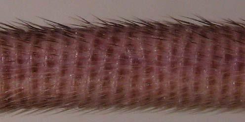


22 Tail tip Tail Fur with guard hairs Ear Footpads and sweat glands Eye EDA-deficient + agonist anti-edar EDA-deficient control Figure 3
INTRODUCTION. Developmental Biology 229, (2001) doi: /dbio , available online at
 doi: /dbio , available online at") Developmental Biology 229, 443 455 (2001) doi:10.1006/dbio.2000.9955, available online at http://www.idealibrary.com on TNF Signaling via the Ligand Receptor Pair Ectodysplasin and Edar Controls the Function
Developmental Biology 229, 443 455 (2001) doi:10.1006/dbio.2000.9955, available online at http://www.idealibrary.com on TNF Signaling via the Ligand Receptor Pair Ectodysplasin and Edar Controls the Function
Localization of Shh expression by Wnt and Eda affects axial polarity and shape of hairs
 Developmental Biology 305 (2007) 246 261 www.elsevier.com/locate/ydbio Localization of Shh expression by Wnt and Eda affects axial polarity and shape of hairs Brigitte Hammerschmidt, Thomas Schlake Max-Planck
Sostdc1 Plays an Essential Role in Mammalian Tooth Patterning: Insight into the Rodent Dental Evolution
 Sostdc1 Plays an Essential Role in Mammalian Tooth Patterning: Insight into the Rodent Dental Evolution PAULIINA MUNNE Developmental Biology Program Institute of Biotechnology University of Helsinki and
Inhibition of Wnt signaling by Wise (Sostdc1) and negative feedback from Shh controls tooth number and patterning
 and negative feedback from Shh controls tooth number and patterning") RESEARCH ARTICLE 3221 Development 137, 3221-3231 (2010) doi:10.1242/dev.054668 2010. Published by The Company of Biologists Ltd Inhibition of Wnt signaling by Wise (Sostdc1) and negative feedback from
RESEARCH ARTICLE 3221 Development 137, 3221-3231 (2010) doi:10.1242/dev.054668 2010. Published by The Company of Biologists Ltd Inhibition of Wnt signaling by Wise (Sostdc1) and negative feedback from
The Beauty of the Skin
 The Beauty of the Skin Rose-Anne Romano, Ph.D Assistant Professor Department of Oral Biology School of Dental Medicine State University of New York at Buffalo The Big Question How do approximately 50 trillion
Fgf20 governs formation of primary and secondary dermal condensations in developing hair follicles and
 Fgf20 governs formation of primary and secondary dermal condensations in developing hair follicles Sung-Ho Huh, 1,3 Katja Närhi, 2,3 Päivi H. Lindfors, 2 Otso Häärä, 2 Lu Yang, 1 David M. Ornitz, 1,4 and
Interactions between Shh, Sostdc1 and Wnt signaling and a new feedback loop for spatial patterning of the teeth
 RESEARCH ARTICLE 1807 Development 138, 1807-1816 (2011) doi:10.1242/dev.056051 2011. Published by The Company of Biologists Ltd Interactions between Shh, Sostdc1 and Wnt signaling and a new feedback loop
Epithelial Bmpr1a regulates differentiation and proliferation in postnatal hair follicles and is essential for tooth development
 Research article 2257 Epithelial Bmpr1a regulates differentiation and proliferation in postnatal hair follicles and is essential for tooth development Thomas Andl 1, Kyung Ahn 3, Alladin Kairo 1, Emily
Vertebrate Limb Patterning
 Vertebrate Limb Patterning What makes limb patterning an interesting/useful developmental system How limbs develop Key events in limb development positioning and specification initiation of outgrowth establishment
Role of Eda and Troy Pathways in Ectodermal Organ Development
 Role of Eda and Troy Pathways in Ectodermal Organ Development MARJA PUMMILA Institute of Biotechnology Developmental Biology Programme and Division of Physiology Department of Biological and Environmental
Development of teeth. 5.DM - Pedo
 Development of teeth 5.DM - Pedo Tooth development process of continuous changes in predetermined order starts from dental lamina A band of ectodermal cells growing from the epithelium of the embryonic
evolution and development of primate teeth
 evolution and development of primate teeth diversity of mammalian teeth upper left molars buccal mesial distal lingual Jernvall & Salazar-Ciudad 07 trends in dental evolution many similar single-cusped
Tooth development in the 'crooked' mouse
 /. Embryo!, exp. Morph. Vol. 41, pp. 279-287, 1977 279 Printed in Great Britain Company of Biologists Limited 1977 Tooth development in the 'crooked' mouse By J. A. SOFAER 1 From the University of Edinburgh,
Sonic hedgehog regulates growth and morphogenesis of the tooth
 Development 127, 4775-4785 (2000) Printed in Great Britain The Company of Biologists Limited 2000 DEV3251 4775 Sonic hedgehog regulates growth and morphogenesis of the tooth Hélène R. Dassule 1, Paula
ODONTOGENESIS- A HIGHLY COMPLEX CELL-CELL INTERACTION PROCESS
 ODONTOGENESIS- A HIGHLY COMPLEX CELL-CELL INTERACTION PROCESS AMBRISH KAUSHAL, MALA KAMBOJ Department of Oral and Maxillofacial Pathology Career Post Graduate Institute of Dental Sciences and Hospital
06 Tooth Development and Eruption
 + 06 Tooth Development and Eruption Tooth development Root development PDL and alveolar bone development Primary tooth eruption and shedding Permanent tooth eruption Q. Where and how tooth starts to form?
MBios 401/501: Lecture 12.1 Signaling IV. Slide 1
 MBios 401/501: Lecture 12.1 Signaling IV Slide 1 Pathways that require regulated proteolysis 1. Notch and Delta 2. Wnt/ b-catenin 3. Hedgehog 4. NFk-B Our last topic on cell signaling are pathways that
Novel EDA mutation resulting in X-linked non-syndromic hypodontia and the pattern of EDA-associated isolated tooth agenesis
 + MODEL Available online at www.sciencedirect.com European Journal of Medical Genetics 51 (2008) 536e546 http://www.elsevier.com/locate/ejmg Original article Novel EDA mutation resulting in X-linked non-syndromic
BCL11B Regulates Epithelial Proliferation and Asymmetric Development of the Mouse Mandibular Incisor
 BCL11B Regulates Epithelial Proliferation and Asymmetric Development of the Mouse Mandibular Incisor Kateryna Kyrylkova 1, Sergiy Kyryachenko 1, Brian Biehs 2 *, Ophir Klein 2, Chrissa Kioussi 1 *, Mark
Molecular Regulation of Embryonic Mammary Gland Development
 DISSERTATIONES SCHOLAE DOCTORALIS AD SANITATEM INVESTIGANDAM UNIVERSITATIS HELSINKIENSIS 3/2015 MARIA VOUTILAINEN Molecular Regulation of Embryonic Mammary Gland Development INSTITUTE OF BIOTECHNOLOGY
Tooth eruption and movement
 Tooth eruption and movement Dr. Krisztián Nagy Diphydont dentition Deciduous dentition primary dentition Diphydont dentition Permanent dentition secondary dentition Mixed Dentition: Presence of both dentitions
A Dual Role for Ikk in Tooth Development
 Developmental Cell, Vol. 6, 219 227, February, 2004, Copyright 2004 by Cell Press A Dual Role for Ikk in Tooth Development Atsushi Ohazama, 1 Yinling Hu, 2,3 buds are initiated by invagination of epithelium
Polarity and Segmentation. Chapter Two
 Polarity and Segmentation Chapter Two Polarization Entire body plan is polarized One end is different than the other Head vs. Tail Anterior vs. Posterior Front vs. Back Ventral vs. Dorsal Majority of neural
Biology Developmental Biology Spring Quarter Midterm 1 Version A
 Biology 411 - Developmental Biology Spring Quarter 2013 Midterm 1 Version A 75 Total Points Open Book Choose 15 out the 20 questions to answer (5 pts each). Only the first 15 questions that are answered
T cell maturation. T-cell Maturation. What allows T cell maturation?
 T-cell Maturation What allows T cell maturation? Direct contact with thymic epithelial cells Influence of thymic hormones Growth factors (cytokines, CSF) T cell maturation T cell progenitor DN DP SP 2ry
The functional investigation of the interaction between TATA-associated factor 3 (TAF3) and p53 protein
 and p53 protein") THESIS BOOK The functional investigation of the interaction between TATA-associated factor 3 (TAF3) and p53 protein Orsolya Buzás-Bereczki Supervisors: Dr. Éva Bálint Dr. Imre Miklós Boros University of
THESIS BOOK The functional investigation of the interaction between TATA-associated factor 3 (TAF3) and p53 protein Orsolya Buzás-Bereczki Supervisors: Dr. Éva Bálint Dr. Imre Miklós Boros University of
Mechanisms and Molecular Regulation of Mammalian Tooth Replacement
 Mechanisms and Molecular Regulation of Mammalian Tooth Replacement ELINA JÄRVINEN Institute of Biotechnology Developmental Biology Programme and Division of Genetics Department of Biological and Environmental
The site of action of the ichthyosis locus (ic) in the mouse, as determined by dermal-epidermal recombinations
 in the mouse, as determined by dermal-epidermal recombinations") /. Embryol. exp. Morph. Vol. 32, 3, pp. 715-721, 1974 715 Printed in Great Britain The site of action of the ichthyosis locus (ic) in the mouse, as determined by dermal-epidermal recombinations BY MARGARET
/. Embryol. exp. Morph. Vol. 32, 3, pp. 715-721, 1974 715 Printed in Great Britain The site of action of the ichthyosis locus (ic) in the mouse, as determined by dermal-epidermal recombinations BY MARGARET
Molecular Mechanisms Regulating Hair Follicle Development
 Molecular Mechanisms Regulating Hair Follicle Development Sarah E. Millar Departments of Dermatology and Cell and Developmental Biology, University of Pennsylvania, Philadelphia, Pennsylvania, U.S.A. Clinical
Insulin Resistance. Biol 405 Molecular Medicine
 Insulin Resistance Biol 405 Molecular Medicine Insulin resistance: a subnormal biological response to insulin. Defects of either insulin secretion or insulin action can cause diabetes mellitus. Insulin-dependent
Supporting Information
 Supporting Information Plikus et al. 10.1073/pnas.1215935110 SI Text Movies S1, S2, S3, and S4 are time-lapse recordings from individually cultured Period2 Luc vibrissa follicles show that circadian cycles
Independent regulation of Dlx2 expression in the epithelium and mesenchyme of the first branchial arch
 Development 127, 217-224 (2000) Printed in Great Britain The Company of Biologists Limited 2000 DEV2450 217 Independent regulation of Dlx2 expression in the epithelium and mesenchyme of the first branchial
Christ Siemens Touraine Syndrome: An unusual Presentation of the ectodermal disorder. Abstract: Introduction:
 Christ Siemens Touraine Syndrome: An unusual Presentation of the ectodermal disorder. Author: Dr.B.Senthilkumar, M.D.S., D.H.M., Asst. Professor, Dept.of Oral Medicine, Diagnosis & Radiology, K.S.R.Institute
BIOL2005 WORKSHEET 2008
 BIOL2005 WORKSHEET 2008 Answer all 6 questions in the space provided using additional sheets where necessary. Hand your completed answers in to the Biology office by 3 p.m. Friday 8th February. 1. Your
Development of the skin and its derivatives
 Development of the skin and its derivatives Resources: http://php.med.unsw.edu.au/embryology/ Larsen s Human Embryology The Developing Human: Clinically Oriented Embryology Dr Annemiek Beverdam School
Missense mutation of the EDA gene in a Jordanian family with X-linked hypohidrotic ectodermal dysplasia: phenotypic appearance and speech problems
 Missense mutation of the EDA gene in a Jordanian family with X-linked hypohidrotic ectodermal dysplasia: phenotypic appearance and speech problems O.F. Khabour 1, F.S. Mesmar 1, F. Al-Tamimi 3, O.B. Al-Batayneh
Cell Cell Communication
 IBS 8102 Cell, Molecular, and Developmental Biology Cell Cell Communication January 29, 2008 Communicate What? Why do cells communicate? To govern or modify each other for the benefit of the organism differentiate
The death receptors: signaling and modulation
 The death receptors: signaling and modulation 1 1 The extrinsic cell death pathway 2 Nat Rev Drug Discov. 2008 Dec;7(12):1001-12. 2 Death receptors Belong to the tumor necrosis factor (TNF) receptor gene
Integumentary System. Integumentary System
 1. General aspects a. The integumentary system consists of several organs major organ of the system is the skin other organs are relatively small and they can be considered as specialized structures of
Toll-like Receptor Signaling
 Toll-like Receptor Signaling 1 Professor of Medicine University of Massachusetts Medical School, Worcester, MA, USA Why do we need innate immunity? Pathogens multiply very fast We literally swim in viruses
Tetrapod Limb Development
 IBS 8102 Cell, Molecular and Developmental Biology Tetrapod Limb Development February 11, 2008 Tetrapod Limbs Merlin D. Tuttle Vicki Lockard and Paul Barry Father Alejandro Sanchez Anne Fischer Limb Patterning
Supplementary Figure 1: Signaling centers contain few proliferating cells, express p21, and
 Supplementary Figure 1: Signaling centers contain few proliferating cells, express p21, and exclude YAP from the nucleus. (a) Schematic diagram of an E10.5 mouse embryo. (b,c) Sections at B and C in (a)
Cell Cell Communication
 IBS 8102 Cell, Molecular, and Developmental Biology Cell Cell Communication January 29, 2008 Communicate What? Why do cells communicate? To govern or modify each other for the benefit of the organism differentiate
Chapter 1 Introduction
 1 Chapter 1 Introduction 2 Craniofacial malformations are serious congenital diseases in humans Craniofacial malformations are very common in humans. Around 75% of patients with congenital birth defects
Hepatogenesis I Liver development
 Hepatogenesis I Liver development HB 308 George Yeoh Room 2.59 MCS Building yeoh@cyllene.uwa.edu.au Topics Early liver development Tissue interaction - role of morphogens and cytokines Liver enriched transcription
Egyptian Dermatology Online Journal Vol. 6 No 2: 16, December Hypohidrotic Ectodermal Dysplasia with Arachnodactyl and Palmoplanter Keratoderma
 Hypohidrotic Ectodermal Dysplasia with Arachnodactyl and Palmoplanter Keratoderma Taseer Ahmed Bhatt Department of Dermatology, STD & Leprosy Govt. Medical College Srinagar, Kashmir Egyptian Dermatology
Expression of Axin2 Indicates a Role for Canonical Wnt Signaling in Development of the Crown and Root During Pre- and Postnatal Tooth Development
 DEVELOPMENTAL DYNAMICS 239:160 167, 2010 SPECIAL ISSUE RESEARCH ARTICLE Expression of Axin2 Indicates a Role for Canonical Wnt Signaling in Development of the Crown and Root During Pre- and Postnatal Tooth
Regulation of Gene Expression in Eukaryotes
 Ch. 19 Regulation of Gene Expression in Eukaryotes BIOL 222 Differential Gene Expression in Eukaryotes Signal Cells in a multicellular eukaryotic organism genetically identical differential gene expression
Primary Cilia Can Both Mediate and Suppress Hedgehog Pathway- Dependent Tumorigenesis (Supplementary Figures and Materials)
") Primary Cilia Can Both Mediate and Suppress Hedgehog Pathway- Dependent Tumorigenesis (Supplementary Figures and Materials) Sunny Y. Wong, Allen D. Seol, Po-Lin So, Alexandre N. Ermilov, Christopher K.
Primary Cilia Can Both Mediate and Suppress Hedgehog Pathway- Dependent Tumorigenesis (Supplementary Figures and Materials) Sunny Y. Wong, Allen D. Seol, Po-Lin So, Alexandre N. Ermilov, Christopher K.
Embryonic hair follicle fate change by augmented β-catenin through Shh and Bmp signaling
 RESEARCH REPORT 367 Development 136, 367-372 (2009) doi:10.1242/dev.021295 Embryonic hair follicle fate change by augmented β-catenin through Shh and Bmp signaling Kentaro Suzuki 1,2, Yuji Yamaguchi 3,
Tooth organogenesis and regeneration
 Tooth organogenesis and regeneration Irma Thesleff and Mark Tummers, Developmental Biology Program, Institute of Biotechnology, PO Box 56, University of Helsinki, FIN-00014, Helsinki, Finland Table of
EDA mutation as a cause of hypohidrotic ectodermal dysplasia: a case report and review of the literature
 EDA mutation as a cause of hypohidrotic ectodermal dysplasia: a case report and review of the literature S.X. Huang 1, J.L. Liang 2, W.G. Sui 3, H. Lin 3, W. Xue 3, J.J. Chen 3, Y. Zhang 3, W.W. Gong 3,
Chapter 4 Opener Pearson Education, Inc.
 Chapter 4 Opener Introduction The integumentary system is composed of: Skin Hair Nails Sweat glands Oil glands Mammary glands The skin is the most visible organ of the body Clinicians can tell a lot about
INTEGUMENTARY 1-Epidermis, 2-Dermis, Structure of thick and thin skin I- Epidermis . Stratum basale
 INTEGUMENTARY The skin (integument, cutis ) and its derivatives constitute the integumentary system. It form the external covering of the body and is the largest organ of the body. The skin consists of
The Epithelial-Mesenchymal Interaction Plays a Role in the Maintenance of the Stem Cell Niche of Mouse Incisors via Fgf10 and Fgf9 Signaling
 The Open Biotechnology Journal, 2008, 2, 111-115 111 The Epithelial-Mesenchymal Interaction Plays a Role in the Maintenance of the Stem Cell Niche of Mouse Incisors via Fgf10 and Fgf9 Signaling Tamaki
Revitalization of a Diastemal Tooth Primordium in Spry2 Null Mice Results From Increased Proliferation and Decreased Apoptosis
 JOURNAL OF EXPERIMENTAL ZOOLOGY (MOL DEV EVOL) 312B:292 308 (2009) Revitalization of a Diastemal Tooth Primordium in Spry2 Null Mice Results From Increased Proliferation and Decreased Apoptosis RENATA
Branching morphogenesis of the lung: new molecular insights into an old problem
 86 Review TRENDS in Cell Biology Vol.13 No.2 February 2003 Branching morphogenesis of the lung: new molecular insights into an old problem Pao-Tien Chuang 1 and Andrew P. McMahon 2 1 Cardiovascular Research
To the root of the stem cell problem. The evolutionary importance of the epithelial stem cell niche during tooth development
 To the root of the stem cell problem The evolutionary importance of the epithelial stem cell niche during tooth development Mark Tummers Developmental Biology Programme Institute of Biotechnology University
CHAPTER 6 SUMMARIZING DISCUSSION
 CHAPTER 6 SUMMARIZING DISCUSSION More than 20 years ago the founding member of the Wnt gene family, Wnt-1/Int1, was discovered as a proto-oncogene activated in mammary gland tumors by the mouse mammary
Toll-like Receptors (TLRs): Biology, Pathology and Therapeutics
: Biology, Pathology and Therapeutics") Toll-like Receptors (TLRs): Biology, Pathology and Therapeutics Dr Sarah Sasson SydPATH Registrar 23 rd June 2014 TLRs: Introduction Discovered in 1990s Recognise conserved structures in pathogens Rely
Toll-like Receptors (TLRs): Biology, Pathology and Therapeutics Dr Sarah Sasson SydPATH Registrar 23 rd June 2014 TLRs: Introduction Discovered in 1990s Recognise conserved structures in pathogens Rely
Subject Index. AXIN2, cleft defects 24, 26
 Subject Index ADAMTS, mouse mutants and palate development 37, 38 Africa, cleft lip and palate prevalence 6, 7 Alcohol dependence, pregnancy risks for cleft 25, 61 Altitude, pregnancy risks for cleft 25,
BMPR1A signaling is necessary for hair follicle cycling and hair shaft differentiation in mice
 Development Development Advance Online epress Articles. online First posted publication online on date 17 March 17 March 2004 as 10.1242/dev.01079 2004 Access the most recent version at http://dev.biologists.org/lookup/doi/10.1242/dev.01079
Problem Set 5 KEY
 2006 7.012 Problem Set 5 KEY ** Due before 5 PM on THURSDAY, November 9, 2006. ** Turn answers in to the box outside of 68-120. PLEASE WRITE YOUR ANSWERS ON THIS PRINTOUT. 1. You are studying the development
Tumor suppressor genes D R. S H O S S E I N I - A S L
 Tumor suppressor genes 1 D R. S H O S S E I N I - A S L What is a Tumor Suppressor Gene? 2 A tumor suppressor gene is a type of cancer gene that is created by loss-of function mutations. In contrast to
Cancer. The fundamental defect is. unregulated cell division. Properties of Cancerous Cells. Causes of Cancer. Altered growth and proliferation
 Cancer The fundamental defect is unregulated cell division. Properties of Cancerous Cells Altered growth and proliferation Loss of growth factor dependence Loss of contact inhibition Immortalization Alterated
Axis Formation and Mesoderm Induction
 Developmental Biology Biology 4361 Axis Formation and Mesoderm Induction October 27, 2005 Amphibian anteroposterior specification polarized eggs animal/vegetal pigment yolk v. clear cytoplasm mitochondrial
Supporting Information
 Supporting Information Voutilainen et al. 0.07/pnas.06709 SI Materials and Methods Animals. The generation and genotyping of K-da-A (K- da) () and N-κ reporter mice () has been described earlier. oth strains
Anatomy and Physiology Homework: Chapters 3-4
 Anatomy and Physiology Homework: Chapters 3-4 CHAPTER 3: Cells and Tissues 1. The smallest unit of living tissue is called a. All living organisms are composed of these basic units where all life processes
Cancer. The fundamental defect is. unregulated cell division. Properties of Cancerous Cells. Causes of Cancer. Altered growth and proliferation
 Cancer The fundamental defect is unregulated cell division. Properties of Cancerous Cells Altered growth and proliferation Loss of growth factor dependence Loss of contact inhibition Immortalization Alterated
Tolerance 2. Regulatory T cells; why tolerance fails. Abul K. Abbas UCSF. FOCiS
 1 Tolerance 2. Regulatory T cells; why tolerance fails Abul K. Abbas UCSF FOCiS 2 Lecture outline Regulatory T cells: functions and clinical relevance Pathogenesis of autoimmunity: why selftolerance fails
B. Incorrect! The ectoderm does not produce the dermis. C. Incorrect! The dermis is derived from the mesoderm.
 Human Anatomy - Problem Drill 04: The Integumentary System Question No. 1 of 10 Instructions: (1) Read the problem and answer choices carefully, (2) Work the problems on paper as 1. From the inner cell
T Cell Development. Xuefang Cao, MD, PhD. November 3, 2015
 T Cell Development Xuefang Cao, MD, PhD November 3, 2015 Thymocytes in the cortex of the thymus Early thymocytes development Positive and negative selection Lineage commitment Exit from the thymus and
Development of the Pharyngeal Arches
 Development of the Pharyngeal Arches Thomas A. Marino, Ph.D. Temple University School of Medicine Competencies: Upon completion of this section of the course, the student must be able to: 1. Recall the
Introduction: 年 Fas signal-mediated apoptosis. PI3K/Akt
 Fas-ligand (CD95-L; Fas-L) Fas (CD95) Fas (apoptosis) 年 了 不 度 Fas Fas-L 力 不 Fas/Fas-L T IL-10Fas/Fas-L 不 年 Fas signal-mediated apoptosis 度降 不 不 力 U-118, HeLa, A549, Huh-7 MCF-7, HepG2. PI3K/Akt FasPI3K/Akt
Developing Molecularly Targeted Therapies for Basal Cell Carcinoma. Ivor Caro, MD, FAAD
 Developing Molecularly Targeted Therapies for Basal Cell Carcinoma Ivor Caro, MD, FAAD Disclosures Genentech, Inc Medical Director, Dermatology (employee) Stock holder Hedgehog Signaling Pathway Fundamental
Apc inhibition of Wnt signaling regulates supernumerary tooth formation during embryogenesis and throughout adulthood
 AND DISEASE RESEARCH ARTICLE 1939 Development 136, 1939-1949 (2009) doi:10.1242/dev.033803 Apc inhibition of Wnt signaling regulates supernumerary tooth formation during embryogenesis and throughout adulthood
Principles of Anatomy and Physiology
 Principles of Anatomy and Physiology 14 th Edition CHAPTER 5 The Integumentary System Introduction The organs of the integumentary system include the skin and its accessory structures including hair, nails,
Fibers and extracellular matrix of hard tissues - Collagen and non-collagen proteins in hard tissues
 Fibers and extracellular matrix of hard tissues - Collagen and non-collagen proteins in hard tissues Dr. Gábor Varga Department of Oral Biology February, 2016 Radiograph of teeth remarkable harmony of
SUPPLEMENTARY INFORMATION
 doi:10.1038/nature10643 Supplementary Table 1. Identification of hecw-1 coding polymorphisms at amino acid positions 322 and 325 in 162 strains of C. elegans. WWW.NATURE.COM/NATURE 1 Supplementary Figure
Signaling Vascular Morphogenesis and Maintenance
 Signaling Vascular Morphogenesis and Maintenance Douglas Hanahan Science 277: 48-50, in Perspectives (1997) Blood vessels are constructed by two processes: vasculogenesis, whereby a primitive vascular
Ontogenesis. Genetics of development
 Ontogenesis Genetics of development 2 Lesson from an experimental animal model 3 Task 1, page 159, Phenotypes of polydactylous rats LEW PD LEW.Lx BN.Lx 4 0 Production of LEW.Lx Lx congenic strain LEW (+/+)
Lecture outline. Immunological tolerance and immune regulation. Central and peripheral tolerance. Inhibitory receptors of T cells. Regulatory T cells
 1 Immunological tolerance and immune regulation Abul K. Abbas UCSF 2 Lecture outline Central and peripheral tolerance Inhibitory receptors of T cells Regulatory T cells 1 The immunological equilibrium:
Dominic J Smiraglia, PhD Department of Cancer Genetics. DNA methylation in prostate cancer
 Dominic J Smiraglia, PhD Department of Cancer Genetics DNA methylation in prostate cancer Overarching theme Epigenetic regulation allows the genome to be responsive to the environment Sets the tone for
Introduction. Skin and Body Membranes. Cutaneous Membranes Skin 9/14/2017. Classification of Body Membranes. Classification of Body Membranes
 Introduction Skin and Body Membranes Body membranes Cover surfaces Line body cavities Form protective and lubricating sheets around organs Classified in 5 categories Epithelial membranes 3 types- cutaneous,
Ex. 7: Integumentary
 Collin County Community College BIOL. 2401 Ex. 7: Integumentary. Skin or Integument Consists of three major regions Epidermis outermost superficial region Dermis middle region Hypodermis (superficial fascia)
Early Embryonic Development
 Early Embryonic Development Maternal effect gene products set the stage by controlling the expression of the first embryonic genes. 1. Transcription factors 2. Receptors 3. Regulatory proteins Maternal
CHAPTER 5 INTEGUMENTARY
 CHAPTER 5 INTEGUMENTARY skin under the skin other stuff cutaneous layer hypodermis (subcutaneous) accessory structures Cutaneous layer = skin epithelial layers = connective tissue layer = dermis Subcutaneous
Skin stem cells: rising to the surface
 JCB: MINI-REVIEW Skin stem cells: rising to the surface Elaine Fuchs Howard Hughes Medical Institute, Laboratory of Mammalian Cell Biology and Development, The Rockefeller University, New York, NY 10065
HLA and antigen presentation. Department of Immunology Charles University, 2nd Medical School University Hospital Motol
 HLA and antigen presentation Department of Immunology Charles University, 2nd Medical School University Hospital Motol MHC in adaptive immunity Characteristics Specificity Innate For structures shared
CHAPTER VII CONCLUDING REMARKS AND FUTURE DIRECTION. Androgen deprivation therapy is the most used treatment of de novo or recurrent
 CHAPTER VII CONCLUDING REMARKS AND FUTURE DIRECTION Stathmin in Prostate Cancer Development and Progression Androgen deprivation therapy is the most used treatment of de novo or recurrent metastatic PCa.
T-cell activation T cells migrate to secondary lymphoid tissues where they interact with antigen, antigen-presenting cells, and other lymphocytes:
 Interactions between innate immunity & adaptive immunity What happens to T cells after they leave the thymus? Naïve T cells exit the thymus and enter the bloodstream. If they remain in the bloodstream,
Author's response to reviews
 Author's response to reviews Title: Wnt1 is epistatic to Id2 in modeling mammary gland development and in causing mammary tumors Authors: Susan Marino (smarino@cmgm.stanford.edu) Claire Romelfanger (clairefrog@yahoo.com)
Alternative splicing. Biosciences 741: Genomics Fall, 2013 Week 6
 Alternative splicing Biosciences 741: Genomics Fall, 2013 Week 6 Function(s) of RNA splicing Splicing of introns must be completed before nuclear RNAs can be exported to the cytoplasm. This led to early
A Genetic Program for Embryonic Development
 Concept 18.4: A program of differential gene expression leads to the different cell types in a multicellular organism During embryonic development, a fertilized egg gives rise to many different cell types
It has been shown that in human embryos and fetuses with cleft lips
 SCIENTIFIC FOUNDATION Crown Morphologic Abnormalities in the Permanent: Dentition of Patients With Cleft Lip and Palate Ma amon A. Rawashdeh, BDS, MScD, FDSRCS (En),*Þ and Emad Omar Abu Sirdaneh, BDS,
The Integumentary System : Embryology & Genetic Bases. Purnomo Soeharso Department of Medical Biology FMUI
 The Integumentary System : Embryology & Genetic Bases Purnomo Soeharso Department of Medical Biology FMUI Tissue organization of the skin (integumentum) : - Epidermis stratified epithelium on the outer
Restriction of sonic hedgehog signalling during early tooth development
 Research article 2875 Restriction of sonic hedgehog signalling during early tooth development Martyn T. Cobourne 1, Isabelle Miletich 2 and Paul T. Sharpe 2, * 1 Department of Craniofacial Development
Programmed Cell Death (apoptosis)
") Programmed Cell Death (apoptosis) Stereotypic death process includes: membrane blebbing nuclear fragmentation chromatin condensation and DNA framentation loss of mitochondrial integrity and release of
Programmed Cell Death (apoptosis) Stereotypic death process includes: membrane blebbing nuclear fragmentation chromatin condensation and DNA framentation loss of mitochondrial integrity and release of
Chapter 2 Tooth Development
 Chapter 2 Tooth Development Experimental research on tooth development or odontogenesis is based very largely on the teeth of murine rodents (Butler 1967 ). Pioneering work by Shirley Glasstone on rat