Università degli Studi di Ferrara
|
|
|
- Marshall Lloyd
- 5 years ago
- Views:
Transcription
1 Università degli Studi di Ferrara DOTTORATO DI RICERCA IN "Biochimica, Biologia Molecolare e Biotecnologie" CICLO XXVI COORDINATORE Prof. Francesco Bernardi Cellular activity of micrornas dysregulated in breast cancer Settore Scientifico Disciplinare: BIO/11 Dottorando Tutore Co-Tutore Dott. Carlotta Zerbinati Prof. Giovanna Marchetti Dott. Stefano Volinia Anni 2011/2013
2 Contents: Page: Abstract Introduction Cancer Breast Cancer Breast Cancer development Classification of Breast Cancer Immunohistochemical classification of breast cancer Molecular Classification of breast cancer: intrinsic subtypes Luminal A and Luminal B Breast Cancers HER2 + Enriched Basal-like and Claudin-Low Normal-like Estrogen and estrogen receptor Therapy for the different subtypes of breast cancer ER-positive breast cancer: Endocrine Therapy Anti EGFR therapy Anti HER2 positive therapy Triple negative breast cancer : TNBC Breast cancer in men Classification of non-coding RNAs Introduction to mirnas Genomic organization of mirnas Current Model of Mammalian mirna Maturation and Processing Molecular mechanisms of mir action MiRNA Classification Experimental techniques for mirna analysis Microarray Next generation sequencing techniques Real time Quantitative PCR MicroRNAs in Tumorigenesis Deregulation of mirnas in cancer MiRNAs as tumor suppressor MiRNAs as oncogenes Therapeutic approaches of mirnas 33 1
3 2. Materials and methods Cell culture Analysis of mutation in breast cancer cell lines MicroRNA microarrays Transient mirna transfection Proliferation assay MTS assay Analysis of data Real-time cell proliferation assay Real Time cell invasion assay Scratch wound healing assay Class comparison Total RNA isolation Quantification of RNA Statistical analysis..41 Rationale Results and discussion Genomic characterization of cell lines Curves of growth for MDA-MB-231 and for MCF Efficiency of transfection Viability regulation by mirnas Effect of co-trasfection of selected mirnas Viability regulation by prognosis-related mirnas Wound healing assay Real time measurement of cell invasiveness and cell proliferation mrna profiling after mirna treatment Cross validation on breast cancer tumors from TCGA cohort Discussion Conclusion 76 References.77 Supplementary Informations.83 2
4 3
5 Abstract Breast cancer is one of the major health problems worldwide and it is the second cause of cancer-related in women. Patients often develop resistance to the current therapies. For this reason, the identification of new specific clinical molecular markers and pharmacologic targets in cancer research is an ongoing challenge. Over the last years, micrornas (mirnas) have become one of the main subjects of study in the area of cancer genomics. They negatively regulate gene expression post-transcriptional by inhibiting translation and causing degradation of target mrna. More than a thousand mirnas exist in the human genome and each one can potentially regulate hundreds of mrnas. By regulating the expression of target genes, mirnas can have a tumor suppressor or oncogenic role. Therefore, mirnas can play an important role in all the phases of cancer, like as initiation, progression, growth, apoptosis, invasion and metastasis. In a previous study based on mirna profiling, in different solid tumors, comprised breast cancer, and normal tissues, several mirna were reported to be over- or down-regulated in solid tumors in comparison to normal tissues (Volinia et al. Genome Res. 2010). In other scientific reports other mirnas were positively or negatively correlated with tumor progression (Volinia S. and Croce CM. PNAS 2013). Starting from these literature data, we decided to analyze the in vitro effect of the administration of this tumor-related mirnas in order to verify their effect on the viability and transcriptional regulation in breast cancer cell line, in order to gain experimental evidences on their actual involvement in the tumor illness. Firstly, we have chosen 10 different cell lines of breast cancer origin (MCF-7, MDA-MB-231, MDA-MB-468, MDA-MB-361, SKBR3, T47D, BT474, ZR75.1, MDA-MB-453, HBL-100) and on 2 cell lines of breast normal epithelium (MCF10A and 184A1). Genomic analysis revealed presence of a complex panel of cancer-related mutations, and the cell lines were different in term of cell growth. We checked also the mirna levels inside cell lines. Two groups of primary solid tumor-related (23 mirnas: hsa-mir-126*, hsa-let-7d*, hsamir-326, hsa-mir-320c, hsa-mir-302a, hsa-mir-222, hsa-mir-218, hsa-mir-210, hsa-mir- 206, hsa-mir-203, hsa-mir-202, hsa-mir-181a, hsa-mir-142, hsa-mir-145, hsa-mir-143, hsa-mir-138, hsa-mir-130b, hsa-mir-126, hsa-mir-99a, hsa-mir-28-5p, hsa-mir-33b, hsa-mir-26b, hsa-mir-21) or progression-related (15 mirnas: hsa-mir-9, hsa-mir-10a, hsa-mir-25, hsa-mir-27, hsa-mir-30a, hsa-mir-93, hsa-mir-103, hsa-mir-148b, hsa-mir- 151, hsa-mir-301a, hsa-mir-328, hsa-mir-484, hsa-mir-615, hsa-mir-874, hsa-mir-1307) mirnas were transiently transfected into cells, and viability was assessed. We were able to 4
6 experimentally identify two groups of mirnas which were able to significantly increase or decrease cell viability. The mirnas that showed to increase cellular viability in five out twelve cell lines were: mir-130b, mir-138, mir-210, mir-148b and mir On the other hand, mirr-93, mir-126 and mir-145 displayed a significant inhibitory effect on cell viability in five out twelve cell lines. These mirnas were further investigated for their capacity to affect cell migration, cell invasion, and genome profiling. The main outcome of our work has been the identification from a wide list of cancer-related mirnas of few of them involved in the in vitro regulation of cell growth and invasion. As a first attempt to identify target genes commonly regulated in vivo and in vitro, we have bioinformatically identified, in a first not exhaustive screening, PTEN and DICER1 genes, which in vivo and in vitro negatively correlated with mir-210 and mir-130b, respectively. 5
7 1. Introduction 1.1. Cancer Cancer is the result of many complex changes occurring in a normal cell, progressing through to malignant and potentially metastatic. The 6 hallmarks of cancer as outlined in Hanahan and Weinburg s review (1) are shown below (Figure 1). Cells commonly become cancerous when they acquire irreparable DNA damage, changing the sequence of genes which code for important regulatory proteins. After replication the mutations are passed down to the next generation of cells which can lead to deregulated growth, tumor progression and invasion through the basement membrane. Figure 1. The 6 hallmarks of cancer. Accumulations of successive DNA mutations arise over the course of a person s life time (somatic gene changes) which is why age is often a risk factor in cancer. Also, lifestyle factors such as diet, alcohol, stress and tobacco, have all been implicated in either causing or increasing the risk of cancer. The environmental exposure to chemical carcinogens and the ultra-violet rays of the sun have also been shown to result in DNA damage and result in cancer. There are examples of gene mutations that are hereditary for instance BRCA1 and BRCA2 which are tumor suppressor genes involved in DNA repair of double stranded breaks. Mutations in these genes can cause instability of the human genome. Women that have heterozygous germ-line mutations in BRCA1 or BRCA2 have a substantially increased risk of highly penetrative breast cancer and ovarian cancer. Patients that are positive for this mutation can chose to have pre-emptive mastectomies or preventative Tamoxifen treatment (2). 6
8 1.2. Breast Cancer Breast cancer (BC) is a complex and heterogeneous disease, characterized by variant genetic alterations and distinct morphologic and molecular features. Despite common histopathological features at diagnosis, BC is noted for disparate clinical behaviors and patient outcomes. The heterogeneity observed among breast cancer reflects the now well accepted notion that is not just one disease, but that instead represents a collection of distinct neoplastic diseases of the breast and the cells composing the breast (3). The character and nature of this diseases can be made through traditional pathological and morphological examination, but only through molecular analyses can be appreciated the extent of diversity among breast cancer. Breast carcinomas can be divided into two major groups: In situ Carcinomas - the tumor cells remain confined to the ducts or lobules and show no evidence of microscopic invasion into the surrounding breast stroma. There are two types of in-situ carcinoma; ductal and lobular, named according to the predominant cell type from which the tumour arises. Invasive carcinoma - the tumor cells invade the breast stroma and have the potential to metastasize to distant sites. The invasive breast carcinomas consist of several histological subtypes; the commonest being infiltrating ductal adenocarcinoma (75-80%), followed in frequency by invasive lobular (10-15%), Mixed ductal-lobular (<5%), Inflammatory (2-3%), Colloid (2-3%), Tubular (<2%), Medullary (<2%), and Papillary (1%) 5. Rarer subtypes, including metaplastic breast cancer and invasive micropapillary breast cancer, all account for less than 5 percent of cases overall. Breast Cancer is a major health problem in the United States and worldwide, it is now the second cause of cancer-related in women (second only to lung cancer) with approximately 40,030 death expected in 2013 for both men and women (or 39,620 deaths among women, representing 14% of all cancer-related deaths among women) and the vast majority of breast cancer-related deaths involve metastatic disease. Metastasis is a multistep process which consists of a cell: - detaching ang migrating out of the primary tumor site; - invading the basement membrane to enter the circulatory system (intravasation); - surviving cell detachment-mediated apoptosis (anoikis); - exiting circulatory vessels at the metastatic site (extravasation); and adapt into a new environment and soil a metastatic tumor. Breast cancer is about 100 times more common in women than in men although males tend to have poorer outcomes due to delays in diagnosis. The American Cancer Society estimates that 232,340 7
9 new cases of invasive ductal breast cancer (IDC) and 64,640 new cases of ductal carcinoma in situ (DCIS) will be diagnosed among women in the United States in Invasive breast cancer accounts for 29% of all cancer diagnoses among women in the United States and 23% of all cancer diagnoses among women worldwide. In the worldwide there is an estimated 1,383,500 new cases of BC will be diagnosed in women in 2013 (4). As such, this disease represents one of the most serious and costly health issues. The incidence of breast cancer increases with age (around 80% of diagnosed cases are in women over the age of 50) and there are other risk factors; such as obesity, high socio-economic group, alcohol use, expression of BRCA1 or BRCA2, ethnicity, early menarche, and childbirth late in life. A family history of breast cancer in female relatives has shown to be an important predisposing factor. In women, the breast cancer susceptibility genes BRCA1 and BRCA2 are thought to account for most hereditary breast cancers. Mutations in these genes confer a 40% to 70% risk for developing breast cancer by age 70 years. Other molecular markers are important in the pathogenesis and prognosis of breast cancer in women, as C-erbB-2, p53, Bcl-2, cyclin D1, and epidermal growth factor receptor (EGFR). The c-erbb-2 protooncogene encodes for a transmembrane receptor of the tyrosine kinase family, which is closely related to EGFR. This protein is expressed in 20% to 30% of breast cancers and may be associated with a poor prognosis. P53 is a tumor suppressor gene that controls cell growth by inducing cell cycle blockade, apoptosis, and cell differentiation. P53 gene alterations are the most common single genetic abnormality in breast cancer and are present in approximately 30% of cases. Bcl-2 is a protooncogene that inhibits apoptosis and thereby promotes cell growth. In breast cancer, expression of Bcl-2 has been associated with favorable prognostic features. Cyclin D1 is involved in cell-cycle regulation and helps control the cell s entry into S phase. In breast cancer this gene is oncogenic but appears to be associated with a favorable prognosis. Epidermal growth factor receptor is a transmembrane glycoprotein that is present in low levels in normal breast tissue and is overexpressed in 35% to 60% of BC. Over expression of EGFR is inversely correlated with estrogen receptor expression and may be a negative prognostic factor. Significant prognostic factors are also axillary lymph node status, tumor size, histological grade and hormone receptor status, as estrogen receptor, progesterone receptor, and ErbB2, these are used to decide the treatment options for the patient. The patient can be classed as triple negative where they do not express any of these 3 receptors. 8
10 Breast Cancer development Breast Cancer develops over a long period of time and requires multiple molecular alterations. The time required for the process of carcinogenesis is not well established for any human cancer, estimates suggest that this multistep process unfolds over many years and possibly several decades. Generally, sporadic breast cancer, in which there is no recognizable strong genetic component, emerge later in life (reflecting mostly postmenopausal breast cancer), whereas hereditary breast cancers occur earlier in life (reflecting the contribution of genetic predisposition) (5). The main hypothesis for the natural history of breast cancer development is stepwise progression from atypical ductal hyperplasia to ductal carcinoma in situ (DCIS), followed by evolution of this pre-invasive lesion to invasive ductal breast cancer (IDC) (Figure 2). DCIS is by definition noninvasive, but can vary from low-grade to high-grade lesions that may contain invasive elements. For this reason, DCIS, especially if high grade, is a risk factor for development of IDC. Many IDC are associated with adjacent DCIS lesions. It is not entirely clear if DCIS is a required precursor for development of IDC, but many IDC at the time of diagnosis are accompanied by DCIS, and there is consensus that DCIS in the absence of intervention will progress to invasive disease. In the progression of the disease might occur genetic and epigenetic alterations between the altered cells of the different morphological stages that contributed at the development of the disease (6). Since the late 1990s, invasive breast cancer have been characterized using gene expression analysis and classified on that basis into several molecular subtypes. Analyses of gene expression patterns between DCIS and IDC, have identified one correspondence in this two subtypes, this suggest that DCIS lesions are likely the direct precursors of invasive cancers. However, some recent molecular analysis suggest that the diversity of subtypes observed in invasive breast cancers derives from an evolution of low-grade to high-grade DCIS lesions. 9
, DCIS, and invasive breast cancer.")
11 Figure 2. Natural history of breast cancer development. Breast cancer develops from normal breast epithelial cells that evolve through atypical hyperplasia (and eventually dysplasia), DCIS, and invasive breast cancer. Multiple molecular alterations occur during this process, involving genetic and epigenetic alterations in precursor and neoplastic cells. Genetic predisposition can contribute to this process, but early molecular alterations (preceding DCIS) have not been well characterized. Original magnification, 20x. Modified from Rivenbark and Coleman (6), with permission from Elsevier. 1.3 Classification of Breast Cancer Immunohistochemical classification of breast cancer The morphological subclassification of invasive ductal carcinomas is accomplished by histopathological immunostaining to detect the differential expression of protein biomarkers to provide a clinical classification for breast cancer and dictates therapeutic approaches for treatment. Routine histopathological subclassification is made for detect the expression of estrogen receptor (ER), progesterone receptor (PR), and human epidermal growth receptor 2 (HER2; alias c-erbb-2 or, in rodents, Neu), as well as HER1 and various cytokeratins (eg, CK5/6) (7). Approximately 70% of invasive breast cancers express ER, and the majority of ER + cancers also express PR. Although the presence of normal PR levels suggests an intact ER signal transduction pathway in the breast cancer cells, discrepant ER and PR expression patterns (ER + /PR - and ER - /PR + ) are sometimes observed, this may be attributable to false positive or false negative results, although technical improvements have reduced errors significantly. Collectively, the ER + malignant neoplasms are classified as luminal cancers. These cancers are further subclassified based on their HER2 status and proliferation rate, giving rise to the ER + /PR + /HER2 + and ER + /PR + /HER2 - subtypes. The ER - breast cancer are subclassified as HER2 + and triple negative based on HER2 overexpression or gene 10

12 amplification status, basal cytokeratin expression, and EGFR (HER1) expression, giving rise to ER - /PR - /HER2 + (HER2-enriched) and ER - /PR - /HER2 - (triple negative) subtypes (Figure 3). Figure 3. Clinical classification of invasive breast cancer based on expression of ER, PR, and HER2. Representative examples of invasive breast cancers that correspond to the general clinical classifications are shown. Cancer histology is depicted using H&E staining; expression of ER, PR, and HER2 is visualized using immunohistochemistry. Breast cancers are generally classified as positive or negative for hormone receptors ER and PR and for HER2, resulting in four major clinical groupings: ER + /PR + /HER2 -, ER + /PR + /HER2 +, ER - /PR - /HER2 +, and the triple-negative ER - /PR - /HER2 -. Original magnification, 40x Molecular Classification of breast cancer: intrinsic subtypes Perou and Sorlie and colleagues (8) studied gene expression profiling using DNA microarrays have grouped breast cancer into 5 distinct molecular classes or intrinsic subtypes named: - Luminal A (ER + ) - Luminal B (ER + /HER2-enriched) - Basal-like - HER2 + -enriched - Normal-like More recently, a further subtypes have been identified: - Claudin-low Significantly, the molecular subtypes of breast cancer revealed by transcriptomic analysis are associated with different clinical outcomes. Although breast cancer classification methods 11
13 show good reproducibility, suggesting that these are robust biological subtypes, breast cancers that are not classifiable are identified with regular frequency (8) Luminal A and Luminal B Breast Cancers ER + breast cancers occur most frequently and comprise two major molecular classifications: Luminal A and Luminal B. Luminal A is the more abundant subtypes and accounts for 40-60% of breast cancer. Predominantly is ER + /PR + /HER2 -, with low histological grade and low expression of proliferative genes. This subtype is associated with lower relapse rate and longer survival, compared with the other subtypes. The TP53 pathway is conserved and the management of this tumors is centered on antiendocrine therapies because this subtype is poor responsive to chemotherapy. Luminal B subtype accounts for 15% of breast cancers, as luminal A is predominantly ER + although it has lower expression of ER-related genes and it is histological high grade with variable PR and HER2 status. Luminal B tumors in contrast with luminal A are more aggressive, with poorer prognosis. The TP53 pathway is frequently inactivated. Although luminal B tumors are ER +, a significant number do not respond to antiestrogen therapy but show greater response to chemotherapy. In general, the two ER + breast cancer subtypes, Luminal A and Luminal B, are associated with a good prognosis and excellent long-term survival, whereas the ER - subtypes (HER2 + and basal-like) are difficult to treat and are associated with poor prognosis HER2 + Enriched The HER2 gene, also known as HER2/neu or c-erbb2, is located on chromosome 17q. It is a proto-oncogene, a normal gene with the potential to become an oncogene as a result of molecular alterations like mutation, amplification or overexpression of its protein product. HER2 is a member of the human epidermal growth factor receptor family, a family of tyrosine kinases, which normally regulate a series of cellular processes, such as proliferation and growth. HER2 is notable for its role in the pathogenesis of breast cancer and as a target of treatment. It is a cell membrane surface-bound receptor tyrosine kinase and is normally involved in the signal transduction pathways leading to cell growth and differentiation. HER2 is thought to be an orphan receptor, with none of the EGF family of ligands able to activate it. However, ErbB receptors dimerise on ligand binding, and HER2 is the preferential dimerization partner of other members of the ErbB family (9). 12
14 The HER2 + -enriched subtype represent approximately 17% of all breast cancers. It is characterized by high expression of HER2 and related genes and it has high genomic instability. HER2 overexpression in breast cancer is associated with poor clinical outcomes, but it is also predictive of positive therapeutic responses to anti-her2 drugs (eg, monoclonal antibody trastuzumab). HER2 + breast cancer are typically ER -, so the treatment for these cancers does not include anti-estrogenic hormonal therapies Basal-like and Claudin-Low Basal-like and Claudin-low together represent subsets of triple negative breast cancers, lacking expression of ER and PR and also lacking amplification of HER2. These tumors represent 10% to 25% of breast cancers, they are highly proliferative and express genes characteristic of normal breast myoepithelial cells, such as cytokeratins 5, 6, and 17, and epidermal growth factor receptor (EGFR). They have a high rate of p53 mutations and are associated with increased genetic complexity. Not all tumors with basal-like gene expression profile are triple negative; 15% to 45% express ER and HER2 or both. The basal-like breast cancer have high rates of cell proliferation and extremely poor clinical outcomes. These cancers are associated with distinct risk factors, including early-onset menarche, younger age at first full-term pregnancy, and abdominal adiposity. Basal-like breast cancers have been shown to be over-represented in patients of certain age and ethnic groups, specifically young Black women (10). Claudin-low subtype breast cancer is enriched for markers of epithelial-tomesenchymal transition (EMT) and stem cell-like and/or tumor-initiating cell features. Although these tumors are chemo responsive, they have shown a poor prognosis across several studies Normal-like The normal-like represents 3% to 10% of breast cancers, it is frequently ER + and has low levels of proliferative genes and low tumor cellular. The normal-like breast cancers are so designated because they tend to cluster closely with normal breast epithelium in microarray studies. It is not yet clear whether this is a distinct molecular subtype of breast cancer or simply a grouping of breast cancers that are not otherwise classifiable because of contaminating normal epithelium. Nevertheless, the prognosis of this group is reported to be intermediate, with better survival than all but luminal A breast cancers. The identification of these molecular signatures has helped to understanding breast cancer and informed the search for novel therapies. However, this classification is a work in progress, 13
15 requiring refinement and standardization before it can be incorporated into clinical practice and decision making. Therefore, despite the progress and better understanding of the drivers of this disease, from a clinical management perspective, breast cancer remains divided into 3 therapeutic categories: 1. ER + disease, which is targeted with antiendocrine strategies 2. HER2 + disease, which is treated with HER2-targeted agents 3. Triple negative breast cancer, which lacks validated targeted therapy options and is treated with traditional cytotoxic therapy 1.4. Estrogen and estrogen receptor Estrogen is an important regulator in the development and progression of breast cancer and also in the development of normal breast. More than a century ago, Scottish surgeon George Beatson performed an oophrectomy and noted this procedure induced regression of breast cancer which was later proven to be due to a reduction in systemic estrogen levels (11). Estrogen mainly originates in the ovaries in pre-menopausal women, whereas in post menopausal women the main source is in the aromatization of androgens in adipose tissue. This accounts for the difference in therapeutic regimen between the two groups of women. Estrogen functions by activating two nuclear steroid receptors: ERα and ERβ. Both receptors bind estrogen and initiate gene transcription through ERE (estrogen response elements) in estrogen target tissues but have distinct functions and tissue distribution (12). In most breast carcinomas (~70%) ERα is highly expressed which results in the increased rate of proliferation without differentiation or apoptosis. ERα is exclusively epithelial (13, 14) whereas the distribution of ERβ in breast cancer is exclusively nuclear but expressed in multiple cell types (stromal fibroblast, endothelial and immune infiltrating cells) its expression has also been shown to correlate with an increase in aggression of the tumor (15). ERβ expression has been reported to range from 26% to 94%, the lowest of which was in a Japanese study, this implicates that the expression could be related to ethnicity. When Caucasian cohorts of patients were compared, the range was less extensive at 74%-94% (14). The expression of ERβ in the breast has been well described although its usual function, clinical value in carcinogenesis, and its relevance to the pathological diagnosis of breast cancer, is yet to be determined (14). Estrogen receptors that are not bound to ligand are inactive and usually sequestered in multi-meric protein complexes organized around the molecular chaperone heat shock protein 90 (HSP90). ER signaling pathways can be classed into 4 main mechanisms of action: Classical, ERE independent, ligand independent and non- 14
16 genomic (Figure 4). Classically, the cytoplasmic ER translocates into the nucleus upon ligand binding. In the cytoplasm, the receptor dimerizes, transcription factors and co-activation proteins are recruited, and the target genes are then activated through an estrogen response element (ERE). Estrogen activates genes that are involved in survival and cell proliferation amongst other actions (16, 17). Estrogen bound ER interaction with Fos and Jun, which dimerize and become part of the activator protein-1 (AP-1) complex modulating gene expression. This ligand bound ER gene modulation can also occur with GC box bound specificity protein -1 (SP- 1) this is ERE independent modulation. ER can act independently of estrogen by being phosphorylated on multiple residues within the receptor after growth factor activation. The phosphorylation of ER leads to the dimerization of the receptor, DNA binding and ultimately activation of transcription (18). Estrogen can also activate membrane bound ER and cause a rapid estrogen signaling response through non-genomic activation (19). The non-genomic simulation of the estrogen receptor is a rapid response to the ligand binding and its response in the cell is independent of the gene transcription. Estrogen receptors act through a complex interplay of signaling cascades; such as insulin-like growth factor 1 receptor (IGF-1 R) (20), EGFR (21), G- proteins, Src, and PI3K. 15
 bound dimers bind to the EREs in target promoter genes; this leads to an up regulation or down regulation of")
17 Figure 4. A simplified view of the 4 mechanisms by which estrogen can exert its action upon a cell (roles and names of co-factors have been omitted). 1, Classical pathway: This is a genomic, ligand dependent mechanism; where estradiol (E2) bound dimers bind to the EREs in target promoter genes; this leads to an up regulation or down regulation of gene transcription and resulting in the estrogenic effects being seen in the cell. 2, ERE independent pathway: this is also a genomic, ligand dependent mechanism where estrogen bound ER dimers bind to alternative gene response elements such as AP-1 through Fos and Jun association and thus lead to gene transcription. 3, Ligand independent mechanism: this is a genomic ligand independent mechanism where the phosphorylation and subsequent activation of the ER, following growth factors and/or signalling molecules stimulation leads to target gene transcription. 4, the non-genomic ligand dependent mechanism; this is where the estradiol binds to a membrane associated receptor activating intracellular signalling generating rapid estrogenic effects in the cell Therapy for the different subtypes of breast cancer Development of new therapeutics for breast cancer has resulted in new agents directed against certain molecular targets such as c-met (22), HSP90 (23), or angiogenesis related targets such as VEGFR2 (24), signalling pathways such as mtor (25) all of which could prove to aid in treatment of the heterogeneous disease and a more unique patient treatment regimen. Presently, the majority of these new therapeutic agents are not clinically available and most are at preliminary laboratory stage of investigation. 16
18 ER-positive breast cancer: Endocrine Therapy Patients with high levels of ER are treated with endocrine therapy (26). Endocrine therapy for breast cancer involves Selective ER Modulators (SERMS) which act as ER antagonists in breast tissue or aromatase inhibitors (AI) which work by inhibiting the action of the enzyme aromatase which converts androgens into estrogens (27). Targeting the ER is well established in the treatment of early and advanced ER + breast cancer. Tamoxifen, a selective ER modulator, was approved in the late 1970 for the treatment of postmenopausal women with breast cancer. Later, its role in preventing disease recurrence was established, and tamoxifen was incorporated into adjuvant therapy, for all women with ER + breast cancer (28). In the mid-1990, anastrozole was the first aromatase inhibitor (AI) developed for postmenopausal women with ER +. Other AIs, letrozole and exemestane, are applied in both the metastatic and adjuvant settings. The AIs have shown increased efficacy compared with tamoxifen, but their benefit is limited to postmenopausal women based on their mechanism of action, which causes a paradoxic estrogen surge in women with functioning ovaries. Fulvestrant, a selective ER downregulator, was subsequently developed for postmenopausal women with ER + following antiestrogen therapy. In premenopausal women, tamoxifen remains the first antiendocrine strategy in both the adjuvant and metastatic setting. The role of ovarian suppression has been explored and in premenopausal women with metastatic breast cancer, the combination of tamoxifen with ovarian suppression improves survival (29). Despite these effective antiendocrine strategies, several patients develop treatment resistance. This may be caused by de novo resistance or acquired resistance shown by refractory disease following an initial period of response. Extensive efforts have been made to better elucidate mechanisms of resistance to identify potential therapeutic targets that may overcome this resistance. Signaling downstream of ER involves multiple pathways, including mitogen-activated protein kinase (MAPK), phosphatidylinositide 3-kinase (PI3K)/AKT/mammalian target of rapamycin (mtor), epidermal growth factor receptor (EGFR), insulin like growth factor receptor (IGF-IR), fibroblast growth factor (FGF) receptor. Inibition of ER may cause up regulation of these alternative pathways, driving resistance to therapy (Figure 5). 17

19 Figure 5. Intracellular ER bound with their ligand estrogen, leads to cell survival and proliferation. Therapy with tamoxifen, AIs and fulvestrant blocks this signaling. However, there is a crosstalk between the ER pathway and other growth pathway as AKT, MAPK, EGFR, and activation of these pathways are implicated in resistance to antiendocrine therapy. Endocrine resistance in breast cancer cells is associated with increased activity of PI3K/AKT (30). There are significant interest in targeting mtor, a downstream mediator of this pathway. Inibition of mtor using inhibitors developed for transplant immunotherapy, restored sensitivity to endocrine therapy in breast cancer cells. The combination of everolimus (an mtor inhibitor) and exemestane (an AI) improved progression-free survival (PFS). There is also interest in targeting PI3K, the upstream driver of the PIK3/AKT/mTOR pathway. PI3K is commonly mutated in breast cancer and activation of this pathway has been implicated in acquired and de novo resistance to endocrine therapy. There is a crosstalk between ER and PI3K/AKT pathways; however they also signal independently, suggesting that dual pathway targeting may be required to optimize outcomes. There are trial that are studying the role of PI3K inhibitor BKM120 with fulvestrant in women who have progressive ER + metastasis breast cancer (MBC) folloving an AI (NCT ). An ongoing trial is studying fulvestrant with or without GDC0941, an alternative PI3K inhibitor in a similar population. The outcome of these studies will further inform the role of targeting this pathway. In addition to mtor, over expression of the HER2 proto-oncogene has been clinically validated as a mediator of resistance to endocrine therapy. But this applies only to a small subgroup because only 10% of ER + are also HER2 +. HER2-driven resistance to antiendocrine 18
20 therapy is mediated via decreased ER level and increased ER phosphorylation, altered ER transcription, and activated downstream PI3K/AKT and MAPK pathways. Dual targeting of both ER and HER2 overcomes this resistance in preclinical models. Crosstalk between the HER2 and ER pathways is thought to drive endocrine therapy resistance. In ER + HER2 - cell lines with acquired resistance, lapatinib, an oral tyrosine kinase inhibitor of HER2, was able to restore sensitivity (31). In a study, women with ER + HER2 + and metastatic breast cancer, the combination of anastrozole (AI) and trastuzumab, a monoclonal antibody to HER2, improved response rate (RR) and progression free survival (PFS) (32) Anti EGFR therapy EGFR is a member of the HER family of receptors, along with HER2. Lapatinib is an oral small-molecule dual inhibitor of both EGFR (HER1) and HER2 kinases. Lapatinib exerts its antitumor effects by inducing growth arrest and or apoptosis, as well as by blocking downstream MAPK and AKT signaling pathways. Cetuximab is a monoclonal antibody that binds to the EGFR with high specificity blocking ligand-induced phosphorilation and activation of EGFR. In preclinical models, EGFR contributes to endocrine resistance; but clinical trials targeting this pathway have had only moderate success. For metastatic breast cancer (MBC) ER +, the addition of gefitinib to anastrozole as first line therapy, improved PFS. However, gefitinib monotherapy show a clinical benefit rate of 54% in tamoxifen resistant MBC (33) Anti HER2 positive therapy The HER2 protein is over expressed and/or its gene is amplified in approximately 20% of invasive breast cancers, and it is associated with more aggressive biology, increased risk for progression of disease, and decreased overall survival (OS). The HER2 receptor is composed of an extracellular ligand binding domain, a single transmembrane domain and an intracellular domain with tyrosine kinase activity. Advances in translational science have led to the development of several therapies that target HER2, including the monoclonal antibody trastuzumab. This antibody selectively binds to the external ligand-binding domain, downregulates the ligand-independent HER2 dimerization and growth factor signaling cascades downstream of HER2, including the PI3K/AKT/mTOR pathway, thereby suppressing HER2 activity. In 2001 was first shown that trastuzumab have clinical benefit for patients that are HER2 positive, amplified or overexpressed, in a study of randomized trial the addition of trastuzumab to chemotherapy was associated with higher overall RR and 19
21 improved PFS and OS, in patients with MBC. Another therapeutic strategy for suppression of HER2 activity, is represents by inhibition of the tyrosine kinase domain with low-molecular weight tyrosine kinase inhibitors (TKI). Examples include lapatinib, gefitinib, erlotinib; however, many tumors either exibit de novo resistance to anti-her2 therapy or acquire resistance over time, leading to disease progression and shortened survival for patients. More recently, novel agents with varying mechanisms of action have been described, and emerging data indicate that combinations of anti-her2 agents may overcome resistance (34) Triple negative breast cancer : TNBC Triple negative breast cancers represents the most problematic subtype with regard to effective management because there are no effective treatment targets. This type of cancer accounts for nearly 20% of all breast cancers. TNBC are tumors that do not express ER, PR, and HER2. TNBC is associated with younger age and more aggressive tumor type. In this subgroup the antiendocrine and anti-her2 targeted therapies are ineffective, and traditional cytotoxic chemotherapy seems to be insufficient. Approximately 19.5% of triple negative patients carry BRCA mutations (35). Preclinical evidence suggests platinum-based therapy for TNBC and BRCA-1 associated malignancy is of benefit because it causes DNA cross-link strand breaks. In cells that lack homologous repair (ie, BRCA1 associated breast cancer), carboplatin or cisplatin have been hypothesized to have particular anticancer activity by leveraging the vulnerability of the cancer cell to DNA damage. In patients with triple negative breast cancers, the use of PARP inhibitors (polyadenosine diphosphate ADP-ribose polymerase) are novel strategy employed in clinical trials Breast cancer in men Carcinoma of the male breast is a relative rare disease that accounts for less than 1% of all cases of cancer in men. The median age at diagnosis is 68 years compared with 63 years in women (36). The risk factors for breast cancer in men involve abnormalities in estrogen and androgen balance, which indicates that breast cancer in men, as in women, may be hormonally driven. An elevated risk has been seen in patients with undescended testes, congenital inguinal hernia, orchitis, testicular injury, infertility, and the Klinefelter syndrome, which is characterized by a 47, XXY karyotype, small testes, azospermia, and gynecomastia. Other possible risk factors that relate to hormonal levels include obesity, which causes increased peripheral aromatization of estrogens, and cirrhosis, which results in a hyperestrogenic state (37). Approximately 15% to 20% of male patients with breast cancer 20
22 have a positive family history (38). In women, the breast cancer susceptibility genes BRCA1 and BRCA2 are thought to account for most hereditary breast cancers (40% to 70% risk for developing breast cancer). In men, BRCA1 does not appear to be associated with a significantly increased risk for breast cancer, however, men with BRCA2 mutations are predisposed to breast cancer. All of the histological subtypes of breast cancer that have been described in women, have also been reported in men. Approximately 90% of all breast tumors in men are invasive carcinomas; the remaining 10% are noninvasive. Almost all of the noninvasive cancers are ductal carcinoma in situ. Carcinomas of the male breast have a higher rate of hormone receptor positivity than do carcinomas of the female breast when matched for tumor stage, grade, and patient age. 81% of breast cancers in men are ER +, and 74% are PR +. In contrast to women, men do not have a higher incidence of estrogen receptor positive tumors with advancing age (39). As in women, lymph node status, tumor size, histological grade, and hormone receptor status have been shown to be significant prognostic factors in men with breast cancer Classification of non coding RNAs Non-coding RNAs (ncrnas) are RNA molecules that do not function by encoding for proteins. They are loosely grouped into two major classes based on their size: small ncrnas less than 200 nucleotides (nt), and long ncrnas (lncrnas) longer than 200 nt. LncRNAs are mrna-like transcripts ranging in length from 200 nt to ~ 100 kilobases (kb) lacking significant open reading frames. Many identified lncrnas are transcribed by RNA polymerase II (RNA pol II) and are polyadenylated. Although only a minority have been characterized in detail, lncrnas participate in diverse biological processes through distinct mechanisms. LncRNAs have been implicated in chromosome dosage-compensation, imprinting, epigenetic regulation, cell cycle control, nuclear and cytoplasmic traffing, transcription, translation, splicing, cell differentiation and others (40). A lncrna could act as a scaffold that keeps proteins together, or as a guide that helps recruit proteins to specific genomic DNA sequences. For instance, several well characterized lncrnas, including AIR, HOTAIR, and XIST, interact with chromatin-remodeling complexes and target them to specific genes, thereby affecting the ability of these complexes to regulate gene transcription. Small ncrnas, ranging from 20 to approximately 300 nt in size, include a broad range of known RNA species that are involved in the most basic cellular mechanisms, such as trna and rrna which are essential for fundamental cellular functions, splicing RNAs (snrnas) that regulates mrna splicing, site specific RNA modification, telomere synthesis, 21
23 transcription, modulation of protein function and regulation of gene expression. In some cases, the molecular mechanisms are well understood, whereas in others they are completely unknown. Transfer RNAs (trnas) serve as key molecules to decode the genetic information stored in mrnas by base pairing with cognate codons on the mrna and delivering amino acids to the translation machinery. However, recent research implied trnas also in signal transduction pathways responding to nutrient deprivation, regulation of apoptosis and in the retroviral life cycle. Small nucleolar RNAs (snornas) are involved in ribosomal RNA (rrna) processing and are responsible for the 2 O-methylation or pseudouridylation of rrnas, snrnas and trnas. Novel reports furthermore show that smaller processed forms of snornas are able to act as mirnas or regulate alternative splicing of the serotonin receptor 2C. Small nuclear RNAs (snrnas) are part of the spliceosome complex directing the accurate removal of intronic sequences of pre-mrnas (41). However, the most extensively studied small RNAs in cancer are micrornas (mirnas) Introduction to mirnas The first mirna was discovered in 1993 by Victor Ambros and colleagues Rosalind Lee and Rhonda Feinbaum. They reported that the Caenorhabditis elegans gene lin-4 coded for a small antisense RNA complementary to a developmentally regulated protein-coding gene lin- 14. The second mirna, let-7, was found 7 years later, also through forward genetics approaches in worms. After the initial discovery, micrornas have undergone a long period of silence. It took indeed several more years to realize that these small RNA molecules are actually expressed in several organisms, including Homo sapiens, and are highly conserved across different species, highly specific for tissue and developmental stage. MicroRNAs play crucial functions in the regulation of important processes, such as development, proliferation, differentiation, apoptosis, metabolism and stress response, as well as human diseases, such as diabetes, immune or neurodegenerative disorders, heart disease, vascular diseases, viral infection and cancer (42). In the last few years, micrornas have indeed took their place in the complex circuitry of cell biology, revealing a key role as regulators of gene expression. To date, more than 1,500 mirnas ( and over 8,000 long non-coding RNAs (lncrnas) are known to be encoded by the human genome. It is estimated that more than 30% of the human genome is targeted by mirna (43). Half of the known mirnas are located inside or close to fragile sites and in minimal regions of loss of heterozygosity, minimal region of 22
24 amplifications, and common breakpoints associated with cancer, suggesting that microrna abnormalities play a broad role in cancer development (44). For example, the mirna cluster is located at 13q31, a region commonly amplified in lymphomas; mir-143 and mir- 145 are located at 5q33, which is frequently deleted in myelodysplastic syndromes; and a rearrangement of mir-125b-1, juxtaposed to the immunoglobulin heavy chain locus, was described in a patient with B cell acute lymphocytic leukemia. Several groups, including our own, have systematically analyzed mirna expression in cancer samples and their corresponding normal tissues (45). In both hematological malignancies and solid tumors, including breast cancer, mirna were discovered aberrantly express, creating a signatures that distinguish between tumoral and normal cells Genomic organization of mirnas MiRNAs are not randomly distributed throughout the genome. It was found early on that mirnas can form polycistronic transcripts consisting of clearly defined clusters within the genome (46). It is often found that clusters were formed by local duplication of an existing mirna locus. Nevertheless, there are also many cases of mirna families with paralogues at different genomic locations, and also mirna clusters containing a wide variety of mirna families. These loci can be found in several different patterns of genomic organization (Figure 6). MiRNA loci can be intergenic, encoded in monocystronic or polycistronic transcripts. They are also frequently found in the introns of protein-coding genes. In rare circumstances, mirnas can also be found in the exons of protein coding genes (47), or be derived from other classes of non-coding RNAs. It is important to note that what we consider to be exonic mirnas is dependent on our knowledge of precise gene splicing patterns. It has also been found that mirnas can form a whole intron by themselves, thus bypassing the requirement of Drosha for their processing. Genomic mirna clusters tend to be relatively small, rarely containing more than five or six distinct loci. Nevertheless there are exceptions. Human chromosome 14 contains the largest known cluster of mirna loci that is conserved among many species, containing 37 mirna loci, belonging to 6 distinct mirna families. Other large clusters have been described, namely the cluster that is present on Human chromosome 19, and is conserved in most other primates that have been sequenced to date. Repeat derived mirnas can be located in locally duplicated clusters along the genome (e.g. mir-427 and mir-430) or be spread in an almost random fashion throughout the genome (e.g. mir-548). 23
25 Figure 6. Possible genomic organization of mirna loci. In rare cases, mirna loci can be contained inside an exon, or be the exclusive feature within an intron, which allows its maturation using the splicing machinery instead of requiring Drosha (mirtron) Current Model of Mammalian mirna Maturation and Processing MicroRNA genes represent approximately 1% of the genome of different species, and each of them has hundreds of different conserved or nonconserved targets: it has been estimated that approximately 30% of the genes are regulated by at least one microrna. MiRNAs may be located either within the introns (mirtron) or exons of protein-coding genes (70%) or in intergenic areas (48) (30%). Mirtrons representing a subspecies of mirnas, were firstly discovered in D. Melanogaster (49). Mirtrons constitute 5-10% of mirna genes in invertebrates and vertebrates, they are expressed at much lower levels than typical canonical microrna (50). Canonical mirna are processed by the endoribonuclease Drosha; mirtrons are instead processed by the spliceosome. MiRNAs are higly conserved, small, noncoding RNA molecules, nucleotides in lengh, which control the expression of genes on the post-transcripional level. MicroRNAs are transcribed for the most part by RNA polymerase II as long primary transcripts, up to several kilobases, characterized by hairpin structures (primicrornas) 5 capped, spliced and polyadenylated, and then processed into the nucleus by the enzyme RNAse III Drosha and its cofactor DGCR8 (DiGeorge syndrome critical region gene 8), into 70- to 100-nts long pre-micrornas. These precursor molecules are exported through the nuclear pores by Exportin 5-mediated mechanism to the cytoplasm, where an additional step mediated by the RNAse III Dicer generates a dsrna of approximately 22 nts, that consists of the leading-strand mirna and its complementary mirna sequence, named 24
, mirgonaute, or microrna-containing RNA-induced")
26 mir/mir*(mirna star). This duplex RNA is unwound by a helicase into a single-stranded mirna. The mature single stranded microrna product is then incorporated in the complex known as microrna-containing ribonucleoprotein complex (mirnp), mirgonaute, or microrna-containing RNA-induced silencing complex (mirisc), whereas the other strand is likely subjected to degradation (Figure 7). However there are also reports of functional mirna* sequences, especially under distinct cellular conditions and in different tissues. For example, in the case of mir-126/mir-126*, both mirnas are stable and mediate characteristic functions (51). Figure 7 Biogenesis of mirnas. mirnas are transcribed as RNA precursor molecules (pri-mirna), which are processed by Drosha and its cofactor DGCR8 into short hairpin structures (pre-mirna). These are exported into the cytoplasm, where they are further processed into mature single-stranded mirnas by the endonuclease Dicer. Those mature mirnas are incorporated into the RISC complex and regulate posttranscriptional gene expression through various mechanisms 25
27 1.10. Molecular mechanisms of mir action The activity of mature mirs is dependent upon the recognition of a target sequence within mrnas. For the most part, the mature 22-nt strand recognizes complementary sequences in the 3 untranslated region (3 UTR) of target mrnas, and are short and often imperfectly complementary. Typically, functional target sites consist of a 6-7 nt sequence complementary to the seed sequence of the mir, followed by an A residue, but may contain further regions of complementarities further upstream. These rules permit mir targets to be predicted from mrna sequences; however such prediction algorithms are imperfectly specific and sensitive, and need to be experimentally validated in each case. Perfect mirna:mrna complementarity leads to cleavage of the mrna by AGO2; this is the small interfering RNA (sirna) pathway, which while important experimentally, is not thought to occur with endogenous mammalian mirs. Instead, the imperfect pairing with mrna causes a downregulation of translation. The mechanism by which this occurs is still not clear; there is evidence supportive of models that translation is affected at initiation (52), or during elongation (53) (Fig. 8). A third possibility is that different mechanisms are used at different times, with the intriguing observation that in one system at least, the nature of the promoter driving mrna synthesis can determine at which stage down-regulation occurs (54). 26
28 Figure 8. Mechanisms of mir action. (A) A stereotypical mir: mrna hybrid. (B) Modes of mir-mediated down-regulation of gene expression MiRNA Classification The discovery of mirnas caused a new surge of research in the field of molecular biology. To ensure the identification of novel mirnas is properly regulated, in 2003, V. Ambros established universal guidelines for proper mirna annotation (55). Briefly, legitimate mirna identification must follow expression and biogenesis criteria, in which evidence is required for the expression of a distinct ~22 nt RNA transcript, together with a mirna precursor that contains the mirna sequence in one arm of the stem-loop structure. Additionally, phylogenetic conservation of the suspected mirna as well as the predicted precursor must also exist, in addition to conservation of the 5 terminus containing the seed region. Furthermore, when Dicer function is purposely disabled, evidence must support increased accumulation of the predicted precursor to ensure that the processing pathway of the mirna involves Dicer. A single one of these conditions is insufficient on its own to annotate a novel mirna, and evidence of both expression and biogenesis must be included (56). 27
29 Mature mirnas are named using a mir prefix, followed by an identification number (ie. mir-31), however if only the mirna precursor has been determined then the mirna will be annotated using mir. Additionally, the gene encoding the mirna uses the same prefix, although it is italicized (ie. mir-31) (56, 57). Letters preceding the mir prefix refer to the organism that the mirna is found in (ie. hsa-mir-31 refers to Homo sapien or mmu-mir-31 refers to Mus musculus). A letter occurring after the identification number refers to its relation to another mirna, which may only differ from another by 1 or 2 nt (ie. mir-200a is related to mir-200b). The designation of 3p or 5p following the identification number refers to the arm from which the mirna is contained in the pre-mirna, while a * in the mirna name refers to the passenger strand mirna, which appears as a minor product in some cell types. However, the most recent version of mirbase (version 17.0) released in 2011 states that mir and mir* annotations will be changing to mir-3p and mir-5p, since accumulating evidence shows that the passenger strand may actually dominate over the supposed mature mirna in some tissue or cell types (55) Experimental techniques for mirna analysis The explosion of interest in mirnas over the past few years necessitates effective tools for detecting their presence, quantification, and functional analysis. Isolation of mirnas from specimens required modification of existing RNA extraction protocols, to take into account their tiny size and unique structure. Column based approaches were adapted to selectively capture and retain both the large and small RNA fractions e.g. using Qiagen RNeasy kits. Co purification methods have also been developed to isolate total RNA, inclusive of the small RNA fraction. MiRNA expression profiling has been facilitated by the advent of highthroughput profiling techniques such as mirna microarrays and bead-based mirna profiling. These methods are far superior to existing low through-put techniques, such as Northern blotting and cloning Microarray Microarray technology has also advanced to facilitate mirna expression profiling. Labelling and probe design have improved to address the poor specificity initially observed when array technology could not distinguish between signals from premirna, pri-mir and mature mirna sequences. Castoldi et al described a novel mirna microarray platform using locked nucleic acid modified capture probes (58, 59). Locked nucleic acid modification improved probe thermostability and increased specificity, thus enabling mirnas with single nucleotide differences to be discriminated an important consideration as sequence-related family 28
30 members may be involved in different physiologic functions (60). An alternative highthroughput mirna profiling technique is the bead-based flow cytometric approach developed by Lu et al (61), a method which offers high specificity for closely related mirnas because hybridisation occurs in solution. The high throughput capability of array based platforms make them an attractive option for mirna studies compared with lower throughput techniques such as Northern blotting and cloning, which remain essential for the validation of microarray data Next generation sequencing techniques One of the limitations of microarray expression profiling is the requirement of prior sequence information to be used for probe design. Until recently, this sequence information has been limited mostly to that found in public databases (e.g. mirbase). These data have been gathered mainly through a combination of bioinformatics, and extensive cloning experiments. In contrast, deep sequencing is not dependent on any prior sequence information. Instead it provides unbiased information about all RNA species in a given sample, thus allowing for discovery of novel mirnas or other types of small RNAs that have eluded previous cloning and standard sequencing efforts. Next generation sequencing utilizes massively parallel sequencing, generating millions of small RNA sequence reads from a sample (62). This provides an excellent tool for those studying species where limited sequence information is currently available. Additionally, new sequence information generated using these techniques can be used to design improved microarray platforms for future large scale expression studies (63). Currently available deep sequencing technologies include the Roche 454 and Illumina s Solexa platforms. Roche 454 s platform utilizes emulsion PCR for template amplification, and pyrosequencing technology on a high well-density picotiter plate. Illumina s Solexa platform uses bridge amplification on glass surface for template preparation and reverse terminator technology for sequencing. Both platforms provide high throughput and high quality sequencing production at low cost. In conjunction with the evolution of next generation sequencing technologies, which generate massive amounts of data, bioinformatic tools have had to evolve in concert. Several bioinformatics analysis programmes have been developed specifically to interpret and interrogate deep sequencing data. Examples include mirdeep, deepbase, mirexpress, and miranalyzer. As these highly sophisticated techniques continue to develop, the extent and significance of mirna regulation of gene expression will become even more evident. 29
31 Real time Quantitative PCR Quantitative real-time PCR (RQ-PCR) methodologies are considered the gold standard for quantification of nucleic acid levels (64) and have been widely applied to mirna investigations. This technique is based on the quantitative relationship between the amount of starting target sample and the amount of PCR product at any given cycle number. The RQ- PCR process involves a reverse transcription (RT) reaction to convert isolated RNA into complementary DNA (cdna), amplification of the cdna using PCR, and quantification of the amplicons in real-time. Whilst the steps involved in RQ-PCR have largely remained constant since its conception (65), the technology has evolved to become increasingly sensitive, specific and versatile. The short length of mature mirnas posed difficulties initially, but with the design of effective primers and probes with adequate specificity, this was overcome. Random primers and poly-t oligonucleotide primers are not suitable for RT of mirna given their short length and the absence of a polyadenylated 3 tail. To overcome this, the extracted small RNA may be polyadenylated using a poly (A) polymerase followed by a RT reaction using a poly-t oligonucleotide primer. Alternatively, a gene-specific, stem-loop RT primer may be used (66). When compared to linear RT primers, the stem-loop RT primers are far superior in discriminating between mirna sequences that differ slightly and are at least 100 times better at discriminating between the mature mirna and its longer precursor. The stem-loop creates steric hindrance that prevents priming of the precursor mirna. To date, the most successful approach in terms of specificity and sensitivity is a two-step approach using looped mirna-specific reverse transcription primers and TaqMan probes from Applied Biosystems, (67). The stem-loop structure is specific to the 3 end of the mature mirna. It extends the short mature mirna and adds a universal 3 priming site for real-time PCR. To correct for variables such as the amount of starting template and enzymatic efficiencies, RQ-PCR data is routinely normalized using endogenous control genes ( housekeeping genes ) which are stably expressed across a sample set. The appropriate choice of endogenous control is critical to ensure validity and accuracy of the results generated. Evidence exists to support particular mirnas as appropriate normalizers for given datasets, depending on the tissue of origin. The many advantages of RQ-PCR in mirna analysis include its efficiency, relatively low cost, low starting mirna requirement, and the fact that both high and low abundance mirnas can be detected. It is therefore particularly useful for validating the data obtained from mirna microarray expression profiling. 30
32 1.13. MicroRNAs in Tumorigenesis Deregulation of mirnas in cancer Since mirnas participate in a vast array of normal functions, it seems logical that they would also be associated with abnormalities in disease states. Indeed, investigations based on this idea have revealed that alterations in mirna genes have a significant involvement in cancer initiation and progression (68). MiRNAs expression is influenced by genomic abnormalities such as chromosomal amplifications or deletions, mutations, and rearrangements, which cause aberrant gene expression when compared to normal tissues. Calin et al. (2004) found that over 50% of mirnas are over-represented at fragile chromosomal regions prone to breakage or rearrangement, and are frequently altered in cancer. For example, two known tumour suppressor mirnas that are clustered together, mir-15a and mir-16-1, are down-regulated in over 70% of human chronic lymphocytic leukemia (CLL) and their genes are situated at the 13q14.3 loci, which is the region of deletion in human CLL. Abnormal mirna expression is also caused by impairments of the mirna processing machinery (69). Drosha up-regulation is apparent in approximately half of all cases of cervical squamous cell carcinoma (SCC), likely caused by an amplification mutation at chromosome 5p where the Drosha gene is located (70, 71). Additionally, Melo et al. (2009) found that mutations in TRPB led to Dicer destabilization in colorectal and endometrial cell lines, resulting in the global downregulation of mirnas. This widespread mirna downregulation has also been determined in other studies. Lu et al. (2005) did a systematic analysis on 217 different mirnas in various tumours samples such as brain, breast, lung, colon, stomach, pancreas, kidney, and found that 129 of them were downregulated in the tumours compared to normal tissues. Other mirna profiling studies such as that performed by Volinia et al. (2006), which looked at characterization of six different tumour types (prostate, lung, breast, colon, pancreas, and stomach) have revealed that an abundance of mirnas are both upregulated and downregulated in cancer cells compared to normal tissue (68). Moreover, many studies have been performed such as that by Liu et al. (2004) on human and mouse RNA profiling, which have revealed a specific pattern of mirna expression in numerous tumour types. For example, Volinia et al. (2006) found that mir-21 was over-expressed in all six tumors types analyzed, while mir-191 was expressed in five MiRNAs as tumor suppressor A mirna, like a protein coding-gene, can act as a tumor suppressor when its function loss can initiate or contribute to the malignant transformation of a normal cell. The loss of function 31
33 of a mirna can be attributed to several mechanism. For example to genomic deletion, mutation, epigenetic silencing and mirna processing alteration. The family of let-7 (let- 7a,b,c,d) of mirnas is down regulated in many tumors including breast cancer (72). Many members of the let-7 family are located in fragile genomic areas associated with lung, breast and cervical cancer. Functionally, let-7 family members inhibit the mrna of wellcharacterized oncogenes, such as the RAS family, HMGA2 and c-myc, and induce apoptosis and cell cycle arrest when overexpressed in lung and colon cancer and in Burkitt lymphoma cell lines. MiR- 29 family members have been shown to be downregulated in CLL, lung cancer, invasive breast cancer, AML, and cholangiocarcinoma. The enforced expression of mir-29b induced apoptosis in cholangiocarcinoma and lung cancer cell lines and reduced tumorigenicity in a xenograft model of lung cancer (73). The tumor suppressor effects of the mir-29 family can be explained in part by the direct targeting of the antiapoptotic protein MCL-1 and the oncogene TCL MiRNAs as oncogenes The amplification or over-expression of mirnas which target tumour suppressors can lead to significant down-regulation of these tumour suppressors, or of genes involved in cell differentiation. This may incite uncontrolled proliferation, loss of apoptotic activity, promote angiogenesis and/or invasion, thus contributing to tumour formation. In this way, mirnas can act as oncogenes. The list of mirnas that function as oncogenes is short, but the evidence of their role is strong. One such oncomir is mir-21. This mirna is up-regulated in a variety of malignancies, including AML, CLL, cancers of the pancreas, prostate, stomach, colon, lung, breast and liver. Over-expression of mir-21 in glioma blocks apoptosis (74) whereas silencing its expression inhibits cell growth and increases apoptotic cell death by targeting genes such as PTEN, PDCD4 (programmed cell death 4), or TPM1 (tropomyosin 1). The polycistronic mir-17/92 cluster represents another mirna with oncogenic function. Over-expression of the seven mirnas in this cluster (mir-17-5p, mir-17-3p, mir-18a, mir-19a, mir-20a, mir-19b, and mir-92 1) have been associated with a variety of malignancies (colon, lymphoma, breast, lung, pancreas, prostate and stomach. The mechanism by which this cluster acts is likely to be due to suppression of PTEN, a tumour suppressor gene and negative regulator of the highly oncogenic prosurvival PI3K/AKT signalling pathway. 32
34 MicroRNA Expression targets Role Function mir-15a mir-16-1 let-7 (a,b,c,d) mir-21 mir-29 (a,-b,-c) mir cluster downregulated in CLL Bcl-2, Wt-1 induce apoptosis and decrease downregulated in lung and breast cancer upregulated in breast, colon, pancreas, lung, prostate, liver, and stomach cancer; AML; CLL; glioblastoma downregulated in CLL, AMLa, lung, breast cancers, cholangiocarcinoma upregulated in lymphomas and in breast, lung, colon, stomach, and pancreas cancers RAS, c-myc, HMGA2 PTEN, PDCD4, TPM1, TP53 TCL-1, MCL1, DNMT3s PTEN, PI3K/AKT tumorigenicity induce apoptosis induces apoptosis and decreases tumorigenicity induce apoptosis and decrease tumorigenicity transgenic mir develop lymphoproliferative disorder Tumor suppressor Tumor suppressor Oncogene Tumor suppressor Oncogene Table 1. Examples of mirnas as oncogenic or tumor suppressor functions in cancer Therapeutic approaches of mirnas There are two approaches to developing mirna-based therapies: antagonists and mimics, depending on mirna function and its status in the diseased tissue. MicroRNA antagonists are generated to inhibit mirnas that acquire a gain of function in human disease. MiRNA antagonists are conceptually similar to other inhibitory approaches, such as sirnas and small-molecule inhibitors. The most common strategy to ablate mirna function is achieved by single-stranded oligonucleotides with mirna complementary sequences. The backbones of these are chemically modified to increase the affinity toward the endogenous mirna and to sequester it in a configuration that is unable to be processed by RISC. Therefore, mirna inhibition occurs upstream of RISC and presumably independently of cellular cofactors. Examples of mirna antagonists are: anti-mir oligonucleotides (AMOs), antagomirs, locked nucleic acids (LNA), and mirna sponges. AMOs are single-stranded molecules that form direct complementarily and thus inhibit specific mirna (75). AntagomiRs are single-stranded molecules that form complementarily to mirnas but may also be modified with a cholesterol conjugated 2 -O-methyl in order to maintain stability while minimizing degradation (75). LNA have a methylene bridge that functionally locks ribose conformation. This change results in increased binding affinity and stability (75). MiRNA sponges represent a newly identified approach to mirna. Sponges function by using 33
35 multiple complementary 3 UTR mrna sites for a specific mirna. Sponges competitively bind to mirna, thus interfering with normal targeting of mirna (76). There are several advantages to mirna sponges, including the ability to target and inhibit a family of mirnas as opposed to single mirna targeting with antisense oligonucleotides. Several in vitro and in vivo mice studies have used AMOs against oncogenic mirnas to induce cancer cell death and inhibit tumor proliferation, migration and invasion. For example, inhibition of mir-17-5 and mir-20a, belonging to the mir mirna cluster (which is over-expressed in lung cancer) using AMOs resulted in the induction of apoptosis in lung cancer cells (77). The only mirna-based treatment tested in people is anti-mir-122 for the treatment of infection with hepatitis C virus (HCV). MiR-122 is an essential mirna for HCV replication and is predominantly in the liver. In 2010, data from a drug trial of an intravenously delivered LNA to remove mir-122 and stop infection in chimpanzees was reported. No resistance or side-effects were noted in the treated animals (78). These results led to the first clinical trials of mirna based treatments in people. A phase trial in healthy volunteers established that anti-mir-122 is safe and identified no dose-limiting toxicities. In contrast, mirna mimics are used to restore mirnas that show a loss of function. This approach, also known as mirna replacement therapy, has attracted much interest as it provides a new opportunity to therapeutically exploit tumor suppressors. MiRNAs mimics, unlike proteins, are smaller and are able to enter cytoplasm of target cells using technologies that are also used for sirnas. MiRNA mimics act downstream of RISC and require enzymatic functions of cellular RISC to be catalytically active. To date, few tumor suppressor mirnas have been discovered for which mirna replacement therapy has been demonstrated in pre-clinical animal models of cancer. The main example of mirna replacement is exemplified by let-7. In lung cancer cells, let-7 is expressed at reduced levels relative to normal lung cells. Studies using cultured lung cancer cells, as well as mouse models of lung cancer demonstrated that the re-introduction of let-7 mimics blocks the proliferation of cancer cells and reduces growth of lung tumors in the animal (79). This preliminary results, are highly encouraging towards translating into human cancer therapy, and indicate that mirna based-treatment will become a central feature of cancer treatment and management. 34
36 2. Materials and methods 2.1. Cell culture The human breast cancer-derived cell lines used in our experiments (Table S1 in supplementary) are: of adenocarcinoma origin: MCF-7, MDA-MB-231, MDA-MB-468, MDA-MB-361, SKBR3; of ductal carcinoma origin: T47D, BT474, ZR75.1; of metastatic origin: MDA-MB-453; of not clarified breast cancer origin: HBL-100; and the breast nontumor cell lines are: of normal epithelium of breast origin: 184A1, MCF10A. All cell lines were purchased from the American Type Culture Collection (ATCC, Rockville, MD). Cells were grown as monolayers in DMEM (MCF-7, MDA-MB-231, MDA-MB-468, MDA.MB- 361, T47D, ZR75.1, HBL100, BT474), in DMEM-F12 (1:1 vol/vols) (184A1, MCF10A), and in McCoy's (SKBR3) (LONZA, Verviers, Belgium), supplemented with 10% of fetal bovine serum (FBS, LONZA) or 10% of horse serum (HS, LONZA) for MCF10A and 184A1, 10µg/ml penicillin-streptomycin (Invitrogen, Carlsbad, CA), and 4mmol/L glutamine (Invitrogen). 184A1 and MCF10A were additionally supplemented with 10µg/ml bovine insulin, 100ng/ml cholera toxin, 0.5µg/ml hydrocortisone, 20ng/ml recombinant human epidermal growth factor (all from SIGMA, St. Louis, MO). Cells were maintained in a 5% CO 2 atmosphere at 37 C. Subconfluent cells were counted daily and cell morphology was evaluated using an inverted phase-contrast microscope (Nikon, Melville, NY). Serum-starved cells were obtained by culturing the different cell lines for 72h or 96h in the presence of 0.1% fetal bovine serum Analysis of mutation in breast cancer cell lines To evaluate which mutations were present in cell lines, DNA was extracted from 10 cell lines (ZR75.1, MCF7, HBL100, MDA-MB-361, 184A1, MCF10A, MDA-MB-453, SKBR3, MDA-MB-231, MDA-MB-468, T47D), according to the manufacturer s protocol (DNA Extraction Kit, QUIAGEN). DNA samples was used to prepare a library using Ion Ampliseq Library kit v.2 (Life Technologies). The sequencing was performed using Ion PGM 200 Sequencing KIT (Ion Chip 314 v.2) with the instrumentation Ion Torrent PGM, the chip contained the genes that are mutated in most breast cancer. These genes are reported in the Table S2 (in supplementary). 35
37 2.3. MicroRNA microarrays The mirna oligo probes are 40 nt long, designed on the sense strand of the mirna hairpin structure to cover both arms for the detection of mature and precursor mirna. The oligo probes, modified with a 5 amine C6 linker (which covalently binds probes onto the chemical matrix of the slide surface by the amine group and increases the accessibility of probes for target hybridization), are printed onto polymer-coated CodeLink-activated slides purchased from GE Healthcare (PN ). A quantity of 2.5µg of total RNA samples, extracted from the following cell lines: MCF7, T47D, BT-474, MCF10A, MDA-MB-231, SKBR3, MDA-MB-468, MDA-MB453, was directly reverse-transcribed with biotin-labeled random octomer primer to obtain labeled target cdna, ready to be hybridized on the mirna microarray chips. After hybridization of the biotin-labeled target cdna with the probes on the array slides, the biotinylated target/probe complexes are stained by affinity binding of Streptavidin-Alexa 647 conjugate to the two biotin molecules present for each target cdna. This allows an eightfold linear signal amplification (there are two biotin molecules per cdna target and four Alexa 647 fluorophores per streptavidin) with a substantial and reliable increase in sensitivity. Signal detection is obtained by laser excitation of the microarray, to allow the measurement of signal intensity Transient mirna Transfection In the transient transfection mirna experiments we used synthetic Ambion Pre-miR mirna Precursor Molecules. This molecules are small, chemically modified double-stranded RNA molecules designed to mimic endogenous mature mirnas, optimized to interact with the RNA-induced silencing-like complex responsible for mirna activity. Pre-miR mirna Precursors are not hairpin constructs and they are different from endogenous pre-mirnas. All the breast cancer cell lines and breast non-tumor cell lines above mentioned, were transfected with either one of 38 mir (mir-21, mir-26b, mir-28-5p, mir-33b, mir-99a, mir-126, mir-126*, mir-130b, mir-138, mir-142-5p, mir-143, mir-181a, mir-202, mir- 203, mir-206, mir-210, mir-218, mir-222, mir-145, mir-301a, mir-302a, mir-320c, mir-326, mir-484, let7d*, mir-93, mir-103, mir-1307, mir-148, mir-328, mir-874, mir- 151, mir-10a, mir-25, mir-30a, mir-615, mir-27a, mir-9). A double-stranded nontargeting control sequence was used as a negative control for the pre-mirs. All sequences were obtained from Ambion (Life Technologies, Austin, TX, USA). Negative controls and pre-mirs were used at a final concentration of 30 or 100 nm. For the experiments of proliferation using MTS assay, cells were seeded and transfected in the same moment, in 96 36
38 well in 0.1% FBS medium for 72h to 96h. Indeed, for the experiments of wound healing, invasion and proliferation with the xcelligence RTCA System, cells were seeded on the day before transfection in 10% FBS medium in 12 well plate to approximately 80% confluence; the day after transfected in 0.1% FBS medium using siport NeoFX Transfection Agent (Invitrogen, Life Technologies, Burlington, ON) and Opti-MEM media (Invitrogen) according to the SiPort protocol. Cells were incubated at 37 C and 5% CO 2 for a range of 48 to 96h. A GFP expression vector was used to assess the performance of various transfection protocols Proliferation assay MTS assay The analysis of cell viability was carried out by performing a colorimetric assay using the MTS (3-(4,5-dimethylthiazol-2-yl)-5-(3-carboxymethoxyphenyl)-2-(4-sulfophenyl)-2Htetrazolium) CellTiter 96 AQueous Reagent Powder (PROMEGA, Madison, USA) and PMS (phenazine methosulfate) (SIGMA), both powder resuspended in phosphate buffered saline in a ratio 20:1. IN The live cells, the mitochondrial enzyme succinate dehydrogenase, metabolizes the MTS in purple formazan directly soluble in the culture medium. The formazan has an absorbance maximum at 490nM. The value of absorbance is directly proportional to the number of living cells. For all breast cancer-derived cell lines, 6 x 10 3 cells were seeded, and for normal ephitelium breast cell lines (MCF10A and 184A1), 12 x 10 3 cells were seeded, into microplates tissue culture 96 wells, flat bottom, in a final volume of 100µl culture medium with 0.1% FBS per well for 72h and/or 96h incubation at 37 C and 5% CO 2. Fifty micro liters of the mix MTS and PMS (final concentration 0.5mg/ml) were then added to each well and the microplates were further incubated for 1-4h at 37 C and 5% CO 2. The absorbance (OD) of the samples was read with an SUNRISE TM ELISA reader operating at 492 nm (Tecan, Mannedorf, CH). All mirnas have been transfected in quadruplicate and the experiment was repeated from 3 to 5 folds. As negative control a scramble mirna was used. The blank, consisting of the medium and of siport agent, was subtracted from each OD values Analysis of data For the analysis of the experiments, once obtained the mean of the quadruplicates samples for each mirna, we calculated the ratio between the value of each mirna and the median of all the samples. This procedure was repeated for all the experiment performed, generating 37
39 several mean/median ratios. To obtain a value representative for all the experiments performed with one mirna, we evaluated the median of all the mean/median ratios obtained (mirna median). We evaluate also a cumulative median, which was the median of all the mirna medians obtained for all the mirna tested. We evaluated also, for every mirna median, the absolute deviation from the cumulative median, utilizing the MAD (median absolute deviation) equation. We defined significant all the mirna medians that we greater (or smaller) than the cumulative median plus (or minus) 2 MADs. The experiments on different cell lines were plotted as log2 of mirna median. In addition, to better compare the mirna effects on cell lines, we summarized all the results in tables, showing in red the mirna median exceeding the cumulative median plus 2 MAD, and in green the mirna median lowest than the cumulative median minus 2 MAD Real-time cell proliferation assay Cell proliferation was determined by means of the xcelligence RTCA System (Real-Time Cell Analyzer System, Roche Applied Science, Mannheim, Germany), developed to monitor cell events in real time, without incorporation of dyes, by measuring electrical impedance created by cells (80). RTCA station was placed inside the incubator at 37 C and 5% CO2. Cells were seeded in a E-Plate 96 microtitre plate device, as suggested by manufacter's specification. Briefly, background measurements were taken by adding 100µl of culture medium to the wells of the E-Plate 96. Subsequently, RTCA Software version 1.2 was used to calibrate the plates. A volume of 100 µl of cell suspension, at a concentration of 5000 cells well, was then added to wells of the E-Plate 96 and allowed to settle at the bottom of wells, at room temperature for 30 min, before placing the E-Plate 96 on the RTCA Station. Each condition was performed in quadruplicate with a programmed signal detection every 15 min for a total of 96 h. Impedance values were converted into a dimensionless parameter termed cell index (CI) Real-time cell invasion assay Experiments for determining cell invasion were carried out using the xcelligence RTCA system, according to the instructions of the supplier. Briefly, cell invasion was assessed using specially designed 16-well plates, CIM-plate 16, with 8 mm pores with the micro-electrodes located on the underside of the membrane of the upper chamber. The upper side of the membranes was covered with a layer of Matrigel. The Matrigel (BD Biosciences) was diluted 1:20 with ice-cold serum-free medium. Preparation of a Matrigel layer on the CIM-Plate 16 upper chamber membranes was carried out by adding 50 µl of the dilution followed by 38
40 immediate? removal of 30 µl, leaving a total of 20 µl Matrigel dilution. The coated membranes in the upper chambers were then incubated at 37 C for at least 4h to ensure homogeneous gelification. The wells of the bottom chamber were then filled with 160 µl of 10% serum containing medium and the top and bottom portions of the CIM-16 plates were assembled together. The assembled CIM-16 plate was allowed to equilibrate for 1h at 37 C, 5% CO2 after the addition of 50 μl of serum-free containing medium to the top chamber wells. Subsequently, cells well were seeded onto the top chambers of CIM-16 plates and placed into the xcelligence system for data collection every 15 min for a period of 24 h. Each condition was performed in quadruplicate with a programmed signal detection every 15 min for a total of 24h. Impedance values were converted into a dimensionless parameter termed cell index (CI) Scratch wound healing assay For to test the effects of selected mirnas on cell migration and cell proliferation, two key process in the development and progression of cancer, we have used the scratch wound healing assay. This method is widely used because it is simple and inexpensive. The basic principle of the assay consist in creating a wound gap in a cell monolayer by scratch, followed by monitoring the healing of this gap by cell migrating and growth towards the centre of the gap, hereby filling up the gap. Factors that alter the motility and/or growth of the cell can lead to increased or decreased rate of healing of the gap. After the experiments of transfection and MTS assay, we have selected the mirnas that gave the greater effect in a largest number of lines and analyzed their effect on migration. This mirna are: mir-138, mir-130b, mir-21, mir-210, mir-26b, mir-148, mir-1307, mir-99, mir-222, mir-126. We performed the wound healing assay on T47D, cells form MDA-MB- 468, SKBR3, MDA-MB-453, MCF7, MDA-MB-231, MCF10A, ZR751. Cell lines were seeded into 12 well plates in complete medium (10% FBS), then when they reached 80% of confluence, medium was removed and substituted with medium containing 0.1% FBS and the complex of transfection plus the selected mirna or negative control (mirna scramble) at the concentration of 100 nm. Samples were in quadruplicate. Incubation was performed for 48h. Then, using a 200 μl pipette tip, we performed a gently and slowly a straight vertical scratch. After scratching, we carefully washed the wells once with PBS to remove the detached cells; after that we replaced with fresh 0.1% FBS medium. For each well, we took three images with an inverted phase-contrast microscope at time T0, T24, T48h. By Image J software, we have measured the area of the scratch and the variation percent of the area during time. 39
41 2.7. Class comparison We analyzed gene expression data of every breast cancer cell line treated by six mirnas ( hsa-mir-130b, -145, -210, -21, -126, -26b). First, we performed a class comparison analysis blocked by cell line type ( p-value < 0.05 ) to obtain the list of genes down-regulated and upregulated by the specific mirna transfected versus the scramble vector. Second, we performed a Spearman Correlation analysis ( p-value < 0.05 ) on the expression dataset obtained by the integration profiles of mirna and RNA sequencing data of more than 500 TCGA breast tumor samples. We found the positively and negatively genes correlated to the expression of a specific mirna, that we have transfected in breast cancer cell lines. Therefore, we made an intersection of the two list of genes down-regulated, then we applied the same process for the up-regulated genes. Finally, we had a list of the genes that were down-regulated in vitro and in vivo (patients) that can be possible targets of each mirnas selected. Fisher's two-sided exact tests were used to compare categorical and continuous variables between two groups Total RNA isolation Total RNA was extracted using TRIzol reagent (Invitrogen Life Technologies), according to the manufacturer s protocol. The adherent cells were lysed directly in 6-well plates with 1 ml of TRIzol. The samples were incubated for 5 min at room temperature to permit a complete dissociation of the nucleoprotein complex. 1ml lysate was transferred to a fresh tube, where 0.2 ml chloroform was added, vortexed thoroughly and incubated for 10 min at room temperature, for phase separation. The samples were centrifuged at 12000g and 4 C for 15 min, which led to the separation of the mixture into three phases (upper colourless aqueous RNA phase, middle DNA phase and lower red protein phase). The upper layer was carefully removed and transferred to a new tube, and 0.5 ml isopropanol was added and then mixed well. The mixture was allowed to stand at room temperature for 10 min, and then centrifuged at 12000g and 4 C for 20 min. The supernatant was removed and the RNA pellet was washed two times with ice-cold 75% ethanol and then centrifuged at 7500g for 5 min at 4 C. All leftover ethanol was completely discarded, RNA pellets were dried (not completely) for 5 min in the heat block at 65 C, then dissolved in RNAase-free water and measured in a spectrophotometer to determine the concentration and purity of RNA. 2 μl sample was diluted in 98 μl of DEPC-water and 50 μl of dilution was transferred to a spectrophotometer tube then measured. The A260/A280 absorbance was above 1.7. Some samples were measured using a 40
42 Nanodrop spectrophotometer where only 2 μl was added directly to the machine without the need to dilute it; a RNAase free-water was used as blank Quantification of RNA The concentration of RNA (ng/μl) for each sample was also measured using the NanoDrop 1000 Specrophotometer (Thermo Scientific, Wilmington, DE, USA). RNA purity was measured with the A260/A280 absorbance ratio on the NanoDrop with acceptable RNA purity ranging from 1.9 to Statistical analysis Data are presented as the mean ± standard error of mean All experiments were done in quadruplicate and repeated. The Student t-test was used to assess the significance of independent experiments of wound healing The criterion p < 0.05 was used to determine statistical significance. 41
43 Rationale Breast cancer is one of the major health problems worldwide and it is the second cause of cancer-related in women. Patients often develop resistance to the current therapies. For this reason, the identification of new specific clinical molecular marker and pharmacologic targets in cancer research is an ongoing challenge. Over the last years, micrornas have become one of the main subjects of study in the area of cancer genomics. By regulating the expression of target genes, micrornas can have a tumor suppressor or oncogenic role. Therefore, micrornas are related to cancer process, including initiation, progression, growth, apoptosis, invasion and metastasis. A panel of 38 candidate mirnas was chosen to investigate their effect on viability in 10 different cell lines of breast cancer origin, from the line more aggressive to the line minus aggressive, and 2 cell lines of breast normal epithelium. These mirnas were chosen for two reasons. Firstly, a previous study on a microrna microarray experiment, conducted by Volinia et al. (81) on different solid tumors and normal tissues, identified several microrna which were differentially expressed in solid tumor, comprised breast cancer, compared to normal tissues. Secondly, several published reports also linked the mirnas in this panel to breast cancer. Aim of the present study was to evaluate the effect of the selected mirnas on several in vitro parameters, like as cell viability, proliferation, invasion and their capacity of wound healing. In addition, we have analyzed the influence of some mirnas which resulted to in vitro influence cell growth, on transcription both in cell lines and patients. 42
44 3. Results and discussion 3.1. Genomic characterization of cell lines As first step, we characterized genomic integrity of the lines selected for this study, in order assess if they actually correspond to literature reports. We used Ion PGM 200 Sequencing KIT and the instrumentation Ion Torrent PGM and compared the results with articles from literature (Somatic mutation and gain of copy number of PIK3CA in human breast cancer. Guojun Wu 2005) and with 2 databases: Cosmic.org ( and TP53 databases ( results in Table 2. We found that 11 seminal mutations were differently represented in the 11 cell lines evaluated. MTOR, the mammalian target of rapamycin, was present in 5 lines over 11. MTOR is the catalytic subunit of two structurally distinct complexes: MTORC1 and MTORC2, and in particular is a serine/threonine protein kinase, which belongs to the phosphatidylinositol 3- kinase-related kinase protein family and regulates cell growth, cell proliferation, cell motility, cell survival, protein synthesis, and transcription. MTOR integrates the input from upstream pathways, including insulin, growth factors (such as IGF-1 and IGF-2), and amino acids, senses cellular nutrient, oxygen, and energy level. MTOR stimulates several proteins, including AkT/PKB, and PKC, strongly affecting autophagy, apoptosis and tumor metastasis. We found mutations of PIK3CA in 6 cell lines. The phosphatidylinositol-4,5-bisphosphate 3- kinase, catalytic subunit alpha, also called p110α protein, is a class I PI 3-kinase catalytic subunit, which uses ATP to phosphorylate phosphatidylinositols (PtdIns), PtdIns4P and PtdIns(4,5)P2. This gene has been found to be oncogenic and has been implicated in cervical cancers. PTEN is present in a mutated form only in 1 cell line. PTEN acts as a tumor suppressor gene through the action of its phosphatase protein product. The protein encoded by this gene is a phosphatidylinositol-3,4,5-trisphosphate 3-phosphatase this protein preferentially dephosphorylates phosphoinositide substrates. It negatively regulates intracellular levels of phosphatidylinositol-3,4,5-trisphosphate in cells and regulating Akt/PKB signaling pathway. It contains a tensin-like domain as well as a catalytic domain similar to that of the dual specificity protein tyrosine phosphatases and actually behaves as a dual-specificity protein phosphatase, dephosphorylating tyrosine-, serine- and threonine-phosphorylated proteins. PTEN functions as a tumor suppressor by negatively regulating the PI3K-AKT/PKB signaling 43
45 pathway through phosphoinositides dephosphorylation and thereby modulating cell cycle progression and cell survival. KIT mutations occur in 2 cell lines. KIT is a Tyrosine-protein kinase that acts as cell-surface receptor for the cytokine KITLG/SCF and plays an essential role in the regulation of cell survival and proliferation, stem cell maintenance, and other cell functions. In response to KITLG/SCF binding, KIT can activate several signaling pathways: it Phosphorylates PIK3R1, PLCG1, SH2B2/APS and CBL, activates the AKT1 signaling pathway by phosphorylation of PIK3R1, the regulatory subunit of phosphatidylinositol 3-kinase. The Activated KIT also transmits signals via GRB2 and activation of RAS, RAF1 and the MAP kinases MAPK1/ERK2 and/or MAPK3/ERK1. Promotes activation of STAT family members STAT1, STAT3, STAT5A and STAT5B. Mutations in the proto-oncogene MET occur in 2 cell lines. MET (Mesenchymal epithelial transition factor), also known as hepatocyte growth factor receptor (HGFR) is a protooncogenic receptor tyrosine kinase. The endogenous ligand for MET is hepatocyte growth factor/scatterfactor (HGF), a disulfide-linked heterodimeric molecule produced predominantly by mesenchymal cells. In the adult, MET expression is limited to stem and progenitor cells and is necessary for wound healing and hepatocyte regeneration. In the embryo, MET receptors are expressed on cells of epithelial origin. They are essential for invasive growth and mediate epithelial-mesenchymal transition (EMT). Aberrant activation of the HGF/MET pathway leads to a variety of cancers. MET mutation is associated with a poor prognosis as it can trigger tumor growth, angiogenesis and metastasis. Four cell lines showed mutations on TP53. TP53 encodes for a transcription factor, p53, whose protein levels and post-translational modification state change in response to cellular stress (such as DNA damage, hypoxia, spindle damage). Activation of p53 begins through a number of mechanisms including phosphorylation by ATM, ATR, Chk1 and MAPKs. KDR, (5 lines on 11) (kinase insert domain receptor) is a gene encoding for a tyrosine-protein kinase that acts as a cell-surface receptor for VEGFA, VEGFC and VEGFD. Though the activation of PLCG1 it leads to the production of the cellular signaling molecules diacylglycerol and inositol 1,4,5-trisphosphate and the activation of protein kinase C. KDR mediates activation of MAPK1/ERK2, MAPK3/ERK1 and the MAP kinase signaling pathway, as well as of the AKT1 signaling pathway. IDH2, (1 line on 11) is a isocitrate dehydrogenases which catalyzes the oxidative decarboxylation of isocitrate to 2-oxoglutarate at mitochondrial level. It plays a role in intermediary metabolism and energy production. This protein may tightly associate or interact with the pyruvate dehydrogenase complex. MLH1, (1 line on 11) was identified as a locus 44
46 frequently mutated in hereditary nonpolyposis colon cancer (HNPCC). It is a human homolog of the E. coli DNA mismatch repair gene mutl. It heterodimerizes with PMS2 to form MutL alpha or MutS beta binding to a dsdna mismatch, then MutL alpha is recruited to the heteroduplex. MutL-MutS-heteroduplex ternary complex in presence of RFC and PCNA, activate endonuclease activity of PMS2. It introduces single-strand breaks near the mismatch and thus generates new entry points for the exonuclease EXO1 to degrade the strand containing the mismatch. It is also implicated in DNA damage signaling, a process which induces cell cycle arrest and can lead to apoptosis in case of major DNA damages. Only one line has a mutation in BRAF. This gene encodes a protein belonging to the raf/mil family of serine/threonine protein kinases. This protein plays a role in regulating the MAP kinase/erks signaling pathway, which affects cell division, differentiation and secretion. Mutations in this gene are associated with cardiofaciocutaneous syndrome, a disease characterized by heart defects and mental retardation. Mutations in this gene have also been associated with various cancers, including non-hodgkin lymphoma, colorectal cancer, malignant melanoma, thyroid carcinoma, non-small cell lung carcinoma, and adenocarcinoma of lung. KRAS results mutated only in one cell line. This gene, a Kirsten ras oncogene homolog from the mammalian ras gene family, encodes a protein that is a member of the small GTPase superfamily. A single amino acid substitution is responsible for an activating mutation. The transforming protein that results is implicated in various malignancies, including lung adenocarcinoma, mucinous adenoma, ductal carcinoma of the pancreas and colorectal carcinoma. We have evaluated the expression level of 34 mirnas in 8 breast cancer cell lines. We have employed mirna microarrays. The results are reported in Table 3. 45
47 Cell line ZR75.1 MCF7 HBL100 MDA- MB A1 MCF10A MDA-MB- 453 SKBR3 MDA-MB- 231 MDA-MB- 468 T47D ERα+, PR+ ERα+, ERα-, ERα+, TN TN ERα- PR- ERα-, TN TN ERα+, Receptor HER2- Status PR+ HER2+ PR- HER2+ PR+ HER2+ HER2+ PR- HER2+ PR+ HER2- MTOR het, E2363G Wt wt het, E1427Q het, Q1627K wt deletion Wt wt Wt hom, L955X MTOR (2nd) het, K1753R het, L955X A954fs het, het, het, PIK3CA het, E545K het, E545K I391M E545K wt wt het, H1047R Wt wt Wt H1047R het, KIT het, R49C Wt wt wt M541L wt wt Wt wt Wt wt MET het, V1218A Wt het, E168D wt wt wt wt Wt wt Wt wt TP53 wt Wt wt wt wt wt wt het, R43H het, R148K het, R141H het, L62F TP53 (2nd) het, R280K het, R273H PTEN wt Wt wt wt wt wt het, E307K Wt wt wt wt IDH2 wt Wt wt wt het, M131V wt wt Wt wt Wt Wt KDR wt Wt het, N274S wt wt wt het, V297I Wt het, V297I hom, V297I hom, V297I MLH1 wt Wt wt wt wt wt wt het, S165N wt Wt Wt BRAF wt Wt wt wt wt wt wt Wt het, G464V Wt Wt KRAS wt Wt wt wt wt wt wt Wt het, G13D Wt Wt Table 2. In table 2 is reported the list of the cell lines, with the estrogen receptor ERα positive (+) or negative (-), progesterone receptor PR ( + positive, - negative), human epidermal growth factor receptor 2 (HER2 + positive or negative), TN indicate triple negative for ERα, PR and HER2, and the correspondent list of mutant genes. In each square it is indicated the status of the protein (wt = wild type), the zygosity (hom = homozygosy, het = hetrozygosity) and the letters and the number indicate the position and the change of the correspondent aminoacid. 46
48 MCF 7 T47 D BT- 474 MCF- 10A MDA-MB- 231 SKBR 3 MDA-MB- 468 MDA-MB- 453 Complessiv e let-7d mir mir-106a mir-106b mir-10a mir mir-130b mir mir mir-148b mir mir mir-181a mir-18a mir mir mir mir mir mir mir mir mir mir-26b mir-27a mir-28-5p mir-301a mir-30a mir mir mir mir mir mir-99a Table 3. Level of expression of a list of mirnas in breast cancer cell lines indicated. indicated absent level of mirna, + indicates normal level, and ++ represent an over-expression of mirna in cell lines reported. 47
49 3.2. Curves of growth for MDA-MB-231 and for MCF7 To test the ideal number of cells to seed into 96 well, for the following experiment of transfection of selected mirnas, we performed curves of growth with different concentrations of cells at time T0, T24, T48, T72h. We used MDA-MB-231 and MCF7 because these cells are representative of all other cell lines, the first is the smaller with a diameter of 6 µm and the second is the bigger with a diameter of 16 µm. To see the effect of a mirna on viability, the cells into the well had to be at 50% of confluence. The cells were seed in 10% FBS medium at concentration of 4x10 3, 6x10 3, 8x10 3, 1x10 4 into 96-plate, each in quadruplicated. The experiment was repeated three time. At the time indicated, we measured the OD (optical density) read at 492 nm after 4h of incubation with MTS salt. In Fig. 9, A and B we reported the mean of OD, for MDA-MB-231 and MCF-7. For both cell lines, MDA-MB-231 and MCF7, as shown in the histograms, the best concentration of cells seeded in 96 well is 6x10 3, because allowed a linear curve of growth is during the explored time, while at the concentration of 8x10 3 and 1x10 4 OD reading reached confluence plateau. 48
50 OD (λ 492 nm) OD (λ 492 nm) A MDA-MB-231 2,5 2 1,5 1 to t24 t48 t72 0, Number of seeded cells B MCF7 2 1,5 1 0,5 t0 t24 t48 t Number of seeded cells Figure 9. Histogram representation of curves of growth for MDA-MB-231 (A) and MCF7 (B) Efficiency of transfection To test the transfection efficiency reached in the cell line that we have used in this study, we have seed cells in 24 well, and when cells reached the concentration of about 80%, using the agent lipidic transfectant siport, we have introduced into cells a plasmid vector containing the green fluorescence protein EGFP. After 48h and 72h from transfection, cells appeared green under fluorescence microscopy. The results have shown that the mean efficiency of transfection for all cell lines explored was 70+/-5%. As shown in Figure 10, after 72h from transfection, the major part of the cell line MDA-MB-231, MCF7 and BT474 appeared green in fluorescence, demonstrating an high transfection percentage. 49

51 Figure 10. In figure are shown the images of cells: A- MDA-MB-231; B- MCF7; C- BT474, transfected with EGFP Viability regulation by mirnas The functions of micrornas in regulating cell growth and proliferation have been well established in recent years because they can target genes involved in cell proliferation. For example, down regulation of mir-143 and mir-145 in different types of cancers suggests their role in controlling cell proliferation serving as tumor suppressor. In contrast, the up regulation of mir-21 and mir-210 in many cancers suggests their role as oncogene. For this reason, we have investigated the effect of 23 selected mirnas (see Table 4) that were over- 50
52 or down-expressed in different types of tumors respect to normal tissues (81), on breast cancer cell lines. mirna Expression in solid tumors hsa-mir-126* Down-expressed hsa-let-7d* Down-expressed hsa-mir-326 Down-expressed hsa-mir-320c Down-expressed hsa-mir-302a Over-expressed in embryonic tissues hsa-mir-222 Over-expressed hsa-mir-218 Down-expressed hsa-mir-210 Over-expressed hsa-mir-206 Down-expressed hsa-mir-203 Down-expressed hsa-mir-202 Down-expressed hsa-mir-181a Down-expressed hsa-mir-142-5p Down-expressed hsa-mir-145 Down-expressed hsa-mir-143 Down-expressed hsa-mir-138 Down-expressed hsa-mir-130b Over-expressed hsa-mir-126 Down-expressed hsa-mir-99a Down-expressed hsa-mir-28-5p Down-expressed hsa-mir-33b Down-expressed hsa-mir-26b Over-expressed hsa-mir-21 Over-expressed Table 4. Selected mirna used for transfection experiments. It is reported the level of expression up or downregulated in solid cancers respect to normal tissue. To simplify data presentation, a comprehensive report is given in supplementary results (S3), while Table 5 shows only the results of the transient transfection of 13 mirnas all having a significant effect on cell viability in more than one cell line. The presented data show the mirnas having an outlier 2 effect, e.g. whose effect on cell viability, (measured as MTS absorbance) was greater (red boxes) or lower (green boxes) than median plus or minus 2 MAD. In addition, in the table the star * indicates which mirna has an outlier 3 effect while mirna name in bold indicates mirna listed as up-regulated in Table 5. It is possible to see that mirnas up-regulated in tumors (mir-26, mir-130b and mir-210) increase cell viability 51
53 and so cluster in the group of growth-stimulating mirnas, while the down-regulated mirnas (mir-145, mir-28-5p, mir-126, mir-181a, mir-203, mir-206, mir-326), when administered to cells, resulted to give a decrease in cell proliferation. MiR-99a, mir-138 and mir-143, although down-regulated in tumors, when restored in cell line culture, originated an apparently paradoxical effect by determinating an increase of cell viability. It is to note that mir-99a is reported in literature to be highly expressed in several cell lines. This suggest that in cell lines at least mir-99a, and possibly also mir-138 and mir-143 act as growthstimulating factors, and, when added, further exert this action. In addition, we can see as mir-99a and mir-210 increased cell viability preferentially in p53wt cell lines, while mir-138 had is effect on 4 p53mut over 5 cell lines. MiR-130b and mir-143 did not show any preference equidistributing their effect on p53wild type or mutant cell lines, while mir-26 increased cell proliferation only in p53mut, triple negative lines. For mirnas able to decrease cell viability, only mir-28-5p and mir-206 exerted their effect only in p53mut lines, while all the others did not show preference for a particular p53 status. 52
54 ZR75.1 MCF7 HBL100 BT474 MDA-MB A1 MCF10A MDA-MB- 453 SKBR3 MDA- MB-231 MDA- MB-468 T47D Tot P53+ P53+ P53+ P53+ P53+ P53+ P53+ P53- P53- P53- P53- P53- cell lines ERα+ PR+ ERα+ PR+ ERα- PR- ERα+ PR+ ERα+ PR+ TN TN ERα- PR- ERα- PR- TN TN ERα+ PR+ HER2- HER2+ HER2+ HER2+ HER2+ HER2+ HER2+ HER2- responsive NI NI NI hsa-mir-26b * 2 hsa-mir-99a 4 hsa-mir-130b * * 5 hsa-mir-138 * * * * 5 hsa-mir-143 * 2 hsa-mir-210 * * 5 hsa-mir-145 * * 5 hsa-mir-28-5p * 2 hsa-mir-126 * * 5 hsa-mir-181a * * * 3 hsa-mir-203 * * 3 hsa-mir-206 * * 2 hsa-mir-326 * * 2 Table 5. Table 5 reports the mirna having an outlier 2 effect, e.g. whose effect was greater (red boxes) or lower (green boxes) than median plus or minus 2 MAD, as reported in M&M. The star indicates which among mirna shows also an outlier 3 effect. The mirna name in bold indicates that mirna is up-regulated in solid tumor, as indicated in table 2. For space pressure, the mirna having significant effect only in 1 cell lines or no effect at all are not showed in this table. Complete data are in supplementary material (S3). 53
55 3.5. Effect of co-transfection of selected mirnas Once measured the regulative effect of mirna transfection on cell viability, of, we have selected three mirnas that resulted to have a stimulatory role on cell viability,, and three giving an opposite effect in the most part of cell lines analyzed. The three proliferating mirs in the most part of cell lines tested were: mir-130b, mir-138, mir-210; the three inhibitory mirs were: mir-26b, mir-126, mir-145. So, we performed experiments of co-transfection using the selected mirnas or alone or pooled together in a 3 mirna mix and evaluated their effects on cell viability. We choose for these experiment MCF7, MDA-MB-468, MCF10A, HBL100, MDA-MB-361,MDA-MB-453, being these cell lines more sensitive to the above reported mirna. In these cell lines mir-130b and mir-210 are normally over-expressed. Nevertheless, and consistently with Table 5, we saw that mir-130b, mir-138, mir-210, administered as mirnas alone increased cell viability. We observed also that, administering all the three mirnas in a whole mix, with a total mirna concentration (100 nm) identical to the one used for mirna alone, there was no additive effect, suggesting that all the three mirna evaluated probably act on similar targets, perhaps already maximally stimulated, or on targets located on the same pathways. Result could be explained noting that in cell lines the mirna resulted already to be normo- or over-expressed; it is so reasonable that the gain of viability obtained by transfecting single mirnas cannot be over-passed. Furthermore, when we used the mix of the three inhibiting mirnas, we saw a significant decrease of living cells respect to the inhibiting mirnas administered alone. This significant gain of effect obtained with anti-proliferating mirnas suggests either that these mirna can act on different parts of the cell machinery regulating cell viability, or that at the concentration used in these experiments they are again far to exert their maximal inhibitory activities. In particular, this significant effect has been seen for MCF7, HBL100, MDA-MB-361 and MDA-MB-453, but this not in MCF10A and MDA-MB-468. These results are reported in Fig
56 hsa-mir-130b hsa-mir-21 hsa-mir-210 mix A hsa-mir-26b hsa-mir-126 hsa-mir-145 mix B scramble log2 mean/median 0,6 0,4 0,2 0,0-0,2-0,4-0,6-0,8-1,0-1,2-1,4 CO-TRASFECTION * * * ** MCF7 MDA-MB-468 MCF10A HBL100 MDA-MB-361 MDA-MB-453 Figure 11. Figure 11 reports MTS results of the three selected mirnas whose effect increase cell viability (mir-130b, mir-21, mir-210) or decreased it (mir-26b, mir-126, mir-145. These mirnas were administered alone or r in combination (mir-130b, mir-21, mir-210, mix A, and mir-26b, mir-126, mir-145, mix B) a) in MCF7, MDA-MB-468, MCF10A, HBL100, MDA-MB-361, MDA-MB-453. All experiment were performed in triplicate Viability regulation by prognosis-related mirnas We also have tested the effect of others 15 mirnas that are implicated in the prognosis of the breast cancer (Table 6) (82). These mirna are: hsa-mir-93, hsa-mir-1307, hsa-mir-148b, hsa-mir-328, hsa-mir-484, hsa-mir-874, hsa-mir-10a, hsa-mir-27, hsa-mir-615, hsa-mir- 9, hsa-mir-25, hsa-mir-301a, hsa-mir-30a, hsa-mir-151, has-mir-103. These mirnas adversely impacted on prognosis, i.e. an increase of expression decreased patients overall survival. Among these mirnas, mir-10a and mir-30a are related to lymph node metastasis. In fact mir-10a is over-expressed in primary tumors of patients who have positive lymph nodes (N+ stage), while mir-30a have the opposite behavior. We have performed experiment of transfection using 100 nm concentration of mirnas. Results are summarized in Table 7, which shows only the results of the transient transfection of 11 mirnas all having a significant effect on cell viability in more than 1 cell line. The presented data show the mirnas having an outlier 2 effect, e.g. whose effect on cell viability was greater (red boxes) or lower (green boxes) than median plus or minus 2 MAD. In addition, the table indicates which mirna has an outlier 3 effect (*). The first consideration is that, differently from what reported for proliferation-related mirnas, the prognosis-related mirnas did not show any apparent clustering according to p53 status. In fact, they exert their role independently from the presence or absence of p53 mutations, and are randomly scattered in the whole table (Table 7). Among the mirnas, mir-148 and mir-1307 resulted to promote cell growth in 5 55
57 different lines, and with 3 and 2 outlier each one, respectively. MiR-484 and mir-27 stimulated cell growth in just 2 cell lines, while mir-301 and mir-9 gave non uniform results, behaving as stimulatory in some lines and anti proliferating in others. Unexpectedly, mir-93, mir-30a, mir-103 and mir-874 inhibited instead of stimulate cell proliferation. This effect was particularly evident for mir-93, which decreased cell viability on 5 lines over 10, also with outlier 3 effects. mirna hsa-mir-9 hsa-mir-10a hsa-mir-25 hsa-mir-27 hsa-mir-30a hsa-mir-93 hsa-mir-103 hsa-mir-148b hsa-mir-151 hsa-mir-301a hsa-mir-328 hsa-mir-484 hsa-mir-615 hsa-mir-874 hsa-mir-1307 Association with prognosis Over expressed and associated with metastasis in BC (ref. Wang J et al. Cancer Biotherapy & Radiopharmaceuticals. Overexpressions of MicroRNA-9 and MicroRNA-200c in Human Breast Cancers Are Associated with Lymph Node Metastasis. May 2013, 28(4): Associated with Lymph Node Metastasis (Volinia et al, unpublished results) Increased level of mir-25 expression is associated with a poor overall survival (Xiaojun Li et al. Med Oncol (2014) 31:781 The expression of mir-25 is increased in colorectal cancer and is associated with patient prognosis. 2012, 7(12): e51702 High mir-27a expression is associated with poor overall survival in patients with breast cancer (Wei Tang et al. PLOS ONE MiR-27 as a Prognostic Marker for Breast Cancer Progression and Patient Survival Associated with Lymph Node Metastasis (Volinia et al, unpublished results) Association with overall survival (Cox Regression) (Volinia et al, unpublished results) Association with overall survival (Cox Regression) (Volinia et al, unpublished results) down-expressed in lines!! Association with Drug Resistance (Cox Regression) (Volinia et al, unpublished results) down-expressed in lines!! Association with Drug Resistance (Cox Regression) (Volinia et al, unpublished results) Association with overall survival (Cox Regression) (Volinia et al, unpublished results) Association with overall survival (Cox Regression) (Volinia et al, unpublished results) Association with overall survival (Cox Regression) (Volinia et al, unpublished results) Association with overall survival (Cox Regression) (Volinia et al, unpublished results) Association with overall survival (Cox Regression) (Volinia et al, unpublished results) Table 6. In table 6 are reported the list of mirnas that are related to prognosis of cancers. 56
58 ZR75.1 MCF7 HBL100 MDA-MB A1 MCF10A MDA- MB-453 SKBR3 MDA-MB- 231 MDA- MB-468 T47D Tot Responsive Cells hsa-mir-9 * nd * 5 hsa-mir-27 nd * 2 hsa-mir-30a 2 hsa-mir-93 * Nd * 5 hsa-mir-103 * Nd 2 hsa-mir-148b * * * * 5 hsa-mir-301a nd * 3 hsa-mir hsa-mir-615 * nd * * 4 hsa-mir-874 Nd 2 hsa-mir-1307 * * 5 Table 7. Table 7 reports the mirna having an outlier 2 effect, e.g. whose effect was greater (red boxes) or lower (green boxes) than median plus or minus 2 MAD, as reported in M&M. The star indicates which among mirna shows also an outlier 3 effect. For space pressure, the mirna having significant effect only in 1 cell lines or no effect at all are not showed in this table. Complete data are in supplementary material (S4). 57
59 In Table 8 are presented some summary data, including mirna expression level in tumors, in cells and the mirna effect on cell viability, detected as MTS assay. mirna Tumors Cell lines Viability hsa-mir-326 Down-expressed - hsa-mir-145 Down-expressed - hsa-mir-126 Down-expressed - hsa-mir-28-5p Down-expressed - hsa-mir-126* Down-expressed - ns hsa-mir-218 Down-expressed - ns hsa-mir-138 Down-expressed + hsa-mir-99a Down-expressed + hsa-mir-206 Down-expressed + hsa-let-7d* Down-expressed + ns hsa-mir-203 Down-expressed ++ hsa-mir-181a Down-expressed ++ hsa-mir-320c Down-expressed ++ ns hsa-mir-143 Down-expressed nd hsa-mir-202 Down-expressed nd ns hsa-mir-142-5p Down-expressed nd ns hsa-mir-33b Down-expressed nd ns hsa-mir-210 Over-expressed + hsa-mir-130b Over-expressed ++ hsa-mir-26b Over-expressed ++ hsa-mir-222 Over-expressed ++ ns hsa-mir-21 Over-expressed ++ ns Table 8. This table summarizes previously presented data regarding the mirna expression levels in tumor, cell lines, and mirna effect on cell viability after transfection. +: normal level of expression; ++: over-expression of mirna; - mirna absent or down-expressed; nd: not determined. ns: not significant change : increase of cell viability (2 MAD outlier); decrease of cell viability (2 MAD outlier). Five mirna are over-expressed in tumors, among them 4 are also over-expressed in cell lines, and 3 (mir-210, mir-130b and mir-26b) strongly increased cell viability, while no one decreased it. In the group of 17 mirnas down-expressed in tumors, 6 are also downexpressed in cell lines. When these are restored by transfection in cells, 4 of them (hsa-mir- 326, hsa-mir-145, hsa-mir-126, hsa-mir-28-5p), could significantly depress cell viability. Overall, 7 mirna over 17 decreased cell viability, and only 3 mirna strongly stimulated cell viability. Starting from the whole data, the Fisher Exact Probability Test estimates as not significant the association between viability stimulation (or depression) and the tumor level. 58
60 3.7. Wound healing assay From analysis of MTS assay, we have selected the mirna that gave a major effect in a great number of lines and analyzed their regulatory role on cell migration. This mirna awere: mir-138, mir-130b, mir-21, mir-210, mir-26b, mir-148, mir-1307, mir-99, mir-222, mir-126. By wound healing assay, we have tested their activity on the following cell lines: T47D, MDA-MB-468, SKBR3, MDA-MB-453, MCF7, MDA-MB-231, MCF10A, ZR751. Table 9 summarizes the wound healing assay results. In the cell lines tested, both mir-1307 and mir-21 increased both cell proliferation and migration, while mir-138, and mir-26 had a positive effect on proliferation but didn t show any effect on migration. MiR-130b significantly increased cell migration in SKBR3, line in which it was shown to increase also cell proliferation. On the other hand, the same mirna failed to significantly increase cell migration in MCF7, MDA-MB-231, and ZR75.1, while in all these lines it significantly increased cell proliferation, suggesting a prevalence of the role of the mirna on proliferation. MiR-210 in MCF7, ZR75.1 and MDA-MB-468 increased cell viability but showed a different effect on cell migration in the 3 different lines tested: in fact, in the first cell line it increased cell migration while in the second significantly decreased it and finally in the third had no effect. MiR-148 decreased cell migration in 3 of the 6 lines tested, had no effect on 2, and just in 1 augmented migration, while it increased proliferation in 5 lines over 6. The images from which it was summarized the Table 9, are reported in supplementary materials (figure S1). 59
61 ZR75.1 MCF7 HBL100 MCF10A MDA-MB-453 SKBR3 MDA-MB-231 MDA-MB-468 T47D hsa-mir-9 nd nd nd nd nd nd nd nd hsa-mir-10a nd nd nd nd nd nd nd hsa-mir-21 nd nd nd nd nd nd nd nd hsa-mir-126 nd nd nd nd nd nd nd nd hsa-mir-130b nd nd nd hsa-mir-138 nd nd nd nd nd nd nd hsa-mir-148 nd nd nd hsa-mir-210 nd nd nd nd hsa-mir-1307 nd nd nd nd nd nd Table 9. Wound healing assay. In red are shown the mirnas that gave a significant (t testp<0.05)increase of migration when analyzed by wound healing assay, in green the mirnas that give a significant (t testp<0.05) decrease of migration. In blank are indicated the mirnas that do not give any effect on migration. nd = not done. To make comparison easier, the mirnas effects on viability, and already reported in tables 4 and 6, were superimposed. Arrows refer to significant changes in cell viability measured with MTS assy: viability increase; viability decrease. The result of all the mirnas tested are reported in supplementary (S5). 60
62 3.8. Real time measurement of cell invasiveness and cell proliferation We have chosen mirnas that showed significant effect on viability and on migration after MTS and wound healing assay, and have investigated their activity on proliferation and on invasion through a matrix using an electronic, impedance-based, measurement systems designed to evaluate changes in cell behavior. Changes in cellular morphology or growth rate can be early indicators of adverse cellular events. Detection and quantization of these events with impedance-based cell sensing measurement systems appear to offer improved, real-time, label-free and non-invasive analysis of key cellular events. When adherent cells attach and spread across the sensor surface of an electrode, increases in impedance are recorded. Conversely, cells that round up or detach even for a short time will cause impedance values to drop. In addition, different setting of this system can measure simple cell proliferation or cell motility through a Matrigel support. The apparatus is the xcelligence System from Roche Applied Science (Real- Time Cell Analyzer System, Roche Applied Science, Mannheim, Germany). For these experiments we used the poorly invasive ERα-positive, p53 wild type, and luminal epitheliallike cell line MCF7. MCF7 were treated with mir-148, and mir-126 and control (mirna scramble), to test their capability to stimulate cell movement through Matrigel layer (cell invasiveness). On the other hand, to independently confirm previous data on cell viability using this proliferation-specific approach, we tested mir-9, mir-130, mir-138, mir-145, mir MiR-126 was under-expressed both in tumors and in MCF7; when restored by transfection, it showed to slightly but significantly decreased cell spreading, as measured by xcelligence System. The MCF7 cells were also treated with mir-148,which showed a positive effect on cell invasion. This is consistent with the proliferative effect revealed by MTS assay, but not with results obtained by wound healing screening. This can be partly explained by the fact that wound healing assay was performed in the presence of 0.1% serum. In this conditions mir-148 was showed to increase proliferation, but impair cell spreading. In the xcelligence System, the invasion assay was performed using as chemo attractor medium containing 10% serum. This particular condition can have sustained cell spreading more than we have detected in wound healing assay in the presence of 0.1% serum 61
63 To further confirm the results obtained with MTS assay, we used xcelligence System also to measure cell proliferation. In this case, culture conditions employed 48h of transfection in 0.1% serum followed by 72h growth in 10% serum after. Interestingly, both mir-9 and mir-145, which strongly decreased cell growth in 0.1% serum, actually reduced cell impedance also in the presence of 10% serum. Similarly, mir-130, mir-138 and mir- 1307, a strong proliferative mirna, were able to further increase cell impedance also in the presence of 10% serum. Each condition was performed in quadruplicate with a programmed signal detection every 15 min for a total of 96 h. Impedance values were converted into a dimensionless parameter termed cell index (CI) M&M. 62

64 63
65 3.9. mrna profiling after mirna treatment MiR-145 appear to display a coherent behavior in different models: in fact, it is downregulated in tumors and in BC cell lines, this suggesting a possible role in the inhibition of tumor growth; consistently, it in vitro reduced strongly BC cell line viability and proliferation, as established by MTS and xcelligence assays. In the attempt to better characterize the inhibitory activity that mir-145 exerted on cancer cell growth, we analyzed the gene expression profiling of several cell lines, MDA-MB-231 (p53 mut), MCF7 (p53 wt), MDA- MB-361 (p53 wt), MDA-MB-453 (p53 wt) versus scramble treated cells. MiR-145 showed to be greatly interestingly, because it induced a significant (p< 0.001) variation of the expression of 736 genes in mir-145 versus scramble-treated cell lines. Among them, mir-145 induced a strong increase in mrna expression of a group of polymorphic genes located on chromosome 6. These genes are all related to immune response, and belong to the MDH I and II family. In fact, we saw an increase in the HLA-A, HLA-B, HLA-C, HLA-E, HLA-F, HLA-G, HLA-H, HLA-J genes, all members of MDH I family. We found also up-regulation of butyrophylin 3, a MDH I-associated gene, and of TAP1, a gene contained in the MHC I region codifying for a membrane transporter involved in the pumping of degraded cytosolic peptides across the endoplasmic reticulum into the membrane-bound compartment where class I molecules assemble. We found also an increase in HLA-DRA and HLA-DRB1, genes belonging to MHC II family and codifying for both the subunits alpha and beta of the dimeric MDH II complex. Complement Factor B and proteasome subunits 8 and 9, other genes in the MHC II region, were also found to be upregulated. In particular, the two IFN-inducible proteasome subunits exert a role in the process of antigen processing and presentation, which constitute an indispensable preliminary step in MHC activity. In addition, several IFN-related genes were found to be increased, and in particular interferon-induced proteins 6, 27, 35, 44 and 44-like (this one encoding a minor histocompatibility antigen), interferon-induced tricopeptides 1, 2, 3 and 5, and interferoninduced transmembrane proteins 1, 2 and 4. In addition, several other IFN-dependent proteins related to immune response were overexpressed, like as ligands for CXCR3 receptors and ligand for CXCR5 receptor. These chemokines can originate a pleiotropic response, including stimulation and induction of migration of monocytes, NK- and T-cells, and in particular CCR5 ligand, normally produced by CD8+ cells, has a recognized HIV-suppressive action. Also DEAD polypeptides 58 and 60-like were found to be increased. The first one is a putative RNA helicase which is implicated in a number of cellular processes involving RNA binding and alteration of RNA 64
66 secondary structure. It is involved in viral double-stranded RNA recognition and the regulation of immune response. Another DEAD box protein, IFIH1, is a RNA helicase upregulated by IFN beta and mezerein, a PKC activator. Stimulation of melanoma by both these compound was reported to be able to reprogram cells to a non invasive, differentiated status. We found also up-regulation of IL29 and IL8, the related Toll-like receptor 3 and of 2'-5'- oligoadenylate synthetase 2 and 3, normally induced by IFN, both implicated in the resistance to viral infection. Additionally, mir-145 stimulated the transcription of a member of the carcinoembryonary antigens, the carcinoembryonic antigen-related cell adhesion molecule 1, CEACAM 1. The gene product belongs to the immunoglobulin superfamily/cell-cell adhesion molecule detected on leukocytes, epithelia, and endothelial cells. It can have roles in the differentiation and arrangement of tissue three-dimensional structure, angiogenesis, apoptosis, tumor suppression, metastasis, and the modulation of innate and adaptive immune responses. Although all the reported expression changes constitute a coherent scenario suggesting the possibility that mir-145 can act in vivo inducing cells to attract members of immune response, or conversely that a reduction of mr-145 levels can help the tumor to escape immune response, anyway they do not explain the causes of in vitro mir-145 toxicity. Different insights may come from different expression variations, that can have a more direct effect in regulating citotoxicity. In fact, we found up-regulation of STAT1 and 2, two activators of transcription that, after homo or heterodimerization, can bind to an interferon stimulated promoter, activating the expression of Interferon Stimulated Genes. STAT1and 2 can also interact with a wide number of molecules, and behave also as cell-death promoting agents. In fact, depending upon stimuli or cell types, STAT1 can modulate a broad spectrum of apoptotic and non-apoptotic pathways. STAT1-dependent regulation of cell death is largely dependent on a transcriptional mechanism such as the activation of death-promoting genes. However, non-transcriptional mechanisms such as STAT1 interaction with TRADD, p53, or HDAC have been implicated in the regulation of cell death by STAT1 (83). In addition, another possibility to increase cell death came from XIAP associated factor 1, a gene strongly up-regulated after mir-145 treatment. This gene encodes a protein which binds to and counteracts the inhibitory effect of a member of the IAP (inhibitor of apoptosis) protein family. In particular, IAP proteins bind to and inhibit caspases which are strong apoptosis effectors. The balancing between IAPs and their counteracting proteins modulate the progress of the apoptosis signaling pathway, and XAF1 over-expression can actually increase the cell death rate. 65
67 Another possible route may came from over-expression of sterile alpha motif domain containing 9, whom we found up-regulated in our samples, while other authors found involved in cytotoxicity induced in gliomas by IFN beta or Sendai virus particles (84). This finding is of particular interest, because furnish an additional evidence that part of the cellular response to viral infection could reduce the viability of cancer cell too. In addition, we found that mir-145 strongly up-regulated 4 members of the PARP family, PARP9, 10, 12 and 14. The functions of PARP-1, which promotes DNA repair but also mediates a caspase-independent form of apoptosis in response to stressors such as irradiation, are well known. However, the biologic function of most other PARPs is not known. PARP- 14 is known to plays a fundamental role mediating protection against apoptosis in IL-4 treated B cells, including that after DNA damage, and mediates IL-4 effects on the levels of gene products that regulate cell survival, proliferation, and lymphomagenesis (85). PARP-9 has recently been discovered in patients with certain types of diffuse large B-cell lymphomas (DLBCL). It has been described as a new nuclear protein encoded by the BAL gene, which is expressed at a significantly higher level in fatal high-risk DLBCLs and may be implicated in B-cell migration. Up-regulated by IFN-gamma in B-cell lymphoma cell lines. We have seen also up-regulation of DTX3L, in concert with PARP9, may play a role in PARP1-dependent DNA damage repair. PARP1-dependent PARP9-DTX3L-mediated ubiquitinization and promotes the rapid and specific recruitment of 53BP1/TP53BP1, UIMC1/RAP80, and BRCA1 to DNA damage sites. Characteristically, mir-145 strongly increased also mrna for phospholipid scramblase 1, a key enzyme responsible for the translocation of phosphatidylserine (PS) from the internal leaflet to the external cell surface. Being PS almost completely segregated at the cytoplasmic layer of cell membrane, the PS externalization constitutes a specific signal able to trigger procoagulation reactions and to provide a phagocytic signal to the macrophages that engulf and clear the apoptotic cells. The up-regulated PPM1K gene encodes a mitochondrial serine/threonine phosphatase that is essential for the regulation and nutrient-induced activation of the branched-chain alpha-keto acid dehydrogenase complex (BCKD or BCKDC), which catalyzes the breakdown of branched-chain amino acids (BCAA). PPM1K regulates the mitochondrial permeability transition pore and is essential for cellular survival and development. Optineurin, which transcript is doubled by mir-145, is able to interact with adenovirus E3-14.7K protein and may utilize tumor necrosis factor-alpha or Fas-ligand pathways to mediate apoptosis. MiR-145 directly stimulated up-regulation of caspase 4, a key effector of apoptotic 66
68 response. In addition to these wide effect on transcription up-regulation, mir-145 reduced also the transcription of several genes. Being the most usual action of a mirna, the number of the down-regulated genes was higher than up-regulated ones (453 vs 283); consequently the almost totality of cellular functions were apparently affected. Although is quite reasonable that this wide down-regulation can pleiotropically affect the cell metabolism and functions, here we pointed out several more interesting gene transcription alterations, dividing them into two groups: 1) Impairment of mitochondrial functions: Down-regulation affected several genes related to energetical metabolism and mitochondrial functionality, and in particular coenzyme A (CoA) a molecule widely used to activate acyl groups. In fact, 4% of cellular enzymes use it or CoA-activated compounds. In addition, mir-145 reduced mrnas for pantothenate kinase 3, which is a regulatory enzyme in the biosynthesis of CoA. Additionally, aconitase 1, a cytosolic protein that represents as an essential enzyme in the TCA cycle catalyzing the citrate to isocitrate isomerization. A reduction in the activity of these enzyme could impair the TCA cycle balancing. Other genes regulating the level of Acetyl-CoA are acetyl-coa acetyltransferase 1 encoding a mitochondrially localized enzyme that catalyzes the reversible formation of acetoacetyl-coa from two molecules of acetyl-coa, and pyruvate dehydrogenase kinase, located in the mitochondrial matrix, which regulates PD complex activity by phosphorylating it. PDC convert pyruvate in acetyl-coa, which is oxidated in the TCA cycle to produce energy. Other mitochondrion-related gene affected by mir-145 are: glutamate dehydrogenase, 1 which encodes glutamate dehydrogenase protein; a mitochondrial matrix enzyme that catalyzes the oxidative deamination of glutamate to alpha-ketoglutarate and ammonia; mitochondrial ribosomal protein S25;mitochondrial fission regulator 1; peroxiredoxin 3. which encodes a protein with antioxidant function and is localized in the mitochondrion. Finally, 2) solute carrier family 25, member of the mitochondrial carrier subfamily of solute carrier protein genes, which product functions as a gated pore that translocates ADP from the cytoplasm into the mitochondrial matrix and ATP from the mitochondrial matrix into the cytoplasm. Impairment of signal transduction machinery Signal transduction also underwent to mir-145-induced down-regulation. In fact, a wide number of genes were affected, including those coding for the isoform b of protein kinase, camp-dependent(pka), protein phosphatase 3, (protein tyrosine phosphatase, receptor type, F), MAPK9 and phospholipase C, isoform b4. All these enzymes are strongly involved in cell signaling and in the regulation of survival related pathways. Another enzyme involved in 67
69 second messenger metabolism is inositol polyphosphate multikinase. It has 3-kinase, 5- kinase and 6-kinase activities on phosphorylated inositol substrates, and plays an important role in the biosynthesis of inositol 1,3,4,5,6-pentakisphosphate, and has a preferred 5-kinase activity. Cell cycle progression could be affected not only through signal transduction impairment but also through down-regulation of cyclin G1 and U2AF 1, an homology motif kinase which encodes a serine/threonine protein kinase that promotes cell cycle progression through G1 by phosphorylation of the cyclin-dependent kinase inhibitor 1B (p27kip1) Cross validation on breast cancer tumors from TCGA cohort To gain further insights on the role of mirnas in cancer pathology, we intersected the expression data obtained in cell line versus data obtained from breast cancer patients. In particular, down-expressed genes in cell culture were intersected with inversely related to mirna levels in BC patients. This strategy could be effective with mirna which levels result to be up regulated in the tumor illness, like as the mir-26, mir-130b and mir-210. In accordance with the well known inhibitory mrna action on transcription, it can allow to detect the down-regulated genes in of the presence of high levels of mirnas, both in vivo and in vitro. This strategy yielded two tables, (supplementary S6) which listed several genes: among them the more interesting resulted be PTEN, down-regulated by mir-210, and DICER1, down-regulated by mir-130b. The importance of PTEN is quite obvious, because PTEN is a tumor suppressor gene which can dephosphorylate phosphatidylinositol-3,4,5- trisphosphate to its lipid precursors; additionally, PTEN can act also as dual specificity protein tyrosine phosphatases. As a whole it can negatively regulate the PI3K-AKT/PKB and thereby modulate cell cycle progression and cell survival e this protein preferentially. This action has been demonstrated in several tumors and specifically in BC. It is to note that the cell lines selected for Affymetrix analysis were all PTEN wt lines, so the identification of a gene not only common to these lines and tumor patients, but probably not mutated in the majority of patients, is of particular interest, because it strongly suggests an active role of mirna involvement in the maintaining of the tumor phenotype. This consideration can correctly reframe the involvement of DICER1 in the tumor illness. DICER1 is a RNAse III enzyme able to cleave the pre-mirna hairpin at the 3' end, cutting away the loop joining the 3' and 5' arms, and yielding a mirna duplex of the desired length. This step is crucial for the correct activity of the RNA silencing complex, so any downregulation of DICER1 levels can greatly affect and impair the normal epigenetic control. 68
70 4. Discussion The general feature of cancer is the loss of cell identity and aberrant proliferation. Characteristically, cancer initiation and progression is driven by the accumulation of mutations in protein encoding genes functioning like oncogenes or tumor suppressors. A new class of non-protein coding, endogenous, small RNAs has been discovered, and were found to be important regulatory molecules in animals and plants. These molecules, named micrornas, are important post-transcriptional regulators of gene expression that control diverse physiological and pathological process. This regulation allows for fine tuning of cellular processes, including regulation of proliferation, differentiation and apoptosis, while its unbalancing is now supposed to play a key role in cancer. MicroRNAs and their targets seem to form complex regulatory networks. For examples, a single microrna can bind to and regulate many different messenger RNA targets and, conversely, several different mirnas can bind to and cooperatively control a single mrna target. It was reported that more than one third of all human genes are regulated by mirnas. Since the discovery of a functional RNA interference (RNAi) strategy in mammals, significant efforts have been undertaken to develop therapeutics that utilize this pathway. While progress has been made toward the design and delivery of short interfering and short hairpin RNAs for therapeutic gene silencing, accumulating evidence indicates that the modulation of mirna activity also represents an attractive strategy. The therapeutic application of mirnas involves two strategies. One strategy is directed against a gain of function and aims to inhibit oncogenic mirnas, whereas the second strategy, microrna replacement, involves the reintroduction of a tumor suppressor mirna to restore a loss of function. In this work, we have examined the mirnas delivery in vitro, testing their effect on twelve cell lines of breast origin. We have selected the mirnas on the basis of two criteria. Firstly, we have choose the mirnas that were down or up-regulated in most part of solid tumors respect to normal tissue; secondly, the other group of mirnas selected were chosen because their expression correlated with the disease progression. By the fact that the mirna level could directly or inversely correlate with presence or advancement of tumor does not indispensably imply a causative role in the disease, we decided to transiently transfect the selected mirna in vitro model for breast cancer. Our goal was to investigate their effect, in order to identify mirna able to actually affect the viability level of cells. The ratio, according on what stated above, is that pro-oncogenic mirna, which are expected to be over-expressed into tumors and probably in our tumor-model cell lines, once transfected should positively increase cell growth. On the other hand, mirna with tumor suppressor activity are expected 69
71 to be down-regulated into tumors and tumor-model cell lines, so it is reasonable to argue that the increase of intracellular mirna levels by transfection should originate a decrease in cell viability. In order to have the more complete amount of starting information, we decided, as preliminary step, to investigate the mutational level of the cell lines and the level of the intracellular mirnas. To do so, we performed a next generation sequencing analysis on a Ion Torrent analyzer, in order to get data on genome integrity of our cell lines, and carefully reviewed the literature and online databanks in order to get data on mirna levels by microarray in cell and to compare information on gene mutations. Our results clearly shows that the 13 selected breast cancer cell lines show a mutational profile superimposable to what reported in literature for the same cell lines. The more frequent mutations occurred in PI3KCA (6 out of 11), and MTOR (5 out of 11), two genes strongly related to tumor illness, while PTEN, which is functionally correlated on the previous mutations, occurred only in 1 cell line. As a whole, these three genes are involved in the regulation of the level of phosphatidylinositol-3,4,5-trisphosphate in cells and of the Akt/PKB signaling pathway. Other common mutations affected TP53 (4 out of 11) and KDR (5 out of 11) a gene encoding for a tyrosine-protein kinase that acts as a cell-surface receptor for VEGF able to stimulate protein kinase C and MAPK and AKT pathways through the activation of inositide cycles. In addition, we carefully revisited existing databases and literature to collect the published data on mirna levels in eight of our cell lines, and the obtained data, categorized as absence or low expression of mirna, normal expression or over-expression, are reported in Tab 3. One interesting finding is that only 6 mirnas out of 34 (mir-9, mir-10, mir-126, mir-138, mir-203,mir-181a) showed heterogeneous expression levels in the different lines, while each one of the remaining 28 mirna showed the same level of expression in all the lines evaluated. After that, we analyzed the effect originated by mirna transient transfection on cell viability. Among the first group of 23 tumor-associated mirna, we identified 13 mirnas which significantly changed cell viability. Among them, 6 increased and 7 decreased cell viability in more than 1 cell line, as reported in Table 5. Apparently, no clustering for p53 mutation or receptor status was observed. In particular, mir-126 and mir-145 gave the higher viability effects, and co-transfection with three inhibitory mirna, mir-9, mir-126 and mir-145 gave significant increase in cell death, while co-transfection with proliferating mirna did not show any increase. These results can be explained in different ways: being proliferative mir- 130b and mir-21 often over-expressed, in our cell lines, it is possible that transfection could have easily reached the maximum effective dose of mirna, or, more simply, the cell 70
72 machinery was forced to the maximum obtainable in our culture conditions. On the other hand, while mir-9, mir-126 and mir-145 are quite always under-expressed in cell line, leaving the possibility to have cumulative effects. Alternatively, they could regulate independent pathways with possibility of additive interaction. Successively, we pooled together the data concerning the level of expression in tumors, cell lines and the effect on viability, and obtained Table 8. The table illustrate the notion, already reported in the literature, that, the majority of mirna seems to be down-regulated in tumors. This table showed that 6 mirnas are over-expressed in tumors, among them 4 were also over-expressed in cell lines while 1 resulted norm-expressed. Of them, mir-210, mir-130b and mir-26b strongly increased cell viability, while no one decreased it. It is anyway to note that, while the data showed in each group are by itself highly significant, the fact that of 6 mirnas over-expressed in tumors only 3 increased cell viability does not allow to reject the null hypothesis, and making possible that the increase in viability occurred just by chance. This does not allow us to say that of the over-expressed mirna can to increase cell line viability. Anyway, some of the tested mirna are well known in the scientific literature, and are reported to positively involved in tumor growth. Mir-130b is a mirna that results over-expressed in a large number of solid tumors, as hepatocellular carcinoma, lung carcinoma, and gastric cancer. Mir-130b results to enhance cell proliferation in 5 cell lines when analyzed by MTS assay. This results was confirmed also by xcelligence RTCA system. Indeed, mir-130b increased the cell migration only in 2 cell lines when tested by wound healing assay. So, we can supports the notion that it acts as an onco-mirna. The analysis of patients with different types of breast cancer and of breast cancer cell lines treated with mir-130b, shown that Dicer1 is one gene that is down regulated. Dicer1 is a ribonuclease type III, that is required for the post-transcriptional processing of mirnas to their mature, functionally active form. In fact, the global mirna levels are often reduced in human tumor specimens and Dicer has been identified as a tumor suppressor gene. The primary tumor development is accelerate when the mirna biogenesis machinery components are inhibited (86). Previously study of Bi-Lan Li et al. (87) shown that overexpression of mir-130b empower cell motility through targeting DICER1, increased cell viability and reduced cell death and apoptosis, leading to deregulation of mir-200 and other EMT-related genes. A similar effect was seen in colorectal cancer (CRC). Colangelo T et al. (Neoplasia, 2013), provide the evidence that mir-130b plays a pivotal role in colon tumorigenesis because it is frequently up-regulated in CRC, and expressed at higher levels in advanced tumor stages, and strongly linked to distant metastasis. 71
73 In the group of 17 down-regulated mirnas in tumors, 6 were also down- regulated in cell lines. When these were restored by transfection in cells, 4 of them (hsa-mir-326, hsa-mir- 145, hsa-mir-126, hsa-mir-28-5p), significantly depressed cell viability. In summary, 7 mirna over 15 (41%) decreased cell viability, while only 3 mirna (18%) stimulated it, while the remaining 33% did not have any effect. When we consider only the group of mirnas capable to exert any effect on viability (7 + 3 mirnas), we can see that 70% of mirna depresses viability, and 30% increases it, independently from the expression level on cell lines. It is to note that, when we examined the viability variations generated in cell lines by tumor over-expressed mirna versus tumor down-regulated mirna. We found that differences between these two groups could be considered not significant by Fisher Exact Probability Test. (p= ). This means that, when we compare the variation of cell viability originated by tumor up- or down-regulated mirna, we find that these variations are not significantly different in these two groups. In other word, the distribution of viability variation (3 lines up and 0 down for tumor over-expressed mirna, versus 7 lines down and 3 up in tumor downregulated mirna) could be due to chance. It is to note that this correlation is found when we restrict our comparison only to the mirna having a positive or negative effect on viability. Data on tumor related mirnas can be found in Table 7, which showed the effect on cell line viability of a group of mirna which expression levels in tumors had showed an inverse correlation with disease-free and overall survivals and were associated with shorter time-tometastasis. The tested mirna are not exhaustive of the number related-to-prognosis mirnas (Table 6) and include mir-21 and mir-210. The oncogenic mir-21, in addition to being a master regulator of cell survival and proliferation, is also implicated in the regulation of the breast cancer metastatic process by directly modeling the cell cytoskeleton via TPM1 suppression, and by indirectly regulating the expression of the pro-metastatic UPAR (via maspin and PDCD4 direct suppression) and of matrix metalloproteinases (via TIMP and PTEN inhibition). MiRNAs whose expression negatively correlate with prognostic endpoints are mir-210 and mir-21. MiR-210 is upregulated by HIF-1 transcription factor in breast cancer hypoxic regions. In breast cancer patients, mir-210 expression levels showed an inverse correlation with disease-free and overall survivals and were associated with shorter time-to-metastasis in all three subgroups (ER+, ER-, triple-negative) of lymph node negative patients. MiR-21 is one of the mirnas most frequently associated with poor cancer prognosis, independently of cancer type. In 72
74 breast cancer patients, mir-21 over-expression was correlated with advanced tumor stage, lymph node metastasis, and high grade and negative hormone receptor status. In table 7 is possible to see that in the group of 15 of prognosis-related mirna, 11 mirna over 15 resulted able to affect cell viability, 6 increasing and 5 decreasing it. The strongest effect was obtained wit mir-148b, mir-615 and mir-1307, which had the most significant effect on the higher number of cell lines. MiR-9 and mir-93 resulted to be the most effective inhibitors of cell viability. No relevant correlations were found between the level in tumor and the effect on cell line viability, and in this sense is important to note that the numerosity of the table elements was lower than for Table 5. Although the distribution of data indicates an increase of viability diminution in tumor down-regulated mirnas, and an increase in the other group, the p value of 0.076, although borderline, does not allow us to reject the null hypothesis. To get further confirmation of viability data through independent evaluations, we performed real time measurement of cell growth and spreading. Although restricted to some among the more interesting genes, and only to MCF-7 cell line, this high throughput methods originated interesting results. Differently from viability data, which were obtained transfecting and growing line in 0.1% of serum, real time measurement of cell proliferation had been performed transfecting for 48h in 0.1% serum, and after detaching them and seeding known amount of cells over the electrodes in the presence of 10% of serum. The data showed that mir-1307 could stimulate cell growth also in the presence of serum: this confirmed the viability stimulating effect already detected in MCF-7, 184A1, MCF10A, SKBR and T47D, and suggested the possibility that this effect could be additive to the serum-induced stimulation, and so a specific mirna contribution rather than a merely reactivation of an unstimulated pathway. Similarly, mir-9 and mir-145 showed, also in presence of growthstimulating serum concentrations, their capability to reduce cells, confirming that the reduction of viability was associated to a reduction of cell proliferation. It is to note that, in presence of serum, the inhibitory effect on cell growth resulted to be greater than measured by MTS in 0.1% serum. This is presumably due to the positive effect of serum on the growth of control cells, but further strengthen the importance of these mirna level in the regulation of cell proliferation. To further validate the data obtained by cell viability evaluation, we analyzed the mirna effect on cell invasiveness using a wound healing assay. We chose some mirnas on the basis of patient studies and evaluated their effect on cell spreading capability. It is to note that such an assay, being performed over 72h after transfection, cannot avoid the possibility that cell could proliferate, so in principle this assay 73
75 does not correctly estimate cell spreading by itself, but the healing can be additively influenced by cell number increase. It is also to note that the healing underwent in 0.1% serum, a condition that should minimize cell growth. Over the 12 mirna tested, we have seen that hsa-mir-1307 in MCF01A and T47D increased significantly margin healing in addition to stimulate cell growth. Similar results were obtained with mir-210 in MCF-7 and HBL100, mir-148 in MCF10A, mir-130b in HBL100 and SKBR3, and mir-21 in MDA- MB-231. It is to note that mir-210 in ZR75.1 and mir-148 in MCF-7 and MDA-MB-453, although having had a stimulatory effect in cell viability, decreased wound healing, indicating that the mirna effect on proliferation can be disjointed from the effect on migration, and that the same mirna can have different effects on different cell lines. In a similar way, two mirna, mir-126 in MB-453 and mir-10 in HBL100, although having an inhibitory effect on viability, showed to be able to stimulate cell migration, suggesting that migration can occur also in absence of stimulation of cell proliferation. Finally, mir-9 in MCF-7 and mir- 148 in MDA-MB-468 showed to be able to reduce, besides to cell viability, also cell migration. In addition to data on cell viability, proliferation and invasiveness, to get more insight on the role of mirna cell lines viability regulation, we investigated also the effect of mirna administration on cell transcription. We found that mir-21, mir-26, mir-126, mir-130b, mir-145 and mir-210 induced in a pool of different cell lines, the transcription of specific set of genes, common to all the lineages explored. Among the different results, the mir-145 induced transcription changes were surely the most interesting. This mirna, one of the strongest inhibitor of cell proliferation, induced both the up-regulation and the downregulation of coherent groups of genes. Among the up-regulated genes, there is complete set of genes related to immune surveillance stimulation, typical only of this mirna among all the explored ones. This set includes members of MDH I family, like as HLA-A, HLA-B, HLA-C, HLA-E, HLA-F, HLA-G, HLA-H, HLA-J genes. We found also an increase of transcription of genes belonging to MDH II family, like as HLA-DRA and HLA-DRB1. Upregulated were also several IFN- related genes, like as IFN-dependent proteins related to immune response were over-expressed, and STAT1 and 2. These up-regulated genes could suggest an in vivo role for mir-145, but were apparently counteracted by the fact that the intersection of the in vitro data with the data obtained from patient did not confirm these results. This contradictory findings admits two explanations. Firstly, the in vivo pleiotropic alterations occurring in the mirna web is able to dampen the mir-145-induced changes that in vitro can be more easily highlighted. Secondly, the 74
76 particular structure of the mirna used for this test could mimic sequence of viral RNA, able to stimulate a strong immune response, not actually related to tumor immune-surveillance. To explain the in vitro toxicity of mir-145, that clearly cannot depend upon recruitment of immune system cells, could be other up-regulated or down-regulated genes. Among them, we could identify up-regulation of XIAP associated factor 1, a gene involved in caspases regulation, 4 members of the PARP family, PARP9, 10, 12 and 14, a mitochondrial serine/threonine phosphatase and optineurin, a protein which may utilize tumor necrosis factor-alpha or Fas-ligand pathways to mediate apoptosis. In addition to these wide effects on transcription up-regulation, mir-145 reduced also the transcription of several genes. Being this the most usual action of a mirna, is not surprising that the number of the downregulated genes was found higher than up-regulated ones (453 vs283); consequently is quite reasonably that this wide down-regulation can pleiotropically impair the cell metabolism and functions. Anyway we pointed out two major possible routes: 1) alteration of mitochondrial functions, obtained through impairment of biosynthesis of CoA and diminution of level of mrna for several mitochondrial proteins 2) impairment of signal transduction machinery, obtained through down-regulation of mrna for PKA, protein phosphatase 3, MAPK9 and phospholipase C, inositol polyphosphate multikinase, cyclin G1 and U2AF 1 To further refine the insights on mirna-induced gene regulation, we intersected these results with the level 3 data of more than 500 breast cancer patients of the Tissue Cancer Genome Atlas, an online available database. The intersection files showed a list of data for mir-130b, mir-145 and mir-210: the shared gene has a p value given by the product of the p value of the intersected genes, whom were chosen at the significance level of This originated three lists of genes, each one with a p=0,0025. Among them, we have pinpointed as genes of outstanding interest, PTEN and DICER1, respectively found in three lineage of breast cancer cell lines and in breast cancer samples. We have also valued the capacity of migration of the cells after the administration of the mirnas proliferating by wound healing assay. MiR-130b in SKBR3 increases the proliferation, evaluated by MTS, and increases the migration, evaluated by wound healing method. The same result, obtained for mir-130b, is found for mir-210 in MCF7. Over-expression of mir-148 in MCF7 increases the proliferation, analyzed by MTS, and increases the invasion, evaluated by xcelligence. The other mirnas of the group of proliferating, mir-1307, give an effect of increase of proliferation and an increase of migration in MCF10A and in T47D. The other group of mirnas inhibiting, when transfected 75
77 into breast cancer cell lines, give the following result: mir-145, whose endogenous expression is absent in almost part of cell lines, except for T47D, BT-474 and MCF10A in which is normal level present, once over-expressed in cell lines, decrease viability on five cell lines. Among these, MCF7 after mir-145 treatment, shown decrease of viability both with MTS assay both with xcelligence. Mir-126, another mirna that results down-expressed in almost cell lines and in most part of solid tumors, decrease the viability analyzed by MTS in five cell lines and also decrease the invasion on MCF7 when tested by xcelligence. Mir-9 is another mirna that give a strong effect on proliferation; in particular decrease the viability tested by MTS, the proliferation tested by xcelligence system, and decrease the migration analyzed by wound healing method, in MCF7. MiR-93 was analyzed only by MTS assay, and shown a decrease on viability in five cell lines. Despite that certain mirnas are over-expressed in cancer cells, the vast majority seems to be down-regulated in tumors. PTEN is a phosphatase that catalyzed the conversion of the lipid second messenger PtdIns(3,4,5)P3 to phosphatidylinositol (4,5)-bisphosphate (PtdIns(4,5)P2). PTEN mutations occur frequently in a variety of human cancers. We can see a non-synonymous mutation of PTEN only in MDA-MB-453. PTEN has been reported to be regulated by a variety of mirnas, such as mir-21, mir-22, and mir-26a (87). We have found that PTEN is downregulated in breast cancer patients with high level of mir-210 and in breast cancer cell lines treated with mir-210. Aberrant mir-9 levels have been reported in many types of cancer, suggesting that mir-9 is involved in tumor formation or progression. We have evaluated the mirnas levels by microarray in some breast cancer cell lines. MiR-9 level is absent in MCF7, MDA-MB-231, SKBR3, it is normal level in T47D, BT474, MDA-MB-468 and MDA-MB-453. MiR-9 level is nearly high expressed in MCF10A. Over-expression of mir-9 in breast cancer cell lines, yield a decrease of viability in MCF7, HBL100, MDA-MB-468 and MCF10A, and also produce a reduced capacity of migration in MCF7 when analyzed by wound healing assay and by excelligence RTCA system. A similar result is reported in human neuroblastoma and medulloblastoma cells, where the over-expression of mir-9 inhibits cell growth (88, 89) and mir-9 expression is also significantly lower in metastatic than primary brain tumors. The analysis of patients with different types of breast cancer and of breast cancer cell lines treated with mir-130b, shown that Dicer1 is one gene that is down regulated. Dicer1 is a ribonuclease type III, that is required for the post-transcriptional processing of mirnas to their mature, functionally active form. In fact, the global mirna levels are often reduced in human tumor specimens and Dicer has been identified as a tumor suppressor gene. The 76
78 primary tumor development is accelerated when the mirna biogenesis machinery components are inhibited (90). Conclusion In my thesis, I investigated the possible causal role of micrornas associated to breast cancer. Seven mirnas which were down-regulated in tumors from breast cancer patients had also a repressive effect on cell viability: hsa-mir-326, hsa-mir-145, hsa-mir-126, hsa-mir-28-5p, hsa-mir-206, hsa-mir-203, and hsa-mir-181a. On the other hand, three mirnas which were up-regulated in human tumors had a positive effect on cell viability in vitro: hsa-mir-210, hsa-mir-130b, hsa-mir-26b. Furthermore, I investigated the in vitro activity of mirnas which are related to patient prognosis. The assumption was that these mirnas would be involved in tumor cell proliferation and survival. Four mirnas which were negatively affecting prognosis activated cell viability in vitro: mir-148b, mir-484, mir-615, mir Finally, I can conclude that using breast cancer cell lines I was able to dissect and prioritize the functional role of mirnas associated to cancer. 77
79 References 1 Hanahan. D and Weinberg R.A. Hallmarks of Cancer: The Next Generation, Cell 2011, 144(5): Reding K.W. et al. Adjuvant systemic therapy for breast cancer in BRCA1/BRCA2 mutation carriers in a population-based study of risk of contralateral breast cancer, Breast Cancer Res Treat. 2010, 123(2): Polyak K. et al. Breast cancer: origins and evolution, J Clin Invest, 2007, 117: Siegel R. et al. Cancer statistics, Cancer J Clin, 2013, 63: Van der Groep P. et al. Pathology of hereditary breast cancer, Cell Oncol, 2011, 34: Rivenbark A.G. et al. Field cancerization in mammary carcinogenesis-implications for prevention and treatment of breast cancer, Exp Mol Pathol, 2012, 93: Patani N. et al. Biomarkers for the clinical management of breast cancer: international perspective, Int J Cancer, 2013, 133: Perou C.M. et al. Molecular portraits of human breast tumors, Nature, 2000, 406: Olayioye M.A. Update on HER-2 as a target for cancer therapy: intracellular signaling pathways of ErbB2/HER-2 and family members, Breast Cancer Res, 2001, 3(6): Carey L.A. et al. Race, breast cancer subtypes, and serviva in the Carolina Breast Cancer Study, Jama, 2006, 295: Stockwell S. George Thomas Beatson, M.D. ( ), Cancer J Clin, 1983, 33(2): Sommer S. and Fuqua S.A. Estrogen receptor and breast cancer, SeminCancerBiol, 2001,11(5): Saunders P.T. et al. Expression of oestrogen receptor beta (ERbeta1) protein in human breast cancer biopsies, Br J Cancer, 2002, 86(2): Speirs V. et al. Oestrogen receptor beta: what it means for patients with breast cancer, Lancet Oncol, 2004,5(3): Younes M. and Honma N. Estrogen receptor β, Arch Pathol Lab Med, 2011,135(1): Elliston J.F. et al. Superactive estrogen receptors. Potent activators of gene expression, J Biol Chem, 1990, 265(20): Frasor J. et al. Profiling of estrogen up- and down-regulated gene expression in human breast cancer cells: insights into gene networks and pathways underlying estrogenic control of proliferation and cell phenotype, Endocrinology, 2003,144(10):
80 18 Le Goff P. et al. Phosphorylation of the human estrogen receptor. Identification of hormone-regulated sites and examination of their influence on transcriptional activity, J Biol Chem, 1994, 269(6): Song R.X. and Santen R.J. Membrane initiated estrogen signaling in breast cancer, Biol Reprod, 2006, 75(1): Song R.X. et al. Estrogen utilization of IGF-1-R and EGF-R to signal in breast cancer cells, J Steroid Biochem Mol Biol, 2010, 118(4-5): Fox E.M. et al. Signal transducer and activator of transcription 5b, c-src, and epidermal growth factor receptor signaling play integral roles in estrogen-stimulated proliferation of estrogen receptor-positive breast cancer cells, Mol Endocrinol, 2008, 22(8): Eder J.P. et al. Novel therapeutic inhibitors of the c-met signaling pathway in cancer, Clin Cancer Res, 2009, 15(7): Wang R.E. Targeting heat shock proteins 70/90 and proteasome for cancer therapy, Curr Med Chem, 2011, 18(27): De Luca A. and Normanno N. Tivozanib, a pan-vegfr tyrosine kinase inhibitor for the potential treatment of solid tumors, IDrugs, 13(9): O Regan R. and Hawk N.N. mtor inhibition in breast cancer: unraveling the complex mechanisms of mtor signal transduction and its clinical implications in therapy, Expert Opin Ther Targets, 2011, 15(7): Normanno et al. Mechanisms of endocrine resistance and novel therapeutic strategies in breast cancer, Endocr Relat Cancer, 2005, 12(4): Collaborative Group, Polychemotherapy for early breast cancer: an overview of the randomised trials. Early Breast Cancer Trialists' Collaborative Group, Lancet, 1998, 352(9132): Sainsbury R. The development of endocrine therapy for women with breast cancer, Cancer Treat Rev, 2013, 39(5): Klijn J.G. et al. Combined treatment with buserelin and tamoxifen in premenopausal metastatic breast cancer: a randomized study, J Nat Cancer Inst, 92(11): Clark A.S. et al. Constitutive and inducible Akt activity promotes resistance to chemotherapy, trastuzumab, or tamoxifen in breast cancer cells, Mol Cancer Ther, 2002, 1(9):
81 31 Leary A.F. et al. Lapatinib restores hormone sensitivity with differential effects on estrogen receptor signaling in cell models of human epidermal growth factor receptor 2- negative breast cancer with acquired endocrine resistance, Clin Cancer Res, 2010, 16(5): Kaufman B. et al. Trastuzumab plus anastrozole versus anastrozole alone for the treatment of postmenopausal women with human epidermal growth factor receptor 2-positive, hormone receptor-positive metastatic breast cancer:results from the randomized phase III TAnDEM study, J Clin Oncol, 2009, 27(33): Gutteridge E. et al. The effects of gefitinib in tamoxifen-resistant and hormone-insensitive breast cancer: a phase II study, Int J Cancer, 2010, 126(8): Cuello M. et al. Down-regulation of the erbb-2 receptor by trastuzumab (herceptin) enhances tumor necrosis factor-related apoptosis-inducing ligand-mediated apoptosis in breast and ovarian cancer cell lines that overexpress erbb-2, Cancer Res, 2001, 61(12): Gonzales-Angulo A.M. et al. Incidence and outcome of BRCA mutations in unselected patients with triple receptor-negative breast cancer, Clin Cancer Res, 2011, 17: Sharon H. et al. Breast cancer in men, 2002, 137: Thomas D.B. et al. Breast cancer in men: risk factors with hormonal implications, Am J Epidemiol, 1992, 135: Goss P.E. et al. Male breast carcinoma: a review of 229 patients who presented to the Princess Margaret Hospital during 40 years: , Cancer, 1999, 85: Dawson P.J. et al. Immunocytochemical characterization of male breast cancer, Mod Pathol, 1992, 5: Gibb E.A. et al. The functional role of long non-coding RNA in human carcinomas, Mol Cancer, 2011, Matera A.G. et al. Non coding RNAs: lessons from the small nuclear and small nucleolar RNAs, Nat Rev Mol Cell Biol, 2007, 8: Zhang B. et al. micrornas as oncogenes and tumor suppressor, Dev Biol, 2007, 302: Croce C. M. Causes and consequences of microrna dysregulation in cancer, Nat Rev Genet, 10: Calin G.A. et al Human microrna genes are frequently located at fragile sites and genomic regions involved in cancers, Proc Natl Acad Sci USA, 101: Volinia S. et al A microrna expression signature in human solid tumors defines cancer targets. Proc. Natl. Acad. Sci. USA, 103: Lagos Quintana et al., Rodriguez et al.,
82 48 Bartel DP et al, Genomics, biogenesis, and function. Cell, 2004;116: Ruby J.G. et al. Intronic microrna precursors that bypass Drosha processing, Nature, 2007,448: Berezikov E. et al. Mammalian mirtron genes, Mol Cell, 2007,28: Meister J. and Schmidt H.H. mir-126 and mir-126*: New Players in Cancer, The Scientific World Journal, 2010,10: Pillai et al., Petersen et al., Kong et al., Kozomara and Griffiths-Jones, Ambros, Kozomara and Griffiths-Jones, Castoldi, M. et al. michip: an array-based method for microrna expression profiling using locked nucleic acid capture probes, Nat Protoc, 2008, 3: Castoldi, M. et al. A sensitive array for microrna expression profiling (michip) based on locked nucleic acids (LNA), RNA, 2006, 12: Abbott, A.L., et al. The let-7 MicroRNA family members mir-48, mir-84, and mir-241 function together to regulate developmental timing in Caenorhabditis elegans., Dev Cell, 2005, 9: Lu, J., et al. MicroRNA expression profiles classify human cancers, Nature, 2005, 435: Creighton C.J. et al. Expression profiling of micrornas by deep sequencing, Brief Bioinform, 2009,10: Bar M. et al. MicroRNA discovery and profiling in human embryonic stem cells by deep sequencing of small RNA libraries, Stem Cells, 2008, 26: Heid C.A. et al. Real time quantitative PCR, Genome Res, 1996, 6: Higuchi R. et al. Kinetic PCR analysis: real-time monitoring of DNA amplification reactions, Biotechnology (NY), 1993, 11: Chen C., et al. Real-time quantification of micrornas by stem-loop RT PCR, Nucleic Acids Res, 2005,33: e Lao K. et al. Multiplexing RT-PCR for the detection of multiple mirna species in small samples, Biochem Biophys Res Commun, 2006, 343: Calin and Croce, Melo and Esteller, Muralidhar et al.,
83 71 Melo and Esteller, Iorio M.V. et al. MicroRNA gene expression deregulation in human breast cancer, Cancer Res, 2005,65: Mott J.L. et al. Mir-29 regulates Mcl-1 protein expression and apoptosis, Oncogene, 2007, 26: Chan J.A. et al. MicroRNA-21 is an antiapoptotic factor in human glioblastoma cells, Cancer Res, 2005, 65: Garzon R. et al.targeting micrornas in cancer: rationale, strategies and challenges, Nat Rev Drug Discov, 2010, 9: Ebert M.S. et al. MicroRNA sponges: progress and possibilities, RNA, 2010,16: Matsubara H. et al. Apoptosis induction by antisense oligonucleotides against mir-17-5p and mir-20a in lung cancers overexpressing mir-17-92, Oncogene, 2007, 26: Lanford R.E. et al. Therapeutic silencing of microrna-122 in primates with chronic hepatitis C virus infection, Science, 2010, 327: Kumar M.S. et al. Suppression of non-small cell lung tumor development by the let-7 microrna family, Proc Natl Acad Sci USA, 2008, 105: Limame R. et al. Comparative nalysis of Dynamic Cell Viability, Migration and Invasion Assessments by Real-Time Technology and Classic Endpoint Assays, Plos One, 2012, 7:e Volinia et al. PNAS Volinia S. et al. Prognostic microrna/mrna signature from the integrated analysis of patients with invasive breast cancer.,pnas, 2013, 110(18): Cellular Signalling, Volume 19, Issue 3, March 2007, Pages Int J Cancer Apr 15;126(8): Cho S.H. et al. PARP-14, a member of the B aggressive lymphoma family, transduces survival signals in primary B cells, Blood, 2009,113(11): Bi-Lan Li et al. mir-130b is an EMT-related microrna that targets DICER1 for aggression in endometrial cancer, Med Oncol, Huse JT et al. The PTEN-regulating microrna mir-26a is amplified in high grade glioma and facilitates gliomagenesis in vivo. Genes Dev 2009, 23: Laneve P. et al. A minicircuitry involving REST and CREB controls mir-9-2 expression during human neuronal differentiation, Nucleic Acids Res, 2010; 38: Nass D. et al. Mir-9 seems to be a useful marker for tumor metastasis, but its role in this process is also dependent on the type of cancer
84 90 Kumar M.S. et al. Impaired microrna processing enhances cellular transformation and tumorigenesis, Nat Gene,t 2007, 39:
85 Supplementary Information Table S1. Breast cancer and normal breast cell lines with the ERα (estrogen receptor), PR (progesterone receptor), HER2 (human epidermal receptor 2) status: + positive, - negative; TP53 wt (wild type), mut (mutate), and tumorigenic potential Cell line ERα status PR status HER2 status TP53 Tumorigenic MCF wt yes MDA-MB mut yes MDA-MB mut yes MDA-MB mut yes SKBR mut yes T47D mut yes BT wt yes ZR wt yes MDA-MB mut no HBL wt yes 184A wt no MCF10A wt no 84
86 Table S2. The genes, the chromosome number, the full name and a short description, for which were analyzed the mutation status by Ion Torrent PGM. Gene Chromosome Official full name Description MTOR chr1 mechanistic target of The protein encoded by this gene belongs to a family rapamycin of phosphatidylinositol kinase-related kinases. These (serine/threonine kinase) kinases mediate cellular responses to stresses such as DNA damage and nutrient deprivation MPL chr1 myeloproliferative The gene c-mpl encoded a protein homologous with leukemia virus oncogene NRAS chr1 neuroblastoma RAS viral (v-ras) oncogene homolog ALK chr2 anaplastic lymphoma receptor tyrosine kinase SF3B1 chr2 splicing factor 3b, subunit 1, 155kDa IDH1 chr2 isocitrate dehydrogenase 1 (NADP+), soluble ERBB4 chr2 v-erb-b2 avian erythroblastic leukemia viral oncogene homolog 4 85 members of the hematopoietic receptor superfamily. This is an N-ras oncogene encoding a membrane protein that shuttles between the Golgi apparatus and the plasma membrane. This gene encodes a receptor tyrosine kinase, which belongs to the insulin receptor superfamily. This gene encodes subunit 1 of the splicing factor 3b protein complex. Splicing factor 3b, together with splicing factor 3a and a 12S RNA unit, forms the U2 small nuclear ribonucleoproteins complex (U2 snrnp). The protein encoded by this gene is the NADP(+)- dependent isocitrate dehydrogenase found in the cytoplasm and peroxisomes. This gene is a member of the Tyr protein kinase family and the epidermal growth factor receptor subfamily. It encodes a single-pass type I membrane protein with multiple cysteine rich domains, a transmembrane domain, a tyrosine kinase domain, a phosphotidylinositol-3 kinase binding site and a PDZ domain binding motif. The protein binds to and is activated by neuregulins and other factors and induces a variety of cellular responses including mitogenesis and differentiation. MLH1 chr3 mutl homolog 1 This gene was identified as a locus frequently mutated in hereditary nonpolyposis colon cancer (HNPCC). It is a human homolog of the E. coli DNA mismatch repair gene mutl. CTNNB1 chr3 catenin (cadherinassociated protein), beta 1, 88kDa PIK3CA chr3 phosphatidylinositol-4,5- bisphosphate 3-kinase, catalytic subunit alpha FGFR3 chr4 fibroblast growth factor receptor 3 PDGFRA chr4 platelet-derived growth factor receptor, alpha polypeptide The protein encoded by this gene is part of a complex of proteins that constitute adherens junctions (AJs). AJs are necessary for the creation and maintenance of epithelial cell layers by regulating cell growth and adhesion between cells. The encoded protein also anchors the actin cytoskeleton and may be responsible for transmitting the contact inhibition signal that causes cells to stop dividing once the epithelial sheet is complete. Phosphatidylinositol 3-kinase is composed of an 85 kda regulatory subunit and a 110 kda catalytic subunit. The protein encoded by this gene represents the catalytic subunit, which uses ATP to phosphorylate PtdIns, PtdIns4P and PtdIns(4,5)P2. This gene has been found to be oncogenic and has been implicated in cervical cancers. This gene encodes a member of the fibroblast growth factor receptor (FGFR) family. This gene encodes a cell surface tyrosine kinase receptor for members of the platelet-derived growth factor family. This gene plays a role in organ development, wound healing, and tumor progression. Mutations in this gene have been associated with idiopathic hypereosinophilic syndrome, somatic and
87 KIT chr4 v-kit Hardy-Zuckerman 4 feline sarcoma viral oncogene homolog KDR chr4 kinase insert domain receptor (a type III receptor tyrosine kinase) FBXW7 chr4 F-box and WD repeat domain containing 7, E3 ubiquitin protein ligase APC chr5 adenomatous polyposis coli CSF1R chr5 colony stimulating factor 1 receptor NPM1 chr5 nucleophosmin (nucleolar phosphoprotein B23, numatrin) EGFR chr7 epidermal growth factor receptor 86 familial gastrointestinal stromal tumors, and a variety of other cancers. This gene encodes the human homolog of the protooncogene c-kit. C-kit was first identified as the cellular homolog of the feline sarcoma viral oncogene v-kit. This protein is a type 3 transmembrane receptor for MGF (mast cell growth factor, also known as stem cell factor). Vascular endothelial growth factor (VEGF) is a major growth factor for endothelial cells. This gene encodes one of the two receptors of the VEGF. This receptor, known as kinase insert domain receptor, is a type III receptor tyrosine kinase. It functions as the main mediator of VEGF-induced endothelial proliferation, survival, migration, tubular morphogenesis and sprouting. This gene encodes a member of the F-box protein family which is characterized by an approximately 40 amino acid motif, the F-box. The F-box proteins constitute one of the four subunits of ubiquitin protein ligase complex called SCFs (SKP1-cullin-F-box), which function in phosphorylation-dependent ubiquitination. This gene encodes a tumor suppressor protein that acts as an antagonist of the Wnt signaling pathway. It is also involved in other processes including cell migration and adhesion, transcriptional activation, and apoptosis. Defects in this gene cause familial adenomatous polyposis (FAP), an autosomal dominant pre-malignant disease that usually progresses to malignancy. The protein encoded by this gene is the receptor for colony stimulating factor 1, a cytokine which controls the production, differentiation, and function of macrophages. The encoded protein is a tyrosine kinase transmembrane receptor and member of the CSF1/PDGF receptor family of tyrosine-protein kinases. This gene encodes a phosphoprotein which moves between the nucleus and the cytoplasm. The gene product is thought to be involved in several processes including regulation of the ARF/p53 pathway. The protein encoded by this gene is a transmembrane glycoprotein that is a member of the protein kinase superfamily. This protein is a receptor for members of the epidermal growth factor family. EGFR is a cell surface protein that binds to epidermal growth factor. Binding of the protein to a ligand induces receptor dimerization and tyrosine autophosphorylation and leads to cell proliferation. MET chr7 met proto-oncogene The proto-oncogene MET product is the hepatocyte growth factor receptor and encodes tyrosine-kinase activity. The primary single chain precursor protein is post-translationally cleaved to produce the alpha and beta subunits, which are disulfide linked to form the mature receptor. SMO chr7 smoothened, frizzled family receptor The protein encoded by this gene is a G proteincoupled receptor that interacts with the patched protein, a receptor for hedgehog proteins. The encoded protein tranduces signals to other proteins after activation by a hedgehog protein/patched protein complex.
88 BRAF chr7 v-raf murine sarcoma viral oncogene homolog B EZH2 chr7 enhancer of zeste homolog 2 (Drosophila) FGFR1 chr8 fibroblast growth factor receptor 1 This gene encodes a protein belonging to the raf/mil family of serine/threonine protein kinases. This protein plays a role in regulating the MAP kinase/erks signaling pathway, which affects cell division, differentiation, and secretion. This gene encodes a member of the Polycomb-group (PcG) family. PcG family members form multimeric protein complexes, which are involved in maintaining the transcriptional repressive state of genes over successive cell generations. The protein encoded by this gene is a member of the fibroblast growth factor receptor (FGFR) family, where amino acid sequence is highly conserved between members and throughout evolution. JAK2 chr9 Janus kinase 2 This gene product is a protein tyrosine kinase involved in a specific subset of cytokine receptor signaling pathways. It has been found to be constituitively associated with the prolactin receptor and is required for responses to gamma interferon. CDKN2A chr9 cyclin-dependent kinase inhibitor 2A This gene is frequently mutated or deleted in a wide variety of tumors, and is known to be an important tumor suppressor gene. 87
89 Table S3. The list of all mirna tested in the correspondent cell lines. Tot. ZR75.1 MCF7 HBL100 BT A1 MCF10 A MDA- MB-453 SKBR3 MDA- MB-231 MDA- MB-468 T47D MDA- MB-361 Cell lines responsive hsa-mir-21 * 1 hsa-mir-26b * 2 hsa-mir-99a 4 hsa-mir-126* * 1 hsa-mir-130b * * 5 hsa-mir-138 * * * * 5 hsa-mir-142-5p 0 hsa-mir-143 * 2 hsa-mir hsa-mir hsa-mir-210 * * 5 hsa-mir hsa-mir301a 1 hsa-mir-302a 1 hsa-let-7d* * 1 hsa-mir-145 * * 5 hsa-mir-28-5p * 2 hsa-mir-33b 1 hsa-mir-126 * * 5 hsa-mir-181a * * * 3 hsa-mir hsa-mir-203 * * 3 hsa-mir-206 * * 2 hsa-mir-302a * 1 hsa-mir-326 * * 2 hsa-mir-484 * 1 MiRNA having an outlier 2 effect, e.g. whose effect was greater (red boxes) or lower (green boxes) than median plus or minus 2 MAD, as reported in M&M. The star indicates which among mirna shows also an outlier 3 effect. The mirna name in bold indicates that mirna is up-regulated in solid tumor 88
90 ZR75.1 MCF7 HBL100 MDA- MB A1 MCF10A MDA- MB-453 SKBR3 MDA- MB-231 MDA- MB-468 T47D Tot cell lines responsive hsa-mir-9 * nd * 5 hsa-mir-10a * 2 hsa-mir-25 1 hsa-mir-27 nd * 2 hsa-mir-30a 2 hsa-mir-93 * nd * 5 hsa-mir-103 * nd 2 hsa-mir- 148b * * * * 5 hsa-mir hsa-mir- 301a nd * 3 hsa-mir hsa-mir hsa-mir-615 * nd * * 4 hsa-mir-874 nd 2 hsa-mir * * 5 Table S4. In table are reported all the mirna tested in the cell lines. MiRNA having an outlier 2 effect, e.g. whose effect was greater (red boxes) or lower (green boxes) than median plus or minus 2 MAD 89
91 Table S5. Table reports the wound healing assay. ZR75.1 MCF7 HBL100 MCF10A MDA-MB-453 SKBR3 MDA-MB-231 MDA-MB-468 T47D hsa-mir-9 nd nd nd nd nd nd nd nd hsa-mir-10a nd nd nd nd nd nd nd hsa-mir-21 nd nd nd nd nd nd nd nd hsa-mir-26 nd nd nd nd nd nd hsa-mir-99 nd nd nd nd nd nd nd hsa-mir-126 nd nd nd nd nd nd nd nd hsa-mir-130b nd nd nd hsa-mir-138 nd nd nd nd nd nd nd hsa-mir-148 nd nd nd hsa-mir-210 nd nd nd nd hsa-mir-222 nd nd nd nd nd nd nd nd hsa-mir-1307 nd nd nd nd nd nd In red are shown the mirnas that gave a significant (t testp<0.05) increase of migration when analyzed by wound healing assay, in green the mirnas that give a significant (t testp<0.05) decrease of migration. In blank are indicated the mirnas that do not give any effect on migration. nd = not done. Arrows refer to significant changes in cell viability measured with MTS assy: viability increase; viability decreas 90
92 91 mir-210 mir- 130b ABAT ACADSB ABCC3 ADCY1 ABHD6 AIG1 ADIPOR2 ANXA9 AGL AREG AKAP2 ARL3 AKAP9 C14orf45 ANK3 C2orf15 ANKS1B C7orf63 ARHGAP12 CCDC146 ARHGAP18 CD44 ARHGAP32 CDS1 ARHGEF12 CELSR2 ARHGEF38 CLCN3 ARID2 CLSTN2 ARRDC4 CPPED1 ATP7A CYB5D1 BAZ2B DCDC2 BBX DHRS2 BCKDHB DHX40 BHLHE41 DICER1 BTN3A1 DYNLRB2 BTN3A3 EEF2 C12orf35 EFHC1 C20orf96 ELOVL2 C7orf63 EML1 CD59 ENPP4 CDS1 FBXL17 CGN FHL2 CHD6 FLJ42627 CHM GDAP1 CMYA5 GPR39 COL4A3BP GREB1 COMMD10 IFT122 CPEB2 IFT46 CPNE8 IRX3 DDB2 ITGB5 DOCK8 ITPK1 DUSP16 KATNAL1 EFEMP1 LDLRAD3 ELF1 LMLN ELOVL2 LONP2 EPB41L5 LPGAT1 EPM2AIP1 LRRC49 ERAP1 MACF1 FAM102A MEF2C FAM126A MGMT FGD4 MTL5 FIG4 MTX3 GLCE MYB GLRB NBPF22P GPD1L NBPF4 GRAMD1C NBPF6 HMCN1 NCKAP5 HOXC6 NPY1R ICA1L NR1D2 IFIT5 NRIP1 INHBB PAN3 ITPR2 PCNX JMJD1C PDZK1 KIF16B PDZK1P1 KITLG PLA2G12A KLF8 PNMA1 KLHL2 PNPO KLHL5 PTGR2 KLHL9 PTPRG LASP1 RAB31 LIMA1 SBF2 LIPH SCUBE2 LNPEP SETD7 LOC SLC22A5 LOC SLC4A7 LPGAT1 SNX24 LPIN2 SPA17 LRBA SPATA6 MAST4 SPRY2 MBNL2 STXBP4 MECOM SYTL5 MED13L TEX2 MET TEX9 MGP TGFBR2 MMP16 TMEM67 MPP7 TOM1L1 MSI2 TRIM45 MYCBP2 TRIM68 MYO5C TSHZ1 MYO6 UHMK1 MYOF VTCN1 NCKAP5 WDR19 NCOA2 WDR35 NFIC WDR52 NR1D2 WRN NSF ZNF185 PAIP2B ZNF418 PARP4 ZNF559 PBX1 ZNF594 PDE5A PDZK1 PDZK1P1 PER3 PHF11 PIK3C2A PION PJA2 PKD2 PLA2R1 PLK2 PPP1R9A PRLR PROS1 PTEN PTPN21 PTPRG RAB27B RAB8B RAPGEF6 RBL2 RBPMS RICTOR RNF43 RSC1A1 SAMD12 SCARA3 SEMA3C SETBP1 SH3D19 SHROOM3 SIDT2 SLC26A2 SLC35A3 SMPDL3A SP100 SPIN3 SPRED2 SRGAP1 SYTL2 SYTL5 TBC1D9 TET2 TEX9 THBS1 TMEM117 TMEM144 TMEM154 TMTC2 TP53BP1 TRAK2 TRANK1 TRIB1 TRIM44 TRIM5 TSPAN1 TTC21B UTRN VPS13C WDR44 WDR52 ZBTB38 ZHX2 ZNF134 ZNF224 ZNF772 ZNF827 Table S6. In the table the genes that are negatively regulated in breast cancer tumors and in breast cancer cell lines transfected with mir-210 and mir-130b.

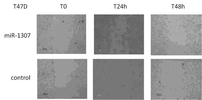
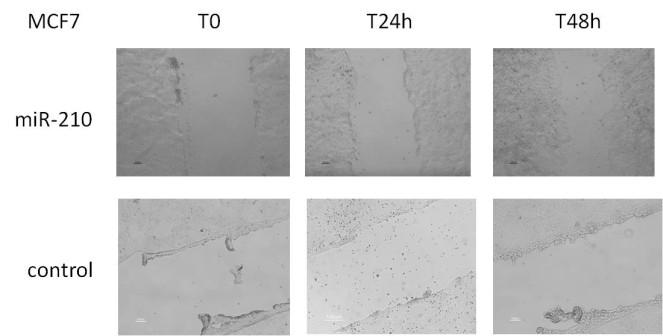


93 Figure S1. the experiments of wound healing assay, for the indicated cell lines and mirnas. 92
mirna Dr. S Hosseini-Asl
 mirna Dr. S Hosseini-Asl 1 2 MicroRNAs (mirnas) are small noncoding RNAs which enhance the cleavage or translational repression of specific mrna with recognition site(s) in the 3 - untranslated region
Basement membrane in lobule.
 Bahram Memar, MD Basement membrane in lobule. Normal lobule-luteal phase Normal lobule-follicular phase Lactating breast Greater than 95% are adenocarcinomas in situ carcinomas and invasive carcinomas.
Multimedia Appendix 6 Educational Materials Table of Contents. Intervention Educational Materials Audio Script (version 1)
") Multimedia Appendix 6 Educational Materials Table of Contents Intervention Educational Materials... 1 Audio Script (version 1)... 1 Text (version 1)... 5 Slides (version 1)... 17 Audio Script (version
Multimedia Appendix 6 Educational Materials Table of Contents Intervention Educational Materials... 1 Audio Script (version 1)... 1 Text (version 1)... 5 Slides (version 1)... 17 Audio Script (version
Maram Abdaljaleel, MD Dermatopathologist and Neuropathologist University of Jordan, School of Medicine
 Maram Abdaljaleel, MD Dermatopathologist and Neuropathologist University of Jordan, School of Medicine The most common non-skin malignancy of women 2 nd most common cause of cancer deaths in women, following
MicroRNA expression profiling and functional analysis in prostate cancer. Marco Folini s.c. Ricerca Traslazionale DOSL
 MicroRNA expression profiling and functional analysis in prostate cancer Marco Folini s.c. Ricerca Traslazionale DOSL What are micrornas? For almost three decades, the alteration of protein-coding genes
Mechanisms of hormone drug resistance
 Mechanisms of hormone drug resistance Ljiljana Stamatović Institute for Oncology and Radiology of Serbia Tenth UMOS Conference, Belgrade, 16-17 th May 2015. Hormone receptor-positive breast cancer (HR+
Intro to Cancer Therapeutics
 An Intro to Cancer Therapeutics Christopher R. Chitambar, MD Professor of Medicine Division of Hematology & Oncology Froedtert and Medical College of Wisconsin Clinical Cancer Center cchitamb@mcw.edu Intro
number Done by Corrected by Doctor Maha Shomaf
 number 19 Done by Waseem Abo-Obeida Corrected by Abdullah Zreiqat Doctor Maha Shomaf Carcinogenesis: the molecular basis of cancer. Non-lethal genetic damage lies at the heart of carcinogenesis and leads
MicroRNA in Cancer Karen Dybkær 2013
 MicroRNA in Cancer Karen Dybkær RNA Ribonucleic acid Types -Coding: messenger RNA (mrna) coding for proteins -Non-coding regulating protein formation Ribosomal RNA (rrna) Transfer RNA (trna) Small nuclear
Triple Negative Breast Cancer
 Triple Negative Breast Cancer Prof. Dr. Pornchai O-charoenrat Division of Head-Neck & Breast Surgery Department of Surgery Faculty of Medicine Siriraj Hospital Breast Cancer Classification Traditional
It is a malignancy originating from breast tissue
 59 Breast cancer 1 It is a malignancy originating from breast tissue including both early stages which are potentially curable, and metastatic breast cancer (MBC) which is usually incurable. Most breast
Recent advances in breast cancers
 Recent advances in breast cancers Breast cancer is a hetrogenous disease due to distinct genetic alterations. Similar morphological subtypes show variation in clinical behaviour especially in response
Contents 1 The Windows of Susceptibility to Breast Cancer 2 The So Called Pre-Neoplastic Lesions and Carcinoma In Situ
 Contents 1 The Windows of Susceptibility to Breast Cancer... 1 1.1 Introduction... 1 1.2 Risk Factor and Etiological Agents... 2 1.3 The Concept of the Windows of Susceptibility to Carcinogenesis... 5
Outline of the presentation
 Outline of the presentation Breast cancer subtypes and classification Clinical need in estrogen-positive (ER+) metastatic breast cancer (mbc) Sulforaphane and SFX-01: the preclinical evidence STEM Phase
Claudin-4 Expression in Triple Negative Breast Cancer: Correlation with Androgen Receptors and Ki-67 Expression
 Claudin-4 Expression in Triple Negative Breast Cancer: Correlation with Androgen Receptors and Ki-67 Expression Mona A. Abd-Elazeem, Marwa A. Abd- Elazeem Pathology department, Faculty of Medicine, Tanta
Diabetes Mellitus and Breast Cancer
 Masur K, Thévenod F, Zänker KS (eds): Diabetes and Cancer. Epidemiological Evidence and Molecular Links. Front Diabetes. Basel, Karger, 2008, vol 19, pp 97 113 Diabetes Mellitus and Breast Cancer Ido Wolf
Crosstalk between Adiponectin and IGF-IR in breast cancer. Prof. Young Jin Suh Department of Surgery The Catholic University of Korea
 Crosstalk between Adiponectin and IGF-IR in breast cancer Prof. Young Jin Suh Department of Surgery The Catholic University of Korea Obesity Chronic, multifactorial disorder Hypertrophy and hyperplasia
Diseases of the breast (2 of 2) Breast cancer
 Breast cancer") Diseases of the breast (2 of 2) Breast cancer Epidemiology & etiology The most common type of cancer & the 2 nd most common cause of cancer death in women 1 of 8 women in USA Affects 7% of women Peak at
Diseases of the breast (2 of 2) Breast cancer Epidemiology & etiology The most common type of cancer & the 2 nd most common cause of cancer death in women 1 of 8 women in USA Affects 7% of women Peak at
Breast Cancer. Dr. Andres Wiernik 2017
 Breast Cancer Dr. Andres Wiernik 2017 Agenda: The Facts! (Epidemiology/Risk Factors) Biological Classification/Phenotypes of Breast Cancer Treatment approach Local Systemic Agenda: The Facts! (Epidemiology/Risk
Computer Science, Biology, and Biomedical Informatics (CoSBBI) Outline. Molecular Biology of Cancer AND. Goals/Expectations. David Boone 7/1/2015
 Outline. Molecular Biology of Cancer AND. Goals/Expectations. David Boone 7/1/2015") Goals/Expectations Computer Science, Biology, and Biomedical (CoSBBI) We want to excite you about the world of computer science, biology, and biomedical informatics. Experience what it is like to be a
Goals/Expectations Computer Science, Biology, and Biomedical (CoSBBI) We want to excite you about the world of computer science, biology, and biomedical informatics. Experience what it is like to be a
Breast Cancer. Excess Estrogen Exposure. Alcohol use + Pytoestrogens? Abortion. Infertility treatment?
 Breast Cancer Breast Cancer Excess Estrogen Exposure Nulliparity or late pregnancy + Early menarche + Late menopause + Cystic ovarian disease + External estrogens exposure + Breast Cancer Excess Estrogen
Index. Note: Page numbers of article titles are in boldface type.
 Note: Page numbers of article titles are in boldface type. A Adjuvant therapy, for early-stage triple-negative breast cancer, 740 742 in older early-stage breast cancer patients, 790 795 anti-her2-directed
LAPATINIB-Resistance to small Molecule ErbB2 Tyrosine Kinase Inhibitor (TKI)
") LAPATINIB-Resistance to small Molecule ErbB2 Tyrosine Kinase Inhibitor (TKI) Prim Mr Sc Dr Suzana Vasović Institute for oncology and radiology of Serbia UMOS, X Conference, 16.05.2015 Belgrade How do we
LAPATINIB-Resistance to small Molecule ErbB2 Tyrosine Kinase Inhibitor (TKI) Prim Mr Sc Dr Suzana Vasović Institute for oncology and radiology of Serbia UMOS, X Conference, 16.05.2015 Belgrade How do we
10/15/2012. Biologic Subtypes of TNBC. Topics. Topics. Histopathology Molecular pathology Clinical relevance
 Biologic Subtypes of TNBC Andrea L. Richardson M.D. Ph.D. Brigham and Women s Hospital Dana-Farber Cancer Institute Harvard Medical School Boston, MA Topics Histopathology Molecular pathology Clinical
Molecular Characterization of Breast Cancer: The Clinical Significance
 Molecular Characterization of : The Clinical Significance Shahla Masood, M.D. Professor and Chair Department of Pathology and Laboratory Medicine University of Florida College of Medicine-Jacksonville
Mechanisms of Resistance to. Lisa A. Carey, M.D. University of North Carolina at Chapel Hill Lineberger Comprehensive Cancer Center
 Mechanisms of Resistance to Hormonal Therapy Lisa A. Carey, M.D. University of North Carolina at Chapel Hill Lineberger Comprehensive Cancer Center Antagonizing Estrogen Dependent Growth Premenopausal
609G: Concepts of Cancer Genetics and Treatments (3 credits)
") Master of Chemical and Life Sciences Program College of Computer, Mathematical, and Natural Sciences 609G: Concepts of Cancer Genetics and Treatments (3 credits) Text books: Principles of Cancer Genetics,
Master of Chemical and Life Sciences Program College of Computer, Mathematical, and Natural Sciences 609G: Concepts of Cancer Genetics and Treatments (3 credits) Text books: Principles of Cancer Genetics,
Introduction. Cancer Biology. Tumor-suppressor genes. Proto-oncogenes. DNA stability genes. Mechanisms of carcinogenesis.
 Cancer Biology Chapter 18 Eric J. Hall., Amato Giaccia, Radiobiology for the Radiologist Introduction Tissue homeostasis depends on the regulated cell division and self-elimination (programmed cell death)
MicroRNA dysregulation in cancer. Systems Plant Microbiology Hyun-Hee Lee
 MicroRNA dysregulation in cancer Systems Plant Microbiology Hyun-Hee Lee Contents 1 What is MicroRNA? 2 mirna dysregulation in cancer 3 Summary What is MicroRNA? What is MicroRNA? MicroRNAs (mirnas) -
RAS Genes. The ras superfamily of genes encodes small GTP binding proteins that are responsible for the regulation of many cellular processes.
 ۱ RAS Genes The ras superfamily of genes encodes small GTP binding proteins that are responsible for the regulation of many cellular processes. Oncogenic ras genes in human cells include H ras, N ras,
BREAST PATHOLOGY. Fibrocystic Changes
 BREAST PATHOLOGY Lesions of the breast are very common, and they present as palpable, sometimes painful, nodules or masses. Most of these lesions are benign. Breast cancer is the 2 nd most common cause
7/6/2015. Cancer Related Deaths: United States. Management of NSCLC TODAY. Emerging mutations as predictive biomarkers in lung cancer: Overview
 Emerging mutations as predictive biomarkers in lung cancer: Overview Kirtee Raparia, MD Assistant Professor of Pathology Cancer Related Deaths: United States Men Lung and bronchus 28% Prostate 10% Colon
Deregulation of signal transduction and cell cycle in Cancer
 Deregulation of signal transduction and cell cycle in Cancer Tuangporn Suthiphongchai, Ph.D. Department of Biochemistry Faculty of Science, Mahidol University Email: tuangporn.sut@mahidol.ac.th Room Pr324
Introduction to Cancer Biology
 Introduction to Cancer Biology Robin Hesketh Multiple choice questions (choose the one correct answer from the five choices) Which ONE of the following is a tumour suppressor? a. AKT b. APC c. BCL2 d.
Molecular classification of breast cancer implications for pathologists. Sarah E Pinder
 Molecular classification of breast cancer implications for pathologists Sarah E Pinder Courtesy of CW Elston Histological types Breast Cancer Special Types 17 morphological special types 25-30% of all
Genetics and Genomics in Medicine Chapter 6 Questions
 Genetics and Genomics in Medicine Chapter 6 Questions Multiple Choice Questions Question 6.1 With respect to the interconversion between open and condensed chromatin shown below: Which of the directions
6/22/2017 TARGETING THE TARGETS IN 2017 TARGETING THE TARGETS IN 2017
 TARGETING THE TARGETS IN 2017 Primary Care Focus Symposium July 1, 2017 Grace Wang MD I do not have any relevant financial relationships to disclose at this time TARGETING THE TARGETS IN 2017 What are
BREAST CANCER BREAST CANCER
 BREAST CANCER George Raptis, M.D., M.B.A Division of Medical Oncology & Hematology College of Physicians & Surgeons Columbia University BREAST CANCER Epidemiology - Commonest cancer in women - About 235,000
DOES THE BRCAX GENE EXIST? FUTURE OUTLOOK
 CHAPTER 6 DOES THE BRCAX GENE EXIST? FUTURE OUTLOOK Genetic research aimed at the identification of new breast cancer susceptibility genes is at an interesting crossroad. On the one hand, the existence
Understanding and Optimizing Treatment of Triple Negative Breast Cancer
 Understanding and Optimizing Treatment of Triple Negative Breast Cancer Edith Peterson Mitchell, MD, FACP Clinical Professor of Medicine and Medical Oncology Program Leader, Gastrointestinal Oncology Department
Breast Cancer. Most common cancer among women in the US. 2nd leading cause of death in women. Mortality rates though have declined
 Breast Cancer Most common cancer among women in the US 2nd leading cause of death in women Mortality rates though have declined 1 in 8 women will develop breast cancer Breast Cancer Breast cancer increases
Open Clinical Trials: What s Out There Now Paula D. Ryan, MD, PhD
 Open Clinical Trials: What s Out There Now Paula D. Ryan, MD, PhD Hanahan and Weinberg, 2000 Acquired Capabilities of Cancer Clinical Trials When should I consider a clinical trial? How do I find the right
Osamu Tetsu, MD, PhD Associate Professor Department of Otolaryngology-Head and Neck Surgery School of Medicine, University of California, San
 Osamu Tetsu, MD, PhD Associate Professor Department of Otolaryngology-Head and Neck Surgery School of Medicine, University of California, San Francisco Lung Cancer Classification Pathological Classification
The Biology and Genetics of Cells and Organisms The Biology of Cancer
 The Biology and Genetics of Cells and Organisms The Biology of Cancer Mendel and Genetics How many distinct genes are present in the genomes of mammals? - 21,000 for human. - Genetic information is carried
Dr Rodney Itaki Lecturer Anatomical Pathology Discipline. University of Papua New Guinea School of Medicine & Health Sciences Division of Pathology
 Neoplasia Dr Rodney Itaki Lecturer Anatomical Pathology Discipline University of Papua New Guinea School of Medicine & Health Sciences Division of Pathology General Considerations Overview: Neoplasia uncontrolled,
Regulation of Gene Expression in Eukaryotes
 Ch. 19 Regulation of Gene Expression in Eukaryotes BIOL 222 Differential Gene Expression in Eukaryotes Signal Cells in a multicellular eukaryotic organism genetically identical differential gene expression
Breast Cancer. Saima Saeed MD
 Breast Cancer Saima Saeed MD Breast Cancer Most common cancer among women in the US 2nd leading cause of death in women 1 in 8 women will develop breast cancer Incidence/mortality rates have declined Breast
CELL BIOLOGY - CLUTCH CH CANCER.
 !! www.clutchprep.com CONCEPT: OVERVIEW OF CANCER Cancer is a disease which is primarily caused from misregulated cell division, which form There are two types of tumors - Benign tumors remain confined
Carcinoma mammario: le istologie non frequenti. Valentina Guarneri Università di Padova IOV-IRCCS
 Carcinoma mammario: le istologie non frequenti Valentina Guarneri Università di Padova IOV-IRCCS Histological diversity of breast adenocarcinomas Different histological types are defined according to specific
 Radiation therapy Radiation therapy is a cancer treatment that uses high-energy x-rays or other types of radiation to kill cancer cells or keep them from growing. Chemotherapy Chemotherapy is a cancer
Dr C K Kwan. Associate Consultant, Queen Elizabeth Hospital, HKSAR Honorary member, Targeted Therapy Team, ICR, UK
 HA Convention 2013 Dr C K Kwan MBChB, FRCR, FHKCR, FHKAM, MPM, CPI, MAppMgt(Hlth) Associate Consultant, Queen Elizabeth Hospital, HKSAR Honorary member, Targeted Therapy Team, ICR, UK To personalize cancer
LESSON 3.2 WORKBOOK. How do normal cells become cancer cells? Workbook Lesson 3.2
 For a complete list of defined terms, see the Glossary. Transformation the process by which a cell acquires characteristics of a tumor cell. LESSON 3.2 WORKBOOK How do normal cells become cancer cells?
oncogenes-and- tumour-suppressor-genes)
") Special topics in tumor biochemistry oncogenes-and- tumour-suppressor-genes) Speaker: Prof. Jiunn-Jye Chuu E-Mail: jjchuu@mail.stust.edu.tw Genetic Basis of Cancer Cancer-causing mutations Disease of aging
Special topics in tumor biochemistry oncogenes-and- tumour-suppressor-genes) Speaker: Prof. Jiunn-Jye Chuu E-Mail: jjchuu@mail.stust.edu.tw Genetic Basis of Cancer Cancer-causing mutations Disease of aging
Immunohistochemical classification of breast tumours
 Immunohistochemical classification of breast tumours Workshop in Diagnostic Immunohistochemistry September 19 th - 21 th 2018 Anne-Vibeke Lænkholm Department of Surgical Pathology, Zealand University Hospital,
UNIVERSITY OF MEDICINE AND PHARMACY CRAIOVA PhD SCHOOL. PhD THESIS
 UNIVERSITY OF MEDICINE AND PHARMACY CRAIOVA PhD SCHOOL PhD THESIS THE IMPORTANCE OF TUMOR ANGIOGENESIS IN CEREBRAL TUMOR DIAGNOSIS AND THERAPY ABSTRACT PhD COORDINATOR: Prof. univ. dr. DRICU Anica PhD
Corporate Medical Policy
 Corporate Medical Policy Molecular Analysis for Targeted Therapy for Non-Small Cell Lung File Name: Origination: Last CAP Review: Next CAP Review: Last Review: molecular_analysis_for_targeted_therapy_for_non_small_cell_lung_cancer
Page. Objectives: Hormone Therapy Resistance: Challenges and Opportunities. Research Support From Merck
 Hormone Therapy Resistance: Challenges and Opportunities Pamela. N. Munster, MD University of California, San Francisco Financial Disclosures Research Support From Merck Objectives: Understanding the current
Oncogenes and Tumor Suppressors MCB 5068 November 12, 2013 Jason Weber
 Oncogenes and Tumor Suppressors MCB 5068 November 12, 2013 Jason Weber jweber@dom.wustl.edu Oncogenes & Cancer DNA Tumor Viruses Simian Virus 40 p300 prb p53 Large T Antigen Human Adenovirus p300 E1A
Lecture 8 Neoplasia II. Dr. Nabila Hamdi MD, PhD
 Lecture 8 Neoplasia II Dr. Nabila Hamdi MD, PhD ILOs Understand the definition of neoplasia. List the classification of neoplasia. Describe the general characters of benign tumors. Understand the nomenclature
Concise Reference. HER2 Testing in Breast Cancer. Mary Falzon, Angelica Fasolo, Michael Gandy, Luca Gianni & Stefania Zambelli
 Concise Reference Testing in Breast Cancer Mary Falzon, Angelica Fasolo, Michael Gandy, Luca Gianni & Stefania Zambelli Extracted from Handbook of -Targeted Agents in Breast Cancer ublished by Springer
Circular RNAs (circrnas) act a stable mirna sponges
 act a stable mirna sponges") Circular RNAs (circrnas) act a stable mirna sponges cernas compete for mirnas Ancestal mrna (+3 UTR) Pseudogene RNA (+3 UTR homolgy region) The model holds true for all RNAs that share a mirna binding
Circular RNAs (circrnas) act a stable mirna sponges cernas compete for mirnas Ancestal mrna (+3 UTR) Pseudogene RNA (+3 UTR homolgy region) The model holds true for all RNAs that share a mirna binding
Precision Genetic Testing in Cancer Treatment and Prognosis
 Precision Genetic Testing in Cancer Treatment and Prognosis Deborah Cragun, PhD, MS, CGC Genetic Counseling Graduate Program Director University of South Florida Case #1 Diana is a 47 year old cancer patient
Question 1 A. ER-, PR-, HER+ B. ER+, PR+, HER2- C. ER-, PR+, HER2- D. ER-, PR-, HER2- E. ER-, PR+, HER2+
 Triple Negative Breast Cancer Laura C. Collins, M.D. Department of Pathology Beth Israel Deaconess Medical Center and Harvard Medical School, Boston, MA Question 1 The tumor depicted on the next slide
microrna Presented for: Presented by: Date:
 microrna Presented for: Presented by: Date: 2 micrornas Non protein coding, endogenous RNAs of 21-22nt length Evolutionarily conserved Regulate gene expression by binding complementary regions at 3 regions
Predictive Assays in Radiation Therapy
 Outline Predictive Assays in Radiation Therapy Radiation Biology Introduction Early predictive assays Recent trends in predictive assays Examples for specific tumors Summary Lecture 4-23-2014 Introduction
1. Basic principles 2. 6 hallmark features 3. Abnormal cell proliferation: mechanisms 4. Carcinogens: examples. Major Principles:
 Carcinogenesis 1. Basic principles 2. 6 hallmark features 3. Abnormal cell proliferation: mechanisms 4. Carcinogens: examples Carcinogenesis Major Principles: 1. Nonlethal genetic damage is central to
Ch. 18 Regulation of Gene Expression
 Ch. 18 Regulation of Gene Expression 1 Human genome has around 23,688 genes (Scientific American 2/2006) Essential Questions: How is transcription regulated? How are genes expressed? 2 Bacteria regulate
Development of Carcinoma Pathways
 The Construction of Genetic Pathway to Colorectal Cancer Moriah Wright, MD Clinical Fellow in Colorectal Surgery Creighton University School of Medicine Management of Colon and Diseases February 23, 2019
Personalized Genetics
 Personalized Genetics Understanding Your Genetic Test Results Tracey Evans, MD September 29, 2017 Genetics 101 Punnett Square Genetic Pedigree 2 Genetics 101 Punnett Square Genetic Pedigree 3 It s not
Cancer genetics
 Cancer genetics General information about tumorogenesis. Cancer induced by viruses. The role of somatic mutations in cancer production. Oncogenes and Tumor Suppressor Genes (TSG). Hereditary cancer. 1
Breast cancer: Molecular STAGING classification and testing. Korourian A : AP,CP ; MD,PHD(Molecular medicine)
") Breast cancer: Molecular STAGING classification and testing Korourian A : AP,CP ; MD,PHD(Molecular medicine) Breast Cancer Theory: Halsted Operative breast cancer is a local-regional disease The positive
Breast cancer: Molecular STAGING classification and testing Korourian A : AP,CP ; MD,PHD(Molecular medicine) Breast Cancer Theory: Halsted Operative breast cancer is a local-regional disease The positive
Johns Hopkins Clinical Update Webinar
 Johns Hopkins Clinical Update Webinar Ben Ho Park, M.D., Ph.D. Department of Oncology Johns Hopkins University February 2015 This presentation is the intellectual property of the author/presenter. Contact
José Baselga, MD, PhD
 i n t e r v i e w José Baselga, MD, PhD Dr Baselga is Physician-in-Chief at Memorial Sloan-Kettering Cancer Center in New York, New York. Tracks 1-15 Track 1 Track 2 Track 3 Track 4 Track 5 Track 6 Track
UK Interdisciplinary Breast Cancer Symposium. Should lobular phenotype be considered when deciding treatment? Michael J Kerin
 UK Interdisciplinary Breast Cancer Symposium Should lobular phenotype be considered when deciding treatment? Michael J Kerin Professor of Surgery National University of Ireland, Galway and Galway University
Breast Cancer: the interplay of biology, drugs, radiation. Prof. L. Livi Università degli Studi di Firenze. Brescia, October 3rd 4th, 2013
 Breast Cancer: the interplay of biology, drugs, radiation Prof. L. Livi Università degli Studi di Firenze Brescia, October 3rd 4th, 2013 BACKGROUND (1) The complex interactions between tumor-specific signaling
Corporate Medical Policy
 Corporate Medical Policy Ado-Trastuzumab Emtansine (Trastuzumab-DM1) for Treatment of File Name: Origination: Last CAP Review: Next CAP Review: Last Review: ado_trastuzumab_emtansine_(trastuzumab-dm1)_for_treatment_of_her-2_positivemalignancies
Contemporary Classification of Breast Cancer
 Contemporary Classification of Breast Cancer Laura C. Collins, M.D. Vice Chair of Anatomic Pathology Professor of Pathology Beth Israel Deaconess Medical Center and Harvard Medical School Boston, MA Outline
 Mdi Medical Management of Breast Cancer Morbidity and Mortality Aug 13, 2009 Irina Kovatch, PGY3 Introduction Metastatic disease is the principal cause of death from breast cancer Metastatic events often
The PI3K/AKT axis. Dr. Lucio Crinò Medical Oncology Division Azienda Ospedaliera-Perugia. Introduction
 The PI3K/AKT axis Dr. Lucio Crinò Medical Oncology Division Azienda Ospedaliera-Perugia Introduction Phosphoinositide 3-kinase (PI3K) pathway are a family of lipid kinases discovered in 1980s. They have
Recent Update in Management of Breast Cancer: Medical Oncology. Jin Hee Ahn, M.D., PhD. 23-April-2015
 2015 GBCC & 4 th IBCS 1/37 Recent Update in Management of Breast Cancer: Medical Oncology Jin Hee Ahn, M.D., PhD. 23-April-2015 Department of Oncology, Asan Medical Center, UUCM, Seoul, Korea 2/37 3/37
Biochemistry of Carcinogenesis. Lecture # 35 Alexander N. Koval
 Biochemistry of Carcinogenesis Lecture # 35 Alexander N. Koval What is Cancer? The term "cancer" refers to a group of diseases in which cells grow and spread unrestrained throughout the body. It is difficult
TUMOR M ARKERS MARKERS
 TUMOR MARKERS M.Shekarabi IUMS Definition Many cancers are associated with the abnormal production of some molecules l which h can be measured in plasma. These molecules are known as tumor markers. A good
Targeting CDK 4/6. Jee Hyun Kim, M.D., Ph.D. Seoul National University College of Medicine
 2016.04.30 GBCC Education Symposium Targeting CDK 4/6 Jee Hyun Kim, M.D., Ph.D. Seoul National University College of Medicine Contents Cyclins -CDKs in cell cycle control CDK 4/6 in breast cancer Preclinical
MicroRNA and Male Infertility: A Potential for Diagnosis
 Review Article MicroRNA and Male Infertility: A Potential for Diagnosis * Abstract MicroRNAs (mirnas) are small non-coding single stranded RNA molecules that are physiologically produced in eukaryotic
Breast Cancer Statistics
 1 in 8 Breast Cancer Statistics Incidence Mortality Prevalence 2 Breast Cancer Incidence Breast Cancer Mortality Breast Cancer Prevalence ~$100,000 Female Breast Anatomy Breasts consist mainly of fatty
Epigenetic Principles and Mechanisms Underlying Nervous System Function in Health and Disease Mark F. Mehler MD, FAAN
 Epigenetic Principles and Mechanisms Underlying Nervous System Function in Health and Disease Mark F. Mehler MD, FAAN Institute for Brain Disorders and Neural Regeneration F.M. Kirby Program in Neural
Clinical Oncology - Science in focus - Editorial. Understanding oestrogen receptor function in breast cancer, and its interaction with the
 Clinical Oncology - Science in focus - Editorial TITLE: Understanding oestrogen receptor function in breast cancer, and its interaction with the progesterone receptor. New preclinical findings and their
Novel Strategies in Systemic Therapies: Overcoming Endocrine Therapy Resistance
 Novel Strategies in Systemic Therapies: Overcoming Endocrine Therapy Resistance Richard S. Finn, MD Division of Hematology/ Oncology Director, Translational Oncology Laboratory Geffen School of Medicine
Mousa. Israa Ayed. Abdullah AlZibdeh. 0 P a g e
 1 Mousa Israa Ayed Abdullah AlZibdeh 0 P a g e Breast pathology The basic histological units of the breast are called lobules, which are composed of glandular epithelial cells (luminal cells) resting on
Determination Differentiation. determinated precursor specialized cell
 Biology of Cancer -Developmental Biology: Determination and Differentiation -Cell Cycle Regulation -Tumor genes: Proto-Oncogenes, Tumor supressor genes -Tumor-Progression -Example for Tumor-Progression:
Clinico- Pathological Features And Out Come Of Triple Negative Breast Cancer
 Clinico- Pathological Features And Out Come Of Triple Negative Breast Cancer Dr. HassanAli Al-Khirsani, MBChB, CABM, F.I.C.M.S AL-Sadder teaching hospital, oncology unit Dr. Nasser Ghaly Yousif, MBChB,G.P.
Breast Cancer Carcinogenesis: Mechanisms and Pathways in Hormone Receptor Positive Disease
 Breast Cancer Carcinogenesis: Mechanisms and Pathways in Hormone Receptor Positive Disease 1 Nearly Two-Thirds of Metastatic Breast Cancers Express Hormone Receptors Breast cancer tumors are often classified
PHARMACOGENETICS OF BREAST CANCER
 PHARMACOGENETICS OF BREAST CANCER MALGORZATA JAREMKO, PhD Mount Sinai School of Medicine, Department of Genetics and Genomic Sciences Outlines Breast cancer therapeutic situation Pharmacogenetics of antiestrogen
Genome of Hepatitis B Virus. VIRAL ONCOGENE Dr. Yahwardiah Siregar, PhD Dr. Sry Suryani Widjaja, Mkes Biochemistry Department
 Genome of Hepatitis B Virus VIRAL ONCOGENE Dr. Yahwardiah Siregar, PhD Dr. Sry Suryani Widjaja, Mkes Biochemistry Department Proto Oncogen and Oncogen Oncogen Proteins that possess the ability to cause
Overview: Conducting the Genetic Orchestra Prokaryotes and eukaryotes alter gene expression in response to their changing environment
 Overview: Conducting the Genetic Orchestra Prokaryotes and eukaryotes alter gene expression in response to their changing environment In multicellular eukaryotes, gene expression regulates development
September 20, Submitted electronically to: Cc: To Whom It May Concern:
 History Study (NOT-HL-12-147), p. 1 September 20, 2012 Re: Request for Information (RFI): Building a National Resource to Study Myelodysplastic Syndromes (MDS) The MDS Cohort Natural History Study (NOT-HL-12-147).
Introduction to Genetics
 Introduction to Genetics Table of contents Chromosome DNA Protein synthesis Mutation Genetic disorder Relationship between genes and cancer Genetic testing Technical concern 2 All living organisms consist
Subtype-directed therapy of TNBC Global Breast Cancer Conference 2015 & 4th International Breast Cancer Symposium Jeju Island, Korea, April 2015
 Subtype-directed therapy of TNBC Global Breast Cancer Conference 2015 & 4th International Breast Cancer Symposium Jeju Island, Korea, April 2015 Ruth M. O Regan, MD Visiting Professor and Division Chief
Histological Type. Morphological and Molecular Typing of breast Cancer. Nottingham Tenovus Primary Breast Cancer Study. Survival (%) Ian Ellis
 Ian Ellis") Morphological and Molecular Typing of breast Cancer Ian Ellis Molecular Medical Sciences, University of Nottingham Department of Histopathology, Nottingham University Hospitals NHS Trust Histological Type
Morphological and Molecular Typing of breast Cancer Ian Ellis Molecular Medical Sciences, University of Nottingham Department of Histopathology, Nottingham University Hospitals NHS Trust Histological Type
Tumor suppressor genes D R. S H O S S E I N I - A S L
 Tumor suppressor genes 1 D R. S H O S S E I N I - A S L What is a Tumor Suppressor Gene? 2 A tumor suppressor gene is a type of cancer gene that is created by loss-of function mutations. In contrast to