The nature of miniature melanocytes in murine epidermis
|
|
|
- Moris George
- 6 years ago
- Views:
Transcription
1 University of Richmond UR Scholarship Repository Master's Theses Student Research The nature of miniature melanocytes in murine epidermis John Thomas Earnhardt Follow this and additional works at: Recommended Citation Earnhardt, John Thomas, "The nature of miniature melanocytes in murine epidermis" (1973). Master's Theses. Paper 472. This Thesis is brought to you for free and open access by the Student Research at UR Scholarship Repository. It has been accepted for inclusion in Master's Theses by an authorized administrator of UR Scholarship Repository. For more information, please contact
2 The Nature of Miniature Melanocytes in Murine Epidermis by John Thomas Earnhardt B. S. Lenoir Rhyne College 1971 A thesis submitted to the faculty of the Graduate School of the University of Richmond in partial fulfillment of the requirements for the degree of Master of Science in Biology April 1973 LISR/\i':Y.UNIVC:RS!TY o::- t :1c; l ''.:J\','D Vil::G! i J I/\
3 The Nature of Miniature Melanocytes in Murine Epidermis by John Thomas Earnhardt Approved Examining Committee
4 I ' TABLE OF CONTENTS I Abstract ii II Acknowledgements iii III Introduction 1-4 IV Materials and Methods 5-10 V Results VI Discussion VII References VIII Figures IX Vita 34
5 ABSTRACT In the epidermis of PET/Wmr mice the population of melanocytes reaches a peak and begins to decline during the first postnatal week, and has disappeared within four weeks. During this period a small number of weakly DOPA reactive miniature melanocytes are seen dispersed among the generally large, highly DOPA reactive melanocytes typical of the animal. These miniature melanocytes appear early, are among the last to disappear, and their population remains relatively constant against the drastically changing population of typical melanocytes. In attempts to determine the nature of the miniature melanocytes, heterologous grafting was employed whereby the melanocytes were subjected to morphogenetic impacts. The evidence suggests that these miniature melanocytes are relatively resistant to transformation into typical melanocytes, and may constitute a distinct cell type.
6 -iii- ACKNOWLEDGEMENTS I wish to express my sincere thanks to Dr. W. M. Reams for his guidance throughout this investigation and for the association which has afforded that which may not have been. I would also like to thank Dr. F. B. Leftwich for his helpful suggestions in the preparation of this communication and otherwise in the course of graduate work. My appreciation is expressed to Dr. W. S. Woolcott for his review of the manuscript and his encouragement throughout my graduate studies. I also thank Dr. William H. Leftwich for his assistance in preparation of statistical analysis. I would like to thank my wife, Marian and daughter, Heather for their understanding throughout.
7 INTRODUCTION It is well known that the melanocytes of the skin and hair of mammals are derived from melanoblasts which migrate from the neural crest early in embryonic development (Rawles,1947). That mammalian epidermal melanocytes vary somewhat as to shape, size, and color is well established (Markert and Silvers, 1956). Although the mode by which melanocyte morphology is expressed is genotypically dependent, the tissue environment does modify cellular expression. Markert and Silvers (1959) examined mela~ocyte morphology as related to tissue environment and have shown that cells with a dilute (dd) genotype exhibit a non-dilute morphology if grown in the anterior chamber of the eye. Rovee (1965) has shown that cell size is related to cell density in the melanocyte system of the mouse in that the greater the density, the smaller the size of the melanocytes. In the melanocyte pattern of an area of freckled epidermis covering a stretched scar, Breathnach (1958) noted the presence of small, weakly DOPA reactive
8 -2- melanocytes in the pale area of the scar. Also in man, the white macules of tuberous sclerosis have melanocytes which are small and weakly DOPA reactive as compared to normal melanocytes (Fitzpatrick et al, 1968, Figures 5 and 6). The chorioallantoic membrane (CAM) of chick embryos possesses extensive vascularity which can support the cultivation of isolated tissues (New, 1966). The grafting process involves direct vascular connections between the graft and the chorioallantoic circulation (Coulombre, 1967). The early development of the procedure for CAM grafting (Willier, 1924) has been modified somewhat since its conception. Although the tissue explant is isolated from its original melieu, it does come under hormonal and other influences present in the blood of the host (Rugh, 1962). One factor which eminates in the blood has been described by Bullough and Lawrence (1962} with the term chalone. In an earlier investigation by Bullough and Lawrence (1960), the question of the presence of growth controlling, tissue specific inhibitors was reviewed. It is now generally accepted that such inhibitors of mitotic activity are present in various
9 -3- tissue and control cell proliferation by negative feedback inhibition. The action of chalones persists in vitro and in vivo and although tissue specificity exists, species specificity does not (Maugh, 1972). Reams (1963) has described a "morphogenetic factor" present in humoral fluid of chick embryos that can evoke branching of donor pigment cells within the coelomic lining of the host. In that same investigation the morphogenetic factor has been shown to be non-species-specific with regard to the mouse and the chick. In the epidermis of the PET/Wmr mice, the melanocyte population reaches a peak and begins to decline during the first postnatal week of life. During this period, a small number of miniature melanocytes (figure 8) have been noted dispersed among the generally large melanocytes (figure T) typical of the animal. The purpose of this investigation was to attempt to determine the nature of the miniature melanocytes by subjecting them to the impact of the circulating titres of chalone and morphogenetic factor present in the chick embryo via the CAM. The pigment cell terminology used throughout this communication conforms to that of Fitzpatrick et al,
10 -4- (1966): Melanoblast- a cell which is the precursor of the melanocyte. Melanocyte- any cell that produces melanin; a cell which synthesizes a specialized melanin-containing organelle known as the melanosome. A melanoblast becomes a melanocyte with the formation of melanosomes.
11 MATERIALS AND METHODS Mice of the PET/Wmr strain maintained at the University of Richmond were used throughout this investigation. In order to determine the normal pigment cell complement of the epidermis of the mice, microscopic examination of whole mount preparations was carried out. Mice.were selected for use when at least three separate litter births occured within 6 hours. At least one animal from each of the three litters was killed by decapitation at the following ages: O, 1, 2, 3, 4~ 5, 6, 7, 8, 9, 10, 15, days and adult. Litters were allowed to age in order that the time period between each day would more closely approximate 24 hours. A specimen of skin between 6 and 10 nnn per side was removed from the mid dorsum of each mouse. The hair of the mice in age groups 6 days and older was removed by electric clippers and/or a commercially available depilatory chemical. Each skin section was removed along with the underlying hypodermis and scraped to remove the subcutaneous tissue. To prevent the skin from rolling up, it was placed dermal side down on a square of
12 Whatman #1 filter paper slightly larger than the skin specimen. The sodium bromide method for epidermal stripping was employed: the skin was placed in 2 M NaBr for approximately 45 minutes at 37 C after which the epidermis was separated from the dermis with the aid of a dissecting microscope (20 X) and Dumont #5 forceps. The epidermis was then placed in 5% formaldehyde solution (ph 7.2 with NaHC03) for 30 minutes with a successive washing in distilled water (5 minutes). The skin section was then placed into a 0.1% buffered solution of 1: 3-4 dihydroxyphenylalinine (DOPA). In order to prepare this solution, phosphate buffer was prepared by combining 20 parts of 0.1 M KH2P04 and 80 parts 0.1 M Na2HP04. This stock buffer solution (ph 7.4) was used in the preparation of DOPA solutions throughout, all of which were compounded just prior to use. Tissues were incubated with the DOPA solution for one hour at which time the DOPA solution was replaced with freshly prepared DOPA solution and incubated for an additional three hours. All incubations were carried out at 37 C. Subsequent to the DOPA staining, the tissue was plac~d in 5% formalin and allowed to stand overnight.
13 -7- The section was then placed in a position with the hair follicles upward. The follicles were removed by "plucking" with Dumont #5 forceps utilizing magnification (20 X). After washing in distilled water for 15 minutes, the epidermal sheets were dehydrated in successive solutions of 70, 95, and 100% ethanol for a minimum of 15 minutes each. The tissue was then cleared in xylene and a permanent slide prepared with balsam. The number of melanocytes was determined by microscopically examining five areas on each whole mount preparation of skin using an ocular grid of known dimensions. The areas examined on each control slide were identical although orientation of the slide in the particular plane may have varied. Slides were marked in such a manner as to keep epidermal sheets of the same litter in corresponding groups. Mean values for each age group were determined and a graphic relationship of age versus melanocytes/mm 2 established. To prepare grafts of mouse skin, newborn PET mice (6 hours: postnatal age) were obtained and sacrificed by decapitation. A small square of skin (5-8 mm/side) was removed from the mid-dorsum and placed in sterile saline containing.06 mg of penicillin (Penicillin G Sodium, Nutritional Biochemicals Company) and.05 mg
14 -8- of streptomycin (Streptomycin Sulfate, Nutritional Biochemicals Company) per cubic centimeter. The ph of the saline/antibiotic solution was adjusted to 7.2 with NaHC0 3. The skin sections were scraped on the dermal side to remove the hypodermis and left in the saline/ antibiotic solution while hosts were made ready for receiving the grafts. In order to determine the effects of environmental change upon the melanocyte population, CAM grafting was employed. Fertile eggs of the White Leghorn fowl were obtained from a local poultry farm. The age of the embryo was determined by establishing the hour of iniation of incubation as the base line for the onset of development. Fertile eggs were incubated in a Davis Bradley cabinet incubator maintained at 38 C with a consistant level of humidity. Eggs incubated for days were used as hosts. Each egg was candled in a darkened room to determine the position of the major blood vessels of the CAM. The area on the shell overlying one of the blfurcations of major blood vessel in the CAM was penciled. Each egg was then placed on a nest of cotton in a Syracuse watch glass and cleaned with cotton soaked in 70% ethanol.
15 -9- A small triangular hole (lcm/side) was carefully cut in the shell using a 10 cm section of a fine toothed hacksaw blade. The shell was removed, leaving the shell membrane intact. The shell membrane was moistened with saline/antibiotic solution to reduce the tendancy for adherence to the chorioallantois. The shell membrane was removed with extreme care to avoid puncturing the CAM. Previously prepared grafts were removed from the saline/antibiotic, placed onto the flat end of a blunted glass stirrring rod, and carefully lowered onto the CAM with the dermis side down. Orientation of the graft on a well vascularized area between bifurcating blood vessels was accomplished with the aid of forceps. Aseptic technique was used throughout the grafting procedure. The triquetral opening was sealed with cellophane tape with the edges sealed by applying melted paraffin with a small brush. The eggs were returned to the incubator with the cellophane window directed upward. The grafts were recovered after 4, 5, 7, and 8 days of incubation. The epidermis was removed by the sodium bromide method and the resulting epidermal sheet subjected to the DOPA procedure explained previously. Whole mount preparations were made and examined micr~scopically with an ocular grid employed for determining melanocyte populations. Five separate
16 -10- areas were counted in each epidermal preparation. Mean values were obtained and collated with control values of melanocyte numbers in groups comparable in age to the CAM graft period. In a preliminary survey, it was found that there was a good correlation between the general size of a melanocyte and the size of its nucleus. Therefore, to qualify as a miniature melanocyte, a pigment cell had to have a nuclear volume no greater than half that of a typical melanocyte.
17 -11- RESULTS The control population of melanocytes is presented as a relationship between age of the mice and the number of melanocytes per square millimeter. It can be seen (figure 1) that in the population of macromelanocytes (typical melanocytes) the peak number of cells is reached at day 3 after which there is a gradual decline in the melanocyte population until a relatively constant rate is seen after day 15. From the graph (figure 1) it is evident that the range is much greater for mice in the age groups of 2-3 day. The mean values for each group (day) were obtained from a total of fifteen areas in three different mice. Figure 1 also relates the relative constancy of the miniature melanocytes from day 0 to day 10 although the group with the largest mean value (day 3) coincides with the peak obtained in macromelanocyte counts. The miniature melanocyte population after day 10 shows mean values that are similar. A total of 107 CAM grafts were attempted. It is readily apparent from figure 2 that the macromelanocyte population shows a marked increase in melanocyte numbers beginning with an average of 89.7 cells/mm2 after 4 days of CAM incubation to an average of cells/mm2 after
18 -l:l- 8 days of CAM incubation and no apparent peak in the number of cells/mm2. The graph (figure 2) shows the 7 day CAM graft to have the widest range. The miniature melanocyte population increased slightly before declining with 8.2, 9.3, 10.2, and 7.9 cells calculated per mm2 for respective CAM incubation periods of 4, 5, 7, and 8 days. Figure 3 depicts the relationship between the macromelanocyte control population and the macromelanocyte CAM graft population. It is evident that the population of melanocytes in the CAM grafts rises later when an overall comparative picture is presented (see figure 9). Mean values approximate each other at day 5 with the control population averaging cells/mm2 and the CAM graft population averaging cells/mm2. The control range of this group is contained within the range of the CAM graft population; However, the graft indicates divergence at this point. Statistical analysis employing the t-test revealed significance in 3 of 4 in macromelanocyte population when control melanocyte numbers for a given day were compared with CAM graft populations for the corresponding day. Statistically significant figures were recorded for days 4, 7, and 8 when total populations were compared.
19 . -LS- A comparison of miniature melanocyte populations can be seen in figure 4. Although the values seem to show only slight differences, significant values we~e recorded for days 4 and 7. In both of these populations the range of values overlapped when control and experimental groups were compared. t! The standard deviation for each population in the ; ~ miniature group was 'determined 'in corder to obtain a better understanding of the relationship involved.
20 -14- DISCUSSION The present study establishes a normal population of epidermal melanocytes present in the dorsum of the PET/Wmr mice. The macromelanocytes were c onsidered to be the normal type since their size and DOPA reactivi.ty compared favorably with that of previous investigations. Quevedo et al. (1966) in relating the number of epidermal melanocytes per mm2 in mid-dorsum to the age of dilute black (dd) mice and intense black (DD) mice has shown a graphic relationship with the peak of melanocyte population seen at age 2 days with a steady decline thereafter. The populations presented by Quevedo show ~hat DOPA reactive epidermal melanocytes in the intense black mice averaged 163:± 6/mm 2 at birth and increased to a maximum of /mm2 at age 2 days. In dilute black mice the number averaged /rmn2 at birth and reached a peak of SSS + 48/mm2 at day 2. In the ventral skin of PET mice, Rovee and Reams (1964) have shown peak population of melanocytes present 3 days after birth. The relative relationship between melanocyte populations for a given day when relating this investigation to others, compares favorably in a way as
21 -15- to show a general trend in the pattern of the melanocyte population in the first 10 days of postnatal life. Although small melanocytes have been noted in scat~ tered reports, this investigation brings together their presence in a way which relates them to the control population of melanocytes. Perhaps the lack of their being reported generally has been due to the assumption that small cells become larger when they mature. The control group results indicate a relatively constant population of small melanocytes, suggesting they they are not an intermediate cell type in the sense of the melanoblast. It has been established that the melanoblasts migrate from the neural crest of the embryo of the mouse (Rawles, 1947). After invasion into an area, t'he melanoblast is capable of 1) Immediate proliferation, 2) differentiation into a melanocyte, or 3) latentcy with subsequent proliferation or maturation. This varied potentaility of the melanoblast is effected by two main factors: the genotype of the cell and the environment in which the cell resides (Markert and Silvers, 1959). Quevedo et al. (1966) have shown that many melanocytes which are active shortly after birth continue in an arnelanotic state after the early postnatal life. It is possible also that pigment cells may fail to be identified
22 even by the DOPA reaction since the latent form of the cell is not always expressed by chemical techniques (Mayer, 1965). This may account for the gradual decline in the melanocyte population of the control group. That is, the pigment cells are present but are in an amelanotic form. This suggestion is supported by Quevedo et al. (1966) who have noted the presence of melanocytes showing various degrees of DOPA reactivity. Since the control population of miniature melanocytes remains constant, it is proposed that there are melanoblasts which give rise only to small melanocytes. Reams (1956) has shown that in the chick embryo some tissues are able to evoke branching of unbranched melanocytes more effectively than others. That investigation has also shown the coelomic lining as a tissue has an inhibitory effect, holding the branching of a pigment cell in check. Reams (1957) has shown the skin to be an initiator of pigment cell branching and the principle source of a pigment cell branching effector substance. Nichols and Reams (1960) have shown that unbranched melanocytes of PET/MCV mouse embryos changed from the unbranched to branched form when the chick hosts were 15 days old. Reams (1963) has interpreted this response to indicate the time at which a titre of the effector substance sufficient to evoke
23 -17- branching is present. From data presented in the above mentioned investigation, the present study incorporated fertile eggs incubated days in order to extend the full impact of the morphogenetic factor upon the host. The CAM grafts resulted in pigment cell populations which included miniature as well as macromelanocytes after DOPA incubation. If the titre of chalone and morphogenetic factor was present in the blood of the chick embryo, the effects of the non-species-specific factors should bestow their influence upon the epidermal sheet. Since grafts which c~e in contact with the CAM were initially close to the newborn age, the regulator factors should influence the entire complement of pigment cells. That is, since miniature melanocytes are present in the first few postnatal days of the control group population the regulator factors should inhibit the mitotic activity of pigment cells in.the grafts. Therefore, if the smaller,cells were precursors of the. larger cells, miniature., me~~mocytes should ~tur:e into macromelanocytes without further mitotic activity. This would result in only large cells seen in the CAM graft situation, but this was not the case. The population levels of the macromelanocytes of the CAM grafts show ~orresponding daily increases directly proportional to the length of CAM incubation.
24 -18- The population of macromelanocytes in the CAM grafts shows statistically significant differences at days 4, 7, and 8; the population at day 4 being lower while 7 and 8 are higher. Day 7 CAM shows more than a fourfold increase with day 8 CAM showing a ninefold increase over the corresponding daily control group. The data could indicate a stimulatory mitotic effect rather than an inhibitory reaction of the grafting procedure, or merely activation of latent, non-dividing melanoblasts into melanocytes. Although the population of CAM grafted epidermis is much higher, it is within the earlier mentioned range of a previous investigation. In that same investigation, Quevedo et al~ (1966) have shown that ultraviolet stimulation of pigment cells evokes the latent po.pulation into expression. This would indicate that ultraviolet irradiation would extend.the population to its highest attainable peak. For comparable daily population counts, the melanocyte number presented in the communication at hand is well within maximum attainable limits. Another point can be_ made about the comparison of the macromelanocyte population. The slope of the line of the CAM graft melanocyte numbers between day 4 and day 8 closely parallels the slope of the line formed by the control population between day 1 and day 3. The controls
25 -19- and CAM grafts differ here only in that there is continued melanocyte expression throughout the CAM culture period while in the controls there is a later regression. An even more interesting point can be made concerning the miniature melanocyte population. Since day 4 CAM corresponds to day 1, graphically speaking, the difference is three days. If the graphic representation of the CAM miniature melanocyte population is then moved back three days and superimposed on the control miniature melanocyte population, the values are strikingly similar. The overall picture presented here may indicate that the activity of chalone and other regulator factors is present in that it represses the population so that it leaves the "starting block" late and with much more impact. The delay is possibly due to the post operative recovery period of the graft. Breathnach (1957, 1958a and b) has demonstrated that pigment cells of a freckle are larger than normal while those of a scar are smaller than normal. These melanocytes are described as having different lineage and provides evidence that pigment cells in certain areas may comprise distinct cell lines.
26 -20- It is evident from this investigation that the miniature melanocytes are resistant to transformation into typical melanocytes by the method employed and it is conceivable that they likewise represent a distinct cell line.
27 -2 - REFERENCES Breathnach, A. S. (1957). Melanocyte distribution in forearm epidermis of freckled subjects. Dennat., 29; Breathnach, A. S. (1958a). Observations on tyrosinase activity in melanocytes of freckled human epidermis. J. Invest. Dermat., 30; 153. Breathnach, A. S. (1958b). Melanocyte pattern of an area of freckled epidermis covering a stretched scar. J. Invest. Dermat., 31; Bullough, W. S. and Laurence, E. B. (1960). The control of epidennal mitotic activity in the mouse. Proc. Roy. Soc. Biol., 151; Bullough, W. S. (1962). The control of mitotic activity in adult mammalian tissues. Biol. Rev., 37; 307. Coulombre, A. J. (1967). In: Methods in Developmental Biology. Wilt, F. H. and Wessells, N. K. (eds.). Thomas Y. Crowell Co., New York. pp
28 -22- Fitzpatrick, T. B., Quevedo, W. C., Levene, A. J., McGovern, A. G., Mishima, Y. and Oettle, A.G. (1966). Terminology of vertebrate melanin-containing cells. Science, 152; Fitzpatrick, T. B., Szabo, G., Hori, Y., Simone, A. A., Reed, W. B., and Greenberg, M. H. (1968). White leaf shaped macules. Arch. Denn.at., 98; 1-6. Markert, C. L. and Silver~, W. K. (1956). The eff~cts of genotype and cell environment on melanoblast differentiation in the house mouse. Genetics, 41; Markert, C. L. and Silvers, W. K. (1959). Effects of genotype and cellular environment on melanocyte morphology. In: Pigment Cell Biology. Gordon, M. (ed.). Academic Press, New York. pp Maugh, T. H. (1972). Chalones: Chemical regulation of cell division. Scien6e, 176; Mayer, T. C. (1965). The development of piebald spotting in mice. Develop. Biol., 11; New, D. A. T. (1966). The Culture of Vertebrate Embryos. Logos Press. London. pp
29 ~~ Nichols, S. E. and Reams, W. M. (1960). The occurence and morphogenesis of melanocytes in the connective tissues of the PET/MCV mouse strain. J. Emb. Exp. Morph., 8; Quevedo, W. C., Youle, M. C., Rovee, D. T., and Bienieki, T. ~ - (1966). The developmental fate of melanocytes in murine skin. In: Structure and Control of the, Melanocyte. Por.ta, G. D. and Muhlbock, O. (eds.). Springer-Verlag. Berlin. pp Rawles, M. E. (1947). Origin of pigment cells from the neural crest in the mouse embryo. Physiol. Zool., 20; Reams, W. M. (1956). An experimental study of the development of pigment cells in the coelomic lining of the chick embryo. J. Morph. 99; Reams, W. M. (1957). Tissue differentials and the differentiation of pigment cells in the chick embryo. Anat. Rec. 127;; Reams, W. M. (1963). Morphogenesis of pigment cells in the connective tissue of the PET mouse. Annals. N. Y. Acad. Sci., 100;
30 -24- Rovee, D. T. (1965). Relation of cell size to cell density in the melanocyte system of the mouse. ASB Bull., 12; s1. Rugh, Roberts (1968). Experimental Embryology. Burgess Publishing Co. Minneapolis. pp Willier, B. H. (1924). The endocrine glands in the development of the chick. Amer. J. Anat., 33;
31 -25- Figure 1. Melanocytes per mm2 versus age (days) for control populations of macromelanocytes and miniature melanocytes.
32 300 ~MACRO MINIATURE C"4 E E... \I\ 150 w... >- u 0 z :: 100 LI.I ~ ~ 0... AGlt. (oavs) 20 ADULT FIGURE 1
33 -26- Figure 2. Melanocytes per mm2 versus. chorioallantoic graft culture period (days) for populations of macromelanocytes and miniature melanocytes.
34 -Q-- MACRO MINIATURE N 400 E E -... V'I w I- >- 300 u 0 z ~ _, w :: s AGE (oays) FIGURE 2
35 -27- Figure 3. Melanocytes per rrnn 2 versus age (days) for populations of macromelanocytes in control and chorioallantoic membrane graft... groups.
36 CONTROL 600 *CAM GRAFT N E E... V\ w... >- 300 U. 0 z < ~ w ~ 200 I I I I I I I I I I I J I I i I /.,. I I 100 I / I, s AGE (DAYS) FIGURE 3
37 -28- Figure 4. Melanocytes per rrnn 2 versus age (days) for populations of miniature melanocytes in control and chorioallantoic membrane graft groups.
38 CONTROL o CAM GRAFT 15 N E E 10 "..,, w... >- u 0 z <(..J w :E s s I AGE (DAYS) FIGURE 4
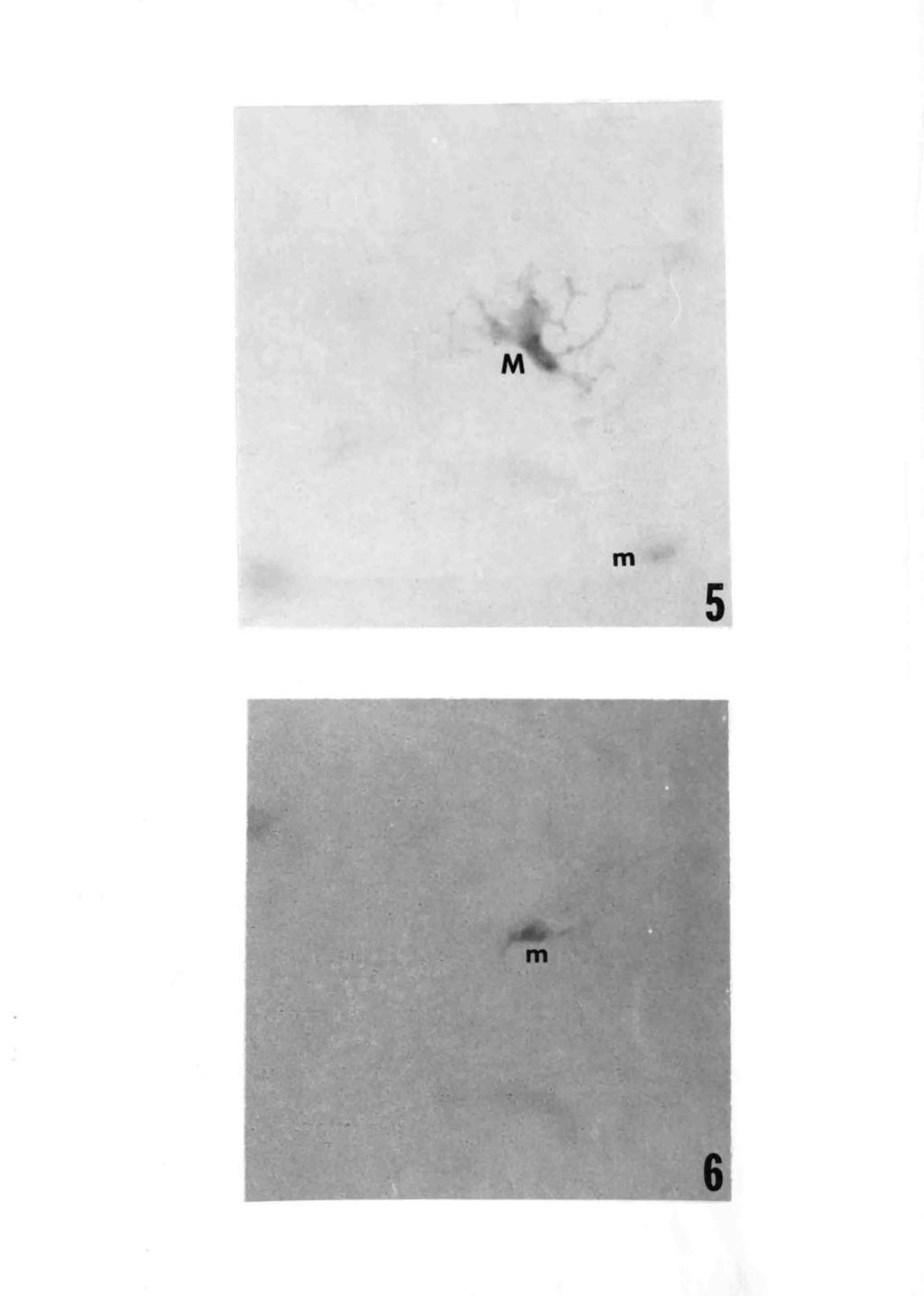
39 -29- Figure 5. A typical epidermal melanocyte (M) from a white macule of an individual with tuberous sclerosis. The dark area (m) is a miniature melanocyte not in focus. DOPA reagent. X Figure 6. A miniature epidermal melanocyte (m) from a white macule of an individual with tuberous sclerosis. DOPA reagent. X 1000.
40 m 5 6
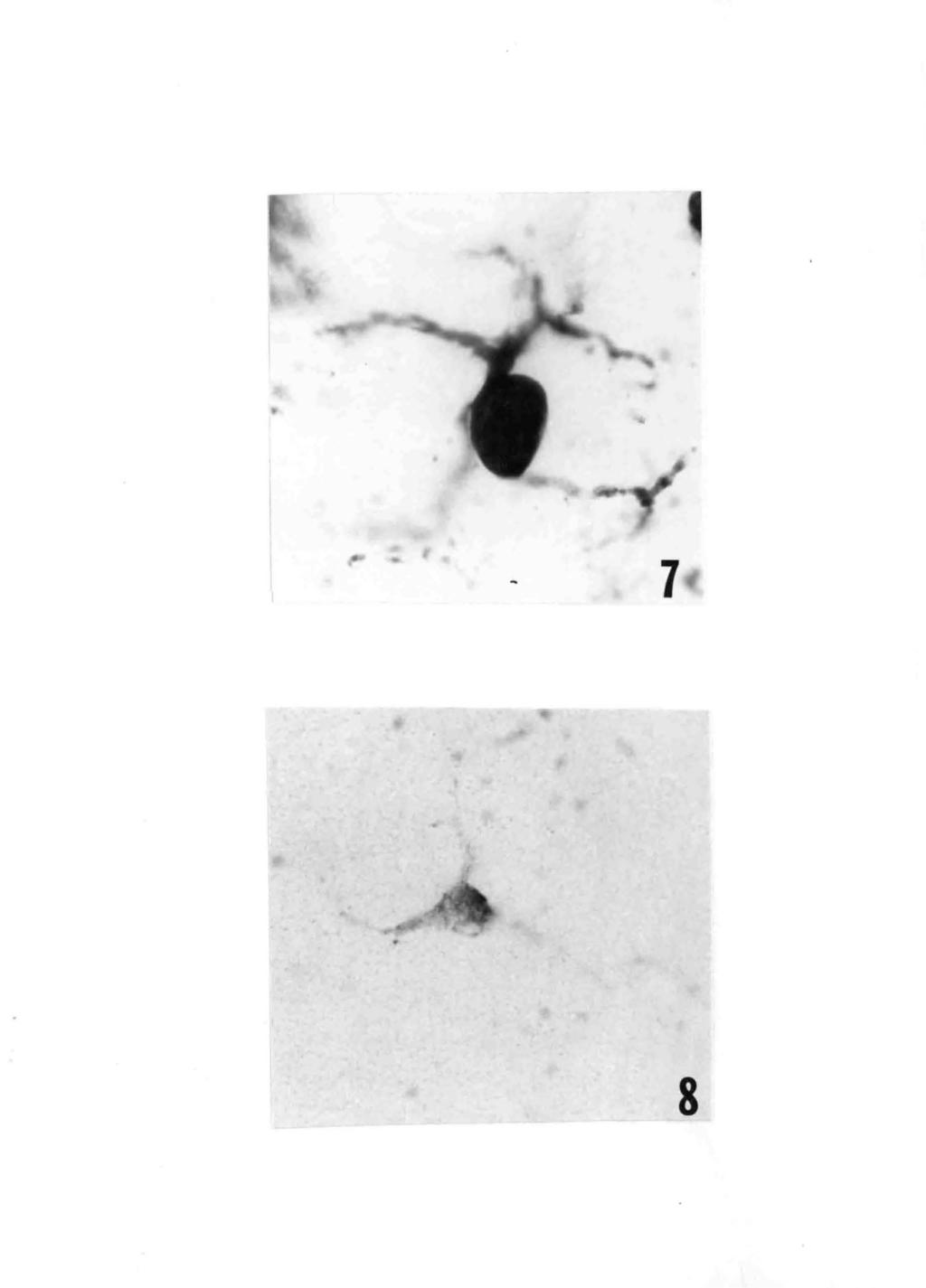
41 -30- Figure 7. A typical epidermal melanocyte in the dorsal region of the PET/Wmr mouse. DOPA reagent. x 450. Figure 8. A miniature epidermal melanocyte in the dorsal region of the PET/Wmr mouse. Note DOPA reactivity and size with respect to figure 7. DOPA reagent. X 450.
42 7 8
43 -31- Figure 9. Epidermal melanocytes of a PET/Wmr dorsal skin section incubated 7 days on the chorioallantoic membrane. DOPA reagent. X 450.

44
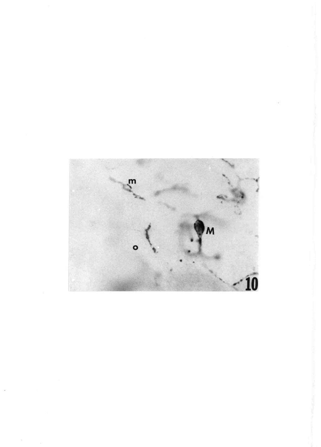
45 -32- Figure 10. A 7 day ~horioallantoic membrane graft of PET/Wrnr dorsal skin showing typical (M) and miniature (m) melanocytes in the epidermis. DOPA reagent. X 450.
46 0 M
47 -33- Figure 11. A miniature melanocyte in the epidermis of a PET/Wmr dorsal skin specimen incubated 7 days on the chorioallanto1c membrane. DOPA reagent. X 450.
48 11
49 -34- VITA John Thomas Earnhardt was born in Salisbury, North Carolina June 6, He received his elementary and secondary education in the Salisbury City School System graduating from Boyden High School in He entered Lenoir Rhyne College in Hickory, North' Carolina and graduated with a B. S. degree in Biology in May In September of the same year he entered the Graduate School of the University of Richmond... During this :time he became a member of Beta Beta Beta Biological Honor Society, was a recipient of a Williams Fellowship, and assisted various laboratories. He was invited and attended the International Pigment Cell Conference at Yale University du~ing his final semester of graduate studies. He received the M. S. degree in Biology in May 1973.
The Occurrence and Morphogenesis of Melanocytes in the Connective Tissues of the PET/MCV Mouse Strain 1
 The Occurrence and Morphogenesis of Melanocytes in the Connective Tissues of the PET/MCV Mouse Strain 1 by STUART E. NICHOLS, JR. 2 and WILLIE M. REAMS, JR. 3 From the Department of Anatomy, Medical College
The effects of ultraviolet irradiation on the pigment cells of the PET/Wmr mouse epidermis
 University of Richmond UR Scholarship Repository Master's Theses Student Research 1973 The effects of ultraviolet irradiation on the pigment cells of the PET/Wmr mouse epidermis Vaughan Henry Howard Jr.
strain), were tested by growing explants of neural fold from the posterior Woronzowa,2' 3 implanting pituitaries in postlarval stages, observed
, were tested by growing explants of neural fold from the posterior Woronzowa,2' 3 implanting pituitaries in postlarval stages, observed") VOL. 35, 1949 GENETICS: H. C. DALTON 277 DE VELOPMENTAL ANAL YSIS OF GENETIC DIFFERENCES IN PIGMENTATION IN THE AXOLOTL By H. CLARK DALTON DEPARTMENT OF GENETICS, CARNEGIE INSTITUTION OF WASHINGTON, COLD
VOL. 35, 1949 GENETICS: H. C. DALTON 277 DE VELOPMENTAL ANAL YSIS OF GENETIC DIFFERENCES IN PIGMENTATION IN THE AXOLOTL By H. CLARK DALTON DEPARTMENT OF GENETICS, CARNEGIE INSTITUTION OF WASHINGTON, COLD
Further studies on the melanophores of periodic albino mutant of Xenopus laevis
 J. Embryol. exp. Morph. 91, 65-78 (1986) 65 Printed in Great Britain The Company of Biologists Limited 1986 Further studies on the melanophores of periodic albino mutant of Xenopus laevis T. FUKUZAWA AND
The Effect of Cortisone on Cell Proliferation and Migration in Peripheral Nerves undergoing Wallerian degeneration
 The Effect of Cortisone on Cell Proliferation and Migration in Peripheral Nerves undergoing Wallerian by G. A. THOMAS 1 From the Department of Anatomy, Guy's Hospital Medical School, London INTRODUCTION
The site of action of the ichthyosis locus (ic) in the mouse, as determined by dermal-epidermal recombinations
 in the mouse, as determined by dermal-epidermal recombinations") /. Embryol. exp. Morph. Vol. 32, 3, pp. 715-721, 1974 715 Printed in Great Britain The site of action of the ichthyosis locus (ic) in the mouse, as determined by dermal-epidermal recombinations BY MARGARET
/. Embryol. exp. Morph. Vol. 32, 3, pp. 715-721, 1974 715 Printed in Great Britain The site of action of the ichthyosis locus (ic) in the mouse, as determined by dermal-epidermal recombinations BY MARGARET
A technique for introducing localized long-lasting implants in the chick embryo
 /. Embryol. exp. Morph. Vol. 39, pp. 261-266, 1977 261 Printed in Great Britain SHORT PAPERS A technique for introducing localized long-lasting implants in the chick embryo By MARIETA B. HEATON 1 From
Supplementary Information
 Supplementary Information Figure S1: Follicular melanocytes in the wound peripheral area migrate to the epidermis in response to wounding stimuli. Dorsal skin of Trp2-LacZ mice stained with X-gal and analyzed
Genetic Factors Controlling the Proliferative Activity of Mouse Epidermal Melanocytes During the Healing of Skin Wounds
 Copyright 0 1988 by the Genetics Society of America Genetic Factors Controlling the Proliferative Activity of Mouse Epidermal Melanocytes During the Healing of Skin Wounds Tomohisa Hirobe Division of Biology,
value as a medium for the in vivo cultivation of different
 THE BEHAVIOR OF THE VIRUS OF EQUINE ENCEPH- ALOMYELITIS ON THE CHORIOALLANTOIC MEMBRANE OF THE DEVELOPING CHICK' ELIZABETH HIGBIE AND BEATRICE HOWITT George Williams Hooper Foundation, University of California,
MELANOCYTE PATTERN OF AN AREA OF FRECKLED EPIDERMIS COVERING A STRETCHED SCAR*
 MELANOCYTE PATTERN OF AN AREA OF FRECKLED EPIDERMIS COVERING A STRETCHED SCAR* Figure 1 illustrates the appearance after eighteen months of the scar resulting from the removal of an area of full thickness
(From the Department of Animal and Plant Pathology of The Rockefeller Institute for Medical Research, Princeton, New Jersey)
") THE YIELD OF RABIES VIRUS IN THE CHICK EMBRYO BY BJORN SIGURDSSON, M.D.* (From the Department of Animal and Plant Pathology of The Rockefeller Institute for Medical Research, Princeton, New Jersey) (Received
THE YIELD OF RABIES VIRUS IN THE CHICK EMBRYO BY BJORN SIGURDSSON, M.D.* (From the Department of Animal and Plant Pathology of The Rockefeller Institute for Medical Research, Princeton, New Jersey) (Received
Primary Isolation and Cultivation of Viruses
 Primary Isolation and Cultivation of Viruses Practical Medical Virology 450 MBIO 2017-18 01/10/2017 Amal Alghamdi Reham Alahmadi Dalia Alsrar 1 Diagnostic Virology Virus Isolation and Cultivation Viral
TRANSFER OF PREMELANOSOMES INTO THE KERATINIZING CELLS OF ALBINO HAIR FOLLICLE
 TRANSFER OF PREMELANOSOMES INTO THE KERATINIZING CELLS OF ALBINO HAIR FOLLICLE PAUL F. PARAKKAL. From the Department of Dermatology, Boston University School of Medicine, Boston, Massachusetts 02118 INTRODUCTION
:1c.c :& Preliminary and Short Report GRANULE FORMATION IN THE LANGERHANS CELL* structure with rounded ends and a striated lamella
 THE JOURNAL OF INVESTIGATIVE DERMATOLOGY Copyright 1566 by The Williams & Wilkins Co. Vol. 7, No. 5 Printed in U.S.A. Preliminary and Short Report GRANULE FORMATION IN THE LANGERHANS CELL* ALVIN S. ZELICKSON,
SKIN. 3. How is the skin structured around the finger joints to allow for flexible movement of the fingers?
 SKIN Objectives for Exam #1: 1. List various skin structures and describe their functions. 2. Describe skin responses to increases and decreases in body temperature. 3. Provide examples of various skin
Tyrosinase Activity in the Pigmented Cells of the Nucleus Substantiae Nigrae. I. Monophenolase and Diphenolase Activity. By C. D.
 4 7 Tyrosinase Activity in the Pigmented Cells of the Nucleus Substantiae Nigrae. I. Monophenolase and Diphenolase Activity By C. D. MARSDEN (From the Department of Anatomy, St. Thomas's Hospital Medical
(Received 12 December 1967)
") J. Physiol. (1968), 195, pp. 755-759 755 With 2 text-figures Printed in Great Britain A NOTE ON TRANSMEMBRANE POTENTIAL IN DERMAL MELANOPHORES OF THE FROG AND MOVEMENT OF MELANIN GRANULES BY A. R. MARTIN
J. Physiol. (1968), 195, pp. 755-759 755 With 2 text-figures Printed in Great Britain A NOTE ON TRANSMEMBRANE POTENTIAL IN DERMAL MELANOPHORES OF THE FROG AND MOVEMENT OF MELANIN GRANULES BY A. R. MARTIN
THE EFFECT OF X-RAY IRRADIATION ON MELANOCYTES IN THE SKIN*
 THE EFFECT OF X-RAY IRRADIATION ON MELANOCYTES IN THE SKIN* The effect of x-rays on skin is described in most textbooks of dermatology. However, the literature contains few reports of their action on melanin
Protocol for Processing parasites
 1 Protocol for Processing parasites By/ Adnan I. Al-Hindi (M.Sc.) Parasitology The Islamic University of Gaza Faculty of Science Department of Biology 28 June, 2000 Staining of parasites 28-6-2000 (Home)
EFFECTS OF SOME PESTICIDES ON THE EMBRYONIC STAGES OF CHICK
 EFFECTS OF SOME PESTICIDES ON THE EMBRYONIC STAGES OF CHICK Subrata Datta Department of Zoology, East Calcutta Girls College, Lake town Kolkata (INDIA) Received August 17, 2007 Accepted January 4, 2007
CELLS CONTAINING LANGERHANS GRANULES IN HUMAN LYMPH NODES OF DERMATOPATHIC LYMPHADENOPATHY*
 THS JOURNAL OF INVEBTIOATIVR DERMATOLOGY Copyright 1969 by The Williams & Wilkins Co. Vol. 93, No. 4 Printed in U.S.A. CELLS CONTAINING LANGERHANS GRANULES IN HUMAN LYMPH NODES OF DERMATOPATHIC LYMPHADENOPATHY*
Epidermal Melanin Units: Melanocyte-Keratinocyte Interactions
 AM. ZOOLOCIST, 12:35-41 (1972). Epidermal Melanin Units: Melanocyte-Keratinocyte Interactions WALTER C. QUEVEDO, JR. Division of Biological and Medical Sciences, Brown University, Providence, Rhode Island
Morphogenesis of the residual body of the mouse testis
 93 Morphogenesis of the residual body of the mouse testis By CASIMIR F. FIRLIT and JOSEPH R. DAVIS (From the Department of Pharmacology and Therapeutics, Stritch School of Medicine, and Graduate School,
Development of retinal synaptic arrays in the inner plexiform layer of dark-reared mice
 /. Embryo/, exp. Morph. Vol. 54, pp. 219-227, 1979 219 Printed in Great Britain Company of Biologists Limited 1977 Development of retinal synaptic arrays in the inner plexiform layer of dark-reared mice
The effects of topical nitrogen mustard on the pigment system in PET/Wmr mouse epidermis
 University of Richmond UR Scholarship Repository Master's Theses Student Research 8-1975 The effects of topical nitrogen mustard on the pigment system in PET/Wmr mouse epidermis Ronald Lee Salisbury Follow
ELECTRON MICROSCOPY OF A SMALL PIGMENTED CUTANEOUS LESION*
 ELECTRON MICROSCOPY OF A SMALL PIGMENTED CUTANEOUS LESION* The description of the lesion in the title of this rcport is intentionally non-committal. Diagnosed clinically as a lentigo, it was removed as
Epidermis. Integumentary system
 Epidermis the doctor mentioned at the begging of the lecture that the slides is from different sources and has information and details that is enough for us so we don t have to go back and read from the
Chorion Allantois Membrane (CAM) Assay
 Assay") Chorion Allantois Membrane (CAM) Assay Introduction: The CAM assay was based on Nguyen et al. (1994). It is a simple and commonly used test to assess the angiogenesis inhibiting or stimulating properties
Chorion Allantois Membrane (CAM) Assay Introduction: The CAM assay was based on Nguyen et al. (1994). It is a simple and commonly used test to assess the angiogenesis inhibiting or stimulating properties
SUMMARY. Keywords: quail, Coturnix japonica, morphology, ovary, oviduct, neurotrophins, immunohistochemistry
 SUMMARY Keywords: quail, Coturnix japonica, morphology, ovary, oviduct, neurotrophins, immunohistochemistry Studies on the development of biological systems have expanded using animal models, always to
SEASONAL CHANGES IN HISTOLOGYOF THE THYROID GLAND CALOTIS VERSICOLOR
 SEASONAL CHANGES IN HISTOLOGYOF THE THYROID GLAND CALOTIS VERSICOLOR M. D. Kulkarni And A. H. Shinde Department of Zoology Yashwantrao Chavan Arts & Science College, Mangrulpir Dist. Washim. (Received
Induction of melanogenesis in the epidermal melanoblasts of newborn mouse skin bymsh
 /. Embryol. exp. Morph. Vol. 37, pp. 79-90, 1977 79 Printed in Great Britain Induction of melanogenesis in the epidermal melanoblasts of newborn mouse skin bymsh ByTOMOHISA HIROBE AND TAKUJI TAKEUCHI 1
Mesenchymal control over elongating and branching morphogenesis in salivary gland development
 J. Embryol. exp. Morph. Vol. 66, pp. 9-, 98 9 Printed in Great Britain Company of Biologists Limited 98 Mesenchymal control over elongating and branching morphogenesis in salivary gland development ByHIROYUKI
FACTORS INFLUENCING VARIOLA VIRUS GROWTH ON THE CHORIOALLANTOIC MEMBRANE OF EMBRYONATED EGGS
 FACTORS INFLUENCING VARIOLA VIRUS GROWTH ON THE CHORIOALLANTOIC MEMBRANE OF EMBRYONATED EGGS NICHOLAS HAHON, MILTON RATNER, AND EDMUND KOZIKOWSKI U. S. Army Chemical Corps, Fort Detrick, Frederick, Maryland
Experimental Neoplastic Formation in Embryonic Chick Brains
 Experimental Neoplastic Formation in Embryonic Chick Brains by BENGT KALLEN 1 From the Tornblad Institute of Comparative Embryology, Lund WITH TWO PLATES IN mammalian teratology, a malformation consisting
Exercise 6. Procedure
 Exercise 6 Procedure Growing of root tips Select a few medium-sized onion bulbs. Carefully remove the dry roots present. Grow root tips by placing the bulbs on glass tubes (of about 3 4 cm. diameter) filled
Changes of organelles associated with the differentiation of epidermal melanocytes in the mouse
 /. Embryol. exp. Morph. Vol. 43, pp. 107-121, 197S ]Ç)J Printed in Great Britain Company of Biologists Limited 1978 Changes of organelles associated with the differentiation of epidermal melanocytes in
Scanning Electron Microscopic Observations on the Sperm Penetration through the Zona Pellucida of Mouse Oocytes Fertilized in vitro
 Scanning Electron Microscopic Observations on the Sperm Penetration through the Zona Pellucida of Mouse Oocytes Fertilized in vitro Masatsugu MOTOMURA and Yutaka TOYODA School of Veterinary Medicine and
ACTIVE.LITE. Patent-Pending Technology + Visibly Perceivable Results in Less than 14 Days. Tomorrow s Vision Today!
 ACTIVE.LITE Patent-Pending Technology + Visibly Perceivable Results in Less than 14 Days Tomorrow s Vision Today! AESTHETIC PERFECTION IS THE STANDARD Aesthetic skin perfection is now the consumer standard
Ch 4. Skin and Body Membranes
 Ch 4 Skin and Body Membranes TITLE HISTOLOGY SLIDES & NOTES ESSENTIAL QUESTION What tissues compose the integumentary system? Stratified Squamous Epithelium Stratified = several layers; Squamous = shape
Histopathology: skin pathology
 Histopathology: skin pathology These presentations are to help you identify, and to test yourself on identifying, basic histopathological features. They do not contain the additional factual information
MITOSIS IN DEVELOPING CARDIAC MUSCLE. FRANCIS J. MANASEK. From the Department of Anatomy, Harvard Medical School, Boston, Massachusetts 02115
 Published Online: 1 April, 1968 Supp Info: http://doi.org/10.1083/jcb.37.1.191 Downloaded from jcb.rupress.org on June 30, 2018 MITOSIS IN DEVELOPING CARDIAC MUSCLE FRANCIS J. MANASEK. From the Department
AN ATTEMPT TO INDUCE "PIGMENT SPREAD" IN FRECKLED HUMAN SKIN*
 AN ATTEMPT TO INDUCE "PIGMENT SPREAD" IN FRECKLED HUMAN SKIN* White skin transplanted on to a pigmented area of a spotted black-and-white guinea-pig becomes blackened, and black skin transplanted on to
Influence of a temporary embryonic testis graft on the regression of Miillerian ducts in female chick embryo
 J. Embryol exp. Morph. Vol. 67, pp. 81-87, 1982 g \ Printed in Great Britain Company of Biologists Limited 1982 Influence of a temporary embryonic testis graft on the regression of Miillerian ducts in
A HISTOCHEMICAL AUTORADIOGRAPHIC METHOD FOR DEMON- STRATION OF TYROSINASE IN HUMAN MELANOCYTES, NEVI AND MALIGNANT MELANOMA*
 A HISTOCHEMICAL AUTORADIOGRAPHIC METHOD FOR DEMON STRATION OF TYROSINASE IN HUMAN MELANOCYTES, NEVI AND MALIGNANT MELANOMA* THOMAS B. FITZPATRICK, M.D., PHI). AND ATSUSHI KUI{ITA, KUKITA, M.T). Ml). Numerous
THE CYTOPATHOGENIC ACTION OF BLUETONGUE VIRUS ON TISSUE CULTURES AND ITS APPLICATION TO THE DETECTION OF ANTIBODIES IN THE SERUM OF SHEEP.
 Onderstepoort Journal of Veterinary Research, Volume 27, Number 2, October, 1956. The Government Printer. THE CYTOPATHOGENIC ACTION OF BLUETONGUE VIRUS ON TISSUE CULTURES AND ITS APPLICATION TO THE DETECTION
Chapter 6 Skin and the Integumentary System. Skin Cells. Layers of Skin. Epidermis Dermis Subcutaneous layer beneath dermis not part of skin
 Chapter 6 Skin and the Integumentary System Composed of several tissues Maintains homeostasis Protective covering Retards water loss Regulates body temperature Houses sensory receptors Contains immune
Hole s Human Anatomy and Physiology Eleventh Edition. Mrs. Hummer. Chapter 6
 Hole s Human Anatomy and Physiology Eleventh Edition Mrs. Hummer Chapter 6 1 Chapter 6 Skin and the Integumentary System Composed of several tissues Maintains homeostasis Protective covering Retards water
Studies on Induced Ovulation in the Intact Immature Hamster. Charles W. Bodemer, Ph.D., Ruth E. Rumery, Ph.D., and Richard J. Blandau, Ph.D., M.D.
 Studies on Induced Ovulation in the Intact Immature Hamster Charles W. Bodemer, Ph.D., Ruth E. Rumery, Ph.D., and Richard J. Blandau, Ph.D., M.D. IT IS WELL KNOWN that gonadotropins are incapable of inducing
Accelerating Embryonic Growth During Incubation Following Prolonged Egg Storage 2. Embryonic Growth and Metabolism 1
 Accelerating Embryonic Growth During Incubation Following Prolonged Egg Storage 2. Embryonic Growth and Metabolism 1 V. L. Christensen, 2 J. L. Grimes, M. J. Wineland, and G. S. Davis Department of Poultry
IN the skin of the mouse we find that the dermal papilla of growing hair
 (240 Cyclic Changes in Polysaccharides of the Papilla of the Hair Follicle By WILLIAM MONTAGNA, HERMAN B. CHASE, JANET D. MALONE, AND HELEN P. MELARAGNO (From Brown University, Providence, Rhode Island,
Lab 2: Investigating Variation Across Spatial Scales
 Lab 2: Investigating Variation Across Spatial Scales What are scales and variation in a biological context? The world around us displays incredible diversity across many scales. Today s lab investigates
Bacterial Interference in Chick Embryos *
 Journal of Clinical Investigation Vol. 46, No. 3, 1967 Bacterial Interference in Chick Embryos * JOHN C. RIBBLE t AND HENRY R. SHINEFIELD (From the Department of Medicine, The New York Hospital-Cornell
THE INDUCTION OF HAIR FOLLICLES BY EMBRYONIC DERMAL PAPILLAE*
 THE JouR AL OF INVESTIGATIVE DERMATOLOGY Copyright 1970 by The Williams & Wilkins Co. Vol. 55, No. 6 Printed in U.S.A. THE INDUCTION OF HAIR FOLLICLES BY EMBRYONIC DERMAL PAPILLAE* EDWARD J. KOLLAR, PH.D.
Human Saliva as a Convenient Source of Ribonuclease. By S. BRADBURY
 Human Saliva as a Convenient Source of Ribonuclease 323 By S. BRADBURY (From the Cytological Laboratory, Department of Zoology, University Museum, Oxford) SUMMARY Saliva, heated to 80 C for 10 minutes
Administrative - Master Syllabus COVER SHEET
 Administrative - Master Syllabus COVER SHEET Purpose: It is the intention of this to provide a general description of the course, outline the required elements of the course and to lay the foundation for
Peeling. Smooth out the skin. Remove the outer skin layers. Erase the fine wrinkles. The regenerated skin is smoother & Less wrinkled - Renewed
 If you foolishly ignore beauty, you will soon find yourself without it. Your life will be impoverished. But if you wisely invest in beauty, it will remain with you all the days of your life Frank Lloyd
THE EFFECT OF UNILATERAL CASTRATION ON THE REMAINING TESTIS OF THE MOUSE
 402 THE EFFECT OF UNILATERAL CASTRATION ON THE REMAINING TESTIS OF THE MOUSE BY I. W. ROWLANDS. (From the Department of Zoology, University College of North Wales, Bangor.) (Received 14th April, 1934.)
Summary. Mouse eggs were fertilized in vitro, in the presence and
 THE R\l=O^\LEOF CUMULUS CELLS AND THE ZONA PELLUCIDA IN FERTILIZATION OF MOUSE EGGS IN VITRO A. PAVLOK and ANNE McLAREN Czechoslovak Academy of Sciences, Laboratory of Animal Genetics, Libechov, Czechoslovakia,
FURTHER STUDIES OF THE CONDUCTING SYSTEM OF THE BIRD'S HEART
 FURTHER STUDIES OF THE CONDUCTING SYSTEM OF THE BIRD'S HEART By FRANCIS DAVIES, M.D. (LONDON) Anatomy Department, University College, London INTRODUCTION T1HE histological investigation of the conducting
The Structure of Viruses of the Newcastle Disease- Mumps-Influenza (Myxovirus) Group
 Group") 680 * VALENTINE, R. C. & ISAACS, A. (1957). J. gen. Microbiol. 16, 680-685 The Structure of Viruses of the Newcastle Disease- Mumps-Influenza (Myxovirus) Group BY R. C. VALENTINE AND A. IsAAcS National
680 * VALENTINE, R. C. & ISAACS, A. (1957). J. gen. Microbiol. 16, 680-685 The Structure of Viruses of the Newcastle Disease- Mumps-Influenza (Myxovirus) Group BY R. C. VALENTINE AND A. IsAAcS National
PHOSPHORUS METABOLISM OF THE SOFT TISSUES OF THE NORMAL MOUSE AS INDICATED BY RADIOACTIVE PHOSPHORUS '
 PHOSPHORUS METABOLISM OF THE SOFT TISSUES OF THE NORMAL MOUSE AS INDICATED BY RADIOACTIVE PHOSPHORUS ' H. B. JONES, I. L. CHAIKOFF, AND JOHN H. LAWRENCE (From the ~ivision of ~kysiology'of the Medical
Efferent innervation of the retina
 Efferent innervation of the retina II. Morphologic study of the monkey retina' Francisco M. Honrubia and James H. Elliott Silver impregnation of the monkey's retina has been employed to study the presence
The Integumentary System. Mosby items and derived items 2010, 2006, 2002, 1997, 1992 by Mosby, Inc., an affiliate of Elsevier Inc.
 The Integumentary System The Skin Structure two primary layers called epidermis and dermis Epidermis Outermost and thinnest primary layer of skin Composed of several layers of stratified squamous epithelium
Ocular malformations of the chick embryo produced by photocoagulation. David O. Jesberg
 Ocular malformations of the chick embryo produced by photocoagulation David O. Jesberg Heat-produced necrotizing lesions of the chick embryo pigment epithelium and retina are repaired by regeneration in
Tracking skin cancers and melanoma at the microscopic level
 Tracking skin cancers and melanoma at the microscopic level Rosalie Elenitsas, M.D. Professor of Dermatology Director of Dermatopathology Hospital of the University of Pennsylvania May 12, 2017 Outline
CREDIT COURSE OUTLINE: BIO 210 Last Revised and Approved: 12/08/2011 BIO HUMAN ANATOMY 2.00
 BIO 210 - HUMAN ANATOMY Units Lecture Total Hrs Lecture 2.00 Units Lab 2.00 Units Total 4.00 33.00 Total Hrs Lab 99.00 Total Course Hrs 132.00 COURSE DESCRIPTION This course follows a systemic approach
THE PROPAGATION OF A VIRULENT GOAT PLEUROPNEUMONIA-LIKE ORGANISM IN THE CHICK EMBRYO
 THE PROPAGATION OF A VIRULENT GOAT PLEUROPNEUMONIA-LIKE ORGANISM IN THE CHICK EMBRYO RICHARD YAMAMOTO, HENRY E. ADLER, AND DONALD R. CORDY School of Veterinary Medicine, University of California, Davis,
612.6I7.5:612.6I6.I. different, but most of them appear to be satisfactory from a qualitative
 442 612.6I7.5:612.6I6.I SIZE CHANGES IN THE SEMINAL VESICLES OF THE MOUSE DURING DEVELOPMENT AND AFTER CASTRATION. BY RUTH DEANESLY AND A. S. PARKES'. (From the National Institute for Medical Research,
Skin. Kristine Krafts, M.D.
 Skin Kristine Krafts, M.D. Skin Lecture Objectives Describe the functions of skin. Describe the structure, location and function of the cell types found in epidermis: keratinocytes, melanocytes, Langerhans
Stem Cells. Induced Stem Cells
 Induced Stem Cells Stem Cells Mouse and human somatic cells can either be reprogrammed to a pluripotent state or converted to another lineage with a combination of transcription factors suggesting that
Human Pluripotent Stem Cell Cardiomyocyte Differentiation Kit (PSCCDK) Introduction Kit Components Cat. # # of vials Reagent Quantity Storage
 Introduction Kit Components Cat. # # of vials Reagent Quantity Storage") Human Pluripotent Stem Cell Cardiomyocyte Differentiation Kit (PSCCDK) Catalog #5901 Introduction Human pluripotent stem cells (hpsc), including embryonic stem cells (ESC) and induced pluripotent stem
Human Pluripotent Stem Cell Cardiomyocyte Differentiation Kit (PSCCDK) Catalog #5901 Introduction Human pluripotent stem cells (hpsc), including embryonic stem cells (ESC) and induced pluripotent stem
Understanding Skin Colour
 Understanding Skin Colour SKIN COLOUR The natural colour of skin without any pigments is yellowish. However, we are all aware of the different colours of skin and these differences are determined by the
In Vitro Speeds of Bovine Spermatozoa
 In Vitro Speeds of Bovine Spermatozoa A. N. Moeller, M.S., and N. l. VanDemark, Ph.D. THE RATE OF progressive movement of the spermatozoa has been used as one criterion in physiologic studies for evaluation
Introduction. Skin and Body Membranes. Cutaneous Membranes Skin 9/14/2017. Classification of Body Membranes. Classification of Body Membranes
 Introduction Skin and Body Membranes Body membranes Cover surfaces Line body cavities Form protective and lubricating sheets around organs Classified in 5 categories Epithelial membranes 3 types- cutaneous,
CELLULAR KINETICS OF THE ANTI-MRBC RESPONSE IN CHICKENS
 19 CELLULAR KINETICS OF THE ANTI-MRBC RESPONSE IN CHICKENS K. Dagg, S. P. Turner and F. Seto Department of Zoology, University of Oklahoma, Norman, Oklahoma The serum hemagglutinin (HA) titers and the
Skin (Integumentary System) Wheater, Chap. 9
 Wheater, Chap. 9") Skin (Integumentary System) Wheater, Chap. 9 Skin (Integument) Consists of skin and associated derivatives Largest organ of body (21 ft 2 ; 9 lbs.; has 11 miles of blood vessels) Functions: Protection
Skin (Integumentary System) Wheater, Chap. 9 Skin (Integument) Consists of skin and associated derivatives Largest organ of body (21 ft 2 ; 9 lbs.; has 11 miles of blood vessels) Functions: Protection
An in vitro method for screening skin-whitening products
 j. Cosmet. Sci., 49, 361-367 (November/December 1998) An in vitro method for screening skin-whitening products GOPA MAJMUDAR, GEORGE JACOB, YOLANDA LABOY, and LOUIS FISHER, Mary Kay Holding Corporation,
Chapter 5: Integumentary System
 Chapter 5: Integumentary System I. Overview of the Integumentary System A. List the five major functions of the integumentary system: 1. 2. 3. 4. 5. Il. Skin A. Epidermis 1. The epidermis consists of 2.
Gene expression at the pink-eyed dilution (p) locus in the mouse is confirmed to be pigment cell autonomous using recombinant embryonic skin grafts
 locus in the mouse is confirmed to be pigment cell autonomous using recombinant embryonic skin grafts") /. Embryol. exp. Morph. 87 65-73 (1985) 55 Printed in Great Britain The Company of Biologists Limited 1985 Gene expression at the pink-eyed dilution (p) locus in the mouse is confirmed to be pigment cell
/. Embryol. exp. Morph. 87 65-73 (1985) 55 Printed in Great Britain The Company of Biologists Limited 1985 Gene expression at the pink-eyed dilution (p) locus in the mouse is confirmed to be pigment cell
The Effect of Cyclosporin A on the Proliferating Chick Embryo Erythroblasts
 Physiol. Res. 43: 223-228, 1994 The Effect of Cyclosporin A on the Proliferating Chick Embryo Erythroblasts M. PETERKA, Z. LIKOVSKY, R. PETERKOVA Institute o f Experimental Medicine, Academy o f Sciences
SUPPLEMENTARY INFORMATION
 DOI: 10.1038/ncb2535 Figure S1 SOX10 is expressed in human giant congenital nevi and its expression in human melanoma samples suggests that SOX10 functions in a MITF-independent manner. a, b, Representative
THE SITE OF STEROL AND SQUALENE SYNTHESIS IN THE HUMAN SKIN123
 THE SITE OF STEROL AND SQUALENE SYNTHESIS IN THE HUMAN SKIN123 N. NICOLAIDES, PH.D. AND STEPHEN ROTHMAN, M.D. In earlier work (1) it was demonstrated that human scalp skin is an efficient organ for synthesizing
Protocols. Harvesting and Sectioning the Ascending Aorta
 DAUGHERTY LAB Saha Cardiovascular Research Center University of Kentucky Protocols Harvesting and Sectioning the Ascending Aorta Page 1 of 7 Harvesting the ascending aorta Materials: 1. Insulin syringe
Immunohistochemical Study on the C-cells in the Internal Parathyroid Gland of the Goat
 Immunohistochemical Study on the C-cells in the Internal Parathyroid Gland of the Goat Takeshi TSUCHIYA Department of Animal Morphology, Faculty of Agriculture, Tohoku University, Sendai-Shi 980 (Received
Tyrosinase Activity in the Skin of Three Strains of Albino Gecko (Eublepharis macularius)
") Tyrosinase Activity in the Skin of Three Strains of Albino Gecko (Eublepharis macularius) Fig. 1. Adult wild-type leopard gecko. Tony Gamble 1, Jodi L. Aherns 2, and Virginia Card 3 1,3 Metropolitan State
Tyrosinase Activity in the Skin of Three Strains of Albino Gecko (Eublepharis macularius) Fig. 1. Adult wild-type leopard gecko. Tony Gamble 1, Jodi L. Aherns 2, and Virginia Card 3 1,3 Metropolitan State
Integumentary System
 Integumentary System Overview Functions 1. Protection 2. Excretion of wastes 3. Maintenance of T b 4. Synthesis of vitamin D 3 5. Storage of lipids 6. Detection of sensory stimuli Epidermis Tissue types
Minimal Effects When Acute Nicotine Exposure Introduced to 3 Day Old Chick Embryos
 Minimal Effects When Acute Nicotine Exposure Introduced to 3 Day Old Chick Embryos Amy Langevin Independent Research Project Bio254- Developmental Biology November 28, 2007 Introduction Thousands of mothers
The effect of eyestalk removal and eyestalk extract injection on the light and dark adaptation in the crab Sesarma boulengeri Calman
 Marina Mesopotamica Volume 7, Number 2, pp. 241 247 (1992) The effect of eyestalk removal and eyestalk extract injection on the light and dark adaptation in the crab Sesarma boulengeri Calman Abdullah
Integumentary System and Body Membranes
 Integumentary System and Body Membranes The Skin and its appendages hair, nails, and skin glands Anatomy/Physiology NHS http://www.lab.anhb.uwa.edu.au/mb140/corepages/integumentary/integum.htm I. System
MEASURING FERTILITY & EARLY DEADS LEVELS PART 1
 MEASURING FERTILITY & EARLY DEADS LEVELS PART 1 Why measure fertility and early deads? An unfertilised egg cannot produce a chick. Flock fertility is governed by management of males and females on the
Instructions for Use. APO-AB Annexin V-Biotin Apoptosis Detection Kit 100 tests
 3URGXFW,QIRUPDWLRQ Sigma TACS Annexin V Apoptosis Detection Kits Instructions for Use APO-AB Annexin V-Biotin Apoptosis Detection Kit 100 tests For Research Use Only. Not for use in diagnostic procedures.
IN VITRO OF MALIGNANT TUMORS.* BY ALEXIS CARREL AnD MONTROSE T. BURROWS.
 CULTIVATION IN VITRO OF MALIGNANT TUMORS.* BY ALEXIS CARREL AnD MONTROSE T. BURROWS. (From the Laboratories of the Rockefeller Institute for Medical Research, New York.) Pr~AT~ LXXVII. The growth of malignant
Ch. 4: Skin and Body Membranes
 Ch. 4: Skin and Body Membranes I. Body Membranes A. Function of body membranes 1. Cover body surfaces 2. Line body cavities 3. Form protective sheets around organs II. Classification of Body Membranes
Guide to Small Animal Reproductive Imaging using the Vevo 770
 Guide to Small Animal Reproductive Imaging using the Vevo 770 Course Objectives: After completion of this module, the participant will be able to accomplish the following: Recognize reproductive female
Integumentary System
 Integumentary System The integumentary system is commonly known as the Skin Largest organ of human body 10% total body weight and would cover over 20 square feet Functions of Skin 1. Protection Barrier
MATERIALS AND METHOD
 Chapter - 3 Histomorphology, Ecology and Biochemistry of leaf galls of Ficus glomerata Roxb. induced by Pauropsylla depressa Crawford. MATERIALS AND METHOD Field observations were confined in Saharanpur
McClure, C. F. W., On the experimental production of edema in larval and adult Anura, J. Gen. Physiol., , i, 261.
 ON THE EXPERIMENTAL PRODUCTION OF EDEMA BY NEPHRECTOMY. BY W. W. SWINGLE. (From the Laboratory of Comparative Anatomy, Princeton University, Princeton.) (Received for publication, March 27, 1919.) The
Differentially Localized Incorporation of Amino Acids. in Relation to Epidermal Keratinization in the Newborn Rat '
 Differentially Localized Incorporation of Amino Acids - in Relation to Epidermal Keratinization in the Newborn Rat ' KIMIE FUKUYAMA, TOSHIO NAKAMURA AND I. A. BERNSTEIN Department of Dermutology, Tokyo
Overripeness and the Mammalian Ova
 Overripeness and the Mammalian Ova II. Delayed Ovulation and Chromosome Anomalies ROY L. BUTCHER, PH.D., and N. W. FUGO, PH.D., M.D. THE CAUSES of abortion and birth defects are undoubtedly multiple and