Lapatinib and Sorafenib Kill GBM Tumor Cells in a Greater than Additive Manner
|
|
|
- Elaine Edwards
- 6 years ago
- Views:
Transcription


1 Virginia Commonwealth University VCU Scholars Compass Theses and Dissertations Graduate School 2013 Lapatinib and Sorafenib Kill GBM Tumor Cells in a Greater than Additive Manner Seyedmehrad Tavallai Virginia Commonwealth University Follow this and additional works at: Part of the Biochemistry, Biophysics, and Structural Biology Commons The Author Downloaded from This Thesis is brought to you for free and open access by the Graduate School at VCU Scholars Compass. It has been accepted for inclusion in Theses and Dissertations by an authorized administrator of VCU Scholars Compass. For more information, please contact libcompass@vcu.edu.
2 Lapatinib and Sorafenib Kill GBM Tumor Cells in a Greater than Additive Manner By Mehrad Tavallai Major Director: Paul Dent, Ph.D. Department of Biochemistry Virginia Commonwealth University Richmond, Virginia December 2013
3 Acknowledgements I would like to acknowledge my advisor Dr. Paul Dent for providing me with the opportunity to pursue my Master s degree in his laboratory, and also my committee members, Dr. Suzanne Barbour, Dr. Andrei Budanov and Dr. Tomasz Kordula, for mentoring me throughout this project. I would also need to thank my lab mates Dr. Hossein Hamed, Dr. Laurence Booth and Dr. Nichola Cruickshanks for kindly teaching me the techniques required for my research, and for selflessly sharing their knowledge and experience with me. Finally, I need to acknowledge my family whose support has always given me the confidence and the encouragement to improve and to fulfill my goals. 1
4 Table of Contents Page List of Tables and Figures...4 List of Abbreviations...6 Abstract...10 Introduction...11 Glioblastoma Multiforme...11 Conventional Therapy...12 Targeted Therapy...13 Sorafenib...14 Lapatinib...15 Epidermal Growth Factor Receptor Tyrosine Kinases...17 Mitogen-activated Protein Kinase Pathways...18 Ras/Raf/MAPK Pathway...20 PI3K/AKT Pathway...21 Apoptosis: Extrinsic and Intrinsic Pathways...23 Autophagy...27 Endoplasmic Reticulum Stress...33 Materials and Methods...36 Materials...36 Cell Culture...37 Drug Treatment...37 Infection with Adenovirus
5 Plasmid and sirna Transfection...38 Trypan Blue Exclusion Assay...39 Western Blot Analysis...39 LC3-GFP Vesicle Formation Assay...40 Data Analysis...40 Results...41 Western blot analysis...46 The role of autophagy and formation of LC3-GFP vesicle formation in GBM12 cells...49 Knockdown of ATG5 and Beclin 1 reduces the drug combination-mediated toxicity in GBM12 and GBM5 cells...52 Up-regulation of FLIP and Bcl-xL and down-regulation of caspase 9 reduce the drug combination-mediated toxicity in GBM12 and GBM5 cells...55 Knockdown of Fas and FADD reduces the drug combination-mediated toxicity in GBM12 cells...60 Discussion...62 Conclusion...65 Bibliography
6 List of Tables and Figures Table 1: The list of GBM cells used in the study and their mutational characteristics Figure 1: Chemical structure of sorafenib tosylate...14 Figure 2: Chemical structure of lapatinib...16 Figure 3: Four major mammalian MAPK pathways...19 Figure 4: Ras/Raf/MAPK signaling pathway...22 Figure 5: PI3K/AKT signaling pathway...24 Figure 6: Intrinsic and extrinsic apoptotic pathways...28 Figure 7: Autophagosome formation in mammalian cells...32 Figure 8: The mechanism of the unfolded protein response...35 Figure 9: Lapatinib and sorafenib dose response in GBM12 cells...42 Figure 10: Assessment of cell viability in GBM5 cells treated with sorafenib and lapatinib...43 Figure 11: Assessment of cell viability in GBM12 cells treated with sorafenib and lapatinib...44 Figure 12: Assessment of cell viability in GBM6 and GBM14 cells treated with sorafenib and lapatinib...45 Figure 13: Western blot analysis of GBM12 cells 3, 6, 12 and 24 hours after exposure to sorafenib and lapatinib...48 Figure 14: LC3-GFP vesicle formation assay in GBM12 cells treated with sorafenib and lapatinib...50 Figure 15: Knockdown of ATG5 and Beclin-1 reduces drug combination-mediated toxicity in GBM12 cells
7 Figure 16: Knockdown of ATG5 and Beclin-1 reduces drug combination-mediated toxicity in GBM5 cells...54 Figure 17: Overexpression of FLIP and Bcl-xL reduces the toxicity of the drug combination in GBM12 cells...56 Figure 18: Down-regulation of caspase 9 reduces the toxicity of the drug combination in GBM12 cells...57 Figure 19: Up-regulation of FLIP and Bcl-xL and down-regulation of caspase 9 reduce the toxicity of the drug combination in GBM5 cells...58 Figure 20: Up-regulation of FLIP and down-regulation of caspase 9 dramatically reduce the toxicity of the drug combination in GBM12 cells during 48 hour-drug exposure...59 Figure 21: Knockdown of CD95 (Fas) and FADD reduce the toxicity of the drug combination in GBM12 cells
8 List of Abbreviations Full Name Activating transcription factor 4 Activating transcription factor 6 Apoptosis-inducing factor Apoptotic protease-activating factor 1 Autophagy-related gene Bax-interacting factor-1 B-cell lymphoma 2 B-cell lymphoma-x long Bcl-2-associated X protein Bcl-2 homologous antagonist/killer Bcl-2 interacting mediator of cell death BH3 Interacting Domain death agonist Binding Immunoglobin Protein Caspase-activated deoxyribonuclease c-jun NH 2 -terminal kinase Cluster of differentiation 95 Cytomegalovirus Death domain Death effector domain Death-inducing signaling complex Dulbecco s Modified Eagle s Medium Abbreviation ATF4 ATF6 AIF Apaf-1 ATG Bif-1 Bcl-2 Bcl-xL Bax Bak BIM Bid BiP CAD JNK CD95 CMV DD DED DISC DMEM 6
9 Dimethyl Sulfoxide Endoplasmic Reticulum Epidermal Growth Factor Receptor Eukaryotic initiation factor 4E Eukaryotic initiation factor 4E inhibitory binding protein Eukaryotic translation initiation factor 2 alpha Extracellular signal-regulated kinase FLICE-inhibitory protein Fas-associated death domain Fas receptor Glioblastoma multiforme Glucose-regulated protein 78 Glyceraldehyde-3-phosphate dehydrogenase Growth factor receptor-bound protein 2 GTPase-activating proteins Guanine nucleotide exchange factors Guanosine diphosphate Guanosine triphosphate DMSO ER EGFR eif4e 4EBP eif2α ERK FLIP FADD FasR GBM Grp78 GAPDH GRB2 GAP GEF GDP GTP Half maximal inhibitory concentrations IC 50 Inositol requiring enzyme 1 Mammalian target of rapamycin MAPK/ERK Kinase IRE1 mtor MEK Micro (10-6 ) µ 7
10 Microtubule-associated protein 1 light chain 3 Milli (10-3 ) Mitogen-acitvated Protein Kinase Molar Myeloid cell leukaemia-1 NADPH oxidase activator Neurofibromin 1 O 6 -methylguanine-dna methyltransferase p53 unregulated modulator of apoptosis Phosphatase and tensin homolog Phosphatidylinositol 3 -kinase Phosphatidylinositol 4,5-biphosphate Phosphatidylinositol 3,4,5-triphosphate Phosphatidylinositol 3-phosphate Phosphatidylinositol-dependent kinase 1 PKR-like eukaryotic initiation factor 2α kinase Platelet-derived growth factor receptor-β Protein kinase B Receptor tyrosine kinase Renal cell carcinoma Sons of sevenless Spliced X-box binding protein Src homology 2 LC3 m MAPK M Mcl-1 Noxa NF1 MGMT Puma PTEN PI3K PIP2 PIP3 PI3P PDK1 PERK PDGFRβ PKB(AKT) RTK RCC SOS XBP-1 SH2 8
11 Temozolomide Trans-Golgi Network Truncated Bid Tumor Necrosis Factor Receptor Ultraviolet radiation resistance-associated gene Unc51-like kinase 1 Unfolded Protein Response Vascular endothelial growth factor receptor TMZ TGN tbid TNFR UVRAG ULK1 UPR VEGFR 9
12 Lapatinib and Sorafenib Kill GBM Tumor Cells in a Greater than Additive Manner By Mehrad Tavallai Major Director: Paul Dent, Ph.D. Department of Biochemistry Abstract Glioblastoma multiforme (GBM) is the most common and malignant brain tumor in adults, affecting thousands of people worldwide every year, with a life expectancy, post diagnosis of 12 months. Surgery, radiotherapy and chemotherapy together, result in an overall mean survival not exceeding 15 months. Targeted therapeutic agents sorafenib, an oral multi kinase inhibitor, and lapatinib, an epidermal growth factor receptor (EGFR) inhibitor, used in combination have been shown to kill GBM cells be through inhibition of major growth mediating signaling pathways that are frequently over expressed in gliomas, including mitogen-activated protein kinase (MAPK) and phosphatidylinositol 3- kinase/ protein kinase B (PI3K/AKT). Sorafenib can restore lapatinib induced cytotoxicity by down regulation of myeloid cell leukaemia-1 (Mcl-1) expression. Prior studies have shown Mcl-1 to play an important role in resistance to lapatinib. Furthermore, data indicated that this drug combination is able to trigger activation of autophagic and apoptotic pathways and induce endoplasmic reticulum (ER) stress response in GBM cells, collectively resulting in cell death. In conclusion, data presented here demonstrates that the combination of sorafenib and lapatinib can kill GBM cells in a greater than additive fashion, through induction of autophagy, apoptotic events (extrinsic and intrinsic) and ER stress. 10
13 Introduction Glioblastoma Multiforme Glioblastoma multiforme (GBM) is the most common and lethal primary malignant brain tumor in adults. 18,54,64 Its prognosis remains dismal since the majority of glioblastomas develop very rapidly without any clinical evidence with the median survival time not exceeding 15 months, even with the available therapies. 18,54,63,64 In the United States, about 20,000 people are diagnosed with GBM annually and the number of patients diagnosed with this aggressive malignancy is increasing globally. 63 The main reasons for the lethality of glioblastomas include the highly invasive nature of tumor cells, the resistance of these cells to chemotherapeutics in the brain micro-environment and the existence of blood-brain barrier, which makes it difficult for therapeutic agents to reach and remain in the brain tissue adequately. 57 Therefore, there is an urgent need for new therapies which can fight this fatal disease more effectively. The advent of molecular targeted therapies was a turning point in the battle against cancer, and these novel molecular inhibitors have been subject to many in vitro and in vivo studies ever since. Currently, there are several targeted agents in clinical trial for the treatment of patients with GBM as individual agents or in combination with radiation and chemotherapy and their efficacy is yet to be determined. 18 These therapeutic agents are selected based upon their capacity to modulate the signal transduction pathways that are commonly dysregulated in GBM cells such as mitogen-activated protein kinase (MAPK) and phosphatidylinositol 3-kinase (PI3K) which result in abnormal cellular growth and proliferation. 58,64 Table 1 provides the names and characteristics of four GBM cell lines used in this study. 11
14 Cell Line Characteristics GBM5 GBM6 GBM12 GBM14 Over expresses PDGFRα, Mutant PI3K Mutant Variant - EGFR viii Expresses mutant active full length EGFR Mutant PTEN Table 1. The list of GBM cells used in the study and their mutational characteristics. Conventional Therapy The conventional therapy for patients with GBM include surgical removal of tumor to the extent feasible, followed by adjuvant high-dose radiotherapy and chemotherapy. 18,54 Currently, the common chemotherapeutic used in this treatment is temozolomide (TMZ), an oral alkylating agent. TMZ induces the formation of O 6 - methylguanine in DNA, which mispairs with thymine during the next cycle of DNA replication resulting in double strand breaks and eventually activation of apoptotic pathways. 5,20 The previous studies have shown that the median survival time of patients receiving radiotherapy and TMZ concomitantly was 14.6 months as compared to 12.1 months with radiotherapy alone. 54 Unfortunately, this clinically meaningful survival benefit is overshadowed by the harsh toxicity and devastating side effects in GBM patients. The limited success of TMZ is mainly due to chemoresistance and the inability of TMZ to induce apoptosis. 5 The ability of alkylating agents to induce apoptosis depends upon O 6 -methylguanine-dna methyltransferase (MGMT) activity and the activity of various survival pathways. 5 MGMT is an important DNA repair enzyme, and methylation of its promoter has been shown to extend the median survival time of GBM patients who received concurrent radiotherapy and TMZ by 6.4 months. 20 Tumor tissue samples from 12
15 patients receiving TMZ treatment indicated that AKT and Extracellular Signal-Regulated Kinase (ERK)1/2 were phosporylated. 5 In a similar study, Hirose et al 21 showed that activation of AKT protects GBM cells from TMZ-induced cytotoxicity. Moreover, it was reported that the chemo resistance of GBM cells to TMZ was partially diminished with inhibitors of PI3K/AKT and ERK1/2/ MAP kinase pathways. 5 Collectively, the findings presented here elucidate the significance of these cellular survival pathways in GBM cells and suggest that they can potentially serve as targets for future therapeutic treatment. Targeted Therapy Typically, conventional chemotherapy does not discriminate between rapidly dividing normal cells and cancer cells, has low therapeutic efficacy and often produces palliative and unpredictable responses. In contrast, targeted therapies only interfere with molecular targets within tumor cells, have a high specificity and cause less toxicity. 3 Therefore, targeted therapy represents a more promising approach based upon the molecular understanding of tumorigenesis, which may potentially replace conventional cytotoxic chemotherapy in the near future. A major setback to this novel therapy is the potential for crosstalk between cellular survival pathways. This may result in the cell activating an alternative survival pathway in the event of blockade of a specific pathway; thus, leading to drug resistance. For instance, recent clinical trials revealed that EGFR tyrosine kinase inhibitors, used as single agents, are of little therapeutic benefit in patients with GBM, which commonly possess deregulated EGFR signaling pathway. 58 Therefore, there is growing evidence that the use of targeted therapeutics in combination provides a more rational strategy to increase the efficacy of treatment. 13
16 There are multiple types of targeted therapeutics available, including monoclonal antibodies, antisense inhibitors of growth factor receptors, and most importantly, inhibitors of tyrosine kinases. 3 The two targeted therapeutics used in this study, sorafenib, a multi serine/threonine/tyrosine kinase inhibitor, and lapatinib, an inhibitor of EGFR tyrosine kinase, will be introduced in the following section. Sorafenib Sorafenib (BAY , Nexavar) is an oral multi kinase inhibitor, which was primarily designed to inhibit Raf kinase, a serine/threonine kinase, but was shown to have an inhibitory effect on mutant Raf and a variety of tyrosine kinases including vascular endothelial growth factor receptor (VEGFR) 2/3, platelet-derived growth factor receptorβ (PDGFRβ), FLT3, Ret, and c-kit with half maximal inhibitory concentrations (IC 50 ) in the nanomolar ranges. 24,30,40,41,53,61 The development cycle of sorafenib took approximately 11 years and it was approved by Food and Drug Administration (FDA) in December 2005 for the treatment of advanced renal cell carcinoma (RCC). 60 Figure 1. Chemical structure of sorafenib tosylate
17 Crystallographic analyses of sorafenib interaction with the kinase domain of B- Raf revealed that the inhibitor bound to the ATP-binding pocket, thus inhibiting substrate binding and phosphorylation. 42 In vivo and in vitro studies have indicated the ability of sorafenib to inhibit tumor growth and tumor microvasculature through anti-proliferative, anti-angiogenic, and proapoptotic effects. 61 Recent studies concerned with the mechanism of action of sorafenib have shown the induction of cell death through a process involving induction of ER stress leading to down-regulation of anti-apoptotic protein, Mcl-1. 40,41 Sorafenib toxicity has been well tolerated by patients who received up to 800 milligrams (mg) of the drug per day and the side effects have been limited to fatigue, diarrhea and hand or foot skin reactions. 50,52 Lapatinib Lapatinib (GW , Tykerb) is a tyrosine kinase inhibitor which inhibits EGFR/ErbB-1 and Her-2/ErbB-2 signaling, with IC 50 values of 10.2 and 9.8 nanomolar respectively. 6,32,58,62 In January 2010, lapatinib was granted accelerated FDA approval to be used in combination with letrozole for the treatment of hormone receptor positive metastatic breast cancer that over-express ErbB-2 receptor. 32,56 Lapatinib exhibits reversible, non-covalent inhibition of EGFR and ErbB-2 by binding in the ATP-binding cleft located on the intracellular kinase domain of these receptors; thus leading to inactivation of downstream signal proteins. 32,62 Computational studies have shown that binding of lapatinib to the ATP binding pocket of EGFR receptors perturbs their three dimensional structure and results in accumulation of inactive EGFR dimmers on the cell 15

18 surface. These inactive dimers serve as ligand traps, since they are able to bind the growth factor molecules without receptor phosphorylation at the kinase domain. 46 Although EGFR is amplified or mutated in nearly half of GBM cells, molecular inhibitors of this receptor have not yielded high efficacy in clinical trials, indicating the existence of resistance mechanisms. 7 Consequently, a phase I/II clinical study of lapatinib in recurrent GBM failed to produce any appreciable benefit. 58 Resistance to EGFR inhibitors occurs mainly through secondary mutations in the receptors, through activation of alternative tyrosine kinase pathways, or through over-expression of anti-apoptotic proteins, specifically Mcl-1. 8,34 Recent studies have demonstrated that lapatinib resistance is mediated by elevated expression of Mcl-1 and not by secondary mutation of EGFR receptors Lapatinib caused minimal toxic effects in patients who received up to 1500 mg of the drug per day with the adverse events being limited to diarrhea, rash, nausea, and fatigue. 6 Figure 2. Chemical structure of lapatinib
19 Epidermal Growth Factor Receptor Tyrosine Kinase The EGFR/ErbB family is a critical component in the autocrine growth regulation of carcinoma, and is comprised of four different receptors; EGFR/ErbB1, ErbB2/HER2, ErbB3/HER3, and ErbB4/HER4. 47 All of these trans-membrane proteins are within the kilo Dalton (kda) size range and are composed of three different domains; the extracellular ligand-binding domain, hydrophobic trans-membrane domain, and intracellular catalytic domain. 27,47 The hydrophobic domain anchors the receptor in the membrane and connects the cysteine-rich growth factor binding extracellular domain to the intracellular tyrosine kinase catalytic domain. 47 Unlike ErbB1, 2 and 4 which are catalytically active, ErbB3 does not have an active tyrosine kinase domain, but remains competent for ligand binding and signal transduction. 62 Ligand binding or receptor overexpression induces homo- or hetero-dimerization of ErbB receptors, which results in trans-phosphorylation of tyrosine residues located on the intracellular catalytic domain. 47 The phospho-tyrosine residues on the cytoplasmic side of the receptor act as docking sites for Src homology 2 (SH2) domain-containing signaling proteins that are involved in activation of multiple downstream metabolic signaling cascades including PI3K/AKT and Ras/Raf/MAPK. 17,38,47 EGFR is amplified in approximately 40-50% and over-expressed in more than 60% of glioblastomas. Nearly 40% of the GBMs with EGFR amplification possess a constitutively active mutant form of the receptor, EGFRvIII. 36,38 This mutation leads to deletion of exons 2-7 of the EGFR gene, which makes the receptor incapable of binding to any known ligand. 16 It has been shown that EGFRvIII plays a significant role in chemo-resistance, most notably by activating PI3K/AKT signaling pathway and its 17
20 downstream targets such as mammalian target of rapamycin (mtor). 16 As a result, new approaches to the treatment of recurrent GBM mostly include combinations of targeted therapeutics that can inhibit both EGFRs and their downstream signaling cascades simultaneously. 36 Mitogen-Activated Protein Kinase Pathway (MAPK) MAPK pathways are evolutionary conserved signaling cascades that transduce extracellular signals to the fundamental intracellular processes such as growth, proliferation, migration and apoptosis. 11 MAPK pathways consist of a three-tier kinase module in which a downstream MAPK is activated upon phosphorylation by MAPK kinase (MAPKK), which in turn is activated when phosphorylated by its upstream MAPK kinase kinase (MAPKKK). 11,42 There are four major, well characterized mammalian MAPK kinase pathways that activate four distinct terminal serine/threonine kinases, ERK1/2, c-jun amino-terminal kinase (JNK), p38 kinase and ERK5 (Figure 3). 42 While ERK1/2 is normally activated by growth factors, JNK, p38 and ERK 5 usually become activated in response to environmental stress such as osmotic shock and ionizing radiation. 11,42 Recently, two more mammalian MAPK pathways have been discovered that lead to activation of ERK3/4 and ERK 7/8 while the rest of the components of these pathways are currently unknown. 11 Among all, the ERK1/2 MAPK signaling pathway, commonly deregulated in GBM, has drawn a great deal of attention due to its pivotal role in vital cell functions including growth, differentiation, survival migration and angiogenesis, and has grown as a major target for cancer drug discovery. 11,39 18

21 Figure 3. Four major mammalian MAPK pathways. 42 MAPK pathways are comprised of three-tier kinase module in which a MAPKKK phosphorylates the downstream MAPKK, which in turn phosphorylates its downstream serine/threonine MAPK. Among the MAPKs activated by these four pathways, only ERK1/2 responds to growth factors. ERK5, JNK and p38 are mainly activated due to environmental stress such as osmotic shock and ionizing radiation. 19
22 Ras/Raf/MAPK Pathway When growth factors bind to their receptors, they induce dimerization and autophosphorylation of tyrosine residues on the catalytic domains of these receptors. These phospho-tyrosines act as docking sites for growth factor receptor-bound protein 2 (GRB2), an adaptor protein, and sons of sevenless (SOS), an exchange factor, which together result in the activation of Ras (Figure 4). 25 Ras GTPases act as molecular switches that mediate activation of many signaling pathways. 11 Inactive guanosine diphosphate (GDP)-bound Ras activity is regulated by guanine nucleotide exchange factors (GEFs), such as SOS, which promote the formation of active guanosine triphosphate (GTP)-bound Ras, whereas GTPase-activating proteins (GAPs), such as neurofibromin 1 (NF1), induce GTP hydrolysis and formation of inactive GDP-bound Ras. 11,42 Activated GTP-bound Ras then recruits Raf from the cytosol to the cell membrane where a multi step activation process occurs. 25 Although Ras is commonly mutated in a variety of cancers, mutations in Ras have rarely been reported in GBM. 39 Raf serine/threonine kinases are direct effectors of Ras and work at the apex of this pathway. There are different mutant forms of Raf whose structures are similar, but differ considerably in their modes of regulation, tissue distribution, and the ability to activate MAPK/ERK kinase (MEK). 11 Raf-1 is expressed in all tissues, whereas A-Raf is predominantly found in urogenital tissue and B-Raf in neural tissue and testis. 25 All three isoforms are able to phosphorylate MEK with B-Raf being the strongest activator and A- Raf the weakest. 25 Both wild type B-Raf and constitutively active mutant B-Raf V600E, 20
23 that is overexpressed in 15-20% of high-grade pediatric gliomas, are inhibited by sorafenib. 10,42,60 Activated Raf then phosphorylates MEK1/2 at two serine residues located on the kinase domain, which in turn binds to and phosphorylates ERK1/2. Activated ERK1/2 can translocate to the nucleus and induce genetic responses that regulate processes such as proliferation, differentiation, survival, migration and angiogenesis. 11,25 PI3K/AKT Pathway When growth factors bind to the membrane receptor tyrosine kinases including EGFR, PDGFR and Insulin-like growth factor 1 receptor (IGFR), they induce receptor phosphorylation and membrane recruitment of PI3K. 9,45 PI3K is a lipid kinase that is comprised of two separate subunits, a regulatory p85 and a catalytic p110, which heterodimerize upon activation. 9,29 Activated PI3K phosphorylates the lipid phosphatidylinositol 4,5-biphosphate (PIP2) generating the second messenger phosphatidylinositol 3,4,5-triphosphate (PIP3). 9,29,45 PIP3 in turn recruits both phosphatidylinositol-dependent kinase 1 (PDK1) and AKT to the membrane, where AKT becomes phosphorylated by PDK1 and mtorc2/rapamycin-insensitive companion of mtor(rictor) to become fully active (Figure 5). 9,45 Activated serine/threonine AKT induces several different cellular responses, such as protein synthesis, survival, migration and apoptosis through phosphorylation of multiple downstream effectors. 4 Among all of the AKT targets, mtor has a critical role as a nutrient sensor and is generally deregulated in a variety of cancers including GBM. 9,45 Activated mtor phosphorylates 21

24 Figure 4. Ras/Raf/MAPK signaling pathway. Upon ligand binding, the tyrosine residues on the kinase domain of EGFRs become transphosphorylated, which act as docking sites for the adaptor protein GRB2. SOS binds to GRB2 and activates Ras by exchanging GDP for GTP. GTP-bound Ras recruits Raf from cytosol to the membrane and activates it, which in turn activates MEK1/2 by phosphorylating two distinct serine residues. Activated MEK1/2 then phosphorylates ERK1/2 that translocates in the nucleus and induces multiple cellular responses. 22
25 p70s6 kinase (p70s6k) and eukaryotic initiation factor 4E (eif4e)-inhibitory binding protein (4EBP) resulting in activation of p70s6k and eif4e and increase in protein synthesis consequently. 9,45 The PI3K/AKT pathway is negatively regulated by phosphatase and tensin homolog (PTEN), a lipid phosphatase that dephosphorylates PIP3 to PIP2. 4,9,45 Mutations of PTEN occurs in 15-40% of GBM, and results in constitutive activation of AKT pathway and cannot be compensated for by any other tumor suppressor. 45 Apoptosis: Intrinsic and Extrinsic Pathways Apoptosis, also referred to as programmed cell death type I (PCDI), is an energydependent multi-step biochemical process that plays a crucial role in tissue homeostasis in multicellular organisms, and its deregulation contributes to many diseases, including cancer, autoimmunity and AIDS. Morphologically, apoptosis is characterized by membrane blebbing, cell shrinkage, nucleus fragmentation, chromatin condensation and DNA degradation. 13,48,51 There are two distinct apoptotic pathways: the extrinsic or death receptor pathway and the intrinsic or mitochondrial pathway. However, new evidence suggested that these two pathways are connected and the components of one pathway can influence the other. 13 Both extrinsic and intrinsic pathways converge on the activation of specific intracellular poteases, the caspase family. Caspases are intracellular cysteine proteases that cleave proteins next to aspartate residues. Caspases, typically categorized into initiators and executioners, are synthesized as inactive zymogens and become activated upon cleavage by their upstream modulators. 13,65 Caspase 3 is the most 23

26 Figure 5. PI3K/AKT signaling pathway. The lipid kinase PI3K binds the kinase domain of the EGFR receptor, and phosphorylates PIP2 to generate PIP3, a second messenger. PIP3 recruits both PDK1 and AKT to the membrane where PDK1 phosphorylates AKT. Activated AKT induces protein synthesis and survival through interaction with various substrates. mtor, a major AKT target that acts as a nutrient sensor, phosphorylates p70s6k and 4EBP. Released eif4e and phospho-p70s6k translocate to the nucleus and induce protein synthesis. This pathway is negatively regulated by phosphatase PTEN, which dephosphorylates PIP3 to PIP2. 24
27 important of the executioner caspases, and is activated by any of the initiator caspase 8, 9 and 10 in both extrinsic and intrinsic pathways. Executioner caspases (caspase 3, 6 and 7) cleave various substrates including cytoskeletal and nuclear proteins, and also activate other proteases and endonucleases involved in protein degradation and DNA fragmentation. 13,65 Unlike the extrinsic pathway that is mediated by death receptors, the intrinsic pathway is strictly controlled by the B-cell lymphoma-2 (Bcl-2) family of proteins. 13,65 The Bcl-2 family consists of three different classes: the anti-apoptotic group I, the proapoptotic group II and group III proteins that bind and regulate the activity of antiapoptotic group II proteins. Group I family members such as Bcl-2, Bcl-x long (Bcl-xL) and myeloid cell leukaemia-1 (Mcl-1) directly bind and inhibit pro-apoptotic group II family members including Bcl-2-associated X protein (Bax) and Bcl-2 homologous antagonist/killer (Bak). Whereas, the group III family members including p53 unregulated modulator of apoptosis (Puma), NADPH oxidase activator (Noxa), BH3 interacting domain death agonist (BID) and Bcl-2 interacting mediator of cell death (BIM) interact with pro-apoptotic group II family members and induce their insertion in the mitochondrial membrane. 13,48,65 Recent studies have demonstrated a role for the tumor suppressor p53 in synthesis of Puma and Noxa, linking DNA damage to apoptotic cell death. 37,66 The intrinsic pathway is activated by various stimuli such as viral infection, DNA damage and absence of certain growth factors, hormones and cytokines. After exposure to these stimuli, Bax and Bak are inserted in the outer membrane of mitochondria leading to mitochondrial membrane permeabilization, formation of pores, and release of 25
28 cytochrome-c and other pro-apoptotic proteins, such as caspase-activated deoxyribonuclease (CAD), apoptosis-inducing factor (AIF) and endonuclease G, from the inter-membrane space into the cytosol. 13,65 In the cytosol, cytochrome-c binds apoptotic protease-activating factor-1 (Apaf-1), which in turn binds pro-caspase 9 to form a complex known as the apoptosome. Binding to Apaf-1 induces conformational change and activation of caspase 9, which proteolytically activates executioner caspase 3. 13,65 Besides its proteolytic activity in the cytosol, cleaved caspase 3 can also activate caspase 6, another executioner caspase, and CAD by cleaving its inhibitor (ICAD). CAD alongside AIF and endonuclease G, which unlike CAD function in a caspase-independent manner, translocate to the nucleus where they lead to DNA fragmentation. 13,51,65 The extrinsic signaling pathway is activated when death receptors bind their natural ligands from the tumor necrosis factor (TNF) family. These death receptors, which belong to the TNF receptor family, consist of a cysteine-rich extracellular domain for ligand binding and a cytoplasmic domain of 80 amino acids called the death domain (DD) involved in signal transduction. 13,48,51 The best-characterized member of this family is Fas receptor, also known as cluster of differentiation 95 (CD95). The Fas receptor is a 45-kDa trans-membrane protein that binds to its ligand (FasL) through its cysteine-rich extracellular domain. Ligand binding induces conformational changes in the receptor structure that allows Fas to recruit an adaptor protein called Fas-associated death domain (FADD). FADD contains another important motif, the death-effector domain (DED) that binds initiator caspases 8 and 10 through complementary DED domains. 13,48,51 This death-inducing signaling complex (DISC) leads to auto-proteolytic cleavage and activation of caspases 8 and 10, which subsequently activate executioner caspases 3 and 7 26
29 to induce apoptotic response (Figure 6). 13,48,51 The extrinsic pathway may also result in the release of cytochrome-c and the induction of the intrinsic pathway through activation of BID that serves as a substrate for caspase 8. Upon activation at the DISC, truncated BID (tbid) translocates to the mitochondria and induces the release of apoptotic proteins from the intermembrane space into the cytosol. 13,28,48 The extrinsic apoptotic pathway is regulated at early stage by FLICE-inhibitory proteins (FLIP), which bind to the DISC and inhibit activation of caspase 8. 48,51 Autophagy Autophagy is an evolutionary conserved degradative mechanism that is required for maintaining cellular homeostasis by recycling and turnover of cytoplasmic components. Autophagy is associated with various physiological and pathological processes including development, aging, cancer, neurodegenerative disorders and infectious diseases. 2,31,55 Cancer therapeutics also have the ability to induce autophagy predominantly through interruption of EGFR pathway, activation of MAPK signaling pathways and induction of ER stress. 2 Autophagy occurs in three different modes: macroautophagy, microautophagy and chaperone-mediated autophagy. Macroautophagy (hereafter referred to as autophagy) is the main lysosomal rout for recycling long-lived macromolecules and also organelles, such as mitochondria and peroxisomes, when damaged or in excess. It is characterized by formation of double-membrane vesicles named autophagosomes around targeted cellular components, which directly fuse with lysosomes for enzymatic degradation. 27

30 Figure 6. Intrinsic and extrinsic apoptotic pathways. 19 The intrinsic pathway is controlled by Bcl-2 family of proteins that induce release of cytochrome c from the intermembrane space of mitochondria into the cytosol. Cytochrome c causes Apaf-1 to bind and activate initiator caspase 9. Cleaved caspase 9 then activates executioner caspase 3. On the other hand, the extrinsic pathway is initiated by binding of TNFs to the TNFRs. The TNFRs then recruit adaptor protein that can bind and activate initiator caspases 8 and 10. These initiator caspases in turn activate caspases 3 and 7. Caspase 8 can also activate BID, which can translocate to the mitochondria and induce release of cytochrome c. 28
31 Microautophagy involves direct engulfment of cytoplasmic components into the lysosome through invagination of the lysosomal membrane, whereas chaperone-mediated autophagy is the selective degradation of cytoplasmic proteins, which contain a specific motif that can be recognized by lysosomal receptors. 2,31,55 Out of 31 autophagy-related genes (ATG) discovered by yeast genetic studies, 18 genes are involved in autophagosome formation. Since autophagy is an evolutionary conserved process, most of these genes have mammalian homologs with similar functionality. 23,31,55 Mammalian ATG9 is a trans-membrane protein essential for autophagosome formation that has localizes to the trans-golgi Network (TGN). Upon starvation, Baxinteracting factor-1 (Bif-1) co-localizes with ATG9 and induces fragmentation of Golgi. The ATG9-containing fragments are dispersed in the cytosol and are utilized for autophagosome formation. 55 This process requires activation of class III PI3K (PI3KC3) complex, which consists of PI3KC3, p150, Beclin-1, ultraviolet radiation resistanceassociated gene (UVRAG) and ATG14L that acts upstream of ATG9 trafficking. PI3KC3 forms a complex with p150 adaptor that tethers the enzyme to the cytoplasmic membrane. PI3KC3 then binds Beclin-1 that serves as a binding partner for UVRAG and ATG14L. Bif-1 binds the complex by interacting with UVRAG. Activation of PI3KC3 complex II is regulated by mtor signaling and is crucial for ATG9 trafficking and initiation of autophagosome formation. 23,31,43,55 Nutrient deprivation inhibits mtor inhibitory effect on Unc51-like kinase 1 (ULK1). Activated ULK1 is recruited by ATG14L to directly phosphorylate Beclin-1 and induce activation of PI3KC3. Activated PI3KC3 phosphorylates phosphatidylinositol (PI) to produce phosphatidylinositol 3-29
32 phosphate (PI3P) that serves as an anchor for PI3P-binding proteins such as ATG18 to bind to form phagophores. 43 Recent studies have shown that there are two differentpi3kc3 complexes: complex I contains PI3KC3, p150, Beclin-1 and ATG14L whereas in complex II ATG14L is replaced by UVRAG. Complex I is involved in Formation of phagophores while complex II contributes to autophagosome maturation. 23,43 The crescent-shaped phagophores, also known as isolation membranes, are extended to form double-membrane autophagosomes in a process that involves two ubiquitin-like (UBL) conjugation systems. These UBL systems function in a manner that resembles the ubiquitylation process involved in protein degradation, which is composed of a ubiquitin-activating enzyme (E1), a ubiquitin-conjugating enzyme (E2) and a ubiquitin-protein ligase enzyme (E3). 31,35 In the first UBL system, ATG12 is activated by E1-like enzyme ATG7, forming an ATG12-ATG7 thioester intermediate before being transferred to ATG10, an E2-like enzyme. In the last step ATG12 covalently binds ATG5, and ATG12-ATG5 conjugate non-covalently interacts with ATG16L to form the final complex. This complex dissociates from the membrane when autophagosome formation process is complete. 31,35 The second UBL system involves modification and incorporation of microtubule-associated Protein 1 Light Chain 3 (LC3) into the autophagosome membrane. The C-terminal region of LC3 is first cleaved by ATG4 to form LC3-I. E1-like enzyme, ATG7, activates LC3-I, which is then transferred to ATG3, an E2 like enzyme. In the final step, LC3-I is covalently bound to phosphatidylethanolamine (PE) to form the lipid-protein conjugate LC3-II (Figure 7). LC3-II is tightly associated with autophagosomes, and can be used as an autophagic 30
33 marker in mammalian cells. Upon formation, autophagosomes are fused with lysosomes to complete protein degradation. 31,35 In addition to bulk degradation of cytoplasmic macromolecules and excessive organelles as a result of nutrient deprivation, autophagy can also take part in degradation of misfolded proteins. This process is mediated by the adaptor molecule p62 (sequestosome 1), which possesses specific domains to bind both the ubiquitin moiety on the poly-ubiquitinated misfolded proteins and the LC3 on the autophagosome membranes. Lysosomal degradation of autophagosomes results in a decrease in p62 levels, which makes p62 a suitable marker for tracking autophagy in mammalian cells. 12 Autophagy can induce two opposing responses in cancer cells: protection leading to cell survival and cytotoxicity resulting in cell death. Although toxic effects of autophagy had been proposed to be accompanied by apoptosis, a study by Saeki et al 44 demonstrated that knock down of the anti-apoptotic protein Bcl-2 induced caspaseindependent autophagic cell death, also referred to as programmed cell death type II, by increasing the expression of tumor suppressor Beclin-1. This study suggested that autophagy can directly induce cell death without activating apoptotic pathways. The important role of autophagy in tumor development is further supported by the fact that many regulators of this process such as Beclin-1, PTEN, and PI3K/AKT/mTOR pathway are commonly mutated or deregulated in a variety of cancers including GBM. Therefore, the regulators of autophagy have emerged as attractive targets for development of new cancer therapeutics

34 Figure 7. Autophagosome formation in mammalian cells. Inhibition of mtor due to amino acid starvation redepresses ULK1, which then binds ATG14L to phosphorylate Beclin-1. Phosphorylation of Beclin 1 activates PI3KC3 in complex I leading to production of PIP3 that in turn induces nucleation. Autophagosome formation requires two UBL conjugation systems. UBL system 1 produces ATG5-ATG12-ATG16L1 conjugates that attach the isolation membranes and facilitate membrane nucleation. UBL system 2 modifies LC3 and incorporates the final product, LC3-II, into the autophagosome membrane. The final step in this process is fusion of the lysosomes with the autophagosomes that leads to complete degradation of autophagosome contents. 32
35 Endoplasmic Reticulum Stress Proteins targeted for secretory pathway are folded in the lumen of endoplasmic reticulum (ER) by chaperones before being transported to Golgi apparatus for final modification and secretion. Interruption in this process results in accumulation of unfolded proteins in the lumen of ER, referred to as ER stress, and induction of the unfolded protein response (UPR). The UPR is a series of actions that collectively reduce the rate of protein synthesis and activate transcription factors that enhance function of the ER. 49,59 There are three transmembrane proteins in the membrane of ER that sense the accumulation of misfolded proteins and trigger the UPR: PKR-like eukaryotic initiation factor 2α kinase (PERK), inositol requiring enzyme 1 (IRE1) and activating transcription factor-6 (ATF6). This sensory mechanism is mediated by the chaperone protein glucoseregulated protein of 78 kda (GRP78), also known as binding immunoglobulin protein (BiP), present in the lumen of the ER. Under normal conditions, GRP78 is bound to the luminal domains of PERK, IRE1 and ATF6 inhibiting their function. Upon ER stress occurrence, GRP78 is released to bind to the unfolded protein leading to the activation of the three stress sensors. Upon activation, ATF6 is proteolytically cleaved and directly translocated into the nucleus to induce the expression of the genes required for the UPR. However, activation of PERK and IRE1 is associated with dimerization and subsequent autophosphorylation of specific residues on their cytoplasmic kinase domains. 49,59 Activated IRE1, induces formation of the transcription activator spliced X-box binding protein (XBP-1) through splicing of the XBP-1 messenger RNA whereas PERK phosphorylates the α subunit of eukaryotic initiation factor 2 (eif2α) (Figure 8). Normally, GTP-bound eif2 binds to methionyl-transfer RNA and enhances recognition 33
36 of start codon and is released from ribosomal machinery when GTP is hydrolyzed. Phosphorylation of the α subunit of eif2 inhibits the exchange of GDP for GTP; thus, reducing protein synthesis. Furthermore, activated PERK translationally controls the expression of activating transcription factor 4 (ATF4) that induces the expression of variable UPR-related genes involved in amino acid metabolism, regulation of oxidative stress and apoptosis. 59 To prevent aggregation of misfolded proteins in the lumen of ER during ER stress XBP1 and ATF6 increase expression of proteins that facilitate ER-associated degradation (ERAD). ERAD is accomplished by retrotranslocation of misfolded proteins into the cytosol followed by ubiquitination and proteasomal degradation. ER stress can also induce autophagy as an alternate route for protein degradation. 49 As previously stated, this process is regulated by p62, which has the proper domains to bind the ubiquitin moiety of the misfolded proteins as well as the LC3 on the autophagosomes. 12 Severe ER stress can also induce apoptosis by increasing the expression of group III Bcl-2 family of proteins including Puma, Noxa, BIM and BID which induce the insertion of proapoptotic proteins Bax and Bak in the mitochondrial membrane, and consequently result in the release of cytochrome c. 49 It has also been suggested that ER stress-induced apoptosis occurs through cleavage of caspase 4, a member of caspase 1 subfamily that localizes to the ER membrane
 is released from the luminal domains of PERK, ATF6 and IRE1.")
37 Figure 8. The mechanism of unfolded protein response. 19 Upon accumulation of unfolded proteins in the lumen of the ER, the chaperone GRP78 (BiP) is released from the luminal domains of PERK, ATF6 and IRE1. These activated transmembrane proteins then trigger cascades of events that collectively result in expression of UPR-related genes. Severe ER stress can also induce apoptosis through cleavage of ER membranebound caspase 4. 35
38 MATERIALS AND METHODS Materials Sorafenib Tosylate and Lapatinib Ditosylate were purchased from Selleck Chemicals (Radnor, PA). Dr. C.D. James, (UCSF) generously provided primary human GBM cells (GBM5, GBM6, GBM12, GBM14) and their genetic background. Dr. S. Spiegel (VCU) kindly supplied the plasmid to express green fluorescent protein-tagged (GFP) human LC3 for vesicle formation assay. Dulbecco s Modified Eagle s Medium (DMEM), trypsin-edta, penicillin-streptomycin and Phosphate-buffered saline solution (PBS) were all purchased from GIBCOBRL (Invitrogen-GIBCOBRL Life Technologies, Grand Island, NY). Fetal bovine serum (FBS) was purchased from HyClone Laboratories, Inc (Thermo Scientific Hyclone, South Logan, UT). Trypan blue solution, formaldehyde, 6-Diamidino-2-Phenylidole (DAPI), and dimethyl sulfoxide (DMSO) were all obtained from Sigma Chemical (St. Louis, MO). Recombinant adenoviruses to express constitutively activated c-flip-s and Bcl-xL and dominant negative (DN) caspase 9 were purchased from Vector Biolabs, (Philadelphia, PA). Anti-GAPDH (37 kda, 1:1000, mouse monoclonal), anti-phospho-erk1/2 (42, 44 kda, 1:1000, mouse monoclonal), anti-phospho-akt (60 kda, 1:1000, rabbit polyclonal), anti-phospho-eif2α (38 kda, 1:1000, rabbit monoclonal), anti-puma (23 kda, 1:1000, rabbit polyclonal), anti- Mcl-1 (40 kda, 1:1000, rabbit polyclonal), anti-cleaved caspase 3 (17, 19 kda, 1:1000, rabbit monoclonal), anti-lc3 (14, 16 kda, LC3II/LC3I, rabbit monoclonal), anti-p62 (SQSTM1, 65 kda, 1:1000, mouse monoclonal), anti-grp78 (78 kda, 1:1000, rabbit monoclonal), anti Beclin-1 (60 kda, 1:1000, rabbit polyclonal) and anti-atg5 (55 kda, 1:1000, rabbit polyclonal) antibodies were purchased from both Cell Signaling 36
39 Technologies (Worcester, MA), Santa Cruz Biotechnology (Santa Cruz, CA). Secondary antibodies (IRDye 680LT Goat anti-rabbit IgG and IRDye 800CW Goat anti-mouse IgG), and Odyssey infrared imaging system blocking buffer were obtained from LI-COR Biosciences (Lincoln, NE). Validated sirnas were purchased from QIAGEN (Valencia, CA). Methods Cell Culture All established glioma cell lines (GBM5, GBM6, GBM12 and GBM14), originally derived from patients at the Mayo Clinic (Rochester, MN), were cultured in Dulbecco s Modified Eagle s Medium supplemented with 5% (vol/vol) FBS and 100 µg/ml (1% vol/vol) penicillin/streptomycin. Cells were incubated in a humidified atmosphere of 5% CO 2 at 37 C. For cell viability assays and immunoblotting, cells were plated at a density of (per well of a 12-well plate) for hours prior to any treatment. Drug Treatments Plated cells were treated with Lapatinib and Sorafenib, which were taken from a 10mM stock solution and diluted in DMSO to reach the desired concentration. The maximal concentration of vehicle (DMSO) in media was 0.02% (v/v). 37
40 Infection with Adenovirus: Cells were plated in 12-well plates. 24 hours later the media was removed and replaced by 1ml of plain DMEM (lacking FBS and penicillin-streptomycin). Recombinant adenoviruses to express constitutively activate c-flip-s, Bcl-xL and dominant negative caspase 9 or empty vector virus were added at a multiplicity of infection (MOI) of 50. The plates were incubated for 6 hours and then the plain media was replaced by 5% DMEM (supplemented by FBS and penicillin-steptomycin). The cells were then treated with indicated concentration of each drug for 24/48 hours before being subjected to trypan blue exclusion assay. Plasmid and sirna Transfections For transfection, 0.5 µg of each plasmid was diluted into 50 µl of DMEM medium with no added serum or antibiotic and was incubated in solution for 5 minutes at room temperature. Concurrently, 1 µl of Lipofectamine 2000 reagent (Invitrogen) was diluted into 50 µl of the same medium and was given the same incubation time. After 5 minutes, the two solutions were mixed together and incubated at room temperature for 20 minutes. The total mix was added to each well containing 400 µl of the same serum/antibiotic free medium. Cells were incubated for 6 hours before equal volume (500 µl) of 10% DMEM was added to each well. The same procedure was performed for LC3-GFP plasmid transfection. In order to down-regulate the expression of Beclin-1 and ATG5, a 10 nm concentration of validated sirna was diluted in 50 µl 0f serum-free medium. 1 µl of Lipofectamine 2000 reagent was diluted into 50 µl of the same medium simultaneously, 38
41 and both solutions were incubated for 5 minutes. The two solutions were then mixed and were incubated for 20 minutes. The final solution was added to each well already containing serum-free media. After being incubated for 6 hours, an equal volume of 10% DMEM was added to each well. Trypan Blue Exclusion Assay The media from each well of the 12-well plate was transferred into a 15ml tube. Attached cells were harvested by trypsinization with trypsin/edta for 5 minutes at 37 C and then transferred into the corresponding tube. After centrifugation at 1,200 rpm for 5 minutes the supernatant was removed and the pellet was re-suspended and mixed with the vital stain trypan blue. Insertion of trypan blue stain into the cell cytoplasm was used as an indicator of cell death. A total of 500 cells from randomly selected fields per experimental point were counted using a hemocytometer and a light microscope. The percentage of dead cells was expressed as a percentage of the total number of cells counted. Western Blot Analysis Cells were plated in 60 x 15mm dishes for 24 hours prior to treatment. They were treated with the desired concentration of each drug and were incubated for 3, 6, 12 or 24 hours. After incubation, cells were lysed and scraped using whole-cell lysis buffer (0.5 M Tris-HCl, ph 6.8, 2% (w/v) SDS, 10% (v/v) glycerol, 1% (v/v) β-mercaptoethanol, 0.02% (w/v) bromophenol blue). Collected samples were boiled for 10 minutes and then loaded onto 8-14% sodium dodecyl sulfate polyacrylamide gel (SDS-PAGE). Proteins 39
42 were electrophoretically separated and transferred onto 0.45 µm PVDF membrane. The membrane was blocked in Odyssey blocking buffer. The membrane was then exposed to desired primary antibodies overnight. After removal of the primary antibody, the membrane was then incubated with the corresponding goat anti-mouse or rabbit secondary antibody for 2 hours at room temperature. After being washed three times with TBST, the immunoblots were visualized using an Odyssey Infrared Imager (LI-COR Biosciences, Lincoln, NE). LC3-GFP Vesicle Formation Assay Cells were plated in 4-well glass slide and were transfected with the LC3-GFP plasmid. 24 hours after transfection, cells were treated with the indicated drugs and were visualized on a ZeissAxiovert 200 micoscope (Carl Zeiss, Wake Forest, NC) 6, 12 and 24 hours after the treatment. The number of vesicles in 40 cells representing each group was counted, and the average vesicles formed in each group was calculated. Data Analysis The effects of various treatments were analyzed using one-way analysis of variance and a two-tailed Student s t-test. Results with a P value of <0.05 were considered statistically significant. 40
43 Results Sorafenib (also referred to as Sor) and lapatinib (also referred to as Lap) have recently been subjected to numerous clinical trials either individually or in combination with other anticancer agents. First, a dose response test was performed for both drugs within the clinically relevant range (Figure 9). Afterwards, both GBM5 and GBM12 cells were treated with DMSO, serving as the vehicle group (also referred to as Veh), and a combination of either, 1 µm lapatinib and 3 µm sorafenib (not shown) or 2 µm lapatinib and 3 µm or 6 µm sorafenib (Figure 10 and 11). Viability was determined by trypan blue exclusion assay after 48 hours of drug exposure. The combination of 2 µm lapatinib and 6 µm sorafenib in both GBM5 and GBM12 led to cell death in a greater than additive manner (Figure 10B and 11B). Conversely, lapatinib (2 µm) in combination with a lower concentration of sorafenib (3 µm) failed to produce a similar effect (Figure 10A and 11A). Therefore, the combination of 2 µm lapatinib and 6 µm sorafenib was selected for all other experiments performed in this manuscript. This selected therapeutic combination effectively induced cell death in a greater than additive manner regardless of differing cell type dependent mutations. This was made evident after treatment of GBM6 cells, which express EGFRvIII and GBM14 cells, which express mutant PTEN, yielded in similar results (Figure 12). Considering that all GBM cell lines responded similarly to combinational treatment with sorafenib and lapatinib, GBM5 and GBM12 cells were utilized for further experimentation. 41
44 A 100 Percent Cell Death Veh 0.5 um 1 um 1.5 um 2 um 2.5 um 3 um 3.5 um Lapatinib B 100 Percent Cell Death Veh 1 um 3 um 6 um 10 um Sorafenib Figure 9. Lapatinib and sorafenib dose response in GBM12 cells. GBM12 cells were treated with varying concentrations of lapatinib (A) and sorafenib (B) within the clinically relevant range, and cell viability was determined by trypan blue exclusion assay 48 hours after drug exposure. 42
45 A 100 Percent Cell Death B Veh Lap 2 um Sor 3 um Lap+Sor 100 Percent Cell Death Veh Lap 2 um Sor 6 um Lap+Sor Figure 10. Assessment of cell viability in GBM5 cells treated with lapatinib and sorafenib. GBM5 cells were treated with 2 µm lapatinib combined with two different concentrations of 3 µm (A) or 6 µm (B) sorafenib, and cell viability was determined by trypan blue exclusion assay 48 hours after drug exposure. 43
46 A 100 Percent Cell Death Veh Lap 2 um Sor 3 um Lap+Sor B 100 Percent Cell Death Veh Lap 2 um Sor 6 um Lap+Sor Figure 11. Assessment of cell viability in GBM12 cells treated with lapatinib and sorafenib. GBM12 cells were treated with 2 µm lapatinib combined with two different concentrations of 3 µm (A) or 6 µm (B) sorafenib and cell viability was determined by trypan blue exclusion assay 48 hours after drug exposure. 44
47 A 100 Percent Cell Death B 0 Veh Lap 2 um Sor 6 um Lap+Sor 100 Percent Cell Death Veh Lap 2 um Sor 6 um Lap+Sor Figure 12. Assessment of cell viability in GBM6 and GBM14 cells treated with lapatinib and sorafenib. GBM6 (A) and GBM14 (B) cells were treated with either lapatinib (2 µm) or sorafenib (6 µm) alone or the combination and cell viability was determined by trypan blue exclusion assay 48 hours after drug exposure. 45
48 Western blot analysis To investigate the mechanisms involved in the activation of apoptosis and autophagy and to pinpoint the major growth signaling pathways that are affected by the combinatorial effect of sorafenib and lapatinib, GBM12 cell lysates were collected 3, 6, 12 and 24 hours after treatment. The samples were then probed for the proteins involved in endoplasmic reticulum stress, autophagy and apoptosis via western blot analysis. The data indicated that combinational treatment with lapatinib and sorafenib led to the activation of ER stress, as well as the autophagic and apopototic pathways alongside inhibition of MAP kinase and PI3K/AKT pathways. As stated previously, both sorafenib and lapatinib inhibit MAP kinase pathway through inhibition of Raf kinase and EGFR tyrosine kinases respectively. This effect was made evident by the decreasing level of phospho-erk1/2 observed in response to this therapeutic treatment. Phosphorylation of ERK1/2 was modestly inhibited by lapatinib, whereas sorafenib demonstrated a greater inhibitory effect. Conversely, the combination of sorafenib and lapatinib dramatically reduced the phosphorylation of ERK1/2, emphasizing the therapeutic benefit of using these drugs simultaneously. In addition, an increase in phosphorylation of eif2α in response to sorafenib as well as the combination of both drugs is a solid indicator of the induction of the unfolded protein response. Accordingly, the expression of Mcl-1 was down-regulated in the same groups that demonstrated an increase in phosphorylation of eif2α. Furthermore, a rapid decrease in GRP78 level in response to sorafenib and the combination of sorafenib and lapatinib was observed 12 hours after treatment. 46
49 It was also noted that treatment with lapatinib or sorafenib led to inhibition of the PI3K/AKT pathway. Sorafenib individually, and in combination with lapatinib, resulted in more potent inhibition in phosphorylation of AKT compared to lapatinib alone. The induction of autophagy was also investigated by examining the expression of LC3 isoforms (LC3A and LC3B), Beclin-1 and p62 (sequestosome 1). Increasing levels of total LC3A/B and Beclin-1, predominantly due to exposure of cells to sorafenib or the combination of sorafenib and lapatinib, suggested activation of autophagy indicated by the presence of autophagosomes. A decrease in p62 level in cells treated with sorafenib or the combination is an indicator of lysosomal degradation. Interestingly, in lapatinibtreated cells, accumulation of p62 revealed a surprising stall in protein degradation. Finally, an increase in the level of Puma, primarily caused by lapatinib could be assumed as an indicator of apoptosis. 47
, sorafenib (6 µm) or the combination. Cell lysates were collected at 3, 6, 12 and 24 hours after the treatment.")
50 Figure 13. Western blot analysis of GBM12 cells at 3, 6, 12 and 24 hours after exposure to sorafenib and lapatinib. GBM12 cells were treated with lapatinib (2 µm), sorafenib (6 µm) or the combination. Cell lysates were collected at 3, 6, 12 and 24 hours after the treatment. The samples were then probed for target proteins. 48
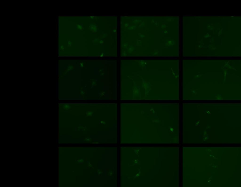
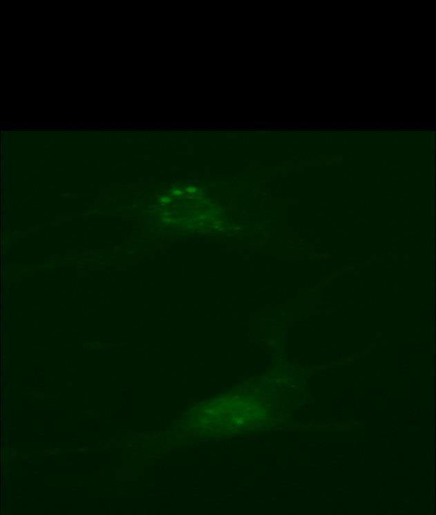
51 The role of autophagy and formation of LC3-GFP vesicle formation in GBM12 cells GBM12 cells plated in four-well chamber slides were transfected with a plasmid containing LC3-GFP construct, and the formation of the autophagic vesicles was assessed by fluorescent microscopy at 6, 12 and 24 hours after the treatment, respectively (Figure 14A). The number of vesicles in 40 cells representing each group was counted, and the average vesicles formed in each group was calculated (Figure 14B). The data showed a significant increase in the formation of autophagic vesicles in GBM12 cells as early as 6 hours after treatment with sorafenib and the combination. This trend stabilized after 12 hours, with vesicle formation remaining higher after combinational treatment. The extent of autophagy in the lapatinib-treated groups was not significant. 49


52 A 50
53 B Mean LC3-GFP Vesicles per Cell Veh Lap Sor Lap+Sor Veh Lap Sor Lap+Sor Veh Lap Sor Lap+Sor 6 hr 12 hr 24 hr Figure 14. LC3-GFP vesicle formation assay in GBM12 cells treated with sorafenib and lapatinib. GBM12 cells were transfected with LC3-GFP plasmid and treated with lapatinib (2 µm), sorafenib (6 µm) and the combination. Cells were then visualized by fluorescent microscopy at 6, 12 and 24 hours after exposure (A). The number of vesicles in 40 cells representing each group was counted, and the average vesicles formed in each group was calculated and plotted (B). 51
54 Knockdown of ATG5 and Beclin 1 reduces the drug combination-mediated toxicity in GBM12 and GBM5 cells In order to investigate the role of autophagy in lapatinib and sorafenib-mediated cell death, Beclin1 and ATG5 were down-regulated. GBM12 and GBM5 cells were transfected with control sirna (SCR), sibeclin1 or siatg5 then subjected to lapatinib, sorafenib and the combination of both agents for 24 hours after which cell viability was assessed by trypan blue exclusion assay. The results revealed a reduction in cellular toxicity in response to sorafenib and sorafenib with lapatinib. Toxicity of lapatinib was not affected by inhibition of autophagy (Figure 15 and 16). These findings agreed with the data gained from the LC3-GFP vesicle formation assay, emphasizing the significance of autophagy in lapatinib and sorafenib combination-mediated toxicity. 52

55 100 Percent Cell Death Veh Lap Sor Lap+Sor Veh Lap Sor Lap+Sor Veh Lap Sor Lap+Sor siscr siatg5 sibeclin1 Figure 15. Knockdown of ATG5 and Beclin-1 reduces drug combination-mediated toxicity in GBM12 cells. GBM12 cells were first transfected with either siatg5 or sibeclin-1 and then treated with lapatinib (2 µm) and sorafenib (6 µm) or the combination. Cell viability was determined by trypan blue exclusion assay 24 hours after exposure. *P<0.05 less than corresponding value in siscr cells. 53
56 100 Percent Cell Death Veh Lap Sor Lap+Sor Veh Lap Sor Lap+Sor Veh Lap Sor Lap+Sor siscr siatg5 sibeclin1 Figure 16. Knockdown of ATG5 and Beclin-1 reduces drug combination-mediated toxicity in GBM5 cells. GBM5 cells were first transfected with either siatg5 or sibeclin-1 and then treated with lapatinib (2 µm) and sorafenib (6 µm) or the combination. Cell viability was determined by trypan blue exclusion assay 24 hours after exposure. *P<0.05 less than corresponding value in siscr cells. 54
57 Up-regulation of FLIP and Bcl-xL and down-regulation of caspase 9 reduce the drug combination-mediated toxicity in GBM12 and GBM5 cells To elucidate the involvement of apoptosis in drug-mediated cell death, GBM12 and GBM 5 cells were infected with adenoviruses carrying an empty vector (CMV) or the constructs designed to either down-regulate caspase 9 via dominant negative mutation (DN9) or up-regulate the expression of FLIP or Bcl-xL. Transfected cells were treated with sorafenib and lapatinib or the combination for 24 hours and cell viability was determined by trypan blue exclusion assay. In GBM12 cells, knockdown of caspase-9, a key component of the intrinsic apoptotic pathway, and overexpression of Bcl-xL, an antiapoptotic protein, reduced the toxicity of lapatinib and the combination whereas the toxicity of sorafenib remained intact. On the other hand, overexpression of FLIP, an inhibitor of caspase 8, decreased cell death caused by both sorafenib and the drug combination, whereas the toxicity of lapatinib was not affected (Figure 17 and 18). In GBM5 cells down-regulation of caspase 9 and up-regulation of Bcl-xL produced similar results. However, overexpression of FLIP reduced the toxicity of sorafenib and lapatinib individually and in combination (Figure 19). The role of the intrinsic and extrinsic apoptotic pathways in drug-mediated cell death was more clearly observed in GBM12 cells after 48 hour exposure (Figure 20). 55

58 100 Percent Cell Death Veh Lap Sor Lap+Sor Veh Lap Sor Lap+Sor Veh Lap Sor Lap+Sor CMV FLIP Bcl-xL Figure 17. Overexpression of FLIP and Bcl-xL reduces the toxicity of the drug combination in GBM12 cells. GBM12 cells were first infected with recombinant adenoviruses to express constitutively activate FLIP and Bcl-xL and then were treated with lapatinib (2 µm), sorafenib (6 µm) and the combination. Cell viability was assessed by trypan blue exclusion assay 24 hours after exposure. *P<0.05 less than corresponding value in CMV cells. 56
59 100 Percent Cell Death Veh Lap Sor Lap+Sor Veh Lap Sor Lap+Sor CMV DN9 Figure 18. Down-regulation of caspase 9 reduces the toxicity of the drug combination in GBM12 cells. GBM12 cells were infected with the recombinant adenovirus expressing mutant dominant negative caspase 9. Cells were then treated with lapatinib (2 µm), sorafenib (6 µm) and the combination. Cell viability was assessed by trypan blue exclusion assay 24 hours after exposure. *P<0.05 less than corresponding value in CMV cells. 57


60 100 Percent Cell Death Veh Lap Sor Lap+Sor Veh Lap Sor Lap+Sor Veh Lap Sor Lap+Sor Veh Lap Sor Lap+Sor CMV DN9 FLIP Bcl-xL Figure 19. Up-regulation of FLIP and Bcl-xL and down-regulation of caspase 9 reduce the toxicity of the drug combination in GBM5 cells. GBM5 cells were infected with recombinant adenoviruses to express constitutively activate FLIP, Bcl-xL and dominant negative caspase 9. Cells were then treated with lapatinib (2 µm), sorafenib (6 µm) and the combination. Cell viability was assessed by trypan blue exclusion assay 24 hours after exposure. *P<0.05 less than corresponding value in CMV cells. 58
61 100 Percent Cell Death Veh Lap+Sor Veh Lap+Sor Veh Lap+Sor CMV DN9 FLIP Figure 20. Up-regulation of FLIP and down-regulation of caspase 9 dramatically reduce the toxicity of the drug combination in GBM12 cells during 48 hour-drug exposure. GBM12 cells were infected with the recombinant adenoviruses expressing mutant dominant negative caspase 9 and constitutively active FLIP. Cells were then treated with lapatinib (2 µm) and sorafenib (6 µm) simultaneously. Cell viability was assessed by trypan blue exclusion assay 48 hours after exposure. *P<0.05 less than corresponding value in CMV cells. 59
62 Knockdown of Fas and FADD reduces the drug combination-mediated toxicity in GBM12 cells Previous data suggested the involvement of receptor-mediated apoptosis in cells treated with sorafenib and the combination of sorafenib and lapatinib. In order to elucidate which members of TNF receptor family are involved in this process, Fas and Fas-associated death domain (FADD) were knocked down by corresponding sirnas and cell viability was assessed by trypan blue exclusion assay 24 hours after the treatment. The results indicated a reduction in toxicity of the drug combination; further confirming activation of extrinsic apoptotic pathways (Figure 21). 60
 and sorafenib (6 µm).")
63 100 Percent Cell Death Veh Lap+Sor Veh Lap+Sor Veh Lap+Sor siscr sicd95 sifadd Figure 21. Knockdown of CD95 (Fas) and FADD reduce the toxicity of the drug combination in GBM12 cells. GBM12 cells were first transfected with either sicd95 or sifadd and then treated with the combination of lapatinib (2 µm) and sorafenib (6 µm). Cell viability was determined by trypan blue exclusion assay 24 hours after exposure. *P<0.05 less than corresponding value in siscr cells. 61
G-Protein Signaling. Introduction to intracellular signaling. Dr. SARRAY Sameh, Ph.D
 G-Protein Signaling Introduction to intracellular signaling Dr. SARRAY Sameh, Ph.D Cell signaling Cells communicate via extracellular signaling molecules (Hormones, growth factors and neurotransmitters
Enzyme-coupled Receptors. Cell-surface receptors 1. Ion-channel-coupled receptors 2. G-protein-coupled receptors 3. Enzyme-coupled receptors
 Enzyme-coupled Receptors Cell-surface receptors 1. Ion-channel-coupled receptors 2. G-protein-coupled receptors 3. Enzyme-coupled receptors Cell-surface receptors allow a flow of ions across the plasma
Cell Quality Control. Peter Takizawa Department of Cell Biology
 Cell Quality Control Peter Takizawa Department of Cell Biology Cellular quality control reduces production of defective proteins. Cells have many quality control systems to ensure that cell does not build
RAS Genes. The ras superfamily of genes encodes small GTP binding proteins that are responsible for the regulation of many cellular processes.
 ۱ RAS Genes The ras superfamily of genes encodes small GTP binding proteins that are responsible for the regulation of many cellular processes. Oncogenic ras genes in human cells include H ras, N ras,
Sildenafil and celecoxib interact to kill breast cancer cells
 Virginia Commonwealth University VCU Scholars Compass Theses and Dissertations Graduate School 2014 Sildenafil and celecoxib interact to kill breast cancer cells Brittany Binion Virginia Commonwealth University,
Signaling. Dr. Sujata Persad Katz Group Centre for Pharmacy & Health research
 Signaling Dr. Sujata Persad 3-020 Katz Group Centre for Pharmacy & Health research E-mail:sujata.persad@ualberta.ca 1 Growth Factor Receptors and Other Signaling Pathways What we will cover today: How
KEY CONCEPT QUESTIONS IN SIGNAL TRANSDUCTION
 Signal Transduction - Part 2 Key Concepts - Receptor tyrosine kinases control cell metabolism and proliferation Growth factor signaling through Ras Mutated cell signaling genes in cancer cells are called
Receptor mediated Signal Transduction
 Receptor mediated Signal Transduction G-protein-linked receptors adenylyl cyclase camp PKA Organization of receptor protein-tyrosine kinases From G.M. Cooper, The Cell. A molecular approach, 2004, third
Signaling Through Immune System Receptors (Ch. 7)
") Signaling Through Immune System Receptors (Ch. 7) 1. General principles of signal transduction and propagation. 2. Antigen receptor signaling and lymphocyte activation. 3. Other receptors and signaling
Signaling Through Immune System Receptors (Ch. 7) 1. General principles of signal transduction and propagation. 2. Antigen receptor signaling and lymphocyte activation. 3. Other receptors and signaling
Introduction to pathology lecture 5/ Cell injury apoptosis. Dr H Awad 2017/18
 Introduction to pathology lecture 5/ Cell injury apoptosis Dr H Awad 2017/18 Apoptosis = programmed cell death = cell suicide= individual cell death Apoptosis cell death induced by a tightly regulated
A particular set of insults induces apoptosis (part 1), which, if inhibited, can switch to autophagy. At least in some cellular settings, autophagy se
, which, if inhibited, can switch to autophagy. At least in some cellular settings, autophagy se") A particular set of insults induces apoptosis (part 1), which, if inhibited, can switch to autophagy. At least in some cellular settings, autophagy serves as a defence mechanism that prevents or retards
A particular set of insults induces apoptosis (part 1), which, if inhibited, can switch to autophagy. At least in some cellular settings, autophagy serves as a defence mechanism that prevents or retards
Cell Signaling part 2
 15 Cell Signaling part 2 Functions of Cell Surface Receptors Other cell surface receptors are directly linked to intracellular enzymes. The largest family of these is the receptor protein tyrosine kinases,
Biol403 MAP kinase signalling
 Biol403 MAP kinase signalling The mitogen activated protein kinase (MAPK) pathway is a signalling cascade activated by a diverse range of effectors. The cascade regulates many cellular activities including
The Tissue Engineer s Toolkit
 The Tissue Engineer s Toolkit Stimuli Detection and Response Ken Webb, Ph. D. Assistant Professor Dept. of Bioengineering Clemson University Environmental Stimulus-Cellular Response Environmental Stimuli
Chapter 15: Signal transduction
 Chapter 15: Signal transduction Know the terminology: Enzyme-linked receptor, G-protein linked receptor, nuclear hormone receptor, G-protein, adaptor protein, scaffolding protein, SH2 domain, MAPK, Ras,
Principles of Genetics and Molecular Biology
 Cell signaling Dr. Diala Abu-Hassan, DDS, PhD School of Medicine Dr.abuhassand@gmail.com Principles of Genetics and Molecular Biology www.cs.montana.edu Modes of cell signaling Direct interaction of a
What would you observe if you fused a G1 cell with a S cell? A. Mitotic and pulverized chromosomes. B. Mitotic and compact G1 chromosomes.
 What would you observe if you fused a G1 cell with a S cell? A. Mitotic and pulverized chromosomes. B. Mitotic and compact G1 chromosomes. C. Mostly non-compact G1 chromosomes. D. Compact G1 and G2 chromosomes.
The elements of G protein-coupled receptor systems
 The elements of G protein-coupled receptor systems Prostaglandines Sphingosine 1-phosphate a receptor that contains 7 membrane-spanning domains a coupled trimeric G protein which functions as a switch
Lipids and Membranes
 Lipids and Membranes Presented by Dr. Mohammad Saadeh The requirements for the Pharmaceutical Biochemistry I Philadelphia University Faculty of pharmacy Membrane transport D. Endocytosis and Exocytosis
MCB*4010 Midterm Exam / Winter 2008
 MCB*4010 Midterm Exam / Winter 2008 Name: ID: Instructions: Answer all 4 questions. The number of marks for each question indicates how many points you need to provide. Write your answers in point form,
CYTOKINE RECEPTORS AND SIGNAL TRANSDUCTION
 CYTOKINE RECEPTORS AND SIGNAL TRANSDUCTION What is Cytokine? Secreted popypeptide (protein) involved in cell-to-cell signaling. Acts in paracrine or autocrine fashion through specific cellular receptors.
Cellular Signaling Pathways. Signaling Overview
 Cellular Signaling Pathways Signaling Overview Signaling steps Synthesis and release of signaling molecules (ligands) by the signaling cell. Transport of the signal to the target cell Detection of the
Apoptosis Chapter 9. Neelu Yadav PhD
 Apoptosis Chapter 9 Neelu Yadav PhD Neelu.Yadav@Roswellpark.org 1 Apoptosis: Lecture outline Apoptosis a programmed cell death pathway in normal homeostasis Core Apoptosis cascade is conserved Compare
Chapter 9. Cellular Signaling
 Chapter 9 Cellular Signaling Cellular Messaging Page 215 Cells can signal to each other and interpret the signals they receive from other cells and the environment Signals are most often chemicals The
Molecular biology :- Cancer genetics lecture 11
 Molecular biology :- Cancer genetics lecture 11 -We have talked about 2 group of genes that is involved in cellular transformation : proto-oncogenes and tumour suppressor genes, and it isn t enough to
Signal Transduction Cascades
 Signal Transduction Cascades Contents of this page: Kinases & phosphatases Protein Kinase A (camp-dependent protein kinase) G-protein signal cascade Structure of G-proteins Small GTP-binding proteins,
Propagation of the Signal
 OpenStax-CNX module: m44452 1 Propagation of the Signal OpenStax College This work is produced by OpenStax-CNX and licensed under the Creative Commons Attribution License 3.0 By the end of this section,
Antibodies for Unfolded Protein Response
 Novus-lu-2945 Antibodies for Unfolded rotein Response Unfolded roteins ER lumen GR78 IRE-1 GR78 ERK Cytosol GR78 TRAF2 ASK1 JNK Activator Intron RIDD elf2α Degraded mrna XB1 mrna Translation XB1-S (p50)
Signal Transduction Pathways. Part 2
 Signal Transduction Pathways Part 2 GPCRs G-protein coupled receptors > 700 GPCRs in humans Mediate responses to senses taste, smell, sight ~ 1000 GPCRs mediate sense of smell in mouse Half of all known
Cell Biology Lecture 9 Notes Basic Principles of cell signaling and GPCR system
 Cell Biology Lecture 9 Notes Basic Principles of cell signaling and GPCR system Basic Elements of cell signaling: Signal or signaling molecule (ligand, first messenger) o Small molecules (epinephrine,
Homework Hanson section MCB Course, Fall 2014
 Homework Hanson section MCB Course, Fall 2014 (1) Antitrypsin, which inhibits certain proteases, is normally secreted into the bloodstream by liver cells. Antitrypsin is absent from the bloodstream of
Practice Exam 2 MCBII
 1. Which feature is true for signal sequences and for stop transfer transmembrane domains (4 pts)? A. They are both 20 hydrophobic amino acids long. B. They are both found at the N-terminus of the protein.
Molecular Cell Biology - Problem Drill 19: Cell Signaling Pathways and Gene Expression
 Molecular Cell Biology - Problem Drill 19: Cell Signaling Pathways and Gene Expression Question No. 1 of 10 1. Which statement about cell signaling is correct? Question #1 (A) Cell signaling involves receiving
Protein Trafficking in the Secretory and Endocytic Pathways
 Protein Trafficking in the Secretory and Endocytic Pathways The compartmentalization of eukaryotic cells has considerable functional advantages for the cell, but requires elaborate mechanisms to ensure
Signal Transduction: G-Protein Coupled Receptors
 Signal Transduction: G-Protein Coupled Receptors Federle, M. (2017). Lectures 4-5: Signal Transduction parts 1&2: nuclear receptors and GPCRs. Lecture presented at PHAR 423 Lecture in UIC College of Pharmacy,
#19 Apoptosis Chapter 9. Neelu Yadav PhD
 #19 Apoptosis Chapter 9 Neelu Yadav PhD Neelu.Yadav@Roswellpark.org Why cells decide to die? - Stress, harmful, not needed - Completed its life span Death stimulation or Stress Cell Survival Death Functions
Mechanisms of Cell Death
 Mechanisms of Cell Death CELL DEATH AND FORMATION OF THE SEMICIRCULAR CANALS Carol M. Troy August 25, 2008 FROM: Fekete et al., Development 124: 2451 (1997) PHENOMENOLOGY OF CELL DEATH I. DEVELOPMENT A.
1. Activated receptor tyrosine kinases (RTKs) phosphorylates themselves
 phosphorylates themselves") Enzyme-coupled receptors Transmembrane proteins Ligand-binding domain on the outer surface Cytoplasmic domain acts as an enzyme itself or forms a complex with enzyme 1. Activated receptor tyrosine kinases
Enzyme-coupled receptors Transmembrane proteins Ligand-binding domain on the outer surface Cytoplasmic domain acts as an enzyme itself or forms a complex with enzyme 1. Activated receptor tyrosine kinases
2013 John Wiley & Sons, Inc. All rights reserved. PROTEIN SORTING. Lecture 10 BIOL 266/ Biology Department Concordia University. Dr. S.
 PROTEIN SORTING Lecture 10 BIOL 266/4 2014-15 Dr. S. Azam Biology Department Concordia University Introduction Membranes divide the cytoplasm of eukaryotic cells into distinct compartments. The endomembrane
Signal Transduction I
 Signal Transduction I Prof. Tianhua Zhou Department of Cell Biology Zhejiang University School of Medicine Office hours by appointment tzhou@zju.edu.cn Signal transduction: Key contents for signal transduction:
Genome of Hepatitis B Virus. VIRAL ONCOGENE Dr. Yahwardiah Siregar, PhD Dr. Sry Suryani Widjaja, Mkes Biochemistry Department
 Genome of Hepatitis B Virus VIRAL ONCOGENE Dr. Yahwardiah Siregar, PhD Dr. Sry Suryani Widjaja, Mkes Biochemistry Department Proto Oncogen and Oncogen Oncogen Proteins that possess the ability to cause
Protein kinases are enzymes that add a phosphate group to proteins according to the. ATP + protein OH > Protein OPO 3 + ADP
 Protein kinase Protein kinases are enzymes that add a phosphate group to proteins according to the following equation: 2 ATP + protein OH > Protein OPO 3 + ADP ATP represents adenosine trisphosphate, ADP
Molecular Cell Biology Problem Drill 16: Intracellular Compartment and Protein Sorting
 Molecular Cell Biology Problem Drill 16: Intracellular Compartment and Protein Sorting Question No. 1 of 10 Question 1. Which of the following statements about the nucleus is correct? Question #01 A. The
Regulation of cell function by intracellular signaling
 Regulation of cell function by intracellular signaling Objectives: Regulation principle Allosteric and covalent mechanisms, Popular second messengers, Protein kinases, Kinase cascade and interaction. regulation
Enzymes Part III: regulation II. Dr. Mamoun Ahram Summer, 2017
 Enzymes Part III: regulation II Dr. Mamoun Ahram Summer, 2017 Advantage This is a major mechanism for rapid and transient regulation of enzyme activity. A most common mechanism is enzyme phosphorylation
Apoptotic Pathways in Mammals Dr. Douglas R. Green
 Apoptotic Pathways in Mammals Douglas R. Green 1 Apoptosis A form of cell death that is defined morphologically, and features a number of biochemical events Programmed cell death Cell death that occurs
Introduction: 年 Fas signal-mediated apoptosis. PI3K/Akt
 Fas-ligand (CD95-L; Fas-L) Fas (CD95) Fas (apoptosis) 年 了 不 度 Fas Fas-L 力 不 Fas/Fas-L T IL-10Fas/Fas-L 不 年 Fas signal-mediated apoptosis 度降 不 不 力 U-118, HeLa, A549, Huh-7 MCF-7, HepG2. PI3K/Akt FasPI3K/Akt
p53 and Apoptosis: Master Guardian and Executioner Part 2
 p53 and Apoptosis: Master Guardian and Executioner Part 2 p14arf in human cells is a antagonist of Mdm2. The expression of ARF causes a rapid increase in p53 levels, so what would you suggest?.. The enemy
Cell Communication. Chapter 11. Biology Eighth Edition Neil Campbell and Jane Reece. PowerPoint Lecture Presentations for
 Chapter 11 Cell Communication PowerPoint Lecture Presentations for Biology Eighth Edition Neil Campbell and Jane Reece Lectures by Chris Romero, updated by Erin Barley with contributions from Joan Sharp
MOLECULAR CELL BIOLOGY
 1 Lodish Berk Kaiser Krieger scott Bretscher Ploegh Matsudaira MOLECULAR CELL BIOLOGY SEVENTH EDITION CHAPTER 13 Moving Proteins into Membranes and Organelles Copyright 2013 by W. H. Freeman and Company
UNIVERSITY OF MEDICINE AND PHARMACY CRAIOVA PhD SCHOOL. PhD THESIS
 UNIVERSITY OF MEDICINE AND PHARMACY CRAIOVA PhD SCHOOL PhD THESIS THE IMPORTANCE OF TUMOR ANGIOGENESIS IN CEREBRAL TUMOR DIAGNOSIS AND THERAPY ABSTRACT PhD COORDINATOR: Prof. univ. dr. DRICU Anica PhD
Revision. camp pathway
 االله الرحمن الرحيم بسم Revision camp pathway camp pathway Revision camp pathway Adenylate cyclase Adenylate Cyclase enzyme Adenylate cyclase catalyses the formation of camp from ATP. Stimulation or inhibition
Tala Saleh. Ahmad Attari. Mamoun Ahram
 23 Tala Saleh Ahmad Attari Minna Mushtaha Mamoun Ahram In the previous lecture, we discussed the mechanisms of regulating enzymes through inhibitors. Now, we will start this lecture by discussing regulation
34 Apoptosis Programmed cell death is vital to the health and development of multicellular organisms.
 Principles of Biology contents 34 Apoptosis Programmed cell death is vital to the health and development of multicellular organisms. Apoptosis is the reason we have separate fingers and toes. During embryonic
Chapter 20. Cell - Cell Signaling: Hormones and Receptors. Three general types of extracellular signaling. endocrine signaling. paracrine signaling
 Chapter 20 Cell - Cell Signaling: Hormones and Receptors Three general types of extracellular signaling endocrine signaling paracrine signaling autocrine signaling Endocrine Signaling - signaling molecules
Cell cycle, signaling to cell cycle, and molecular basis of oncogenesis
 Cell cycle, signaling to cell cycle, and molecular basis of oncogenesis MUDr. Jiří Vachtenheim, CSc. CELL CYCLE - SUMMARY Basic terminology: Cyclins conserved proteins with homologous regions; their cellular
The death receptors: signaling and modulation
 The death receptors: signaling and modulation 1 1 The extrinsic cell death pathway 2 Nat Rev Drug Discov. 2008 Dec;7(12):1001-12. 2 Death receptors Belong to the tumor necrosis factor (TNF) receptor gene
INTRODUCING NOVEL COMBINATORIAL TARGETED THERAPIES IN MULTIPLE TYPES OF CANCER
 Virginia Commonwealth University VCU Scholars Compass Theses and Dissertations Graduate School 2016 INTRODUCING NOVEL COMBINATORIAL TARGETED THERAPIES IN MULTIPLE TYPES OF CANCER Mehrad Tavallai Follow
Summary of Endomembrane-system
 Summary of Endomembrane-system 1. Endomembrane System: The structural and functional relationship organelles including ER,Golgi complex, lysosome, endosomes, secretory vesicles. 2. Membrane-bound structures
Growth and Differentiation Phosphorylation Sampler Kit
 Growth and Differentiation Phosphorylation Sampler Kit E 0 5 1 0 1 4 Kits Includes Cat. Quantity Application Reactivity Source Akt (Phospho-Ser473) E011054-1 50μg/50μl IHC, WB Human, Mouse, Rat Rabbit
Intracellular Compartments and Protein Sorting
 Intracellular Compartments and Protein Sorting Intracellular Compartments A eukaryotic cell is elaborately subdivided into functionally distinct, membrane-enclosed compartments. Each compartment, or organelle,
Lecture: CHAPTER 13 Signal Transduction Pathways
 Lecture: 10 17 2016 CHAPTER 13 Signal Transduction Pathways Chapter 13 Outline Signal transduction cascades have many components in common: 1. Release of a primary message as a response to a physiological
Apoptosis-based Therapies: Mechanisms and Applications
 Apoptosis-based Therapies: Mechanisms and Applications Perspective Bcl-2 family members, potential usage of BH3 domains as drug targets Bcl-2/xL inhibitors - Antisense, inhibitors of protein-protein interactions,
MDA-7/IL-24; A PROMISING CANCER THERAPEUTIC AGENT
 Virginia Commonwealth University VCU Scholars Compass Theses and Dissertations Graduate School 2012 MDA-7/IL-24; A PROMISING CANCER THERAPEUTIC AGENT Hossein Hamed Virginia Commonwealth University Follow
Cell cycle and apoptosis
 Cell cycle and apoptosis Cell cycle Definition Stages and steps Cell cycle Interphase (G1/G0, S, and G2) Mitosis (prophase, metaphase, anaphase, telophase, karyokinesis, cytokinesis) Control checkpoints
Peter Walter, UCSF IRE1 Signaling Affects Cell Fate during the Unfolded Protein Response
 Peter Walter, UCSF IRE1 Signaling Affects Cell Fate during the Unfolded Protein Response Jenn Hou Burke Group Literature Seminar November 19 th 2016 Protein Synthesis Pathway 4. Final Destination: proteins
Annals of RSCB Vol. XVI, Issue 1
 ENDOPLASMIC RETICULUM INVOLVEMENT IN APOPTOSIS OF NORMAL AND TREATED GINGIVAL FIBROBLASTS Ancuţa Goriuc, Raluca Jipu, Roxana Irina Iancu, M. Costuleanu GR. T. POPA UNIVERSITY OF MEDICINE AND PHARMACY,
shehab Moh Tarek ... ManarHajeer
 3 shehab Moh Tarek... ManarHajeer In the previous lecture we discussed the accumulation of oxygen- derived free radicals as a mechanism of cell injury, we covered their production and their pathologic
1. to understand how proteins find their destination in prokaryotic and eukaryotic cells 2. to know how proteins are bio-recycled
 Protein Targeting Objectives 1. to understand how proteins find their destination in prokaryotic and eukaryotic cells 2. to know how proteins are bio-recycled As a protein is being synthesized, decisions
Cell Communication. Local and Long Distance Signaling
 Cell Communication Cell to cell communication is essential for multicellular organisms Some universal mechanisms of cellular regulation providing more evidence for the evolutionary relatedness of all life
Effects of Second Messengers
 Effects of Second Messengers Inositol trisphosphate Diacylglycerol Opens Calcium Channels Binding to IP 3 -gated Channel Cooperative binding Activates Protein Kinase C is required Phosphorylation of many
Apoptosis Oncogenes. Srbová Martina
 Apoptosis Oncogenes Srbová Martina Cell Cycle Control point Cyclin B Cdk1 Cyclin D Cdk4 Cdk6 Cyclin A Cdk2 Cyclin E Cdk2 Cyclin-dependent kinase (Cdk) have to bind a cyclin to become active Regulation
Kinase Inhibitor p21 WAF1/CIP1 in Apoptosis and Autophagy
 Pivotal Role of the Cyclin-dependent Kinase Inhibitor p21 WAF1/CIP1 in Apoptosis and Autophagy Keishi Fujiwara, Shigeru Daido, Akitsugu Yamamoto, Ryuji Kobayash, Tomohisa Yokoyama, Hiroshi Aok, Eiji Iwado,
Chapter 11. Cell Communication
 Chapter 11 Cell Communication Overview: The Cellular Internet Cell-to-cell communication Is absolutely essential for multicellular organisms Concept 11.1: External signals are converted into responses
Principles of cell signaling Lecture 4
 Principles of cell signaling Lecture 4 Johan Lennartsson Molecular Cell Biology (1BG320), 2014 Johan.Lennartsson@licr.uu.se 1 Receptor tyrosine kinase-induced signal transduction Erk MAP kinase pathway
Protein sorting (endoplasmic reticulum) Dr. Diala Abu-Hsasan School of Medicine
 Dr. Diala Abu-Hsasan School of Medicine") Protein sorting (endoplasmic reticulum) Dr. Diala Abu-Hsasan School of Medicine dr.abuhassand@gmail.com An overview of cellular components Endoplasmic reticulum (ER) It is a network of membrane-enclosed
Protein sorting (endoplasmic reticulum) Dr. Diala Abu-Hsasan School of Medicine dr.abuhassand@gmail.com An overview of cellular components Endoplasmic reticulum (ER) It is a network of membrane-enclosed
609G: Concepts of Cancer Genetics and Treatments (3 credits)
") Master of Chemical and Life Sciences Program College of Computer, Mathematical, and Natural Sciences 609G: Concepts of Cancer Genetics and Treatments (3 credits) Text books: Principles of Cancer Genetics,
Master of Chemical and Life Sciences Program College of Computer, Mathematical, and Natural Sciences 609G: Concepts of Cancer Genetics and Treatments (3 credits) Text books: Principles of Cancer Genetics,
#19 Apoptosis Chapter 9. Neelu Yadav PhD
 #19 Apoptosis Chapter 9 Neelu Yadav PhD Neelu.Yadav@Roswellpark.org Why cells decide to die? - Stress, harmful, not needed - Completed its life span Death stimulation or Stress Cell Survival Death Functions
INTERACTION DRUG BODY
 INTERACTION DRUG BODY What the drug does to the body What the body does to the drug Receptors - intracellular receptors - membrane receptors - Channel receptors - G protein-coupled receptors - Tyrosine-kinase
Cell Communication. Chapter 11. Biology Eighth Edition Neil Campbell and Jane Reece. PowerPoint Lecture Presentations for
 Chapter 11 Cell Communication PowerPoint Lecture Presentations for Biology Eighth Edition Neil Campbell and Jane Reece Lectures by Chris Romero, updated by Erin Barley with contributions from Joan Sharp
PROTEIN TRAFFICKING. Dr. SARRAY Sameh, Ph.D
 PROTEIN TRAFFICKING Dr. SARRAY Sameh, Ph.D Overview Proteins are synthesized either on free ribosomes or on ribosomes bound to endoplasmic reticulum (RER). The synthesis of nuclear, mitochondrial and peroxisomal
Lecture #27 Lecturer A. N. Koval
 Lecture #27 Lecturer A. N. Koval Hormones Transduce Signals to Affect Homeostatic Mechanisms Koval A. (C), 2011 2 Lipophilic hormones Classifying hormones into hydrophilic and lipophilic molecules indicates
number Done by Corrected by Doctor Maha Shomaf
 number 19 Done by Waseem Abo-Obeida Corrected by Abdullah Zreiqat Doctor Maha Shomaf Carcinogenesis: the molecular basis of cancer. Non-lethal genetic damage lies at the heart of carcinogenesis and leads
General Principles of Endocrine Physiology
 General Principles of Endocrine Physiology By Dr. Isabel S.S. Hwang Department of Physiology Faculty of Medicine University of Hong Kong The major human endocrine glands Endocrine glands and hormones
C-Phycocyanin (C-PC) is a n«sjfc&c- waefc-jduble phycobiliprotein. pigment isolated from Spirulina platensis. This water- soluble protein pigment is
 is a n«sjfc&c- waefc-jduble phycobiliprotein. pigment isolated from Spirulina platensis. This water- soluble protein pigment is") ' ^Summary C-Phycocyanin (C-PC) is a n«sjfc&c- waefc-jduble phycobiliprotein pigment isolated from Spirulina platensis. This water- soluble protein pigment is of greater importance because of its various
' ^Summary C-Phycocyanin (C-PC) is a n«sjfc&c- waefc-jduble phycobiliprotein pigment isolated from Spirulina platensis. This water- soluble protein pigment is of greater importance because of its various
Supplementary Figures
 Supplementary Figures Figure S1. Validation of kinase regulators of ONC201 sensitivity. Validation and screen results for changes in cell viability associated with the combination of ONC201 treatment (1
Lecture 15. Signal Transduction Pathways - Introduction
 Lecture 15 Signal Transduction Pathways - Introduction So far.. Regulation of mrna synthesis Regulation of rrna synthesis Regulation of trna & 5S rrna synthesis Regulation of gene expression by signals
Zool 3200: Cell Biology Exam 4 Part I 2/3/15
 Name: Key Trask Zool 3200: Cell Biology Exam 4 Part I 2/3/15 Answer each of the following questions in the space provided, explaining your answers when asked to do so; circle the correct answer or answers
Cell Communication. Biology Eighth Edition Neil Campbell and Jane Reece. PowerPoint Lecture Presentations for
 Chapter 11 Cell Communication PowerPoint Lecture Presentations for Biology Eighth Edition Neil Campbell and Jane Reece Lectures by Chris Romero, updated by Erin Barley with contributions from Joan Sharp
Concise Reference. HER2 Testing in Breast Cancer. Mary Falzon, Angelica Fasolo, Michael Gandy, Luca Gianni & Stefania Zambelli
 Concise Reference Testing in Breast Cancer Mary Falzon, Angelica Fasolo, Michael Gandy, Luca Gianni & Stefania Zambelli Extracted from Handbook of -Targeted Agents in Breast Cancer ublished by Springer
Signaling Apoptosis. Scott André Oakes, M.D. Dept. of Pathology Univ. of Calif-San Francisco. Cyt c Release BAX/BAK. Apoptosome Formation
 Signaling Apoptosis Cyt c Release BAX/BAK Apoptosome Formation Caspase Activation Scott André Oakes, M.D. Dept. of Pathology Univ. of Calif-San Francisco Why do we need cell death? Sculpt Organs Control
SUPPLEMENTARY INFORMATION
 Supplementary Figures Supplementary Figure S1. Binding of full-length OGT and deletion mutants to PIP strips (Echelon Biosciences). Supplementary Figure S2. Binding of the OGT (919-1036) fragments with
Signal Transduction: Information Metabolism. Chem 454: Regulatory Mechanisms in Biochemistry University of Wisconsin-Eau Claire
 Signal Transduction: Information Metabolism Chem 454: Regulatory Mechanisms in Biochemistry University of Wisconsin-Eau Claire Introduction Information Metabolism How cells receive, process and respond
Mechanisms of Cell Injury: Loss of Calcium Homeostasis
 Mechanisms of Cell Injury: Loss of Calcium Homeostasis SCPA610: Cellular Pathology Amornrat N. Jensen, Ph.D. amornrat.nar@mahidol.ac.th Leading questions Why is intracellular calcium important for the
Insulin mrna to Protein Kit
 Insulin mrna to Protein Kit A 3DMD Paper BioInformatics and Mini-Toober Folding Activity Student Handout www.3dmoleculardesigns.com Insulin mrna to Protein Kit Contents Becoming Familiar with the Data...
BIOL 4374/BCHS 4313 Cell Biology Exam #1 February 13, 2001
 BIOL 4374/BCHS 4313 Cell Biology Exam #1 February 13, 2001 SS# Name This exam is worth a total of 100 points. The number of points each question is worth is shown in parentheses. Good luck! 1. (2) The
Cell Communication. Chapter 11. PowerPoint Lectures for Biology, Seventh Edition. Lectures by Chris Romero. Neil Campbell and Jane Reece
 Chapter 11 Cell Communication PowerPoint Lectures for Biology, Seventh Edition Neil Campbell and Jane Reece Lectures by Chris Romero Overview: The Cellular Internet Cell-to-cell communication Is absolutely
Cancer and Tyrosine Kinase Inhibition
 Cancer and Tyrosine Kinase Inhibition 1 Motivation (1) In first world countries cancer is the second most common cause of death after cardiovascular diseases. For most tumors, treatment is limited to surgery,
Phospho-AKT Sampler Kit
 Phospho-AKT Sampler Kit E 0 5 1 0 0 3 Kits Includes Cat. Quantity Application Reactivity Source Akt (Ab-473) Antibody E021054-1 50μg/50μl IHC, WB Human, Mouse, Rat Rabbit Akt (Phospho-Ser473) Antibody
CELLS. Cells. Basic unit of life (except virus)
") Basic unit of life (except virus) CELLS Prokaryotic, w/o nucleus, bacteria Eukaryotic, w/ nucleus Various cell types specialized for particular function. Differentiation. Over 200 human cell types 56%
Basic unit of life (except virus) CELLS Prokaryotic, w/o nucleus, bacteria Eukaryotic, w/ nucleus Various cell types specialized for particular function. Differentiation. Over 200 human cell types 56%