Detection of Chemotherapy-Induced Apoptosis in Human Breast Cancer
|
|
|
- Rosalyn Robertson
- 6 years ago
- Views:
Transcription
1 Detection of Chemotherapy-Induced Apoptosis in Human Breast Cancer Item Type text; Electronic Thesis Authors High, Rachel Publisher The University of Arizona. Rights Copyright is held by the author. Digital access to this material is made possible by the University Libraries, University of Arizona. Further transmission, reproduction or presentation (such as public display or performance) of protected items is prohibited except with permission of the author. Download date 13/05/ :50:16 Link to Item
2
3 Abstract Caspase-cleaved keratin 18 (K18) is used as a biomarker of apoptosis to measure chemotherapy-induced cell death. The M30-FITC antibody can be used as a method of detection for caspase-cleaved K18, giving it potential as a prognostic and predictive tool in cancer treatment. This study tests the M30-FITC antibody for use with the human breast cancer cell lines MCF7, SKBR3, and MDA-MB-231 in flow cytometry, with the goal of optimizing the M30- FITC assay for use in human whole blood. The assay was evaluated for use with two different apoptotic pathways: first induced by the chemotherapy docetaxel (Taxotere), and then by the tumor inhibitor staurosporine. Analysis indicates that the M30-FITC antibody requires a specific caspase cleavage product to be produced during apoptosis, which the mechanism of docetaxel-induced apoptosis (mitotic catastrophe) does not appear to produce. Staurosporine treatment appears to induce apoptosis in a manner compatible for use with the M30 antibody, and is sufficient to induce apoptosis in MCF7, SKBR3, and MDA-MB-231. These treated cells are also suitably detected in spiked human whole blood, indicating the potential for clinical relevance of the assay. Introduction Circulating tumor cells (CTCs) are cells that have detached from a primary tumor and entered the bloodstream, playing a critical role in the metastasis of cancer 1., CTCs aide in the progression of disease and makes cancerous growth unpredictable and problematic to contain. Therefore evaluation of CTCs can yield important information about the cancer growth and potentially about responses to treatment 2. When treated with some chemotherapy agents, cancer, cells undergo apoptotis indicating response to treatment. Apoptosis can occur by different mechanisms, depending on the type of cell and the type of chemotherapy to which they were exposed and is characterized by blebbing of the membrane, cell shrinkage, and condensation of chromatin. A family of cysteine proteases, called caspases, are an essential part of this apoptotic pathway. These caspases fall into two groups: initiator caspases (caspases-8 and -9), which can either self-cleave or activate the second type, effector caspases (caspases-3, -6, and -7) 3. Together, caspases catalyze the process of apoptosis through regulation of other proteins in the cell. The chemotherapy agent, docetaxel, used to induce apoptosis in the first part of this study, initiates its apoptotic mechanism through the process of mitotic catastrophe, which inhibits the depolymerization of microtubules in the cytosol 4. The other toxin in this experiment, the kinase inhibitor staurosporine, is not used as a chemotherapy due to its high toxicity, but was used to model a specific apoptotic pathway. It utilizes caspase-3, one of the main caspases involved in apoptosis, in its mechanism of programmed cell death 5. This study investigates apoptosis specifically in three different human breast cancer cell lines: MCF7, SKBR3, and MDA-MB-231, as these cell lines exemplify some of the most common types of breast cancer: estrogen receptor positive, Her2 positive, and triple negative, respectively. Of these cells, the apoptotic mechanism in MCF7 is the most well-documented, and will thus be the focus of discussion. The ability to effectively test for apoptotic CTCs in patient blood would result in a clinically relevant, non-invasive method to monitor the early stages of cancer progression, as well as the recurrence of treated cancers. Currently, CTCs are difficult to detect due to their
4 infrequency of occurrence only 1-10 CTCs may be present per ml of whole blood 2 and are therefore an inefficient method of evaluating cancer development. Flow cytometry is one common method of CTC detection, and involves the detection of fluorescent biomarkers in apoptotic cells. Cells can be fluorescently stained in a sample of blood, and then isolated and characterized by the cytometer, which uses spectroscopy to detect the fluorophores on the cells. Three-colour flow, used in this project, allows the simultaneous detection of intracellular components and surface markers 8 by using differently-coloured labels on specific antibodies. Flow cytometry is also useful to differentiate between apoptosis and necrosis, the distinction between which is important for identification of which cells died in response to the applied chemotherapy, and which underwent a natural death due to other factors 9. Creation of fluorescent antibodies for flow cytometry is a process by which fluorescent dyes are conjugated to antibodies targeted for proteins of interest in the cell. The modified antibodies then bind to their target, the antigen, and fluoresce when detected by the cytometer s laser. Epithelial cell adhesion molecule (EpCAM), conjugated with the red protein-pigment phycoerythrin (PE), is one of these commonly used fluorescent markers. As it is expressed on most epithelial cells, but no other types 10, it is useful for detection of epithelial tumor cells in the blood. In conjunction with this, other fluorescently-tagged antigens are used to detect apoptosis. In this experiment, AnnexinV-fluorescein isothiocyanate (AnnexinV-FITC), Cytokeratin-PE (CK-PE) and 7-Aminoactinomycin D (7AAD) were used to characterize apoptosis and necrosis. AnnexinV binds to the surface marker phosphatidylserine, which is translocated from the inner to the outer cellular membrane during the early stages of apoptosis. The dye with which it is conjugated, FITC, is a green fluorophore and is easily distinguishable from the red PE 11. CK is an extremely useful marker for representing cell progression through apoptosis, as they are released from apoptotic cells during intermediate apoptosis 12. Epithelial cells, and thus epithelial cancers, have relatively large concentrations of both soluble and insoluble cytokeratins 13. 7AAD is a nuclear marker, and thus is one of the last markers to be detected in apoptosis. As 7AAD cannot pass through an intact cell membrane, it will not fluoresce until the nuclear envelope has broken open, indicating the final stages of apoptosis and entry into necrosis 14. While the protocols for the aforementioned antibodies have all been optimized, the associated assays require the use of several antibodies to test for apoptosis in epithelial cells EpCAM to detect the epithelial cells, and AnnexinV to detect early apoptosis, for instance. Another less common assay, which uses an M30 antibody, can perform both these tasks with use of only one antibody and could provide another method of study for cell death in chemotherapytreated cells. The M30 antibody, conjugated with FITC for this experiment, detects caspasecleaved keratin 18 (K18), a biomarker of epithelial apoptosis 15. In previous studies, M30 antigen levels have been correlated with other established prognostic markers 13, indicating the potential for M30 to be successful in characterization of apoptosis. Treatment of normal, tumor-free rats with a kinase inhibitor (AZD1152) yielded no change in baseline values of M30, while treatment of tumor-bearing rats produced a three-fold increase in M30 antigen levels in plasma, compared to controls 16. These findings suggest the prospective uses of M30 as a more prevalent antigen in the detection of apoptotic CTCs in whole blood, an ability which is critical in monitoring cancer progression and evaluating the effectiveness of administered chemotherapies 2. This experiment will examine the efficacy of the M30 antibody and apoptosis assay to determine its value as a potential biomarker of chemotherapy-induced cell death in human breast cancer cells.
5 Methods Cell Culture Human cell lines MCF7, SKBR3, and MDA-MB-231 were cultured in T-75 flasks for adherent cells, and maintained in RPMI 1640 media, supplemented with 10% fetal bovine serum (FBS), 1% penicillin/streptomycin, and 1% glutamate. To prevent overgrowth, cells were split approximately every three days and kept at approximately 50% optical density. For cell splitting, the media was removed, cells were washed with 5 ml Dulbecco s phosphate-buffered saline (DPBS), and incubated at 37 C for three minutes in 1 ml Trypsin. The flask was tapped against the counter to loosen any adherent cells, and 9 mls of RPMI, warmed to 37 C, was added. Cells were then brought into a single-cell suspension and a fraction was discarded into 10% bleach. Fresh RPMI was added to bring the total volume of the flask back to 12 ml and cells were replaced in the incubator at 37 C. Drug Treatment For treatment with docetaxel or staurosporine, cells were collected with Trypsin and counted using Trypan blue. Cells were plated in six-well plates, at 500,000 cells/well, and incubated overnight at 37 C. Cell media was then replaced with media containing the appropriate treatment at a concentration varying from to 2 µm for a duration ranging from 30 min to 24 hrs. Apoptosis Assays Cells from each cell line were collected independently, washed in DPBS, resuspended in DPBS, and added to Epi tubes in 100 ul aliquots. Cells were stained according to manufacturer s specifications. AnnexinV-FITC was used in conjunction with EpCAM-PE, CK- PE, and 7AAD as a control. The M30-FITC CytoDEATH antibody from Peviva was used as an experimental variable. For the M30 assay, methanol was used as a fixative and 1% PBSF was used as an incubation buffer. Flow Cytometry All samples were assayed on a BD Accuri C6 flow cytometer, with 10,000 cells per sample collected for analysis. Apoptotic cells are defined as AnnexinV-FITC +, 7AAD +, or M30- FITC +, necrotic cells as CK-PE + or M30-FITC +, and EpCAM-PE was used as an epithelial marker. Control tubes of untreated and treated cells stained with each individual antibody were run before each group of experimental tubes, to compensate and establish a baseline to determine positivity. Results M30-FITC as an Apoptotic Marker in Docetaxel-Induced Apoptosis The first undertaking in this experiment was to evaluate the potential of M30-FITC to be used as an apoptotic marker, using a previously-optimized AnnexinV apoptosis assay. A secondary goal was to test if the staining of any antibodies was affected by methanol fixation: methanol fixation is required for M30-FITC staining, but is not standard for any of the other antibodies used. The cell line MCF7 was initially used for this experiment, as it was shown in
6 previous experiments to be compatible with the chemotherapy docetaxel, and the required treatment concentration was known: MCF7 was treated for 30 min with 50 µm docetaxel. Both fixed and unfixed cells were stained with AnnexinV-FITC, CK-PE, 7AAD, and M30-FITC individually. Unfixed cells were used as a control to which to compare the fixed, treated cells. The increase in apoptosis from the untreated cells to the treated cells (Fig. 1) determines that the cells responded successfully to the docetaxel treatment: 11.5% apoptosis and 2.3% necrosis in the untreated cells increased to 42.4% apoptosis and 29.4% necrosis in the treated cells as evaluated by AnnexinV-FITC and 7AAD fluorescence. This is in agreement with the goal of ~50% apoptosis in treated cells. Staining of the cells (Fig. 2) indicated that fixation with methanol does not affect the use of any necessary antibodies. However, M30-FITC has little induction in treated cells, with levels of apoptosis for treated cells closely reflecting the levels of apoptosis in untreated cells, despite the knowledge that the treated cells were in fact more apoptotic than the untreated cells, as evidenced by the AnnexinV-FITC assay. Similar results were obtained for the SKBR3 and MDA-MB-231 cell lines. Permeabilizaton of Docetaxel-Treated MCF7 Cells One hypothesis as to why M30-FITC had such little induction was the possibility of poor permeabilization acting as a barrier to effective binding. To test this hypothesis, MCF7 cells, treated with 50 µm docetaxel as in the first experiment, were permeabilized with Tween20 before antibody staining, according to standard protocol. Cells were shown to be ~50% apoptotic/necrotic with a Trypan blue cell count before fixation with methanol. Trypan blue was used to measure apoptosis, as opposed to AnnexinV, to conserve both time and resources. Trypan blue has previously been shown to be a comparable substitute to the AnnexinV apoptosis assay. However, minimal apoptosis (1.6%) was detected in treated by M30-FITC staining, which was equivalent to the amount of apoptosis present in the healthy, untreated cells (Fig. 3). These results suggest that, while apoptosis is being induced by docetaxel in MCF7, the M30-FITC antibody is incapable of recognizing it. Staurosporine-Induced Apoptosis in MCF7 The hypothesis that M30-FITC was effectively recognizing apoptosis, but not apoptosis induced by the chemotherapy docetaxel, was then put under investigation. The kinase inhibitor staurosporine was selected as the new toxin to be tested, as literature searches suggested its compatibility with the M30 antibody 5. In its first experiment, a serial dilution of staurosporine was done overnight (24 hours), starting with 2 µm and decreasing to µm. The treated cells were stained with M30-FITC and run on the cytometer. Not only was M30-FITC capable of recognizing apoptosis, it appeared to stain in a dose-dependent manner, detecting increasing amounts of apoptosis corresponding to the increases in concentration of staurosporine (Fig. 4). This suggests that the mechanism of apoptosis induced by docetaxel is indeed not compatible for use with the M30 antibody, but the apoptotic mechanism brought about by treatment with staurosporine produced the appropriate caspase cleavage product required for M30 binding. Optimization of Staurosporine in MCF7 Now that an effective apoptotic inducer had been recognized, the assay needed to be optimized in order to determine the concentration of staurosporine required to induce approximately 50% apoptosis in the cells. From the previous experiment, 1 µm staurosporine was anticipated to be sufficient for inducing 50% apoptosis, so another serial dilution, from 1 µm to o.0625 µm was performed. The M30-FITC antibody again was able to stain in a dose-
7 dependent manner, and produced clear populations of apoptotic and healthy cells (Fig. 5). One problem that arose in this assay was a high amount of clumping among the apoptotic cells. Clumping is fairly common when large amounts of apoptosis are induced, so to combat this in future experiments, precautions were taken during cell culture. Rather than grow a single large population of cells for treatment, several smaller populations were cultured and then amalgamated after treatment (ie. Instead of one six well plate with 500,000 cells/well, two six well plates with 250,000 cells/well were used), with the intention of preventing overcrowding of the cells. Clumping was not encountered again. This experiment indicated that an overnight treatment of 1 µm staurosporine was sufficient to induce approximately 50% apoptosis, the desired amount for future assays. This concentration was also sufficient in SKBR3 and MDA- MB-231. Discussion Circulating tumor cells are indicators of cancer cell dissemination in blood, and identification and detection of any chemotherapy induced changes in them might be useful for predicting responses to chemotherapy. The M30-FITC antibody is capable of recognizing apoptosis of human breast cancer cells in whole blood, and thus has potential as an apoptotic identifier, but it also has several associated issues that might need to be optimized prior to it being a useful tool in the clinical setting. Most notably, its specific requirement for the epitope formed after caspase cleavage of K18 makes it incompatible with the chemotherapy docetaxel, which is a commonly used treatment. Docetaxel induces apoptosis through mitotic catastrophe, which prevents microtubule destabilization and does not produce the appropriate caspase cleavage product. Thus, the M30 antibody is limited in its application, as the cells to which it could be applied would have to be carefully selected to ensure the necessary cleavage product was present. Despite these limitations, the M30 antibody seems apt at detecting apoptosis in a laboratory setting, and provides a quicker assay than many alternatives. M30 specifically detects epithelial apoptosis with a single antibody, rather than needing several antibodies to detect epithelial cells and apoptosis, such as EpCAM and 7AAD. The use of M30 could facilitate identification of apoptotic tumor cells by offering a relatively simple and speedy assay for laboratory use. Furthermore, this study has confirmed the ability of staurosporine to induce apoptosis in MCF7, SKBR3, and MDA-MB-231, at a treatment concentration of 1 µm for 24 hours. While it was initially thought that the cell line MCF7 might lack caspase-3, other studies have determined alternate pathways that can produce this cleavage product in MCF7, procaspase-6 or caspase-9, for example 6, 7. Our studies confirm the likelihood of an alternate pathway, and per this study, MCF7 appears to be a good candidate for use with staurosporine, and the dose-response data collected suggests that staurosporine is effective at inducing apoptosis in MCF7. The next logical step in the evaluation of the M30 antibody s usefulness would be to test its ability to stain cells spiked in whole blood, as opposed to spiking the blood with previously-stained cells. As the ultimate goal of this endeavor is to create a laboratory relevant assay for M30.
8 References 1. Fidler, I. J., The pathogenesis of cancer metastasis: the 'seed and soil' hypothesis revisited. Nat Rev Cancer 2003, 3 (6), Miller, M. C.; Doyle, G. V.; Terstappen, L. W., Significance of Circulating Tumor Cells Detected by the CellSearch System in Patients with Metastatic Breast Colorectal and Prostate Cancer. J Oncol 2010, 2010, Thornberry, N. A.; Lazebnik, Y., Caspases: enemies within. Science 1998, 281 (5381), Riou, J. F.; Naudin, A.; Lavelle, F., Effects of Taxotere on murine and human tumor cell lines. Biochem Biophys Res Commun 1992, 187 (1), Xue, L. Y.; Chiu, S. M.; Oleinick, N. L., Staurosporine-induced death of MCF-7 human breast cancer cells: a distinction between caspase-3-dependent steps of apoptosis and the critical lethal lesions. Exp Cell Res 2003, 283 (2), Mooney, L. M.; Al-Sakkaf, K. A.; Brown, B. L.; Dobson, P. R., Apoptotic mechanisms in T47D and MCF-7 human breast cancer cells. Br J Cancer 2002, 87 (8), Manns, J.; Daubrawa, M.; Driessen, S.; Paasch, F.; Hoffmann, N.; Löffler, A.; Lauber, K.; Dieterle, A.; Alers, S.; Iftner, T.; Schulze-Osthoff, K.; Stork, B.; Wesselborg, S., Triggering of a novel intrinsic apoptosis pathway by the kinase inhibitor staurosporine: activation of caspase-9 in the absence of Apaf-1. FASEB J 2011, 25 (9), Jung, T.; Schauer, U.; Heusser, C.; Neumann, C.; Rieger, C., Detection of intracellular cytokines by flow cytometry. J Immunol Methods 1993, 159 (1-2), Darzynkiewicz, Z.; Bruno, S.; Del Bino, G.; Gorczyca, W.; Hotz, M. A.; Lassota, P.; Traganos, F., Features of apoptotic cells measured by flow cytometry. Cytometry 1992, 13 (8), Baeuerle, P. A.; Gires, O., EpCAM (CD326) finding its role in cancer. Br J Cancer 2007, 96 (3), Vermes, I.; Haanen, C.; Steffens-Nakken, H.; Reutelingsperger, C., A novel assay for apoptosis. Flow cytometric detection of phosphatidylserine expression on early apoptotic cells using fluorescein labelled Annexin V. J Immunol Methods 1995, 184 (1), Barak, V.; Goike, H.; Panaretakis, K. W.; Einarsson, R., Clinical utility of cytokeratins as tumor markers. Clin Biochem 2004, 37 (7), de Haas, E. C.; di Pietro, A.; Simpson, K. L.; Meijer, C.; Suurmeijer, A. J.; Lancashire, L. J.; Cummings, J.; de Jong, S.; de Vries, E. G.; Dive, C.; Gietema, J. A., Clinical evaluation of M30 and M65 ELISA cell death assays as circulating biomarkers in a drug-sensitive tumor, testicular cancer. Neoplasia 2008, 10 (10), Philpott, N. J.; Turner, A. J.; Scopes, J.; Westby, M.; Marsh, J. C.; Gordon-Smith, E. C.; Dalgleish, A. G.; Gibson, F. M., The use of 7-amino actinomycin D in identifying apoptosis: simplicity of use and broad spectrum of application compared with other techniques. Blood 1996, 87 (6), Greystoke, A.; Dean, E.; Saunders, M. P.; Cummings, J.; Hughes, A.; Ranson, M.; Dive, C.; Renehan, A. G., Multi-level evidence that circulating CK18 is a biomarker of tumour burden in colorectal cancer. Br J Cancer 2012, 107 (9), Cummings, J.; Hodgkinson, C.; Odedra, R.; Sini, P.; Heaton, S. P.; Mundt, K. E.; Ward, T. H.; Wilkinson, R. W.; Growcott, J.; Hughes, A.; Dive, C., Preclinical evaluation of M30 and M65 ELISAs as biomarkers of drug induced tumor cell death and antitumor activity. Mol Cancer Ther 2008, 7 (3),
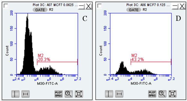
9 Figure Legend Figure 1: Apoptosis in treated and untreated MCF7 cells. (A) An untreated sample of MCF7 cells shows 11.5% apoptosis and 2.3% necrosis. This cellular death is likely due to the natural cell cycle and cells initiating apoptosis due to mistakes during replication or other common factors. These baseline levels establish the distinction between apoptosis in the normal cells and apoptosis due to treatment. (B) A sample of the same cells that had been treated with 50 µm docetaxel for 30 minutes shows an increase to 42.4% apoptosis and 29.4% necrosis. This increase indicates the success of the docetaxel treatment in initiating apoptosis in MCF7. Apoptosis was evaluated by the fluorescence of the AnnexinV-FITC and 7AAD antibodies. Figure 2: Staining of fixed untreated and 50 µm docetaxel treated MCF7. (A, B) Staining with 7AAD and CK-PE in fixed, untreated cells indicates that CK-PE needs to be titrated down in concentration to be brought into scale. 7AAD and CK-PE staining in fixed cells treated with 50 µm docetaxel shows similar results. 7AAD staining is not affected by methanol fixation. (C) Fixed, untreated cells stained with CK-PE and M30-FITC also indicate that CK-PE must be titrated down for use with this assay. (D) Fixed, treated cells stained with CK-PE and M30-FITC show little induction of M30-FITC. (E, F) Staining with 7AAD and EpCAM-PE both in untreated and treated cells demonstrates that EpCAM-PE efficacy is not affected by methanol fixation. (G) Staining with EpCAM-PE and M30-FITC show 12.3% apoptosis in the fixed, untreated sample, which is consistent with the data from the AnnexinV assay 13.7% apoptosis and necrosis. (H) EpCAM-PE and M30-FITC staining in the fixed, treated sample, show little induction of M30- FITC: only 12.5% apoptosis is shown, the same as in the untreated sample. Figure 3: Permeabilization in fixed MCF7. (A) Staining of untreated MCF7 with M30-FITC shows apoptosis present in the healthy population of cells to be 2.0%. (B) Staining of 50 µm docetaxel treated MCF7 with M30-FITC shows little/no induction of M30 the 1.6% apoptosis shown is due to apoptosis from the natural cell cycle as indicated by the baseline provided by (A). Note that the population of cells in the lower left (LL) quadrant has shifted up when compared to the corresponding quadrant in (A). This is likely due to auto-fluorescence caused by permeabilization and is not a confound for the results. (C) Reiteration of the data from (A) shown as a histograph. Cells in M1 (1.9%) are M30-FITC positive. (D) Reiteration of the data from (B). M30-FITC appears to detect apoptosis from cell cycle death, but not docetaxelinduced apoptosis. Figure 4: Dose-response in staurosporine-treated MCF7. M30-FITC proved capable of detecting a dose-dependent response in increasing concentrations of staurosporine treatment. Untreated MCF7 cells were defined as M30-FITC negative and set as a baseline to 0%, while percent apoptosis in treated samples was detected by the fluorescence of M30-FITC. The highest concentration of staurosporine, 2 µm, yielded % apoptosis. The drug was titrated from that concentration to concentrations of 1, 0.5, 0.25, and µm, with levels of apoptosis corresponding to 39.57, 27.16, 16.68, and 11.11% apoptosis, respectively. Figure 5: Optimization of staurosporine in MCF7. (A) Untreated, unstained cells were defined as M30-FITC negative. (B) Untreated, M30-FITC stained cells show 11.7% apoptosis. This apoptosis is indicative of the baseline amount of apoptotic cells in the MCF7 culture. (C) Stained cells treated with µm staurosporine yielded 20.3% apoptosis. (D) µm treated cells were 43.2% apoptotic. There was also a low yield of cells; treated cells were particularly clumpy, likely due to the high levels of apoptosis, and had to be filtered before assaying. (E) Cells treated with 0.25 µm docetaxel also had a high propensity to clump, with 31.7% apoptosis. (F) Cells
10 treated with 0.5 µm docetaxel had 43.7% apoptosis and a moderate amount of clumping. (G) The 1 µm treatment induced 47.5% apoptosis and moderate clumping. The 1 µm staurosporine treatment was selected for further experiments, as it yielded approximately 50% apoptosis.


11 Figures Figure 1

12


13 Figure 2

14 % Apoptosis Figure [Staurosporine] (um) Figure 4

15


16 Figure 5
Annexin V-APC/7-AAD Apoptosis Kit
 Annexin V-APC/7-AAD Apoptosis Kit Catalog Number KA3808 100 assays Version: 04 Intended for research use only www.abnova.com Table of Contents Introduction... 3 Background... 3 General Information... 4
Multi-Parameter Apoptosis Assay Kit
 Multi-Parameter Apoptosis Assay Kit Catalog Number KA1335 5 x 96 assays Version: 05 Intended for research use only www.abnova.com Table of Contents Introduction... 3 Background... 3 Principle of the Assay...
Oncology Cell Death Products from basic research to disease modelling to clinical trials
 Oncology Cell Death Products from basic research to disease modelling to clinical trials US Customer Service: US Technical Service: Peviva Cell Death Products Monoclonal Antibody Products M5 and M6 Keratin
PE Annexin V Apoptosis Detection Kit User Manual KT40001
 PE Annexin V Apoptosis Detection Kit User Manual KT40001 For research use only. Not intended for diagnostic testing. a WuXi AppTec company www.abgent.com.cn PE Annexin-V Apoptosis Detection Kit Product
Protocol for A-549 VIM RFP (ATCC CCL-185EMT) TGFβ1 EMT Induction and Drug Screening
 TGFβ1 EMT Induction and Drug Screening") Protocol for A-549 VIM RFP (ATCC CCL-185EMT) TGFβ1 EMT Induction and Drug Screening Introduction: Vimentin (VIM) intermediate filament (IF) proteins are associated with EMT in lung cancer and its metastatic
Protocol for A-549 VIM RFP (ATCC CCL-185EMT) TGFβ1 EMT Induction and Drug Screening Introduction: Vimentin (VIM) intermediate filament (IF) proteins are associated with EMT in lung cancer and its metastatic
LDL Uptake Flow Cytometry Assay Kit
 LDL Uptake Flow Cytometry Assay Kit Item No. 601470 www.caymanchem.com Customer Service 800.364.9897 Technical Support 888.526.5351 1180 E. Ellsworth Rd Ann Arbor, MI USA TABLE OF CONTENTS GENERAL INFORMATION
Instructions for Use. APO-AB Annexin V-Biotin Apoptosis Detection Kit 100 tests
 3URGXFW,QIRUPDWLRQ Sigma TACS Annexin V Apoptosis Detection Kits Instructions for Use APO-AB Annexin V-Biotin Apoptosis Detection Kit 100 tests For Research Use Only. Not for use in diagnostic procedures.
Interest in any of the products, request or order them at Bio-Connect Diagnostics.
 M3 CytoDEATH ELISA Interest in any of the products, request or order them at Bio-Connect Diagnostics. Bio-Connect Diagnostics B.V. T NL +31 ()26 326 44 6 T BE +32 ()2 52 12 53 Begonialaan 3a F NL +31 ()26
Annexin V APC Assay Kit
 Annexin V APC Assay Kit Item No. 601410 www.caymanchem.com Customer Service 800.364.9897 Technical Support 888.526.5351 1180 E. Ellsworth Rd Ann Arbor, MI USA TABLE OF CONTENTS GENERAL INFORMATION 3 Materials
The Annexin V Apoptosis Assay
 The Annexin V Apoptosis Assay Development of the Annexin V Apoptosis Assay: 1990 Andree at al. found that a protein, Vascular Anticoagulant α, bound to phospholipid bilayers in a calcium dependent manner.
Annexin V-PE Apoptosis Detection Kit
 Annexin V-PE Apoptosis Detection Kit Catalog Number KA0716 100 assays Version: 02 Intended for research use only www.abnova.com Table of Contents Introduction... 3 Background... 3 General Information...
ab Annexin V- mfluor Blue 570 Detection
 Version 1 Last updated 26 March 2018 ab219914 Annexin V- mfluor Blue 570 Detection Reagent For the rapid, sensitive and accurate measurement of PS exposure in live cells This product is for research use
RayBio Annexin V-FITC Apoptosis Detection Kit Plus
 RayBio Annexin V-FITC Apoptosis Detection Kit Plus User Manual Version 1.0 May 25, 2014 RayBio Annexin V-FITC Apoptosis (Cat#: 68FT-AnnVP-) RayBiotech, Inc. We Provide You With Excellent upport And ervice
Large Scale Infection for Pooled Screens of shrna libraries
 Last modified 01/11/09 Large Scale Infection for Pooled Screens of shrna libraries Biao Luo, Glenn Cowley, Michael Okamoto, Tanaz Sharifnia This protocol can be further optimized if cells being used are
Cover Letter. Reviewer 1:
 Cover Letter Michael Yang, M.D., Ph.D. Managing Editor of Cancer Research Frontiers 1188 Willis Ave, #109, Albertson, NY 11507, USA Phone: +1-917-426-1571 http://cancer-research-frontiers.org/ Dear Dr.
Lipid Droplets Fluorescence Assay Kit
 Lipid Droplets Fluorescence Assay Kit Item No. 500001 www.caymanchem.com Customer Service 800.364.9897 Technical Support 888.526.5351 1180 E. Ellsworth Rd Ann Arbor, MI USA TABLE OF CONTENTS GENERAL INFORMATION
Annexin V-FITC Apoptosis Detection Kit with SYTOX
 ab14086 Annexin V-FITC Apoptosis Detection Kit with SYTOX Instructions for Use For the rapid, sensitive and accurate measurement of apoptosis in living cells. This product is for research use only and
7-AAD/CFSE Cell-Mediated Cytotoxicity Assay Kit
 7-AAD/CFSE Cell-Mediated Cytotoxicity Assay Kit Catalog Number KA1293 96 assays Version: 02 Intended for research use only www.abnova.com Table of Contents Introduction... 3 Background... 3 Principle of
Analysis of Apoptosis and Necroptosis by Fluorescence-Activated Cell Sorting
 Protocol Analysis of Apoptosis and Necroptosis by Fluorescence-Activated Cell Sorting Fredrik Wallberg, Tencho Tenev, and Pascal eier 1 The Breakthrough Toby Robins Breast Cancer Research Centre, Institute
L6 GLUT4myc Cell Growth Protocol
 L6 GLUT4myc Cell Growth Protocol Background: Parental L6 cells selected for high fusion (2, 3) were stably transfected with a rat GLUT4 cdna carrying a myc epitope (recognized by the commercially available
Nature Methods: doi: /nmeth Supplementary Figure 1
 Supplementary Figure 1 Finite-element analysis of cell cluster dynamics in different cluster trap architectures. (a) Cluster-Chip (b) Filter (c) A structure identical to the Cluster-Chip except that one
RayBio Annexin V-FITC Apoptosis Detection Kit
 RayBio Annexin V-FITC Apoptosis Detection Kit User Manual Version 1.0 May 25, 2014 (Cat#: 68FT-AnnV-S) RayBiotech, Inc. We Provide You With Excellent Support And Service Tel:(Toll Free)1-888-494-8555 or
BD CBA on the BD Accuri C6: Bringing Multiplexed Cytokine Detection to the Benchtop
 BD CBA on the BD Accuri C6: Bringing Multiplexed Cytokine Detection to the Benchtop Maria Dinkelmann, PhD Senior Marketing Applications Specialist BD Biosciences, Ann Arbor, MI 23-14380-00 Cellular Communication
Rapid antigen-specific T cell enrichment (Rapid ARTE)
") Direct ex vivo characterization of human antigen-specific CD154+CD4+ T cell Rapid antigen-specific T cell enrichment (Rapid ARTE) Introduction Workflow Antigen (ag)-specific T cells play a central role
Direct ex vivo characterization of human antigen-specific CD154+CD4+ T cell Rapid antigen-specific T cell enrichment (Rapid ARTE) Introduction Workflow Antigen (ag)-specific T cells play a central role
Detailed step-by-step operating procedures for NK cell and CTL degranulation assays
 Supplemental methods Detailed step-by-step operating procedures for NK cell and CTL degranulation assays Materials PBMC isolated from patients, relatives and healthy donors as control K562 cells (ATCC,
RayBio Annexin V-Cy5 Apoptosis Detection Kit
 RayBio Annexin V-Cy5 Apoptosis Detection Kit User Manual Version 1.0 Mar 20, 2014 (Cat#: 68C5-AnnV-S) RayBiotech, Inc. We Provide You With Excellent Support And Service Tel:(Toll Free)1-888-494-8555 or
PROTOCOL: OPTIMIZATION OF LENTIVIRAL TRANSDUCTION USING SPINFECTION
 Last Modified: April 2018 Last Review: October 2018 PROTOCOL: OPTIMIZATION OF LENTIVIRAL TRANSDUCTION USING SPINFECTION Table of Contents 1. Brief Description 1 2. Materials and Reagents.1 3. Optimization
The Biochemistry of apoptosis
 The Biochemistry of apoptosis 1 1 The apoptosis is composed of multiple biochemical events 2 2 Biochemical, cellular, and molecular events in Apoptosis 1. Membrane blebbing; phosphatidyl serine exposure
Human Urokinase / PLAU / UPA ELISA Pair Set
 Human Urokinase / PLAU / UPA ELISA Pair Set Catalog Number : SEK10815 To achieve the best assay results, this manual must be read carefully before using this product and the assay is run as summarized
T H 1, T H 2 and T H 17 polarization of naïve CD4 + mouse T cells
 A complete workflow for cell preparation, isolation, polarization and analysis T H 1, T H 2 and T H 17 polarization of naïve CD4 + mouse T cells Introduction Workflow CD4 + T helper (T H) cells play a
HCC1937 is the HCC1937-pcDNA3 cell line, which was derived from a breast cancer with a mutation
 SUPPLEMENTARY INFORMATION Materials and Methods Human cell lines and culture conditions HCC1937 is the HCC1937-pcDNA3 cell line, which was derived from a breast cancer with a mutation in exon 20 of BRCA1
ROS Activity Assay Kit
 ROS Activity Assay Kit Catalog Number KA3841 200 assays Version: 03 Intended for research use only www.abnova.com Table of Contents Introduction... 3 Background... 3 General Information... 4 Materials
Direct ex vivo characterization of human antigen-specific CD154 + CD4 + T cells Rapid antigen-reactive T cell enrichment (Rapid ARTE)
") Direct ex vivo characterization of human antigen-specific CD154 + CD4 + T cells Rapid antigen-reactive T cell enrichment (Rapid ARTE) Introduction Workflow Antigen (ag)-specific T cells play a central
Direct ex vivo characterization of human antigen-specific CD154 + CD4 + T cells Rapid antigen-reactive T cell enrichment (Rapid ARTE) Introduction Workflow Antigen (ag)-specific T cells play a central
Technology Summary: Dansyl Molecular Probe
 Opportunity Statement Cell apoptosis refers to the active death process of cells controlled by genes for multi-cellular organisms to regulate development and maintain homeostasis. It is one of the main
Human Pluripotent Stem Cell Cardiomyocyte Differentiation Kit (PSCCDK) Introduction Kit Components Cat. # # of vials Reagent Quantity Storage
 Introduction Kit Components Cat. # # of vials Reagent Quantity Storage") Human Pluripotent Stem Cell Cardiomyocyte Differentiation Kit (PSCCDK) Catalog #5901 Introduction Human pluripotent stem cells (hpsc), including embryonic stem cells (ESC) and induced pluripotent stem
Human Pluripotent Stem Cell Cardiomyocyte Differentiation Kit (PSCCDK) Catalog #5901 Introduction Human pluripotent stem cells (hpsc), including embryonic stem cells (ESC) and induced pluripotent stem
Plate-Based Assay Methods for the Assessment of Cellular Health
 Plate-Based Assay Methods for the Assessment of Cellular Health Andrew L. Niles, Senior Research Scientist 2012, Promega Corporation. Biological Outcomes in Cell Culture Treatment -Small molecule -Bio-molecule
In vitro bactericidal assay Fig. S8 Gentamicin protection assay Phagocytosis assay
 In vitro bactericidal assay Mouse bone marrow was isolated from the femur and the tibia. Cells were suspended in phosphate buffered saline containing.5% BSA and 2 mm EDTA and filtered through a cell strainer.
B16-F10 (Mus musculus skin melanoma), NCI-H460 (human non-small cell lung cancer
, NCI-H460 (human non-small cell lung cancer") Electronic Supplementary Material (ESI) for ChemComm. This journal is The Royal Society of Chemistry 2017 Experimental Methods Cell culture B16-F10 (Mus musculus skin melanoma), NCI-H460 (human non-small
Electronic Supplementary Material (ESI) for ChemComm. This journal is The Royal Society of Chemistry 2017 Experimental Methods Cell culture B16-F10 (Mus musculus skin melanoma), NCI-H460 (human non-small
7-AAD/CFSE Cell-Mediated Cytotoxicity Assay Kit
 7-AAD/CFSE Cell-Mediated Cytotoxicity Assay Kit Item No. 600120 www.caymanchem.com Customer Service 800.364.9897 Technical Support 888.526.5351 1180 E. Ellsworth Rd Ann Arbor, MI USA TABLE OF CONTENTS
For the rapid, sensitive and accurate measurement of apoptosis in various samples.
 ab14082 500X Annexin V-FITC Apoptosis Detection Reagent Instructions for Use For the rapid, sensitive and accurate measurement of apoptosis in various samples. This product is for research use only and
Gladstone Institutes, University of California (UCSF), San Francisco, USA
, San Francisco, USA") Fluorescence-linked Antigen Quantification (FLAQ) Assay for Fast Quantification of HIV-1 p24 Gag Marianne Gesner, Mekhala Maiti, Robert Grant and Marielle Cavrois * Gladstone Institutes, University of
Fluorescence-linked Antigen Quantification (FLAQ) Assay for Fast Quantification of HIV-1 p24 Gag Marianne Gesner, Mekhala Maiti, Robert Grant and Marielle Cavrois * Gladstone Institutes, University of
Modulating Glucose Uptake in Skeletal Myotubes: Insulin Induction with Bioluminescent Glucose Uptake Analysis
 icell Skeletal Myoblasts Application Protocol Modulating Glucose Uptake in Skeletal Myotubes: Insulin Induction with Bioluminescent Glucose Uptake Analysis Introduction The skeletal muscle is one of the
Procaspase-3. Cleaved caspase-3. actin. Cytochrome C (10 M) Z-VAD-fmk. Procaspase-3. Cleaved caspase-3. actin. Z-VAD-fmk
 Z-VAD-fmk. Procaspase-3. Cleaved caspase-3. actin. Z-VAD-fmk") A HeLa actin - + + - - + Cytochrome C (1 M) Z-VAD-fmk PMN - + + - - + actin Cytochrome C (1 M) Z-VAD-fmk Figure S1. (A) Pan-caspase inhibitor z-vad-fmk inhibits cytochrome c- mediated procaspase-3 cleavage.
A HeLa actin - + + - - + Cytochrome C (1 M) Z-VAD-fmk PMN - + + - - + actin Cytochrome C (1 M) Z-VAD-fmk Figure S1. (A) Pan-caspase inhibitor z-vad-fmk inhibits cytochrome c- mediated procaspase-3 cleavage.
Commercially available HLA Class II tetramers (Beckman Coulter) conjugated to
 conjugated to") Class II tetramer staining Commercially available HLA Class II tetramers (Beckman Coulter) conjugated to PE were combined with dominant HIV epitopes (DRB1*0101-DRFYKTLRAEQASQEV, DRB1*0301- PEKEVLVWKFDSRLAFHH,
Class II tetramer staining Commercially available HLA Class II tetramers (Beckman Coulter) conjugated to PE were combined with dominant HIV epitopes (DRB1*0101-DRFYKTLRAEQASQEV, DRB1*0301- PEKEVLVWKFDSRLAFHH,
Supplementary Data 1. Alanine substitutions and position variants of APNCYGNIPL. Applied in
 Supplementary Data 1. Alanine substitutions and position variants of APNCYGNIPL. Applied in Supplementary Fig. 2 Substitution Sequence Position variant Sequence original APNCYGNIPL original APNCYGNIPL
Overview of methodology, tools and reagents for evaluating cell proliferation and invasion using multicellular tumor spheroids.
 The Next Step in the Evolution of 3D Culture: Utilizing Extracellular Matrix to Enhance Multicellular Tumor Spheroid Models for Proliferation and Invasion Overview of methodology, tools and reagents for
Annexin V-FITC Apoptosis Detection Kit
 ab14085 Annexin V-FITC Apoptosis Detection Kit Instructions for Use For the rapid, sensitive and accurate measurement of Apoptosis in living cells (adherent and suspension). View kit datasheet: www.abcam.com/ab14085
ab Membrane Fractionation Kit Instructions for Use For the rapid and simple separation of membrane, cytosolic and nuclear cellular fractions.
 ab139409 Membrane Fractionation Kit Instructions for Use For the rapid and simple separation of membrane, cytosolic and nuclear cellular fractions. This product is for research use only and is not intended
In vitro human regulatory T cell expansion
 - 1 - Human CD4 + CD25 + regulatory T cell isolation, Workflow in vitro expansion and analysis In vitro human regulatory T cell expansion Introduction Regulatory T (Treg) cells are a subpopulation of T
Detection of Apoptosis in Primary Cells by Annexin V Binding Using the Agilent 2100 Bioanalyzer. Application Note
 Detection of Apoptosis in Primary Cells by Annexin V Binding Using the Agilent 2100 Bioanalyzer Application Note Samuel D. H. Chan Marc Valer and Tobias Preckel, Introduction The Agilent 2100 bioanalyzer
Serafino et al. Thymosin α1 activates complement receptor-mediated phagocytosis in human monocyte-derived macrophages. SUPPLEMENTARY FIGURES
 Supplementary Fig. S1. Evaluation of the purity and maturation of macrophage cultures tested by flow cytometry. The lymphocytic/monocytic cellular fraction was isolated from buffy coats of healthy donors
ab65311 Cytochrome c Releasing Apoptosis Assay Kit
 ab65311 Cytochrome c Releasing Apoptosis Assay Kit Instructions for Use For the rapid, sensitive and accurate detection of Cytochrome c translocation from Mitochondria into Cytosol during Apoptosis in
Human Immunodeficiency Virus type 1 (HIV-1) gp120 / Glycoprotein 120 ELISA Pair Set
 gp120 / Glycoprotein 120 ELISA Pair Set") Human Immunodeficiency Virus type 1 (HIV-1) gp120 / Glycoprotein 120 ELISA Pair Set Catalog Number : SEK11233 To achieve the best assay results, this manual must be read carefully before using this product
Human Immunodeficiency Virus type 1 (HIV-1) gp120 / Glycoprotein 120 ELISA Pair Set Catalog Number : SEK11233 To achieve the best assay results, this manual must be read carefully before using this product
Cathepsin K Activity Assay Kit
 Cathepsin K Activity Assay Kit Catalog Number KA0769 100 assays Version: 03 Intended for research use only www.abnova.com Table of Contents Introduction... 3 Background... 3 General Information... 4 Materials
ab Exosome Isolation and Analysis Kit - Flow Cytometry, Cell culture
 Version 1 Last updated 14 March 2018 ab228564 Exosome Isolation and Analysis Kit - Flow Cytometry, Cell culture For the measurement of human exosomes in cell culture. This product is for research use only
Supplementary Figures
 Supplementary Figures Figure S1. Validation of kinase regulators of ONC201 sensitivity. Validation and screen results for changes in cell viability associated with the combination of ONC201 treatment (1
In vitro human regulatory T cell expansion
 - 1 - Human CD4 + CD25 + CD127 dim/- regulatory T cell Workflow isolation, in vitro expansion and analysis In vitro human regulatory T cell expansion Introduction Regulatory T (Treg) cells are a subpopulation
M30 Apoptosense ELISA. A biomarker assay for detection and screening of NASH
 M30 Apoptosense ELISA A biomarker assay for detection and screening of NASH NASH A Global Disease In the Western countries, Non-Alcoholic Fatty Liver Disease (NAFLD) is the most common liver disease, strongly
Circulating Tumor Cells in non- Metastatic Triple Negative Breast Cancer
 Circulating Tumor Cells in non- Metastatic Triple Negative Breast Cancer Carolyn Hall, Ph.D. Department of Surgical Oncology The University of Texas MD Anderson Cancer Center Triple Negative Breast Cancer
For research or further manufacturing use only. Not for injection or diagnostic procedures.
 PRIME-XV T cell Expansion XSFM PRIME-XV T Cell Expansion XSFM is a xeno-free, serum-free medium optimized for the activation and expansion of human T lymphocytes. This medium contains gentamicin and requires
Peptide stimulation and Intracellular Cytokine Staining
 v Peptide stimulation and Intracellular Cytokine Staining Authors A. Cosma, S. Allgayer MATERIALS Date 01-02-2007 REAGENTS: Version 1.0 - PBMCs - Culture medium - Costimulating antibodies - Peptide pools
LDL Uptake Cell-Based Assay Kit
 LDL Uptake Cell-Based Assay Kit Catalog Number KA1327 100 assays Version: 07 Intended for research use only www.abnova.com Table of Contents Introduction... 3 Background... 3 Principle of the Assay...
Incorporating pharmacodynamic, response and patient selection biomarkers. Paul Elvin PhD Chief Translational Science Officer Aptus Clinical
 Incorporating pharmacodynamic, response and patient selection biomarkers Paul Elvin PhD Chief Translational Science Officer Aptus Clinical 22 Oncology drug development Biomarkers key for: Strong hypothesis
Introduction: 年 Fas signal-mediated apoptosis. PI3K/Akt
 Fas-ligand (CD95-L; Fas-L) Fas (CD95) Fas (apoptosis) 年 了 不 度 Fas Fas-L 力 不 Fas/Fas-L T IL-10Fas/Fas-L 不 年 Fas signal-mediated apoptosis 度降 不 不 力 U-118, HeLa, A549, Huh-7 MCF-7, HepG2. PI3K/Akt FasPI3K/Akt
Green Cathepsin B Kit. For Research Use Only
 Green Cathepsin B Kit For Research Use Only ICT9151-25 Tests Component Storage Conditions Quantity 1 vial Rhodamine 110-(RR) 2-20 o C 25 tests 1 vial Hoechst 33342 (200 µg) 2-8 o C 1 ml 1 bottle 10x Cellular
ab Exosome Isolation and Analysis Kit - Flow Cytometry, Plasma
 Version 1 Last updated 25 May 2018 ab228565 Exosome Isolation and Analysis Kit - Flow Cytometry, Plasma For the isolation/detection of exosomes from human plasma, urine or cell culture media. This product
Supplemental Methods. CD107a assay
 Supplemental Methods CD107a assay For each T cell culture that was tested, two tubes were prepared. One tube contained BCMA-K562 cells, and the other tube contained NGFR-K562 cells. Both tubes contained
ADCC Assay Protocol Vikram Srivastava 1, Zheng Yang 1, Ivan Fan Ngai Hung 2, Jianqing Xu 3, Bojian Zheng 3 and Mei- Yun Zhang 3*
 ADCC Assay Protocol Vikram Srivastava 1, Zheng Yang 1, Ivan Fan Ngai Hung 2, Jianqing Xu 3, Bojian Zheng 3 and Mei- Yun Zhang 3* 1 Department of Microbiology, Li Ka Shing Faculty of Medicine, University
AMPK Phosphorylation Assay Kit
 AMPK Phosphorylation Assay Kit Catalog Number KA3789 100 assays Version: 02 Intended for research use only www.abnova.com Table of Contents Introduction... 3 Intended Use... 3 Background... 3 Principle
ab Exosome Isolation and Analysis Kit - Flow Cytometry, Cell Culture (CD63 / CD81)
") Version 1 Last updated 26 September 2018 ab239682 Exosome Isolation and Analysis Kit - Flow Cytometry, Cell Culture (CD63 / For the isolation and analysis of exosome from cell culture. This product is
Version 1 Last updated 26 September 2018 ab239682 Exosome Isolation and Analysis Kit - Flow Cytometry, Cell Culture (CD63 / For the isolation and analysis of exosome from cell culture. This product is
CytoSelect Tumor- Endothelium Adhesion Assay
 Product Manual CytoSelect Tumor- Endothelium Adhesion Assay Catalog Number CBA- 215 100 assays FOR RESEARCH USE ONLY Not for use in diagnostic procedures Introduction Cancer metastasis comprises several
Cell Lysis Buffer. Catalog number: AR0103
 Cell Lysis Buffer Catalog number: AR0103 Boster s Cell Lysis Buffer is a ready-to-use Western blot related reagent solution used for efficient extraction of total soluble protein in nondenatured state
MATERIALS. Peptide stimulation and Intracellular Cytokine Staining- EFFECTOR 45RA PANEL
 v Peptide stimulation and Intracellular Cytokine Staining- EFFECTOR 45RA PANEL Authors S. Kutscher, A. Cosma, MATERIALS REAGENTS: - PBMCs - Culture medium - Costimulating antibodies - Peptide pools including
To determine the effect of over-expression and/or ligand activation of. PPAR / on cell cycle, cell lines were cultured as described above until ~80%
 Supplementary Materials and Methods Cell cycle analysis To determine the effect of over-expression and/or ligand activation of PPAR / on cell cycle, cell lines were cultured as described above until ~80%
AUTOIMMUNE RESPONSES TO HUMAN TUMOUR ANTIGENS
 510 AUTOIMMUNE RESPONSES TO HUMAN TUMOUR ANTIGENS MADELINE HODKINSON* AND G. TAYLOR From the Immunology Department, Royal Infirmary, Manchester Received for publication May 14, 1969 THE most convincing
Nuclear Extraction Kit
 Nuclear Extraction Kit Catalog Number KA1346 50 assays Version: 07 Intended for research use only www.abnova.com Table of Contents Introduction... 3 Principle of the Assay... 3 General Information... 4
CytoSelect 24- Well Cell Invasion Assay (Basement Membrane, Fluorometric Format)
") Product Manual CytoSelect 24- Well Cell Invasion Assay (Basement Membrane, Fluorometric Format) Catalog Number CBA- 111 12 assays FOR RESEARCH USE ONLY Not for use in diagnostic procedures Introduction
Product Manual CytoSelect 24- Well Cell Invasion Assay (Basement Membrane, Fluorometric Format) Catalog Number CBA- 111 12 assays FOR RESEARCH USE ONLY Not for use in diagnostic procedures Introduction
Annexin V-Cy3 Apoptosis Detection Kit
 ab14142 Annexin V-Cy3 Apoptosis Detection Kit Instructions for Use For the rapid, sensitive and accurate measurement of apoptosis in various samples. This product is for research use only and is not intended
Notch Signaling Pathway Notch CSL Reporter HEK293 Cell line Catalog #: 60652
 Notch Signaling Pathway Notch CSL Reporter HEK293 Cell line Catalog #: 60652 Background The Notch signaling pathway controls cell fate decisions in vertebrate and invertebrate tissues. Notch signaling
SensoLyte pnpp Alkaline Phosphatase Assay Kit *Colorimetric*
 SensoLyte pnpp Alkaline Phosphatase Assay Kit *Colorimetric* Catalog # 72146 Kit Size 500 Assays (96-well plate) Optimized Performance: This kit is optimized to detect alkaline phosphatase activity Enhanced
34 Apoptosis Programmed cell death is vital to the health and development of multicellular organisms.
 Principles of Biology contents 34 Apoptosis Programmed cell death is vital to the health and development of multicellular organisms. Apoptosis is the reason we have separate fingers and toes. During embryonic
Technical Resources. BD Immunocytometry Systems. FastImmune Intracellular Cytokine Staining Procedures
 FastImmune Intracellular Cytokine Staining Procedures BD has developed protocols for the detection of intracellular cytokines in activated lymphocytes and in activated monocytes. The procedures have been
Chapter 7: Modes of Cell Death
 Chapter 7: Modes of Cell Death 7.1. Background Cell death can follow one of two distinct pathways, apoptosis or necrosis, and can occur in response to severe stress conditions or after exposure to toxic
Flow Cytometry Based Exosome Detection and Analysis Using the ZE5 Cell Analyzer
 Flow Cytometry ased Exosome Detection and nalysis Using the ZE5 Cell nalyzer Yasha Talaga, Elizabeth Dreskin, Joyce Lee io-rad Laboratories, Inc. 2000 lfred Nobel Drive, Hercules C 94547 Flow Cytometry
Bead Based Assays for Cytokine Detection
 Bead Based Assays for Cytokine Detection September 27, 2014 6 th EFIS-EJI South East European Immunology School SEEIS 2014 Timisoara, Romania The Cells of the Immune System The Immune Reaction (Th2) (Th1)
Annexin V-Cy3 Apoptosis Detection Reagent
 ab14143 Annexin V-Cy3 Apoptosis Detection Reagent Instructions for Use For the rapid, sensitive and accurate measurement of apoptosis in various samples This product is for research use only and is not
Nature Protocols: doi: /nprot Supplementary Figure 1. Fluorescent titration of probe CPDSA.
 Supplementary Figure 1 Fluorescent titration of probe CPDSA. Fluorescent titration of probe CPDSA (10 um) upon addition of GSH in HEPES (10 mm, ph = 7.4) containing 10% DMSO. Each spectrum was recorded
Primary Adult Naïve CD4+ CD45RA+ Cells. Prepared by: David Randolph at University of Alabama, Birmingham
 Primary Adult Naïve CD4+ CD45RA+ Cells Prepared by: David Randolph (drdrdr@uab.edu) at University of Alabama, Birmingham Goal: To obtain large numbers of highly pure primary CD4+ CD45RO- CD25- cells from
THE CYTOPATHOGENIC ACTION OF BLUETONGUE VIRUS ON TISSUE CULTURES AND ITS APPLICATION TO THE DETECTION OF ANTIBODIES IN THE SERUM OF SHEEP.
 Onderstepoort Journal of Veterinary Research, Volume 27, Number 2, October, 1956. The Government Printer. THE CYTOPATHOGENIC ACTION OF BLUETONGUE VIRUS ON TISSUE CULTURES AND ITS APPLICATION TO THE DETECTION
Figure S1 Time-dependent down-modulation of HER3 by EZN No Treatment. EZN-3920, 2 μm. Time, h
 Figure S1 Time-dependent down-modulation of HER3 by EZN-392 HE ER3 mrna A, %Contr rol 12 No Treatment EZN-392, 2 μm 1 8 6 4 2 2 8 24 Time, h Figure S2. Specific target down-modulation by HER3 (EZN-392)
Figure S1. PMVs from THP-1 cells expose phosphatidylserine and carry actin. A) Flow
 Flow") SUPPLEMENTARY DATA Supplementary Figure Legends Figure S1. PMVs from THP-1 cells expose phosphatidylserine and carry actin. A) Flow cytometry analysis of PMVs labelled with annexin-v-pe (Guava technologies)
SUPPLEMENTARY DATA Supplementary Figure Legends Figure S1. PMVs from THP-1 cells expose phosphatidylserine and carry actin. A) Flow cytometry analysis of PMVs labelled with annexin-v-pe (Guava technologies)
Muse Assays for Cell Analysis
 Muse Assays for Cell Analysis Multiple Assay Outputs for Cell Analysis Cell Health Cell Signalling Immunology Muse Count & Viability Kit Muse Cell Cycle Kit Muse Annexin V & Dead Cell Kit Muse Caspase
Nature Protocols: doi: /nprot Supplementary Figure 1
 Supplementary Figure 1 Traditional electronic gating strategy for analysing cell death based on A5-FITC and 7-AAD. a, Flow cytometry analysis showing the traditional two-stage electronic gating strategy
Apoptotic Pathways in Mammals Dr. Douglas R. Green
 Apoptotic Pathways in Mammals Douglas R. Green 1 Apoptosis A form of cell death that is defined morphologically, and features a number of biochemical events Programmed cell death Cell death that occurs
Cell cycle and apoptosis
 Cell cycle and apoptosis Cell cycle Definition Stages and steps Cell cycle Interphase (G1/G0, S, and G2) Mitosis (prophase, metaphase, anaphase, telophase, karyokinesis, cytokinesis) Control checkpoints
C-Phycocyanin (C-PC) is a n«sjfc&c- waefc-jduble phycobiliprotein. pigment isolated from Spirulina platensis. This water- soluble protein pigment is
 is a n«sjfc&c- waefc-jduble phycobiliprotein. pigment isolated from Spirulina platensis. This water- soluble protein pigment is") ' ^Summary C-Phycocyanin (C-PC) is a n«sjfc&c- waefc-jduble phycobiliprotein pigment isolated from Spirulina platensis. This water- soluble protein pigment is of greater importance because of its various
' ^Summary C-Phycocyanin (C-PC) is a n«sjfc&c- waefc-jduble phycobiliprotein pigment isolated from Spirulina platensis. This water- soluble protein pigment is of greater importance because of its various
Influenza A H7N9 (A/Anhui/1/2013) Hemagglutinin / HA ELISA Pair Set
 Hemagglutinin / HA ELISA Pair Set") Influenza A H7N9 (A/Anhui/1/2013) Hemagglutinin / HA ELISA Pair Set Catalog Number : SEK40103 To achieve the best assay results, this manual must be read carefully before using this product and the assay
Influenza A H7N9 (A/Anhui/1/2013) Hemagglutinin / HA ELISA Pair Set Catalog Number : SEK40103 To achieve the best assay results, this manual must be read carefully before using this product and the assay
BG-4, a bioactive peptide from Momordica charantia, promotes apoptosis in ovarian cancer cells
 University of Tennessee, Knoxville Trace: Tennessee Research and Creative Exchange University of Tennessee Honors Thesis Projects University of Tennessee Honors Program 5-2018 BG-4, a bioactive peptide
Naive, memory and regulatory T lymphocytes populations analysis
 Naive, memory and regulatory T lymphocytes populations analysis Jaen Olivier, PhD ojaen@beckmancoulter.com Cellular Analysis application specialist Beckman Coulter France Introduction Flow cytometric analysis