Role of Human Glutathione S-Transferase Alpha in Modulating Cellular. Stress and Cell Phase Transitions
|
|
|
- Lisa Washington
- 6 years ago
- Views:
Transcription
1 Role of Human Glutathione S-Transferase Alpha in Modulating Cellular Stress and Cell Phase Transitions by Humaira Adnan A Thesis presented to The University of Guelph In partial fulfilment of requirements for the degree of Doctor of Philosophy in Biomedical Sciences Guelph, Ontario, Canada Humaira Adnan, July, 2012
2 ABSTRACT ROLE OF HUMAN GLUTATHIONE S-TRANSFERASE ALPHA IN MODULATING CELLULAR STRESS AND CELL PHASE TRANSITIONS Humaira Adnan University of Guelph, 2012 Advisor: Dr. Gordon M. Kirby As intestinal epithelial cells mature, they continuously transition from proliferation to differentiation to apoptosis under the influence of cell signalling pathways including c-jun N- terminal kinase (JNK). Glutathione S-transferases (GSTs) are cytoprotective detoxification enzymes, some of which, including GSTA1, also sequester and inhibit JNK through complex formation. Thus, GSTA1 may be a key sensor of cellular state and regulator of responses to cell stress stimuli. The focus of this research study was to investigate the functional importance of GSTA1 in two contexts: 1) modulating complex integrity with JNK and activation of JNK by oxidative stress, 2) controlling cellular transitioning between proliferation, differentiation and apoptosis. In the first study, the impact of GSTA1 levels on dissociation of GSTA1-JNK complexes and JNK activation in response to cellular stress was investigated in the human colonic adenocarinoma Caco-2 cells. The pro-oxidant menadione caused GSTA1-JNK complex dissociation in preconfluent Caco-2 cells, whereas postconfluent cells were relatively resistant to this effect. Preconfluent cells were more sensitive than postconfluent cells to menadione-induced cytotoxicity. Additionally, menadione-induced JNK activation was transient since removal of the
3 stimulus resulted in re-association of GSTA1 with JNK and significantly reduced cytotoxicity. Over-expression and knockdown of GSTA1 affected the degree of GSTA1-JNK complex association without altering the JNK activation. However, enhanced GSH levels by N-acetyl cysteine blocked menadione-induced complex dissociation and JNK activation in Caco-2 cells. The results suggest that the mechanism of menadione-mediated JNK activation involves the production of reactive oxygen species, likely superoxide anion, and that the level of intracellular GSH plays an important role in preventing menadione-induced GSTA1-JNK complex dissociation and subsequent JNK activation. The functional importance of GSTA1 in controlling cellular proliferation, differentiation and apoptosis was investigated. Sodium butyrate (NaB) is a short-chain fatty acid, physiologically present in the human large intestine and modulates transitioning of cell states in colon cancer cell lines. GSTA1 levels increased in association with differentiation markers in postconfluent Caco-2 cells. Forced expression of GSTA1 significantly reduced cellular proliferation and sirna-mediated down-regulation of GSTA1 significantly increased cells in S- phase and associated cell proliferation. NaB (1 mm) reduced Caco-2 cell proliferation, increased differentiation and up-regulated GSTA1 activity. In contrast, higher dose of NaB (10 mm) caused toxicity in preconfluent cells via apoptosis through caspase-3 activation in association with reduced GSTA1 activity. GSTA1 down-regulation by sirna did not alter NaB-induced differentiation or the sensitivity of Caco-2 cells to NaB-induced apoptosis. Furthermore, NaB (10 mm) caused GSTA1-JNK complex dissociation but did not affect JNK activation. These findings suggest that GSTA1 levels may play a role in modulating enterocyte proliferation but do not influence differentiation or apoptosis.
4 TABLE OF CONTENTS ABSTRACT... ii TABLE OF CONTENTS... iv ACKNOWLEDGEMENTS... viii DECLARATION OF WORK PERFORMED... x LIST OF FIGURES... xi LIST OF TABLES... xii LIST OF ABREVIATIONS... xiii INTRODUCTION... 1 LITERATURE REVIEW... 3 Glutathione S-Transferases (GSTs)... 3 Glutathione S-Transferase Alpha (GSTA)... 5 Mitogen-Activated Protein Kinases (MAPKs)... 6 c-jun N-Terminal Kinase (JNK)... 7 Oxidative Stress and JNK Activation... 9 Menadione and JNK activation GSTs in cancer development and chemotherapy resistance Differentiation and apoptosis in intestinal epithelial cells Differentiation Apoptosis Sodium butyrate and cellular differentiation and apoptosis Colon Cancer Cell Lines RATIONALE Chapter iv
5 THE EFFECT OF MENADIONE ON GSTA1: JNK COMPLEX DISSOCIATION IN HUMAN COLONIC ADENOCARCINOMA CACO-2 CELLS ABSTRACT INTRODUCTION MATERIAL AND METHODS Materials Cell culture and treatments Immunoprecipitation Detection of GSTA1-JNK complexes Transient transfections SDS-PAGE and Western blot analysis Cytotoxicity assay Lipid peroxidation assay Total Glutathione assay Statistical analysis RESULTS Menadione activates JNK in preconfluent and postconfluent Caco-2 cells Menadione causes GSTA1-JNK complex dissociation only in preconfluent Caco-2 cells Temporary exposure of Caco-2 cells to menadione has a transient effect on GSTA1-JNK complex dissociation and JNK activation Preconfluent Caco-2 cells are more sensitive to menadione-induced cytotoxicity than postconfluent cells Over-expressed GSTA1 forms a complex with JNK but does not impede menadioneinduced complex dissociation Down-regulation of GSTA1 reduces GSTA1-JNK complex formation but does not alter menadione-induced JNK activation v
6 1.7. N-acetyl cysteine (NAC) blocks menadione-activated JNK in Caco-2 cells NAC prevents menadione-mediated GSH depletion and cytotoxicity but lower dose of menadione does not cause lipid peroxidation in Caco-2 cells DISCUSSION Chapter LOW LEVELS OF GSTA1 EXPRESSION ARE REQUIRED FOR CACO-2 CELLS PROLIFERATION ABSTRACT INTRODUCTION MATERIALS AND METHODS Materials Cell culture and treatments RNA isolation and real-time RT PCR analysis Transient transfections GSTA1 activity assay Alkaline phosphatase (AlkP) activity assay Cytotoxicity assay MTS assay BrdU Incorporation Flow cytometry Assessment of GSTA1-JNK complexes SDS-PAGE and Western blot analysis Statistical analysis RESULTS GSTA1 levels increase in differentiating Caco-2 cells vi
7 2.2. Modulation of GSTA1 levels alters proliferation of Caco-2 cells GSTA1 down-regulation affects cell cycle progression GSTA1 activity is altered with NaB-mediated changes in cell cycle phase Modulation of GSTA1 does not affect sodium butyrate-induced differentiation Modulation of GSTA1 does not affect sodium butyrate-induced apoptosis NaB (10 mm) causes GSTA1-JNK complex dissociation without activating JNK in Caco- 2 cells DISCUSSION GENERAL DISCUSSION SUMMARY AND CONCLUSIONS APPENDIX I APPENDIX ΙΙ REFERENCES vii
8 ACKNOWLEDGEMENTS First of all I would like to gratefully and sincerely thank Dr. Gordon Kirby for allowing me to join his lab and providing me opportunities to learn as a scientist. It has been an honour for me to be his student. His guidance, enthusiasm, understanding, and continuous support helped me at every step to move forward towards my goal. I would also like to express warm thanks to the members of my advisory committee Drs Brenda Coomber and Richard Mosser for their keen interest, support and valuable suggestions throughout my PhD research. I would like to express my gratitude to Dr Monica Antenos, whose assistance, training valuable suggestions and encouragement helped me at every step during research and writing of this thesis. I take this opportunity to thank Allison MacKay, Ann Maslen and Yu Gu for their technical assistance. Thanks to Wendy, Sally, Kim, Frances and Lisa for their co-operation. I am thankful to the past and present colleagues of Kirby lab for their help, friendship, support and making the lab environment pleasant and stress-free all the time. Thank you so much all the graduate students and faculty members of the Biomedical Sciences for the friendship you have shown during the last four years. I am thankful for the funding I received during my graduate training including stipend support in the form of a fellowship from Ontario Veterinary College, and research funding through an operating grant from the Canadian Institutes of Health Research (CIHR) awarded to Dr. Kirby. Finally, and most importantly, I have no words to express my appreciation for my children Fizza & Abdullah for their support, quiet patience, unwavering love and prayers. I am heartily obliged to my husband, Adnan without his undeniable support, I couldn t get it done. I am viii
9 indebted to my parents for instilling me with a strong passion for learning, their unmitigated support, encouragement and prayers. I am also thankful to my parents-in law for their generous and unconditional support. I offer my regards to all others who supported me in any respect during the completion of the project. ix
10 DECLARATION OF WORK PERFORMED I declare that with the exception of the work mentioned below, all the worked reported in this thesis was performed by me. I am thankful to Holly Quach, who helped me in optimizing GSTA1 sirna transfections. Dr. Monica Antenos and Holly performed the MTS assay on GSTA1 sirna- and GSTA1-V5-transfected cells reported in Chapter 2. Dr. Antenos performed BrdU incorporation and FACS analysis on GSTA1 down-regulated cells reported in chapter 2. x
11 LIST OF FIGURES Chapter 1 Figure 1. Menadione-mediated JNK activation is time-dependent Figure 2. Menadione causes GSTA1-JNK complex dissociation Figure 3. Menadione-mediated activation of JNK is transient Figure 4. Menadione-induced cytotoxicity is greater in preconfluent Figure 5. Over-expressed GSTA1 forms a complex with JNK Figure 6. Down-regulation of GSTA1 reduces GSTA1-JNK complex formation Figure 7. Menadione depletes glutathione levels Figure 8. N-acetyl cysteine blocks menadione-mediated activation of JNK Chapter 2 Figure 9. GSTA1 levels increase in differentiating Caco-2 cells Figure 10. GSTA1 levels can be modulated in preconfluent Caco-2 cells Figure 11. Modulation of GSTA1 levels mediate changes in Caco-2 cell growth Figure 12. GSTA1 down-regulation increases the percentage of Caco-2 cells in the S phase Figure 13. Distinct doses of NaB differently affect GSTA1 enzyme activities Figure 14. Modulation of GSTA1 does not affect NaB-induced differentiation Figure 15. GSTA1 down-regulation does not affect the sensitivity of Caco-2 cells apoptosis Figure 16. GSTA1 over-expression does not interfere with NaB-induced apoptosis Figure 17. NaB causes GSTA1-JNK complex dissociation in Caco-2 cells xi
12 LIST OF TABLES Table 1. Relative abundance and activity of GSTA1 in transiently transfected Caco-2 cells xii
13 LIST OF ABREVIATIONS 4-AD 4HNE 5-AD AlkP 4-androstene-3,17 dione 4-hydroxynonenal 5-androstene-3,17 dione alkaline phosphates AP-1 activator protein 1 APS ammonium persulfate ASK-1 apoptosis signal-regulating kinase 1 ATF-2 activating transcription factor 2 BrdU BSA cdna CDNB DMEM DNA DNAse dntp DPP-4 DTT E-cad ECL plus EDTA EGF 5-bromo-2-deoxyuridine bovine serum albumin complementary deoxyribonucleic acid 1-chloro-2,4-dinitrobenzene Dulbecco s Modified Eagle s Medium deoxyribonucleic acid Deoxyribonuclease deoxynucleotide triphosphate dipeptidylpeptidise-4 Dithiothreitol E-cadherin enhanced chemiluminescence plus ethylenediaminetetraacetic acid epidermal growth factor xiii
14 EGTA ELISA ERK FBS GAPDH GAR GSH GSR GSSG GST H 2 O 2 HAE HCC HNF IL-1β JNK LDH MAPEG MAPK MAPKK MAPKKK ethylene glycol tetraacetic acid enzyme-linked immunosorbant assay extracellular signal regulated kinase foetal bovine serum glyceraldehyde 3-phosphate dehydrogenase goat anti-rabbit Glutathione glutathione-conjugate glutathione disulfide glutathione s-transferase hydrogen peroxide 4-hydroxyalkenal hepatocellular carcinoma hepatocyte nuclear factor interleukin 1-beta c-jun n-terminal kinase lactate dehydrogenase membrane-associated protein in eicoisanoid and glutathione metabolism mitogen-activated protein kinase mitogen activated protein kinase kinase mitogen activated protein kinase kinase kinase xiv
15 MDA MEF Menadione ( vitamin-k3) M-MLV RT mrna MTS NaB NAC NC sirna PBS PCR PI3K PKC pnp PTEN PUFA RNA RNAi ROS SAPK SCFAs SDS-PAGE Malondialdehyde mouse embryonic fibroblast 2-methyl-1, 4-naphthoquinone murine moloney leukemia virus reverse transcriptase messenger ribonucleic acid 3-(4,5-dimethylthiazol-2-yl)-5-(3- carboxymethoxyphenyl)-2-(4- sulfophenyl)-2h-tetrazolium sodium butyrate N-acetyl cysteine negative control sirna phosphate buffered saline polymerase chain reaction phosphatidylinositol 3-kinase protein kinase C para-nitrophenyl phosphate phosphatase and tensin homolog polyunsaturated fatty acid ribonucleic acid small interfering ribonucleic acid reactive oxygen species stress-activated protein kinase short chain fatty acids sodium dodecyl sulphatepolyacrylamide gel electrophoresis xv
16 shrna SNP TBS TEMED tgsh TNF TPA TTBS UV vhnf-1c short-hairpin ribonucleic acid single nucleotide polymorphism tris buffered saline n,n,n',n'-tetramethylethylenediamine total glutathione tumour necrosis factor 12-o-tetradecanoyl phorbol-13-acetate tween-20- tris buffered saline ultra-violet variant hepatocyte nuclear factor xvi
17 INTRODUCTION Colorectal cancer has emerged as the third most common type of cancer and the second leading cause of cancer-related deaths in North America. The development of resistance to anticancer drugs is one of the primary causes of cancer treatment failure [1]. Improvement in the selectivity and therapeutic activity is a major goal in the development of anticancer agents [2,3]. Glutathione S-transferases (GSTs) are phase II detoxification enzymes that catalyze the conjugation of glutathione to a wide variety of endogenously and environmentally produced electrophilic compounds [4-6]. In humans, GST alpha 1 (GSTA1) is the major cytosolic isozyme present in almost all tissues and represents ~2 % of the total liver protein [7,8]. Typically cancer cells exhibit increased oxidative stress and many antioxidant enzymes, such as GSTAs, are overexpressed in a wide variety of tumours [2,3,9]. Different stimuli induce oxidative stress by generating reactive oxygen species (ROS) and activate the stress cell signalling molecules including c-jun N-terminal kinase (JNK). JNK is a mitogen-activated protein kinase (MAPK) that is an important mediator of cell stress responses and a regulator of pro-apoptotic death signalling events [10]. GSTs also play a role in the modulation of cellular stress signalling responses [11,12]. Specifically, GSTA1 and GSTP1 have been shown to form complexes with JNK thereby inhibiting its activation [13,14]. Therefore, GSTA1 may serve two distinct roles in the development of drug resistance: direct detoxification of chemotherapeutics as well as inhibition of the stress-activated cell signalling and subsequent apoptosis. However, there is limited understanding of the importance of GSTA1-JNK complex dissociation in oxidative stress. 1
18 The human colonic adenocarcinoma cell line, Caco-2, exhibits a unique relationship between growth characteristics and GSTA1 expression. Under normal cellular conditions, preconfluent Caco-2 cells proliferate whereas at confluency proliferation ceases and cells begin to differentiate. Moreover, GSTA1 expression is low in preconfluent cells and progressively increases with cellular confluency. One of the key characteristics of cancer cells is their ability to proliferate continuously without acquisition of cellular characteristics of differentiation and progression to apoptosis. The increased expression of GSTA1 with cellular confluency suggests that this isozyme controls the progression of enterocytes through different cellular states and that modulation of GSTA1 levels may play a role in the onset of differentiation and apoptosis. 2
19 LITERATURE REVIEW Glutathione S-Transferases (GSTs) Glutathione S-transferases (GSTs) are members of a multigene family of isoenzymes expressed in living organisms. GSTs detoxify a broad range of electrophilic compounds including carcinogens, chemotherapeutics, pharmaceuticals, pesticides, herbicides, industrial chemicals and pollutants, products of oxidative damage and natural plant toxins [4-6]. GSTs mediate detoxification of xenobiotics through glutathione (GSH) conjugation, which often results in less reactive and more water-soluble compounds. This minimizes the potential of damage from xenobiotics and eliminates these compounds from the body. Other functions of GSTs include protection against macromolecules such as protein, nucleic acid and lipids oxidized by reactive oxygen species (ROS), regeneration of S-thiolated proteins [15,16], detoxification of products of lipid peroxidation [17] and biosynthesis of physiologically important metabolites [18,19]. GSTs are also involved in the biosynthesis and metabolism of leukotrienes [20], prostaglandins [21], steroids [22] and in the development of resistance to chemotherapeutic agents [4,8,23-25]. GSTs also possess ligandin activity to bind a variety of hydrophilic compounds such as bile acids without catalytic activity and enhance their biliary excretion [26]. This activity is important in sequestration and transportation of non-polar ligands. GSTs modulate cell signalling pathways that regulate cellular proliferation and apoptosis (programmed cell death) [27,28]. In colonic epithelial cells, GSTs play an important role in providing protection against the effects of mutagens, ROS, lipid hydroperoxides and hydroxyalkenals [29,30]. 3
20 Human glutathione S-transferases (hgsts) are divided into three main families: cytosolic, mitochondrial and membrane-bound microsomal [7]. On the basis of structure, genetics, enzymatic properties, and immunological reactivity, there are seven classes of cytosolic GST isoenzymes, named Alpha, Mu, Omega, Pi, Sigma, Theta, and Zeta [1]. GST Kappa is the mitochondrial form in mammals [31]. There are two membrane-bound GSTs found in mammals named as microsomal GST-Ι and microsomal GST-ΙΙ or leukotriene C 4 synthase (LTC 4 S) [32]. Cytosolic and membrane bound GSTs do not share sequence homology [33,34,35]. Many classes of GST show polymorphisms and include numerous subunits. Each subunit ( amino acids in length, KDa) contains a catalytically independent active site that consists of a GSH-binding site ( G-site ) in the amino-terminal domain and a site that binds the hydrophobic substrate ( H-site ) in the carboxy-terminal domain [4-6]. GST isoenzymes can bind to many different substrates in their active site. GSTAs have two domains and an active site is located at the domain-interface; mutations of domain 1 or 2 result in reduced catalytic activity of GSTA1 [36]. The stability of the domain-domain interface particularly the H-site in GSTA1 plays a role in mediating the catalytic functionality of the active site of the enzyme [36]. More than a dozen cytosolic GST subunits have been identified in humans. These subunits exist as homodimers or heterodimers. Heterodimers are usually from the same gene class and only classes A and M of GST exist as both heterodimers as well as homodimers. The isoenzymes are named according to their class and subunit (e.g., GSTA1-2 symbolizes the enzyme composed of subunits 1 and 2 of the alpha class) [37,38]. Studies suggest that GSTs must dimerize in order to be catalytically active in mammalian species [27,39,40]. 4
21 Glutathione S-Transferase Alpha (GSTA) The alpha class GST isoenzymes consist of homo or heterodimers of five major subunits (GSTA1, A2, A3, A4 and A5). Transcripts for hgsta1, A2 and A4 are expressed at high levels in liver, intestine, kidney, adrenal gland and testis whereas expression of hgsta3 is in steroidogenic tissues: ovary, mammary gland, placenta, testis, and adrenal gland, and hgsta5 has not been found in the expressed sequence tag database [1]. hgsta1 is a highly expressed form of hgsta comprising almost 2% of total cytosolic protein in liver. The alpha class possesses a cytoprotective role against oxidative stress produced by various structurally unrelated electrophilic compounds, pro-oxidants and products of lipid peroxidation by catalyzing GSH-conjugation [41]. GSTAs possess GSH-dependent steroid isomerase activity and GSH-dependent selenium-independent peroxidase activity [1]. Epidemiological studies show that increased expression of GSTA has been associated with an increased risk of colorectal cancer, ovarian cancer and renal cell carcinoma [42]. Since GSHconjugates are formed as a result of oxidative stress or xenobiotic insult, it is plausible that these conjugates are indicators of cellular stress [43]. GSTAs have been shown to protect cells from the harmful effects of ROS-induced lipid peroxidation during oxidative stress caused by various agents [44]. These enzymes show high glutathione peroxidase activity towards phospholipid hydroxides generated during lipid peroxidation. For example, GSTA4-4 efficiently metabolizes the product of lipid peroxidation, 4-hydroxynonenal (4-HNE) through conjugating with GSH [45]. GST isozymes also modulate stress-activated signal transduction pathways [27]. Another important function of GSTAs is the inhibition of stress signalling kinases (JNK) and subsequently affecting the cascade of caspase activation and apoptosis in the cell [14]. Indeed, 5
22 over-expression of hgsta2-2 in cells attenuates the cytotoxic effects of hydrogen peroxide (H 2 O 2 ), and protects against H 2 O 2 -induced apoptosis by inhibiting JNK as well as caspase-3 activation [17]. GSTA1 also catalyzes the isomerization of 5-androstene-3,17-dione (AD) into 4- androstene-3,17-dione [46]. This isomerization occurs in the absence of GSH however it is required for increased catalytic activity [46]. GSTA2 has been shown to possess 100-fold less catalytic efficiency towards the isomerization of 5-androstene-3,17-dione than GSTA1 and GSTA4-4 shows 1000-fold less catalytic activity than GSTA1. Because chemical characterization of active sites of GSTA1 and GSTA2 differs by only four amino acid residues, isomerization of 5-androstene-3,17-dione is highly substrate specific reaction for GSTA1 [46]. Mitogen-Activated Protein Kinases (MAPKs) There are complex signalling pathways within cells that are activated in response to different stimuli. The mitogen-activated protein kinases (MAPKs) are a group of signalling proteins. These cell signalling kinases serve as critical regulators of the cellular response to a variety of exogenous and endogenous stresses. [47]. The MAPKs are involved in essential signal transduction cascades, a process that regulates cell growth, differentiation and apoptosis [48]. MAPK activity is regulated by their phosphorylation state that shows the equilibrium between stimulation by upstream kinases and inactivation by phosphatases. Their ultimate biological effect is the activation of specific transcription factors that then modulate cellular gene expression [49]. Three MAPK family members have been implicated in the oxidative stress 6
23 response: JNK, p38- MAPK and extracellular signal-regulated kinase (ERK) [47]. JNK and p38- MAPK activation generally have been shown to promote cell death from ROS [50] whereas ERK is generally activated by growth factors that lead to proliferation and prevent cell death [50,51]. The activation of these kinase pathways is achieved through a phosphorylation cascade [52]. MAPKs are activated by upstream MAPK kinases (MAPKKs) through dual phosphorylation of tyrosine/threonine residues, which are activated by even further upstream MAPK kinase kinases (MAPKKKs) [53]. The activated MAPKs then translocate into the nucleus and regulate the activities of specific transcription factors, ultimately affecting gene expression [54]. The cellular responses to these stimuli are highly dependent on cellular context [55] and can include proliferation, cell survival, differentiation, apoptosis, mitosis, migration and immune responses [56-58]. c-jun N-Terminal Kinase (JNK) JNK, also known as stress-activated protein kinase (SAPK), is an important member of the MAPK family. JNK has three isoforms (JNK1, JNK2, and JNK3) encoded by three different genes. The JNK1 and JNK2 genes are ubiquitously expressed, whereas JNK3 is found to be neural-specific [47,59]. JNK is primarily activated by various environmental stresses, including heat shock, osmotic shock, UV rays, oxidative stress, protein synthesis inhibitors, growth factors, chemotherapeutic agents and pro-inflammatory cytokines such as tumour necrosis factor-α (TNF-α) and interleukin-1β (IL-1β) [60,61]. The growth factors and cytokines activate MAPKs through their specific receptors but so far the mechanism of initiation of MAPKs due to oxidative stress is unclear. The upstream MAPKKKs that are involved in the JNK activation 7
24 cascade are numerous and their regulation is not completely understood [62,63]. Downstream molecules that are activated by phosphorylation of JNK are transcription factors including c-fos, c-jun, p53 and ATF2 [64]. In particular, c-jun, a component of activating protein (AP-1) complex is the most characterized downstream target of JNK. The binding of JNK to the N- terminal region of c-jun causes phosphorylation of c-jun and activates c-jun-dependent transcription [65] that results in the transcriptional upregulation of c-jun as well as an increase in c-jun protein stability [66,67] that has numerous biological consequences. The JNK pathway has also been shown to be important in the control of cell survival and death pathways, and interference with the JNK pathway suppresses the induction of apoptosis by a variety of agents [28]. It is known that the involvement of JNK in controlling diverse cellular functions such as cell proliferation, differentiation, and apoptosis is based on phosphorylation and functional modification of these molecular targets [10,68]. GST isozymes such as GSTP1 and GSTA1-1 are capable of associating with the JNK c- Jun complex [12,14]. GSTA1 forms complexes with JNK and GSTA1 reduces JNK signalling and apoptosis in Caco-2 and MEF3T3 cells [14]. GSTP1 has been identified as a potent JNK inhibitor. It is physically associated with the JNK c-jun complex and prevents the activation of JNK [27]. Binding of an inhibitor or substrate to the active site of GSTP1 induces a conformational change [69], which results in dissociation of the enzyme from the C-terminal domain of JNK [13]. The liberated JNK phosphorylates c-jun, which thereby activates numerous sequential downstream kinases (reviewed in [70]. Transient or low exposure to stress induces cellular proliferation and prolonged or high exposure induces apoptosis [38]. GSTM1-1 has been found to be an endogenous inhibitor of apoptosis signal-regulating kinase 1 (ASK1), an upstream activator of JNK [71]. 8
25 Oxidative Stress and JNK Activation Reactive oxygen species are free radicals and chemically reactive molecules, generated intracellularly due to a variety of stimuli. They have potential to cause damage to cellular macromolecules such as lipids, proteins, and DNA [72]. ROS act as important intermediate signalling molecules that cause a series of biological consequences in the cell [60]. They are produced through a variety of normal cellular processes, for example, as by-products of aerobic metabolism and through exogenous sources as a result of exposure to stimuli [72]. Studies have shown that ROS induce cellular injury and death due to direct biochemical damage and by affecting signal transduction pathways [73]. ROS include partially reduced metabolites of oxygen such as superoxide anion, hydrogen peroxide, and hydroxyl radicals. These metabolites possess higher reactivity than molecular oxygen (reviewed in [74,75]. Transient low level changes in ROS lead to important regulatory functions resulting in cellular proliferation. However, sustained or high levels of ROS can cause severe damage that leads to cell death. A number of responsive intracellular defense systems have been identified that combat the accumulation of ROS. These include various non-enzymatic molecules (e.g., glutathione, vitamins A, C, and E, and flavenoids) and anti-oxidants enzymes (e.g., superoxide dismutase (SOD), catalase, and glutathione peroxide) [76]. When these cellular defense mechanisms are not sufficient to counteract the production of ROS the result is oxidative stress. At the cellular level, oxidant injury elicits a wide spectrum of responses including cell proliferation to growth arrest, to senescence, to cell death. 9
26 Reactive oxygen species elicit many different responses. These responses depend upon the severity of the damage, the cell type, the magnitude of the dose, and the duration of the exposure [76]. Typically, low doses of ROS are mitogenic and promote cell proliferation, intermediate doses result in temporary or permanent growth arrest and severe oxidative stress ultimately causes cell death via either apoptotic or necrotic mechanisms [74,75,77]. Generation of ROS and altered redox status has been observed in cancer cells [78]. Different studies have identified activation of the JNK pathway in radiation-induced apoptosis and implicated the importance of the duration of JNK activation in determining cell fate [68]. Exogenous H 2 O 2 triggers ROS-induced JNK activation [79]. H 2 O 2 has a relatively long half-life, is soluble in both lipid and aqueous conditions and is capable of reaching its cellular targets [80]. Enhanced endogenous ROS production is frequently observed in cells exposed to many stimuli such as ultraviolet light [81,82], ionizing radiation [83,84], cancer chemotherapeutics [76] and TNF-α exposure [85]. JNK activation has been observed in cells treated with ROS precursors which give rise to superoxide radicals (O - 2 ) upon intracellular metabolism [86]. Activation of JNK by endogenously generated ROS is a critically important determinant of cellular fate [87,88]. Menadione and JNK activation Quinones represent a clinically important category of chemotherapeutic agents that promote cell death based on their redox cycling abilities [89]. Quinones target intracellular signalling pathways like JNK and p38 by generating ROS that promote programmed cell death [90]. One example is menadione (2-methyl-1,4-naphthoquinone), or vitamin K3 [91,92]. 10
27 Menadione undergoes redox cycling resulting in the formation of superoxide anions [93-96]. Nontoxic menadione concentrations activate JNK minimally whereas sustained JNK activation results from toxic amounts of this superoxide generator [60]. Menadione depletes GSH, induces apoptosis and inhibits the survival of a variety of animal and human tumour cell lines [86,97]. Menadione-induced cell death can be attenuated by the thiol antioxidant N-acetylcysteine (NAC) [98,99]. As a precursor of GSH, pre-treatment with NAC increases GSH levels and attenuates the generation of menadione-mediated intracellular ROS and associated cytotoxicity [100]. According to Monte et al. (1984) the cytotoxicity of quinones has been related to oxidative stress caused by the redox cycling of these drugs in rat hepatocytes causing hepatotoxicity due to the formation of oxygen radicals. Menadione is reduced to semiquinone radicals that are further oxidized to produce superoxide anions (O - 2 ) [93,101,102]. Superoxide anions (O - 2 ) produce hydrogen peroxide (H 2 O 2 ), which further metabolized to H 2 O by the glutathione peroxidase system due to the presence of GSH [103]. Thus, ROS products of menadione metabolism in isolated hepatocytes are affected by the amount of soluble GSH, the depletion of which increases menadione-induced apoptosis [102]. Menadione has differential effect on cellular function depending on the concentration to which cells are exposed. At a concentration of 5 µm, menadione inhibits proliferation and higher doses (100 µm) induce rapid cell death in H9c2 cardiac muscle cells [104]. The low concentrations of menadione (30 µm) activated JNK or p38 pathways and caspase-3 resulting in apoptosis in a time-dependent fashion. Dunning et al (2009) demonstrated that menadione induced apoptosis in a dose- and time-dependent, but caspase-independent manner in human alveolar epithelial cells [94]. Menadione induces hepatocyte death by a mechanism involving both JNK and ERK MAP kinases in primary cultures of rat hepatocytes [102]. 11
28 Menadione invokes cell death mechanisms that are distinct from that of H 2 O 2 exposure [105]. Whereas menadione causes cell death by apoptosis, H 2 O 2 induces necrotic cell death but does not activate MAPK signalling in primary cultures of rat hepatocytes [105]. Similar findings were observed in the RALA255-10G rat hepatocyte cell line [105,106]. Moreover, hydrogen peroxide does not alter cellular glutathione levels [94]. GSTs in cancer development and chemotherapy resistance Acquired or intrinsic resistance to chemotherapeutic agents is one of the important causes of cancer treatment failure. Chemotherapeutic drug resistance can be due to dysregulation of apoptotic pathways [107], altered expression of multidrug resistance-associated proteins [108,109], altered drug metabolism and/or over-expression of GSTs [43, ]. GSTs play a role in cellular protection by detoxifying electrophiles and products of oxidative stress however in tumour cells this function is the cause of cellular resistance to drugs. Indeed, chemo-resistant tumor cell lines have been shown to over-express GST isozymes [4,8,23-25]. Over-expression leads to an accelerated detoxification of different chemotherapeutic agents and consequently an acquired resistance to those agents. Many studies have provided epidemiological evidence associating GST polymorphisms with cancer incidence and prognosis [42]. The results of these studies show that over-expression of alpha class GSTs have been linked to an increased risk in colorectal cancer, ovarian cancer and clear cell renal cell carcinoma [2,113]. The agents that are specific substrates of GSTs are inactivated through GSH conjugation therefore many cancer drugs that generate electrophilic species can be detoxified via GST-mediated detoxification [114]. 12
29 The non-catalytic function of GSTs as an inhibitor of JNK signalling pathway provides a novel target for anti-cancer drug development. Indeed, GSTP1 inhibitors modulate JNK activity [27,115]. For example, binding of inhibitors to the active site of GSTP1 produce conformational changes that cause the enzyme to dissociate from JNK [13,70]. Elevated levels of GSTP1 reduce the apoptotic response to stimuli such as anti-cancer agents or ROS by inhibiting JNK signalling pathway. Therefore resistance of cancer cells to some anti-cancer drugs can be modulated by exposure to specific GSTP1 inhibitors [115]. For example, endogenous inhibitors of GSTP1 like fatty acids [116] and retinoids [117] bind to the H-site with high affinity and thereby reduce the catalytic activity [118]. However, substrates that bind to active sites of GSTs are better inhibitors [119]. It has been demonstrated that GSH-analogues also act as GST inhibitors because they have higher affinity for the enzyme than GSH [119]. An inhibitor of GSTP1 isoenzyme has been developed to reduce acquired chemoresistance due to over-expression of GSTP1 in tumour cells. TER117 (L-glutamyl-S-(benzyl)-L-cysteinyl-R-phenylglycine) is a GSH-analogue that blocks the active site of GSTP1 and a diethyl ester form (TER 117 DEE, also called TER 199) has also been developed to facilitate the uptake by the cell. Treatment of drug-resistant cell lines with TER117 DEE increases their sensitivity to anticancer drugs such as chlorambucil and melphalan [120]. Oxidative stress and GSTP1 inhibition with TER199 activates JNK and induces apoptosis [115] but TER 199 does not stimulate superoxide anion production [115], rather, it dissociates the protein-protein interaction of the GSTP1 JNK complex [27]. Treatment of HT-4-1 tumor xenografts with TER199 resulted in a growth inhibitory effect of melphalan [120]. It has been demonstrated that GSTA1 also forms complexes with JNK and GSTA1 reduces JNK signalling and apoptosis in Caco-2 and MEF3T3 cells [14]. Therefore GSTs appear to be potential targets to increase responsiveness to chemotherapeutic agents [121]. 13
30 Differentiation and apoptosis in intestinal epithelial cells Differentiation In vivo, colonic epithelial cells are continuously renewing with a systematic replacement of cells. Epithelial cells differentiate and acquire typical features of mature colonocytes. The human adenocarcinoma colorectal cell line, Caco-2 is a frequently used in vitro model of enterocyte development as they undergo spontaneous enterocytic differentiation in culture. Caco-2 cells grow as an epithelial monolayer and undergo enterocyte-like differentiation with associated biochemical changes in culture [122,123]. Differentiation starts immediately after the cells reach confluency and results in polarization. Differentiation is characterised by the formation of domes and a well developed apical brush border membrane (as shown in fig 3) with induction of enzymes such as sucrose-isomaltase, dipeptidyl peptidase 4 (DPP 4), and alkaline phosphatase (AlkP) [7,124]. Many studies using oligonucleotide microarray analyses show the changes in gene expression during the differentiation of Caco-2 cells [17,24,27,42,125] and terminal differentiation results in apoptosis of normal colonocyte in vivo [126]. Apoptosis Apoptosis is programmed cell death that is characterized by specific morphologic and biochemical properties [128]. Morphologically, apoptosis is characterized by a series of structural changes in dying cells such as blebbing of the plasma membrane, condensation of the cytoplasm and nucleus, and cellular fragmentation into membrane apoptotic bodies [129]. 14
31 Caspases, cysteine proteases are critical for the induction of apoptosis in cells [130]. The cascade of caspase pathway plays a functional role in the induction, transduction and propagation of intracellular apoptotic signals [130]. Caspase-3 is activated by apoptotic stimuli and nuclear DNA is fragmented causing inter-nucleosomal DNA cleavage [131]. Caspase-3 activation is the most prominent event in the early stages of apoptosis, which is widely used as a biochemical marker of apoptosis. Dysregulation of apoptosis is a key process in cancer development and progression [ ]. The ability of cancer cells to continue to proliferate and prevent apoptosis is one of the fundamental features of cancer and a target of cancer therapy development [134]. Therefore effective cancer therapy is possible by targeting intrinsic or extrinsic apoptotic pathways to initiate proapototic signals. Sodium butyrate and cellular differentiation and apoptosis Sodium butyrate (NaB) is a short-chain fatty acid and the product of bacterial fermentation of complex carbohydrates in the large intestine. In vivo it is a preferred energy source for normal colonocytes and its absence is associated with atrophy of the normal mucosa [135]. However in colorectal tumor cell lines NaB has also been shown to inhibit proliferation, augment differentiation and induce apoptosis [126]. The anti-proliferative effect of NaB is specific to tumor cells [136]. Studies have indicated that NaB modulates growth and differentiation of colon adenocarcinoma cells in culture [126, ] promoting the expression of differentiation markers such as AlkP [142,143]. NaB has also been shown to influence the morphology and motility of cancer cells in vitro [138]. The mechanisms by which NaB affect cellular differentiation are still not completely clear [144]. NaB inhibits proliferation in a time- and dose- 15
32 dependent manner and induces G1-arrest in a poorly differentiated human gastric carcinoma cell line (AGS) [136] and colon adenocarcinoma HT-29 cells [138]. Ding et al (2001) conducted a study to determine MAPK activity and levels associated with NaB-mediated differentiation and apoptosis in the human colon cancer cell lines Caco-2 and HT-29. AlkP activity increased at 48 h after NaB treatment followed by cell death after 72 h in both cell lines. ERK, p38 and JNK1 protein levels did not change in differentiated Caco-2 cells. But there was a decrease in ERK activity and an increase in JNK and p38 activity with NaB treatment. The combination of the MEK inhibitor, PD98059, with NaB further increased AlkP activity as compared with NaB alone suggesting that NaB induces alterations in the differentiation and apoptotic pathways in intestinal cells [145]. The involvement of the JNK/SAPK and p38-mapk pathway in the induction of apoptosis is well established in colon cancer cells [146]. However, NaB also influences colonocyte differentiation via modulation of the activity of cellular protein kinases [126]. While treatment of two human colon cancer cell lines Caco-2 and HT-29 with low doses (1 mm) of NaB induces differentiation, higher doses (5 and 10 mm) induce apoptosis and fail to stimulate colonocyte differentiation [126,142,143]. The effects of NaB on cellular differentiation and apoptosis are mediated through different intracellular mechanisms [126]. The effect of NaB on AlkP activity is significantly attenuated in the presence of inhibitors of protein kinase C and JNK. Moreover, inhibition of MEK ERK signal transduction pathways augment the impact of NaB on colonocyte differentiation [126]. Other studies using gastric cancer cells (BGC823) and Caco-2 cells demonstrated that NaB induces differentiation through the PTEN/PI3K (phosphoinositide 3-kinase) signalling pathway [147,148]. PTEN also induces growth suppression via cell cycle arrest or induction of apoptosis and inhibits cell adhesion and migration. PTEN is not only involved in the regulation of normal 16
33 cell growth and development, but it also plays an important role in tumorigenesis, progression and metastasis [149]. Mariadason et al (2001) demonstrated that response of Caco-2 to NaB was dependent on their differentiation status. In differentiating cells, the rate of uptake of NaB was fast and it was rapidly metabolised as compared to undifferentiating cells. The contrasting effects of NaB in differentiated and undifferentiated colonic epithelial cells raises the possibility of differing effects of NaB in vivo when exposed to less mature epithelial cells [150]. This hypothetical situation is consistent with clinical observations, in which NaB-enemas reduce the proliferative activity of the epithelium [151,152]. NaB has a protective role in the prevention and progression of colorectal carcinogenesis. However, the effects on apoptosis and proliferation appear to differ between normal and neoplastic tissue [135]. Therefore, effects and mechanisms identified by in vitro models have to be further confirmed. Colon Cancer Cell Lines Human colorectal adenocarcinoma cell lines are known as excellent in vitro models to study enterocyte function [153]. Caco-2 cells are widely used as a model of the intestinal epithelium as they spontaneously differentiate in the postconfluent state and form a monolayer, expressing specialized enzymatic and structural features of the mature enterocyte-like cells [154] as shown in Fig 3. HT-29 cells, isolated from a colon adenocarcinoma, are undifferentiated in standard culture conditions [155]. GST isoforms are expressed in Caco-2 cells and are inducible by NaB in HT-29 cells [24,156] GSTA1 significantly increases as Caco-2 cells differentiate. This distinctive feature makes this cell line particularly appropriate for studying GSTA1 17
34 regulation [157]. For example, Caco-2 cells have been used to assess the potential of food-related substances to modulate GST expression [24,156]. Beaumont et al (1998) studied the relationship between GST expression and resistance to doxorubicin in four human colon adenocarcinoma cell lines (HT-29, LoVo, SW620, and Caco-2) [158]. Caco-2 cells were the most resistant to doxorubicin, showing an IC 50 value approximately 80- to 90-fold higher than HT-29 or LoVo and 600-fold higher than SW620. Total GST catalytic activity was significantly higher in Caco-2 cells compared to the other lines. While there was no significant difference in the level of GSTP1 expression between the four cell lines, Caco-2 cells showed high GSTA1 protein expression that was not detected in the other lines. Since glutathione peroxidase activity was lowest in Caco-2 cells and the multidrug resistanceassociated protein and P-glycoprotein were also not detectable [158] it was concluded that GSTA1-medicated detoxification may play a role in cellular protection and doxorubicin resistance in Caco-2 cells. GSTA1 expression increases during differentiation in Caco-2 cells [12,14] and incubation of Caco-2 cells with NaB significantly increases GSTA1 in proliferating cells [24,156]. Human colon cells constitutively express GSTs mainly GSTA and GSTP [159]. Ebert et al (2003) demonstrated that NaB increased total GST activity in the human colon tumor cell line HT-29 [160]. The comparison of basal expression of cytosolic GSTs in different cell lines such as Caco- 2, MCF-7 and HepG2 cells indicated that GSTP1 and GSTT1, were major subunits in proliferating colon cells. However, expression of GSTA1/2 increases during cell differentiation and with exposure to NaB. An increased expression of GSTs in differentiating Caco-2 cells may reflect the in vivo situation and indicate the potential of NaB to modify intestinal metabolism 18
35 [24,156]. Therefore GSTA1 may be a potential candidate involved in cellular transitioning from proliferation to differentiation. 19
36 RATIONALE In cancer cells, up-regulation of antioxidant capacity in response to intrinsic oxidative stress often results in the development of resistance to antineoplastic agents. Over-expression of GST isozymes is a characteristic adaptive response observed in resistant cells [2,3]. The overexpression of GSTs leads to increased detoxification of antineoplastic agents resulting in acquired resistance to these agents [2,3]. GSTs also play roles in regulating cell signalling kinases. For example, in non-stressed conditions GSTP1 and GSTA1 form complexes with JNK and inhibit activation of JNK. The over-expression of GSTA1 in Tet-responsive MEF3T3 cells reduces oxidative stress-mediated JNK activation. Since cancer cells proliferate continuously and often fail to undergo differentiation and apoptosis, induction of these cellular transitions is a major emphasis in cancer therapy development. Because increased expression of GSTA1 has been observed during differentiation of colonic epithelial cells, GSTA1 may play a functional role in controlling the progression of enterocytes from proliferation, through differentiation to apoptosis. This rationale led to the hypothesis that modulation of GSTA1 influences proliferation, differentiation and apoptosis in the human colonic adenocarcinoma cell line, Caco- 2. The hypothesis was tested with the following two objectives: 1. To examine the effect of the pro-oxidant menadione on GSTA1-JNK complex dissociation and JNK activation 2. To investigate the role of GSTA1 in cellular proliferation, differentiation and the effect of GSTA1 modulation on the sensitivity of Caco-2 cells to apoptosis 20
37 Chapter 1 THE EFFECT OF MENADIONE ON GSTA1: JNK COMPLEX DISSOCIATION IN HUMAN COLONIC ADENOCARCINOMA CACO-2 CELLS ABSTRACT Glutathione S-Transferases (GSTs) act as modulators of mitogen-activated protein kinase signal transduction pathways via a mechanism involving protein protein interactions. We have demonstrated that GSTA1 forms complexes with JNK and modifies JNK activation during cellular stress, but the factors that influence complex association and dissociation are unknown. We hypothesized that menadione causes dissociation of GSTA1-JNK complexes, activates JNK, and the consequences of menadione exposure depend on GSTA1 expression. We demonstrate that menadione causes GSTA1-JNK dissociation and JNK activation in preconfluent Caco-2 cells, whereas postconfluent cells are resistant to this effect. Moreover, preconfluent cells are more sensitive than postconfluent cells to menadione-induced cytotoxicity. Activation of JNK is transient since removal of menadione causes GSTA1 to re-associate with JNK reducing cytotoxicity. While over-expression and knockdown of GSTA1 affects GSTA1-JNK complex association, sensitivity to menadione-induced cytotoxicity is not altered. N-acetyl cysteine prevents GSH depletion and blocks menadione-induced complex dissociation and JNK activation. These results indicate that GSTA1-JNK complex levels do not influence menadionemediated JNK activation. The data suggest that the mechanism of menadione-mediated JNK activation involves the production of reactive oxygen species, likely superoxide anion, and 21
38 intracellular GSH levels play an important role in preventing GSTA1-JNK complex dissociation and subsequent JNK activation. 22
39 INTRODUCTION GSTs are a family of phase II detoxification enzymes that catalyze the conjugation of glutathione (GSH) to a variety of environmentally and endogenously produced electrophilic substances. In particular, the human alpha class GSTs, GSTA1-1, GSTA1-2 [161] and GSTA4-4 [162] are critical in the cellular defence against the deleterious effects of oxidative stress through selenium-independent glutathione peroxidase activity that detoxifies phospholipid and fatty acid hydroperoxides [6]. In addition to enzymatic detoxification, GSTs act as modulators of MAPK signal transduction pathways through a mechanism involving sequestration of c-jun N terminal Kinases (JNK) by protein protein interactions [28,27]. MAPKs including JNK, p38-mapk and extracellular signal-regulated kinase (ERK) serve as critical regulators of responses to perturbations in the cellular environment including oxidative stress [105,163]. Cell-type specific differences exist in MAPK responses to oxidative stress depending on the oxidant [105,164]. ERK activation is thought to act as a protective, prosurvival factor, while JNK and p38-mapk activation generally promote cell death in response to oxidative stress [76,165]. We have demonstrated that GSTA1 forms complexes with JNK and influences the development of apoptosis [14]. In view of the role of alpha class GSTs in the detoxification of products of lipid peroxidation and control of JNK activation in the presence of pro-oxidants, we suspect that GSTA1 is functioning as a sensor for oxidative stress and is an essential component in determining cellular fate. Differential activation of JNK may have important cellular consequences as transient activation is associated with proliferation and persistent activation leads to apoptosis [10]. Thus, the functional role of GSTA1 in oxidative stress and inhibiting 23
40 complex formation with JNK may influence the degree of cellular exposure to pro-oxidants and the level and duration of JNK activation. Menadione (2-methyl-1, 4-naphthoquinone, vitamin-k3) is a precursor in the synthesis of vitamin K that has been utilized widely in models of oxidative damage. Menadione generates intracellular ROS at multiple sites through redox cycling that produces semiquinone radicals [99,166]. Most of the reported cytotoxicities associated with menadione are thought to be a consequence of oxidative damage induced by ROS [167]. Menadione-mediated production of ROS results in activation of MAPKs and the associated cellular responses [90,168,169]. The purpose of this study is to investigate the role of menadione in modulating GSTA1-JNK complex association and the influence of altered GSTA1 expression on menadione-mediated JNK activation in human colon adenocarcinoma cells (Caco-2). We hypothesized that menadionemediated oxidative stress causes dissociation of GSTA1-JNK complexes, activates JNK and that the level of GSTA1 may influence the degree of complex association and menadione-mediated JNK activation. Our results indicate that high endogenous levels of GSTA1 protein inhibit JNK activation and cell death by menadione, whereas preconfluent cells with low GSTA1 expression are prone to dissociation of the GSTA1-JNK complexes and activation of JNK. Modulation of GSTA1 affects GSTA1-JNK complex dissociation. However, protection against menadione-mediated depletion of GSH levels by N-acetyl cysteine (NAC) prevents GSTA1-JNK complex dissociation and blocks JNK activation in preconfluent cells. These results suggest that GSTA1 levels along with GSH play an important role in preventing JNK activation and menadione cytotoxicity. 24
41 MATERIAL AND METHODS Materials Dulbecco s modified eagle s medium (DMEM), fetal bovine serum (FBS), penicillin and streptomycin, menadione sodium bisulfite (Vit K3), N-acetyl cysteine (NAC), anti-human GSTA4-4 rabbit antiserum and a mouse antibody to β-actin were purchased from Sigma-Aldrich (St. Louis, MO). Anti GSTP1 rabbit antibody was purchased from Biotrin (Mississauga, ON). Monoclonal antibodies against JNK and p-jnk and the SAPK/JNK assay kit were purchased from Cell Signaling Technology, Inc. (Pickering, Ontario). The anti GSTA1 antibody, Total Glutathione Microplate Assay Kit and Lipid Peroxidation kit were purchased from Oxford Biomedical Research Inc. (Pickering, Ontario). Protein-A agarose beads, TryPLE Express, Stealth RNAi sequences and pcdna 3.1/V5-His TOPO TA Expression Kit were purchased from Invitrogen (Burlington, ON). CytoTox-ONE kit was purchased from Promega (Whitby, ON). The ECL Plus kit was purchased from GE Health Sciences (Oakville, ON). Cell culture and treatments Human colon adenocarcinoma (Caco-2) cells were cultured in DMEM supplemented with 10% (v/v) FBS and 100 µg/ml of penicillin and streptomycin in 5% CO 2 at 37 C. Preconfluent cells, (approximately 70-80% confluent) and 10 d postconfluent cells were used. Menadione (10 µm to 80 µm) treatments ranged from 10 min to 24 h in both wild-type and GSTA1-modulated Caco-2 cells. All experiments utilized cells between passages
42 Immunoprecipitation The GSTA1-JNK complex was immunoprecipitated from cell lysates using a rabbit anti- JNK antibody or mouse anti-v5 antibody. All protein samples (200µg) were pre-cleared with Protein-A beads. Lysates were then incubated overnight at 4 C with anti-jnk or anti-v5 antibody (1:200), followed by incubation with Protein-A beads for 45 min at 4 C. Samples were centrifuged at g for 2 min at 4 C. The pellet was washed 4 times with lysis buffer and the immunoprecipitates were resuspended with 3XSDS buffer containing 150 mm DTT. Samples were subject to SDS-PAGE. Samples containing Protein-A beads only and normal rabbit IgG were used as controls [170]. Detection of GSTA1-JNK complexes GSTA1-JNK complexes were detected using c-jun fusion protein beads from the SAPK/JNK Kinase assay kit (Cell Signaling Technologies Inc.). Sample protein (350 µg) was incubated with 10 µl of c-jun fusion protein beads overnight at 4 C. Samples were centrifuged at g for 2 min at 4 C. The pellet was washed 4 times with lysis buffer and the immunoprecipitates were resuspended with 3XSDS buffer containing 150 mm DTT. Samples were subjected to SDS-PAGE. Transient transfections For sirna transfections, Caco-2 cells (10 6 cells/6 cm 2 dish) were transiently transfected in suspension using Lipofectamine 2000 in Opti-MEM as recommended by the manufacturer. Preconfluent cells were transfected with non-specific negative control sirna (NS sirna) or 26
43 specific sirna to GSTA1 for 72 h. The sequences of GSTA1 sirna were 20: 5 AAGACUGGAGUCAAGCUCCUCGACG and 3 CGUCGAGGAGCUUGACUCCAGUCUU, GSTA1 sirna 19: 5 AGUUCCACCAGAUGA-AUGUCAGCCC and 3 GGGCUGACAUU- CAUCUGGUGGAACU and GSTA1 sirna 18: 5 UGGACAUACGGGCAGAAGGAGGAUC and 3 GAUCCUCCUUCUGCCCGUAUGUCCA. The final concentration of sirna in all the experiments was 40 nm. Protein and mrna transcript levels of GSTA1 were assessed by western blotting and real-time RT-PCR. The cdna for GSTA1 was cloned in pcdna 3.1/V5-His TOPO using primers 5 AAACCTGAAAATCTTCCTTGCTTCTT and 3 GAAACCTCCAGGAGACTGCTA. A GSTA1 expression vector was cloned and transformed in TOP10 E.coli (Invitrogen). Plasmid inserts were confirmed by DNA sequence analysis at the University of Guelph Laboratory Services. Caco-2 cells (10 6 cells per 6 cm 2 dish) were transfected in suspension for 48 h to overexpress GSTA1 protein. One µg/ ml of GSTA1 pcdna 3.1/V5-His TOPO plasmid was used in all experiments. Empty vector (pcdna 3.1) was used as a transfection control. The anti-v5 antibody was used to confirm over-expression of GSTA1-V5 protein. SDS-PAGE and Western blot analysis JNK activation and GSTA1 expression was assessed by western blot analysis. Cells were harvested with lysis buffer and stored at -80 C. The cell extracts were sonicated on ice for 10 min and centrifuged at 9000 g for 20 min at 4 C. Protein was quantified by the Bradford assay using BSA as a standard. Protein (30 µg) was separated on 12% SDS-PAGE and transferred to nitrocellulose membranes that were then blocked in 5% milk in Tris-buffered saline with 0.1% 27
44 Tween 20 and incubated overnight with either rabbit monoclonal anti p-jnk (1:1000) antibody; rabbit monoclonal anti JNK (1:1000); rabbit polyclonal GSTA1 (1:5000) antibody; mouse anti- V5 antibody (1:5000); rabbit polyclonal GSTP1 (1:500); or rabbit GSTA4-4 antisera (1:500). After 1 h of incubation with a horseradish peroxidase-conjugated anti-rabbit antibody or antimouse antibody, bands were detected by chemiluminescence (ECL Plus) and visualized using a Typhoon 9410 scanner (GE Health Care). The densitometric analysis of protein was determined using Image J (NIH software). Three independent experiments were performed and representative blots are from one experiment. β-actin was detected as a protein loading control. Cytotoxicity assay Cytotoxicity was determined by lactate dehydrogenase (LDH) release using a CytoTox- ONE kit (Promega) according to the manufacturer s protocol. Caco-2 cells (15,000 cells/well) were plated in 96-well clear bottom black plates. Cells were treated with menadione (40 µm and 80 µm) for 4 h and 24 h after which cells were removed from the incubator and left to equilibrate to room temperature for 30 min. LDH release was measured at 544/590 nm using a FluoStar OPTIMA fluorimeter (BMG Labtech). Percent cytotoxicity was calculated as recommended. Lipid peroxidation assay Preconfluent Caco-2 cells ( cells/10 cm 2 dish) were treated with menadione (40µM) for 4 h, 6 h and 24 h. Cells were then harvested with 20 mm PBS supplemented with 5 mm butylated hydroxytoluene to prevent sample oxidation. The protein was quantified by the Bradford assay. Four hundred micrograms of protein of each sample was used in the assay. Lipid peroxidation was then determined for each sample (400 µg) by measuring malondialdehyde 28
45 (MDA) and 4-hydroxyalkenal (HAE) levels as described by the manufacturer (Oxford Biomedical Research Inc.). Total Glutathione assay Preconfluent and 10 d postconfluent Caco-2 cells (10 4 cells/96-well) were treated with menadione (40 µm) for 4 h. In a separate experiment, preconfluent cells (10 4 cells per well in 96 well plate) were preincubated with 20 mm NAC for 2 h followed by menadione (40 µm) treatment for 4 h. Glutathione concentration was determined using a Total Glutathione (tgsh) Microplate Assay kit as described by manufacturer (Oxford Biomedical Research Inc.). Statistical analysis Data are expressed as means ± S.E. Statistical analysis was conducted using ANOVA tests. For single variance, Tukey s post-hoc test was used; for multiple variances Bonferroni s posthoc test was applied. Differences were considered statistically significant at p<0.05. If inhomogeneity of variance was found in the raw data (Brartlett's test; p<0.05), this was corrected before ANOVA by log transformation. 29
46 RESULTS 1.1. Menadione activates JNK in preconfluent and postconfluent Caco-2 cells. We investigated the effect of the pro-oxidant, menadione on JNK activation in preconfluent and postconfluent Caco-2 cells. Cells were incubated with (40 µm) menadione for 0.2 to 24 h, and p-jnk and GSTA1 protein levels were assessed by western blot analysis (Figure 1A and B). Activated JNK was quantified by densitometric analysis and normalized to actin (Figure 1C). In preconfluent cells, p-jnk protein levels increased by 9.1- (p<0.01), 9.3- (p<0.001) and 7.2- (p<0.01) fold after 4, 6 and 24 h respectively of incubation with menadione as compared to control (Figure 1A and C). The fold-change in JNK activation when compared to control was greater in preconfluent cells than postconfluent cells (Figure 1C). Total JNK and GSTA1 levels were unaffected with treatment in preconfluent and postconfluent cells. 30
47 p-jnk levels normalized to β- actin (ratio over control) p-jnk levels normalized to -actin Figure 1 A Preconfluent Time (h) p-jnk JNK GSTA1 β-actin B Postconfluent Time (h) p-jnk JNK GSTA1 β-actin C Preconfluent Postconfluent *** ** ** Time (h) 31
48 Figure 1 2 Figure 1. Menadione-mediated JNK activation is time-dependent in preconfluent Caco-2 cells. Representative western blots of p-jnk (~54KDa and 46KDa) protein levels in (A) preconfluent and (B) postconfluent Caco-2 cells after treatment with menadione (40 µm) for 0.2 to 24 h. Total JNK (~54KDa and 46KDa) and GSTA1 (~25KDa) protein expression were identified and β- actin (~42KDa) was used as a protein loading control. (C) Densitometric analysis of p-jnk levels in menadione-treated preconfluent and postconfluent Caco-2 cells relative to control. Values represent the mean ± S.E of two experiments with three replicates each. Asterisks depict statistical significance from control. (**, p<0.01; ***, p<0.001). 32
49 1.2. Menadione causes GSTA1-JNK complex dissociation only in preconfluent Caco-2 cells. We have previously shown that GSTA1 forms complexes with JNK in Caco-2 cells and that the degree of complex formation varies with cellular confluency and the level of GSTA1 expression [14]. GSTA1-JNK complex formation was assessed in both preconfluent and postconfluent cells by pull-down using c-jun fusion protein beads and the presence of GSTA1 was determined by western blot analysis. Figure 2A shows that GSTA1 is bound to JNK and the level of GSTA1 protein expression in the complex increases with cellular confluency. GSTP1 also formed a complex with JNK but GSTP1 levels in the complex did not change with confluency. We next investigated the effect of menadione on GSTA1-JNK complex formation/dissociation (Figure 2B and C). Immunoprecipitation of GSTA1-JNK complexes in preconfluent cells with anti-jnk antibodies (Figure 2B) revealed that complex dissociation occurs after 4 h of treatment with menadione (40 µm), which corresponds to the time of maximal activation of JNK (see Figure 1A). Figure 2C demonstrates the effect of menadione (40 µm) on GSTA1-JNK complex integrity over time. While the presence of GSTA1 in the complexes diminished to almost undetectable levels by 4 h in preconfluent cells, GSTA1 JNK protein complexes did not dissociate in postconfluent cells. The level of GSTP1 in the complex was not affected by menadione and GSTA4-4 was not detected in the complexes (Figure 2C). 33

50 Figure 3 34
51 Figure 2. Menadione causes GSTA1-JNK complex dissociation in preconfluent Caco-2 cells. JNK and associated binding partners were pulled-down using c-jun fusion protein beads or immunoprecipitated in preconfluent and postconfluent Caco-2 cells. GST proteins in the complexes were detected by immunoblotting. (A) Representative immunoblot of GSTA1 (~25KDa) and GSTP1 (~26KDa) protein forming complexes with JNK in untreated preconfluent and postconfluent Caco-2 cells. (B) Representative western blots of GSTA1-JNK complexes following 4 h of menadione (40 µm) treatment in preconfluent cells. Complexes were immunoprecipitated with anti-jnk antibody and GSTA1 protein in the complexes was determined by immunoblotting. (C) Immunoblot of GSTA1 protein in JNK-GSTA1 complexes in preconfluent and postconfluent cells. Cells were treated with menadione 40 µm for 0.5, 3 and 4 h. The presence of GSTP1 (~26KDa) and GSTA4-4 (~24KDa) in complex with JNK was also assessed. 35
52 1.3. Temporary exposure of Caco-2 cells to menadione has a transient effect on GSTA1-JNK complex dissociation and JNK activation. To determine whether menadione-mediated dissociation of GSTA1-JNK complexes was transient or permanent, we investigated the effect of temporary treatment on p-jnk and GSTA1- JNK complexes by replacing the media containing menadione (40 µm) with fresh complete media after 4 h. Figure 3A indicates that menadione caused an approximately 14-fold increase in p-jnk levels in preconfluent cells at 4 h but removal of menadione from the media for 24 h (indicated by an asterisk in the figure) resulted in a significant decrease in p-jnk levels to a level approximately 2.4-fold greater than control at 24 h (p<0.01) (Figure 3B). This effect of menadione on GSTA1-JNK complex dissociation was also transient as the complexes showed evidence of re-association, 24 h after removal of menadione from the media (Figure 3C). 36
53 p-jnk levels ratio over -actin Figure 3 A 4 24 Time (h) * Menadione p-jnk β-actin B Control Menadione + b 1.0 c* a 4 24 Time (h) a C IP: c-jun beads 4 24 Time (h) * Menadione GSTA1 JNK Figure 4 37
54 Figure 3. Menadione-mediated activation of JNK is transient and GSTA1-JNK complexes reform following menadione removal. (A) Representative western blot of p-jnk protein (~54KDa and 46KDa) levels in preconfluent Caco-2 cells treated with 40 µm menadione for 4 h followed by either cell harvesting or by replacement with fresh complete media without menadione for 24 h (asterisk). β-actin (~42KDa) was used as a protein loading control. (B) Densitometric analysis of p-jnk levels compared to control. Values represent the mean ± S.E of three experiments with three replicates each. Bars with different letters are significantly different (p<0.01). (C) Immunoblot of GSTA1 (~25KDa) and JNK (~54KDa and 46KDa) protein levels in pull down complex using c-jun beads at 4 h and 24 h of menadione treatment and after 24 h (asterisk) of replacing media. 38
55 1.4. Preconfluent Caco-2 cells are more sensitive to menadione-induced cytotoxicity than postconfluent cells. To determine the relative sensitivity of preconfluent and postconfluent Caco-2 cells to menadione-induced cytotoxicity, LDH release was assessed at 4 h and 24 h after menadione treatment at two different concentrations, 40 and 80 µm. The results show that 40 µm menadione caused significant toxicity to 81.7% (p<0.001) in preconfluent cells at 24 h, and that 80 µm menadione caused 23.4% (p<0.001) and 93.6% (p<0.001) cytotoxicity at 4 h and 24 h of incubation respectively (Figure 4A). In postconfluent cells, menadione concentrations of 40 µm and 80 µm doses were not toxic at 4 h. However, a menadione concentration of 80 µm caused 57.4% cytotoxicity at 24 h in postconfluent cells (Figure 4B) that was significantly (p<0.001) less than that of 40 µm or 80 µm concentrations in preconfluent cells at 24 h. We next compared the cytotoxicity in preconfluent cells caused by menadione (40 µm) at 4 h, 24 h and menadione incubation for 4 h, followed by replacement with complete media for 24 h (indicated by an asterisk in Figure 4C). The results demonstrate that menadione 40 µm was not toxic after 4 h as mentioned above and the same dose caused significant toxicity after 24 h (78.9%; p<0.001). However, replacement of menadione with fresh complete media resulted in a significant reduction in cytotoxicity from 78.9% to 49.7% (p<0.001). 39
56 % Cytotoxicity % Cytotoxicity % Cytotoxicity Figure 4 A 150 Preconfluent cells h 24 h c c B b a a a Menadione ( M) Postconfluent cells h 24 h 50 d C 0 a ac ac a bc Menadione ( M) 150 Preconfluent cells h 24 h 24 h* b 50 c a a a a Menadione ( M) 40
57 Figure 5 Figure 4. Menadione-induced cytotoxicity is greater in preconfluent compared to postconfluent Caco-2 cells. Cytotoxicity was assessed in Caco-2 cells, treated with different concentrations (40 µm and 80 µm) of menadione for 4 h or 24 h and % cytotoxicity was calculated for (A) preconfluent cells and (B) postconfluent cells. (C) Cytotoxicity was assessed in preconfluent cells treated with menadione (40 µm) for 4 h, for 24 h or for 4 h followed by replacement with complete media for 24 h (asterisk). Values represent the mean ± S.E. of three independent experiments with six replicates each. Bars with different letters are significantly different (p < 0.001). 41
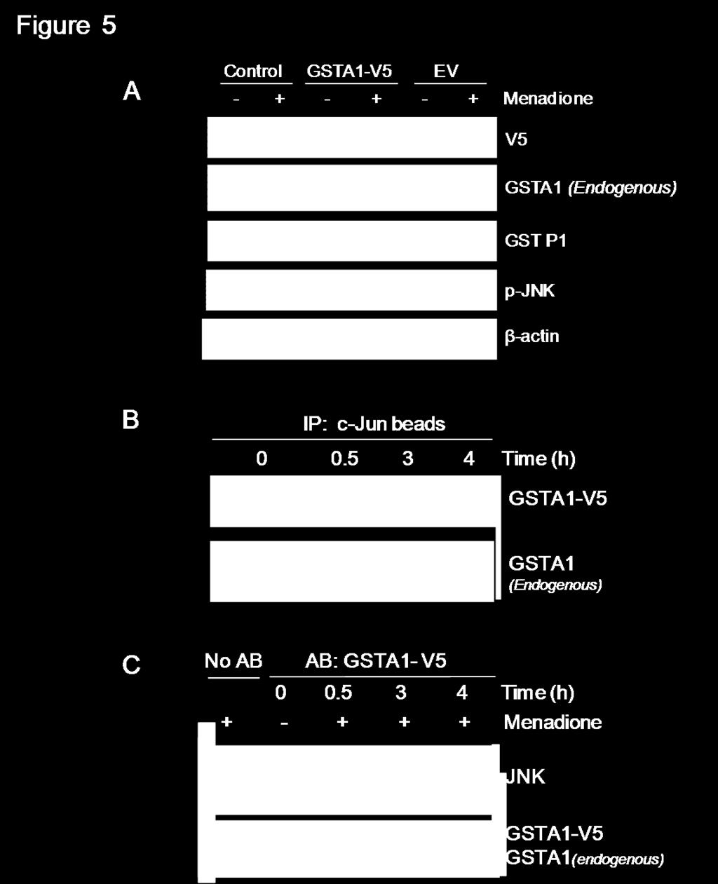
58 1.5. Over-expressed GSTA1 forms a complex with JNK but does not impede menadioneinduced complex dissociation. To determine the specific role of GSTA1 in inhibiting menadione-induced JNK activation, we transiently over-expressed GSTA1 in preconfluent Caco-2 cells. Western blot analysis confirmed that GSTA1-V5 protein was present in cells transfected with GSTA1-V5 plasmid and was not present in cells transfected with empty vector (EV) (Figure 5A). Overexpression of GSTA1 did not reduce menadione-mediated JNK activation because p-jnk protein levels were significantly increased in all cells treated with menadione (40 µm). To determine if transiently over-expressed GSTA1 protein (GSTA1-V5) binds to JNK, GSTA1-JNK complexes were pulled-down using c-jun fusion protein beads (Figure 5B) and immunoprecipitation using a mouse anti-v5 antibody (Figure 5C) in independent experiments. The presence of GSTA1-V5, endogenous GSTA1 and GSTP1 protein in complex with JNK was assessed. GSTA1-V5 protein was bound to JNK in association with endogenous GSTA1 and menadione caused dissociation of both endogenous and overexpressed GSTA1 as well as JNK (Figure 5B and 5C). GSTA1 overexpression did not alter menadione-induced cytotoxicity as LDH release was similar in both GSTA1-V5 and EV-transfected cells (data not shown). 42
59 43
60 Figure 6 Figure 5. Over-expressed GSTA1 forms a complex with JNK but does not impede menadione-induced complex dissociation. Preconfluent Caco-2 cells were transiently transfected with one µg of either GSTA1-V5 or empty vector (EV) followed by menadione (40 µm) treatment for 4 h. (A) Representative western blots for V5 (~26KDa) tagged-protein, p-jnk protein (~54KDa and 46KDa) levels, GST P1 (~26KDa) and, GSTA1 (~25KDa) endogenous protein expression were also assessed and β-actin was used as a protein loading control. (B) Immunoblot of V5 tagged-gsta1 and endogenous GSTA1 protein levels in pull down complex using c-jun beads. (C) Immunoblot of JNK, GSTA1-V5 and GSTA1 endogenous levels in the complexes pulled-down by immunoprecipitation using anti-v5 antibody. 44
61 1.6. Down-regulation of GSTA1 reduces GSTA1-JNK complex formation but does not alter menadione-induced JNK activation. To further investigate the role of GSTA1 on menadione-induced JNK activation and GSTA1-JNK complex dissociation, endogenous GSTA1 was transiently down-regulated in preconfluent Caco-2 cells using a sirna approach. Endogenous GSTA1 protein levels decreased 63% (p<0.01) using GSTA1-specific sirna. No significant decrease was observed in endogenous GSTA1 with non-specific control sirna oligoduplexes (Figure 6A and B). While p- JNK levels significantly increased by nine-fold (p<0.01) with menadione treatment, no significant differences were observed in the amount of JNK activation with GSTA1 downregulation (Figure 6A). GSTA1 down-regulation did not alter menadione-induced cytotoxicity as LDH release was similar in both GSTA1-specific sirna and NS sirna-transfected cells (data not shown). Western blot analysis of GSTA1 in pulled-down GSTA1-JNK complexes revealed that GSTA1 down-regulation reduced GSTA1-JNK complex levels by 58% and 93% in both untreated and menadione-treated cells compared to either cells transfected with nonspecific sirna or to untransfected controls (Figure 6C). GSTP1 expression did not change with GSTA1 down-regulation with or without menadione treatment (Figure 6A and C). 45

62 Figure 7 46
63 Figure 6. Down-regulation of GSTA1 reduces GSTA1-JNK complex formation but does not affect menadione-induced p-jnk levels. (A) Representative western blots of GSTA1 (~25KDa), GSTP1 (~26KDa) and p-jnk protein (~54KDa and 46KDa) levels in Caco-2 cells that were transiently transfected with 40 nm of GSTA1-siRNA or non-specific sirna. After 72 h, cells were treated with 40 µm menadione for 4 h. β-actin was used as a protein loading control. (B) Densitometric analysis of GSTA1 levels in GSTA1 down-regulated cells. Values represent the mean ± S.E of three independent experiments with three replicates each. Bars indicated by b differ significantly from a (p<0.01). (C) Representative western blot of GSTA1 and GSTP1 and JNK protein expression in pull down complexes using c-jun beads in GSTA1 down-regulated cells with and without menadione treatment. 47
64 1.7. N-acetyl cysteine (NAC) blocks menadione-activated JNK in Caco-2 cells. To investigate the mechanism by which menadione activates JNK, we pre-incubated Caco-2 cells with the antioxidants PEG-catalase, N-acetyl cysteine (NAC) and Vitamin E. While menadione caused significant JNK activation (p<0.01) in cells pre-treated with PEG-catalase (2.9-fold) and Vitamin E (3.0-fold) relative to cells without antioxidant pretreatment (2.5-fold), NAC completely inhibited JNK activation by menadione. None of the antioxidants affected GSTA1 protein levels (Figure 7A and B). Pre-incubation of cells with NAC also prevented the dissociation of GSTA1-JNK complexes by menadione (Figure 7C). 48

65 Figure 8 49
66 Figure 7. N-acetyl cysteine blocks menadione-induced activation of JNK. Preconfluent Caco-2 cells were pre-incubated with PEG-catalase (500 U/ml for 1 h), N-acetyl cysteine (NAC, 20 mm for 2 h) and Vitamin E (5 µm/ml for 4 h) followed by 40 µm menadione for 4 h. (A) Representative western blot of p-jnk levels in preconfluent cells with and without antioxidants and menadione. GSTA1 and β-actin protein expression was also analysed. (B) Densitometric analysis of p-jnk protein. Values represent the mean ± S.E of three independent experiments with three replicates each. Bars with different letters are significantly different (p<0.001). (C) Immunoblot of GSTA1 protein expression in GSTA1-JNK complexes with and without NAC and menadione. The GSTA1-JNK complex was pulled-down using c-jun fusion beads. 50
67 1.8. NAC prevents menadione-mediated GSH depletion and cytotoxicity but lower dose of menadione does not cause lipid peroxidation in Caco-2 cells. We next determined if the effects of menadione on Caco-2 cells is related to depletion of total GSH levels (Figure 8A). Our results show that GSH levels were significantly higher by threefold (p<0.001) in postconfluent (48.79 nmol/mg protein) compared to preconfluent cells (16 nmol/mg protein). Menadione (40 µm) treatment for 4 h caused a significant decrease in GSH concentration both in preconfluent and postconfluent cells. In preconfluent cells the GSH concentration dropped from 16 nmol/mg protein to undetectable levels (p<0.001) and in postconfluent cells the GSH levels decreased by 57.1 % (p<0.001); from 48.7 nmol/mg protein to 20.8 nmol/mg protein (40 µm). Furthermore, preincubation of preconfluent and postconfluent cells with NAC prevented the depletion of GSH in menadione-treated cells (Figure 8A). Preincubation of NAC also protected the cell from menadione-induced cytotoxicity in preconfluent cells (Figure 8B). We also assessed the ability of menadione to induce lipid peroxidation in Caco-2 cells since products of lipid peroxidation are activators of JNK. Levels of malondialdehyde (MDA) and 4-hydroxyalkenal (HAE) were not altered in Caco-2 cells treated with menadione (40 µm) after 4 h of treatment (Figure 8C). Higher doses of menadione (100 and 200 µm) for 4 h caused a significant increase in MDA + HAE levels (p<0.001); from 1.48 nmol/mg to 11.7 and 28.9 nmol/mg respectively. 51
68 MDA+HAE + HAE (nmol/mg protein) % Cytotoxicity GSH( mol/mg protein) GSH (nmol/mg protein) Figure 8 A 150 Control NAC b 100 b bc 50 c c B a Preconfluent Postconfluent Control NAC d Menadione ( M) b a C a a a 0 40 Menadione ( M) 30 c 20 b 10 0 a a Menadione ( M) Figure 9 52
69 Figure Figure 8. NAC prevents menadione-mediated GSH depletion and cytotoxicity but lower dose of menadione does not cause lipid peroxidation in Caco-2 cells. (A) GSH concentration (µmol/mg protein) in preconfluent and postconfluent cells (B) % cytotoxicity in preconfluent Caco-2 cells preincubated with NAC (20 mm) for 2 h followed by menadione (40 µm) for 4 h. Values represent the mean ± S.E. of 6 independent replicates each. Bars with different letters are significantly different (p< 0.001). (C) Caco-2 cells were treated with menadione (40, 100 and 200 µm) for 4 h, and lipid peroxidation was determined by assessing malondialdehyde (MDA) and 4-hydroxyalkenal (HAE) levels. The graph indicates MDA+ HAE release (µmol/mg protein) in control and menadione-treated preconfluent cells. Values represent the mean ± S.E. of four independent experiments with six replicates each. 53
70 DISCUSSION We have previously demonstrated complex formation between GSTA1 and JNK and that over-expression of GSTA1 in Tet-responsive MEF3T3 cells reduces JNK activation by oxidative stress [14]. In the current study we investigated whether oxidative stress, due to the pro-oxidant menadione, is a key determinant in mediating GSTA1-JNK complex dissociation and JNK activation. Our results demonstrate that menadione causes GSTA1-JNK complex dissociation and JNK activation. Menadione-mediated JNK activation was significantly greater in preconfluent Caco-2 cells that endogenously express low levels of GSTA1 and GSH compared to postconfluent cells that express higher levels. Transient exposure to menadione in preconfluent cells leads to dissociation of complexes with subsequent re-association and reduction in JNK activation and the degree of cytotoxicity. While menadione caused dissociation of GSTA1-JNK complexes in cells with forced expression or knock-down of GSTA1 levels, there is no effect on menadione-mediated JNK activation or cytotoxicity. However, the antioxidant NAC effectively prevents dissociation of GSTA1-JNK complexes, induction of cytotoxicity and blocks menadione-induced phosphorylation of JNK. These findings suggest that ROS, likely superoxide anion accumulation, is a primary activator of JNK by menadione and that levels of glutathione play a key role in modulating JNK activation during oxidative stress. We demonstrated that menadione (40 µm) rapidly activates JNK in preconfluent Caco-2 cells within 30 min of exposure with peak activation between 4 to 6 h. Similar results were obtained by Osada et al (2008) who demonstrated, in a rat pancreatic cancer cell line (ARIP), that menadione activates JNK at 30 min and that this activation is maintained over 6 h [90]. Dunning et al (2009) has also demonstrated that menadione-induced JNK activation is dose- and 54
71 time-dependent in rat hepatic stellate cells (HSCs) [94,105]. In postconfluent cells, the foldchange in JNK activation is not as great as that observed in preconfluent cells following menadione treatment. This suggests that increased GSTA1 protein levels in postconfluent cells may be responsible for reduced JNK activation following oxidative stress stimulation. These findings are supported by our previous studies in which increased GSTA1 levels in postconfluent Caco-2 cells were associated with increased complex formation with JNK and reduced JNK phosphorylation of c-jun following exposure to the pro-oxidants IL-1β, UV-light and H 2 O 2 [14]. It has been suggested that the biological relevance of increased GSTA1 expression with increasing confluency contributes to a sensor capacity to respond to changes in redox potential during oxidative stress [125]. Menadione-mediated activation of JNK occurs in conjunction with dissociation of GSTA1-JNK complexes in Caco-2 cells. This suggests that complex dissociation is necessary for JNK activation to occur. Moreover, GSTA1-JNK complex dissociation occurs in preconfluent and not in postconfluent cells suggesting that the level of GSTA1 expression may influence the integrity of complex association with JNK. GSTA4-4 expression does not change with menadione treatment and does not form complexes with JNK. While GSTP1 is present in complexes with JNK, GSTP1 levels in the complex do not change with menadione treatment suggesting that increased levels of superoxide anion is not capable of altering the association of GSTP1 with JNK. Whereas direct inhibitory interactions between GSTP1 and JNK [13,27] and between GST Mu and ASK1 [71] have been shown, our results indicate that maintenance of GSTP1-JNK complex integrity following menadione treatment does not prevent JNK activation. While our results suggest that the level of GSTA1 expression establishes the degree of dissociation of GSTA1-JNK complexes in response to oxidative stress, it is possible that other 55
72 factors associated with advancing confluency, such as the antioxidant capacity, may play a modulatory role. We also show that JNK activation by menadione is transient since GSTA1-JNK complexes re-associate and levels of activated JNK significantly decrease after removal of menadione. Transient JNK activation has been demonstrated in studies on cardiac myocytes [68] using H 2 O 2 where p-jnk levels remained elevated with sustained treatment and declined during transient stress. It has previously been shown that superoxide anion produced by menadione initiates signals that result in JNK phosphorylation [94,95]. It is also likely that superoxide anion causes GSTA1 to be released from the complex with JNK. Our data have indicated for the first time that complex formation and disassembly is a reversible process since removal of the stimulus causes JNK to return to a latent state and re-associates with GSTA1. While menadione, at a concentration of 40 µm, causes maximal JNK activation in preconfluent cells at 4 h, cytotoxicity is not observed unless cells are persistently exposed to menadione for 24 h. In addition, postconfluent cells are relatively resistant to menadione-induced cytotoxicity compared to preconfluent cells possibly due to higher GSTA1 levels or other cellular factors, such as glutathione, that prevent JNK activation and subsequent cell death. This suggests that menadione-mediated activation of JNK triggers cytotoxicity which occurs later if the oxidative stress stimulus is persistent. Indeed, transient exposure of Caco-2 cells to menadione results in a significant decrease in cytotoxicity as compared to persistent exposure. This is a reversible process since a decrease in p-jnk levels after removal of menadione slowly allows for cellular recovery. These findings are supported by studies on Jurkat T-cells that 56
73 implicated the involvement of the JNK pathway in radiation-induced apoptosis and the importance of the duration of JNK activation in determining cellular fate [171]. In this study, altered levels of GSTA1 expression (using either sirna technology or a GSTA1 over-expression vector) did not alter the overall level of JNK activation by menadione. While levels of GSTA1 protein complexed with JNK decreased in cells transiently transfected with GSTA1 sirna suggesting that the overall levels of GSTA1 are important for complex formation, levels of activated JNK did not change. In addition, overexpressed GSTA1 can also form complexes with JNK however JNK activation is not significantly different from controls. Futhermore, modulated levels of GSTA1 does not alter levels of menadione-induced cytotoxicity. This suggests that the level of GSTA1 expression is not a determinant of susceptibility to JNK activation. Several lines of evidence indicate the importance of protection against superoxide anion production as a key aspect in minimizing the consequences of menadione exposure. While we had hypothesized that products of lipid peroxidation might be involved in activation of JNK, we have shown that lipid peroxidation does not occur as a result of 40 µm menadione exposure. Our finding that vitamin E does not impede JNK activation corresponds with the inability of 40 µm menadione to cause lipid peroxidation. Petrova (2008) also demonstrated that alpha-tocopherol does not protect cells against ROS damage in rat thymocytes [172]. While the mechanism by which menadione activates JNK in Caco-2 cells is independent of lipid peroxidation, our results suggest that superoxide anion plays a key role. For example, both thiol (NAC) and non-thiol (PEG-Catalase and vitamin E) antioxidants were utilized to block ROS but only NAC-mediated increase in glutathione (GSH) levels blocked menadione-induced JNK activation in Caco-2 cells. 57
74 Others have also shown that GSH is an inducible component of the superoxide adaptive stress response correlating with a reduction in intracellular oxidation [173]. Previous studies in various cell lines indicate that menadione depletes GSH in preconfluent cells [94,174]. Laux (2001) has shown that menadione-induced cytotoxicity in Jurkat T cells is due to altered mitochondrial membrane potential that interferes with one-electron transfer and generates superoxide anion free radicals, an effect that can be reversed by NAC [171]. Moreover, superoxide anion-induced apoptosis in HSC is dependent on JNK activation and glutathione status [94]. The inability of PEG-catalase to block JNK activation demonstrated in the present study suggests that menadione-induced JNK activation is independent of a pathway that involves H 2 O 2. Others have shown that PEG-catalase is not protective against menadione-induced cellular injury in alveolar epithelial cells [175] and that menadione cytotoxicity utilizes mechanisms that are distinct from that of H 2 O 2 exposure [176]. Moreover, it has been demonstrated that menadione, but not hydrogen peroxide, reduces cellular glutathione levels [94]. Glutathione provides a major source of thiols critical to the maintenance of a reduced cellular environment that is conducive to cell survival [177]. The decrease of GSH concentration in postconfluent Caco-2 cells following menadione treatment may imply that the increase of menadione-thiodione metabolism by induction of GSTA1 led to increase the consumption of GSH. Further studies are required to determine the cytoprotective role of GSH-dependent peroxidase activity of GSTA1 during menadione-induced oxidative stress. The results of this study indicate that the mechanism of menadione-mediated JNK activation involves the production of reactive oxygen species, likely superoxide anion. We also provide evidence, for the first time, for a GSH-dependent mechanism in regulating JNK activation and Caco-2 cell death by oxidative stress. Thus, it is clear that the levels of 58
75 intracellular GSH play a more important protective role than the level of GSTA1 expression in menadione cytotoxicity, NAC did increase JNK-GSTA1 complex integrity suggesting that these complexes may play a role in cytoprotection. It is also possible that the decrease in menadioneinduced cytotoxicity observed in postconfluent Caco-2 cells may be mediated by an increase in menadione metabolism due to an increase of GSTA1 expression and GSH conjugation, rather than via an increase in dissociation of GSTA1-JNK complexes. 59
76 Chapter 2 LOW LEVELS OF GSTA1 EXPRESSION ARE REQUIRED FOR CACO-2 CELLS PROLIFERATION ABSTRACT The colonic epithelium continuously regenerates with transitions through various cellular phases including proliferation, differentiation and cell death via apoptosis. Human colonic adenocarcinoma (Caco-2) cells in culture undergo spontaneous differentiation into mature enterocytes in association with progressive increase in expression of glutathione S-transferase alpha-1 (GSTA1). We hypothesize that GSTA1 plays a functional role in controlling proliferation, differentiation and apoptosis in Caco-2 cells. We demonstrate increased GSTA1 levels associated with decreased proliferation and increased expression of differentiation markers alkaline phosphatase, villin, dipeptidyl peptidase-4 and E-cadherin in postconfluent Caco-2 cells. Results of MTS assays, BrdU incorporation and flow cytometry indicate that forced expression of GSTA1 significantly reduces cellular proliferation and sirna-mediated down-regulation of GSTA1 significantly increases cells in S-phase and associated cell proliferation. Sodium butyrate (NaB) at a concentration of 1 mm reduces Caco-2 cell proliferation, increases differentiation and increases GSTA1 activity 4 fold by 72 hours. In contrast, 10 mm NaB causes significant toxicity in preconfluent cells via apoptosis through caspase-3 activation with reduced GSTA1 activity. However, GSTA1 down-regulation by sirna does not alter NaB-induced differentiation or apoptosis in Caco-2 cells. While 10 mm NaB causes GSTA1-JNK complex dissociation, 60
77 phosphorylation of JNK is not altered. These findings suggest that GSTA1 levels may play a role in modulating enterocyte proliferation but do not influence differentiation or apoptosis. 61
78 INTRODUCTION The glutathione S-transferases (GSTs) are a multigene family of drug detoxification enzymes that are important in phase II metabolism by catalyzing the conjugation of glutathione to a variety of electrophilic substances [6]. GSTs are also involved in the biosynthesis and metabolism of prostaglandins [21], steroids [22] and leukotrienes [20], in the detoxification of products of lipid peroxidation [17] and in the acquisition of resistance to chemotherapeutic agents [110]. GST isoenzymes are also known to modulate cell signaling pathways that control cell proliferation and apoptotic cell death [27,28] and have become potential therapeutic targets for the treatment of cancer [38]. Human colonic adenocarcinoma Caco-2 cells are extensively utilized as a model of intestinal epithelial cell differentiation as they have many phenotypic features of enterocytes [178]. In vivo, colonic epithelial cells are continuously renewing with a systematic turnover of cells involving transition between cell proliferation, differentiation and cell death by apoptosis. In culture, Caco-2 cells grow as an epithelial monolayer and undergo enterocyte-like differentiation with concomitant biochemical changes [122,123]. Differentiation of Caco-2 cells is characterized by cell polarization, appearance of intercellular tight junctions and typical brush border microvilli projecting perpendicularly to the surface. The expression and activity of brush border enzymes notably alkaline phosphatase (AlkP), are increased in cellular differentiation [126,141,179,180]. Moreover, the expression of GSTA1 progressively increases as Caco-2 cells differentiate [14]. Terminally differentiated enterocytes undergo apoptosis and are sloughed from the surface epithelium into the intestinal lumen [153]. Therefore apoptosis seems to be a necessary component of colonocyte terminal differentiation [137]. Indeed, neoplastic 62
79 transformation of the colonic epithelium is associated with disordered regulation of cellular differentiation and apoptosis [107]. Numerous factors are involved in the control of intestinal epithelial cell differentiation, including growth factors, hormones, extracellular matrix proteins, vitamins, and luminal nutrients such as short chain fatty acids (SCFAs) [126]. Sodium butyrate (NaB), a short-chain fatty acid present in the human large intestine, is derived from bacterial fermentation of complex carbohydrates. NaB is a preferred energy source for normal colonocytes in vivo but also reduces the growth and motility of colon cancer cell lines and causes dose-dependent cellular differentiation and apoptosis [143,150,181]. GSTs act as mediators of cell signaling kinase pathways involved in cell cycle transition such as proliferation and apoptosis [38]. Progressive increase in GSTA1 expression with cellular confluency in Caco-2 cells may influence responses to cellular stress [14]. Therefore we suspect that GSTA1 may function as a modulator of cell phase transitions. We have previously shown that the incidence of apoptosis stimulated by tumour necrosis factor α and sodium butyrate is significantly higher in preconfluent Caco-2 cells with minimal GSTA1 expression compared to differentiated postconfluent cells with high GSTA1 expression [14]. We hypothesized that low GSTA1 expression was a necessary condition for cell proliferation and that increased expression of GSTA1 is a requirement for Caco-2 cell differentiation. Our results indicate that low concentrations (1 mm) of NaB cause Caco-2 cell differentiation and concomitant GSTA1 induction and high concentrations (10 mm) stimulate apoptosis and down-regulation of GSTA1. Moreover, manipulation of GSTA1 levels by forced expression and sirna technology demonstrated altered cell proliferation but did not affect NaB-mediated differentiation or sensitivity to apoptosis. 63
80 MATERIALS AND METHODS Materials Dulbecco s modified Eagle s medium (DMEM), fetal bovine serum (FBS), penicillin and streptomycin, mouse anti-β-actin antibody, reduced glutathione, para-nitrophenyl phosphate, para-nitrophenol and sodium butyrate were purchased from Sigma-Aldrich (Oakville, ON). The anti-gsta1 rabbit antibody was purchased from Oxford Biomedical Research (Pickering, ON). Anti-GSTP1 rabbit antibody was purchased from Biotrin (Mississauga, ON). Mouse anti-v5 antibodies, TryPLE Express, Stealth sirna sequences and pcdna 3.1/V5-His TOPO TA Expression Kit, TRIzol reagent, MMLV (Moloney-murine-leukaemia virus) RT were purchased from Invitrogen (Burlington, ON). 5-Androstene-3,17-dione (AD) was purchased from Steraloids Inc (Newport, RI). An ECL Plus kit was purchased from GE Health Sciences (Oakville, ON). A DNA Master SYBR Green I kit and 5-bromo-2-deoxyuridine (BrdU) chemiluminescent enzyme-linked immunosorbant assay (ELISA) kit was purchased from Roche Diagnostics (Mississauga, ON, Canada). CytoTox-ONE Assay kits and Aqueous Non- Radioactive Cell Proliferation (MTS) Assay kits were purchased from Promega (Whitby, ON). A SAPK/ JNK Kinase assay kit, and rabbit anti-caspase-3 antibodies were purchased from Cell Signaling Technology, Inc (Pickering, Ontario). Cell culture and treatments Human adenocarcinoma (Caco-2) cells were cultured in DMEM supplemented with 10% (v/v) FBS and 100 µg/ml of penicillin and streptomycin in 5% CO 2 at 37 C. All experiments utilized preconfluent cells with 80% confluency and 10 d postconfluent Caco-2 cells between 64
81 passages Wild type Caco-2 and GSTA1-modulated Caco-2 cells were treated with sodium butyrate (NaB, 1mM and 10mM) in serum-free media for 24 to 72 h. RNA isolation and real-time RT PCR analysis Total RNA was isolated using TRIzol reagent, according to the manufacturer s instructions. Isolated RNA (one μg/20 μl of reaction volume) and one unit of RQ1 DNAse were used to prepare DNase-treated RNA. Complementary DNA (cdna) was then generated from the DNase-treated RNA using 0.1 μg of random primers, 20 units of RNase inhibitor, 10 mm dntps (Invitrogen) and 200 units of MMLV (Moloney-murine-leukaemia virus) RT. Real-time PCR was performed on a Roche Light Cycler using a DNA Master SYBR Green I kit. The PCR reaction was done in a volume of 10 μl, containing 1 μl of SYBR Green I, 5 μm of each primer and 2 mm MgCl 2. Oligonucleotide primers for the differentiation makers AlkP were 5 CTCCAACATGGACATTGACG and 3 TGAGATGGGTCACAGACTGG, villin were 5 AGCCAGATCACTGCTGAGGT and 3 TGGACAGGTGTTCCTCCTTC, dippeptidyl peptidase-4 (DDP-4) were 5 GGCGTGTTCAAGTGTGGAAT and 3 TCTTCTGGAGTTGGGAGACC, E-cadherin (E-cad) were 5 TGATCGGTTACCGTGATCAAAA and 3 GTCATCCAACGGGAATGCA and for glyceraldehyde-3-phosphate dehydrogenase (GAPDH) were 5 CAGTCCATGCCATCACTGCC and 3 GCCTGCTTCACCACCTTCTTG. The PCR parameters were 95 C for 1 min, 1 cycle, and 35 cycles of 95 C for 15 s, 70 C for 5 s and 72 C for 15 s. Messenger RNA levels for these differentiation markers were normalized against GAPDH mrna. 65
82 Transient transfections For sirna transfections, Caco-2 cells (10 6 cells per 6 cm 2 dish) were transiently transfected in suspension using Lipofectamine 2000 as recommended by the manufacturer. Preconfluent cells were transfected with non-specific negative control sirna (NS sirna) or GSTA1-specific sirna. The sequences of GSTA1 sirna were 20: 5 AAGACUGGAGUCAAGCUCCUCGACG and 3 CGUCGAGGAGCUUGACUCCAGUCUU, GSTA1 sirna 19: 5 AGUUCCACCAGAUGAAUGUCAGCCC and 3 GGGCUGACAUUCAUCUGGUGGAACU and GSTA1 sirna 18: 5 UGGACAUACGGGCAGAAGGAGGAUC and 3 GAUCCUCCUUCUGCCCGUAUGUCCA. Transfections were performed in Opti-MEM using a final concentration of 40 nm of sirna for 72 h. GSTA1 protein and enzymatic activity were assessed by western blotting and 5-androstene- 3,17-dione (AD) assay respectively to confirm GSTA1 specific silencing. A GSTA1 expression vector was constructed. Briefly, the cdna for GSTA1 was cloned in pcdna 3.1/V5-His TOPO using primers 5 AAACCTGAAAATCTTCCTTGCTTCTT and 3 GAAACCTCCAGGAGACTGCTA. Plasmid inserts were confirmed by DNA sequence analysis at the University of Guelph, Laboratory Services Division. One µg/ml of GSTA1 pcdna 3.1/V5-His TOPO plasmid was used in all experiments. Empty vector (pcdna 3.1) was used as a transfection control. Caco-2 cells (10 6 cells per 6 cm 2 dish) were transfected in suspension to over-express GSTA1 protein. Protein levels of GSTA1-V5 were assessed by immunoblotting procedures using mouse anti-v5 antibody to confirm over-expression. 66
83 GSTA1 activity assay Cells were harvested with 50 mm Tris HCl ph 8.0 and 0.1 mm EDTA followed by sonication on ice and centrifugation at 9000 g, 4 C for 20 min. GSTA1 activity (nmol/mg/min) was assessed using 10 mm AD as substrate (dissolved in 100 % methanol) in the presence of 50 mm glutathione-reduced (dissolved in 50 mm Tris ph 8) and 200 µg of protein at 248 nm at 30 C [40,46]. All the reagents were prepared fresh and kept on ice. Alkaline phosphatase (AlkP) activity assay To determine AlkP activity, cells were harvested in PBS and whole cell lysates (400 µg of protein) were used in the reaction mixture. AlkP activity (µmol/mg/min) was determined using para-nitrophenyl phosphate as substrate as previously described [41,192]. The alkaline phosphatase activity in each sample was calculated from a para-nitrophenol standard curve. Cytotoxicity assay Cytotoxicity was determined by lactate dehydrogenase (LDH) release using a CytoTox- ONE kit (Promega) according to the manufacturer s protocol. Caco-2 cells (15,000 cells per well) were plated in 96-well clear bottom black plates. Cells were treated with NaB (1 mm and 10 mm) for 24 h to 72 h. LDH release was measured at 544/590 nm using a FluoStar OPTIMA fluorimeter (BMG Labtech). Percent cytotoxicity was calculated by subtracting the background and dividing the LDH release from control wells as recommended by the manufacturer. 67
84 MTS assay Cellular proliferation was determined using a CellTiter 96 Aqueous Non-Radioactive Cell Proliferation Assay MTS kit (Promega) according to the manufacturer s instruction. Caco-2 cells (15,000 cells per well) were plated in 96-well plates. 20 µl of the MTS reagent was added to each well containing 100 µl culture medium. The plate was incubated for 2 h at 37 C in a humidified, 5% CO 2 atmosphere. The plate was then read at 490 nm using a plate reader. BrdU Incorporation Cell proliferation was assessed by determining BrdU incorporation using the 5-bromo-2- deoxyuridine (BrdU) chemiluminescent enzyme-linked immunosorbant assay (ELISA) kit (Roche). Caco-2 cells were incubated with a 10 µm BrdU solution for 2 hours at 37 C, then fixed and denatured and incubated with a peroxidase-conjugated antibody against BrdU (1:100) for 1 hour at room temperature. The cells were then incubated with the hydrogen peroxide substrate solution and fluorescence was read at 460 nm using a Fluostar OPTIMA fluorimeter (BMG Labtech). Flow cytometry Caco-2 cells (10 6 cells per 6 cm 2 dish) were plated for the assay, and flow cytometry analysis was done as described previously [42,193]. Before harvesting, the cells were washed twice with 1 PBS, followed by centrifugation at 6000 g for 10 min. The pellet was resuspended with 70% ethanol for 30 min at 4 C. Cells were again centrifuged at 6000 g for 10 min and were incubated with the DNA-binding dye propidium iodide (PI) solution (containing 0.1% sodium citrate, 0.1% Triton X-100, and 50 mg/l PI in water) for 1 hour at room temperature. 68
85 Cells were then analyzed using a FACS (Fluorescence-activated cell sorting) caliber flow cytometer (BD FACSCalibur, Becton, Dickinson and Company Biosciences, San José, USA). Assessment of GSTA1-JNK complexes GSTA1-JNK complexes were detected from cells using c-jun fusion protein beads from the SAPK/JNK Kinase assay kit (Cell Signaling Technologies Inc.). Sample protein (350 µg) was incubated with 10 µl of c-jun fusion protein beads with gentle rocking overnight at 4 C to pull-down JNK. Samples were centrifuged at g for 2 min at 4 C. The pellet was washed 4 times with lysis buffer and the immunoprecipitates were resuspended with 3X SDS buffer containing 150 mm DTT. Samples were boiled at 95 C for 5 minutes to denature the proteins and subject to SDS-PAGE. SDS-PAGE and Western blot analysis Caspase-3, p-jnk and GSTA1 and GSTP1 expression were assessed by western blot analysis. Cells were harvested with lysis buffer and stored at -80 C. The cell extracts were then thawed and sonicated on ice for 10 minutes and centrifuged at 9000 g for 20 min at 4 C. Protein was quantified by the Bradford assay using BSA as a standard. Protein (30 µg) was separated by 12 % SDS/PAGE and transferred to nitrocellulose membranes that were blocked in 5% milk in Tris-buffered saline with 0.1% Tween (TBS/T) and incubated overnight with either rabbit monoclonal anti-caspase-3 (1:1000) antibody; rabbit polyclonal anti-gsta1 (1:5000) antibody: rabbit polyclonal anti-gstp1 antibody (1:500); or rabbit monoclonal anti p-jnk (1:1000) antibody. After 1 h of incubation with a horseradish peroxidase-conjugated anti-rabbit 69
86 antibody, bands were detected by chemiluminescence (ECL Plus) and visualized using a Typhoon 9410 scanner (GE Health Care). The densitometric analysis of protein was determined using Image J (NIH software). β-actin was detected as a protein loading control. Statistical analysis One-way ANOVA and two-way ANOVA test were used to assess statistically significant differences among treatment groups for the analysis of single variance and multiple variances respectively. For single variance, the Tukey s least significant test was used for comparison and for multiple variances Bonferroni s hoc test was applied. Significance was established at p values <0.05. If inhomogeneity of variance was found in the raw data (Brartlett's test; p<0.05), this was corrected before ANOVA by log transformation. 70
87 RESULTS 2.1. GSTA1 levels increase in differentiating Caco-2 cells Caco-2 cells in culture progressively undergo spontaneous enterocytic differentiation with increasing days of postconfluency. We have previously shown that GSTA1 protein expression increases with confluency in Caco-2 cells [14]. To investigate the relationship between GST expression and Caco-2 cell differentiation we compared GSTA1 and GSTP1 protein levels in preconfluent and postconfluent Caco-2 cells. While GSTA1 protein levels significantly increased in 10 d postconfluent cells, GSTP1 expression did not change (Figure 9A). Moreover, GSTA1 activity increased 6.1-fold (p<0.001) from 13.8 nmol/mg/min in preconfluent cells to 84.1 nmol/mg/min in 10 d postconfluent cells (p< 0.001) (Figure 9B). We verified that specific genes associated with cellular differentiation were transcriptionally up-regulated by examining the mrna transcript levels of various differentiation markers. The mrna levels of alkaline phosphatase (AlkP), villin, dipeptidyl peptidase-4 (DPP-4) and E-cadherin (E-cad) in preconfluent and 10 d postconfluent Caco-2 cells were assessed by real time RT-PCR (Figure 9C). Transcripts of AlkP, villin, DDP-4 and E-cad significantly increased by 3.9- (p<0.001), 3.1- (p<0.001), 3.1- (p<0.001) and 2.7- (p<0.01) fold, respectively, in postconfluent cells (Figure 9C). Moreover, a five-fold change in AlkP activity from 1.2 µmol/mg/min in preconfluent cells to 6.0 µmol/mg/min in 10 d postconfluent cells was observed (p<0.05) (Figure 9D). 71
88 mrna levels (ratio over control) AlkP activity ( mol/mg/min) GSTA1 activity (nmol/mg/min) Figure 9 A B 100 b 80 GSTA1 60 GSTP1 40 β-actin 20 a 0 Preconfluent Postconfluent C D a b a b Preconfluent Postconfluent a b a b a b 0 AlkP Villin DDP-4 E-cad Differentiation markers 0 Preconfluent Postconfluent 72
89 Figure 9. GSTA1 levels increase in differentiating Caco-2 cells. ure 10 Preconfluent and 10 d postconfluent Caco-2 cells were assessed for: (A) protein expression of GSTA1 (~25KDa) and GSTP1 (~26KDa). β-actin was used as a protein loading control; (B) GSTA1 enzyme activity (nmol/mg/min); (C) mrna levels of differentiation markers: AlkP, villin, DPP-4 and E-cad by real time RT-PCR; and (D) AlkP enzyme activity (µmol/mg/min). Values represent the mean ± S.E. of three independent experiments with three replicates each. Bars indicated by different letters differ significantly from one another (p 0.001). 73
90 2.2. Modulation of GSTA1 levels alters proliferation of Caco-2 cells Since GSTA1 levels progressively increase with confluency in differentiating Caco-2 cells, we investigated the relationship between GSTA1 and cellular proliferation. For this purpose, we transiently modulated GSTA1 expression levels in preconfluent cells and confirmed GSTA1 down-regulation or over-expression by western blot analysis and enzyme activity (Figure 10 and Table 1). Preconfluent cells were transiently transfected with GSTA1 sirna and non-specific negative control (NS) sirna for 72 h to down-regulate GSTA1 (Figure 10A). The protein levels significantly decreased by 68% (p<0.001) in GSTA1 sirna-transfected cells as compared to controls (Figure 10A and Table 1). Preconfluent cells were transiently transfected with a GSTA1-V5 expression plasmid and empty vector (EV) for 48 h to overexpress GSTA1. Western blot analysis of transfected cells using an anti-v5 antibody confirmed that expression of GSTA1- V5 occurred only in GSTA1-V5-transfected cells and was absent in EV-transfected cells (Figure 10B). In cells transiently transfected with GSTA1-V5, total GSTA1 activity increased 3.5-fold (p<0.001) from 2.8 nmol/mg/min in cells transfected with EV to 9.9 nmol/mg/min (Table 1). 74
91 Figure10 A NS sirna GSTA1siRNA GSTA1 β-actin B EV GSTA1-V5 V5 β-actin Figure 11 75
92 Figure 10. GSTA1 levels can be modulated in preconfluent Caco-2 cells. (A) Representative western blot of GSTA1 (~25 KDa) protein expression in preconfluent Caco-2 cells that were transiently transfected with 40 nm of GSTA1 sirna or non-specific (NS) sirna for 72 h. (B) Representative western blot of V5 (~26 KDa) protein expression in preconfluent Caco-2 cells that were transiently transfected with one µg of GSTA1-V5 or empty vector (EV) for 48 h. β-actin (~42 KDa) was used as a protein loading control in all panels. 76
93 Table1. Relative abundance and activity of GSTA1 in transiently transfected Caco-2 cells GSTA1 silencing Table 1 GSTA1 protein levels* GSTA1 activity* (nmol/mg/min) GSTA1 sirna 0.31 ± 0.02 b 2.10 ± 0.16 b NS sirna 1 ± 0.12 a 4.50 ± 0.31 a GSTA1 overexpression GSTA1-V ± 0.05 b 9.90 ± 0.42 b empty vector ND 2.80 ± 0.93 a *Each value represents the mean ± S.E of three independent experiments with three replicates each. ND, not detected. Values with different letters are significantly different from each other. 77
94 To examine the effect of GSTA1 knockdown or over-expression on cellular proliferation, a MTS assay was performed for up to 72 h (Figure 11A and B). GSTA1 knockdown significantly increased cell proliferation at 24 (p<0.05), 48 (p<0.01) and 72 h (p<0.01) as compared to controls (Fig 11A). In Caco-2 cells overexpressing GSTA1, a significant decrease in proliferation at 48 h (p<0.05) and 72 h (p<0.01) was observed when compared to controls (Fig 11B). Similar results were obtained when cells were labeled using bromodeoxyuridine (BrdU). BrdU incorporation decreased significantly to 54% of control levels in cells overexpressing GSTA1 (Figure 11C). No significant increase in cytotoxicity was observed due to transfections in GSTA1 knock-down or overexpressed Caco-2 cells (data not shown). 78
95 Figure 12 79
96 Figure 11. Modulation of GSTA1 levels mediate changes in Caco-2 cell growth. Effect of (A) GSTA1 down-regulation and (B) GSTA1-V5 overexpression on Caco-2 cell viability evaluated by MTS assay over three days. Asterisks depict significant differences between controls and the cells with GSTA1 modulated levels (*, p 0.05; and **, p 0.01). (C) Effect of GSTA1-V5 over-expression on cellular proliferation at 72 h as determined by BrdU incorporation in Caco-2 cells. Bars indicated by different letters differ significantly from one another (p 0.001). Values represent the mean ± S.E. of four independent experiments with three replicates each. 80
97 2.3 GSTA1 down-regulation affects cell cycle progression We analysed the changes of cell cycle phase distribution in GSTA1 down-regulated Caco- 2 cells. Forty-eight hours after transfection with GSTA1 sirna, FACS analysis was performed to assess the influence of GSTA1 knockdown on cell cycle progression. While Caco-2 cells transfected with non-specific sirna had a cell cycle distribution identical to untransfected control cells (Fig 12A), the distribution of cell cycle phases was significantly altered in cells in which GSTA1 was knocked down. Fifty-five percent of cells transfected with GSTA1 sirna were in G1 phase of the cell cycle compared 68% in controls and cells transfected with nonspecific sirna (p<0.05) (Figure 12A and 12B). In addition, over 42% of the cells accumulate in the S phase of the cell cycle with GSTA1 knockdown (p<0.01) compared to 24% in controls and cells transfected with non-specific sirna (Figure 12B). 81
98 Figure 12 A Control Control GSTA1 sirna GSTA1 sirna Non-specific sirna Non-specific sirna G % G % 24.51% S 24.51% G2 7.53% G2 7.53% G % G % 41.93% S 41.93% G2 3.35% G2 3.35% G % G % 23.57% S 23.57% G2 8.89% G2 8.89% G1 G1 G1 G1 G1 G1 S G2 G2 S G2 G2 S G2 G2 B Control GSTA1 sirna NS sirna Figure 13 82
99 Figure 12. GSTA1 down-regulation increases the percentage of Caco-2 cells in the S phase. (A) Changes of cell cycle phase distribution in GSTA1 down-regulated Caco-2 cells as compared to controls. (B) Graphic representation of percent of cells in G1, S and G2 phase of cell cycle in non-transfected control, GSTA1 sirna and NS sirna transfected Caco-2 cells. Asterisks depict significant differences between control and GSTA1 down-regulated cells (*, p 0.05; and **, p 0.01). 83
100 2.4. GSTA1 activity is altered with NaB-mediated changes in cell cycle phase Since GSTA1 modulation affected cellular proliferation and induced changes in cell cycle phase distribution, we further investigated the relationship between GSTA1 expression and transition through various cellular states in cells treated with NaB. Two concentrations of NaB that are known to cause either cellular differentiation (1 mm) or apoptosis (10 mm) were used. To determine the effect of NaB on cellular proliferation, a MTS assay was performed on preconfluent Caco-2 cells treated with NaB (1 and 10 mm) for up to 96 h (Figure 13A). A concentration of 1 mm NaB caused a significant decrease in proliferation at 48 h (p<0.05) and at 96 h (p<0.001) compared to controls. A concentration of 10 mm NaB caused a greater reduction in cellular proliferation at 48, 72 and 96 h (p<0.001) compared to controls. We confirmed that reduction in proliferation by 1 mm NaB did not result in cytotoxicity whereas 10 mm NaB caused a significant increase in cytotoxicity in preconfluent cells (49.4%, p<0.001) and not in postconfluent cells (Fig 13B). In preconfluent Caco-2 cells treated with 1 mm NaB for 72 h, cellular differentiation was demonstrated by progressive increases in AlkP activity of 2.9-fold at 48 h (p<0.01) and 4.4-fold at 72 h (p<0.001) (Figure 13C). No significant changes in AlkP activity were observed in untreated cells. NaB differentially affected GSTA1 activity in Caco-2 cells treated with 1 or 10 mm NaB over a 72 h period (Figure 13D). While GSTA1 activity significantly increased by 2.2-fold at 72 h in untreated control cells, a much greater progressive increase in GSTA1 activity (4.4-fold) occurred with 1 mm NaB treatment reaching a maximum activity of 21.9 nmol/mg/min at 72 h, 84
101 (p<0.001). Conversely, at 72 h GSTA1 activity decreased from 5.2 nmol/mg/min in control cells to 2.7 nmol/mg/min with NaB (10 mm) treatment. 85
102 AlkP activity ( mol/mg/min) GSTA1 activity (nmol/mg/min) Fold change (ratio over control) % Cytotoxicity Figure 13 A B Control NaB (1mM) NaB (10mM) *** * ** *** *** Time (h) a Preconfluent Postconfluent b a a a NaB (mm) a C D Control NaB (1mM) b c Control NaB (1mM) NaB (10mM) c d 2 1 a a a a 10 5 b a a a a b a Time (h) Time (h) Figure 14 86
103 Figure 13. Distinct doses of NaB differently affect cell proliferation and AlkP and GSTA1 enzyme activities. Preconfluent Caco-2 cells were treated with NaB (1 mm and 10 mm) in serum-free media. (A) Cellular proliferation was assessed from h. Asterisks depict significant differences between control and NaB treatments (*, p 0.05; **, p 0.01 and ***, p 0.001). (B) Cytotoxicity was determined in preconfluent and postconfluent Caco-2 cells treated with 1 mm and 10 mm NaB at 48 h. Cytotoxicity measured LDH release and presented as % cytotoxicity. (C) AlkP activity (µmol/mg/min) and (D) GSTA1 activity (nmol/mg/min) was determined. Values represent the mean ± S.E. of three independent experiments with six replicates each. Bars indicated by different letters differ significantly from one another (p 0.001). 87
104 2.5. Modulation of GSTA1 does not affect sodium butyrate-induced differentiation The data thus far demonstrates that a low concentration of NaB (1mM) induces differentiation, reduces cellular proliferation, and increases GSTA1 activity. To investigate whether GSTA1 plays a direct role in NaB-induced differentiation, endogenous GSTA1 was either knocked down or over-expressed in preconfluent Caco-2 cells. GSTA1 activity was determined in GSTA1-down-regulated cells with and without NaB (1 mm) treatment (Figure 14A). NaB (1 mm) significantly increased GSTA1 activity at 72 h by 1.4-fold (p<0.05) in control cells. In cells transfected with GSTA1 sirna, a 78 % (p<0.001) and 63 % (p<0.001) decrease in GSTA1 activity was observed with and without NaB (1mM) treatment respectively as compared to cells transfected with non-specific (NS) sirna. NaB did not alter GSTA1 activity in cells transfected with GSTA1 sirna and non-specific sirna (Figure 14A). AlkP activity was assessed in cells with GSTA1 knocked down with and without NaB treatment to determine the effect of GSTA1 knock-down on cellular differentiation (Figure 14B). As expected, NaB caused a significant increase in AlkP activity from 2.4 µmol/mg/min to 4.0 µmol/mg/min (1.7-fold; p<0.001), 3.7 µmol/mg/min (1.4-fold; p<0.01) in nontransfected control cells and NS sirna transfected cells treated with NaB (1mM) respectively. However, AlkP activity in NaB-treated cells transfected with GSTA1 sirna was not significantly different from controls. To determine the effect of GSTA1 over-expression on differentiation, AlkP activity was determined in preconfluent Caco-2 cells transfected with the GSTA1-V5 expression plasmid (Figure 14C). NaB caused a greater than 4-fold increase (p<0.001) in AlkP activity in all cells 88
105 indicating that NaB-mediated increases in AlkP activity were independent of the level of GSTA1. 89
106 AlkP activity ( mol\mg\min) AlkP activity (µmol/mg/min) AlkP activity ( mol\mg\min) AlkP activity (µmol/mg/min) GSTA1 activity (nmol/mg/min) Figure 14 A a c a Control NaB (1mM) a 4 2 b b 0 Control GSTA1 sirna NS sirna B 5 4 b Control NaB (1mM) b 3 a a ab a Control GSTA1 sirna NS sirna C 5 4 b b Control NaB (1mM) b a a a 0 Control GSTA1-V5 EV 90
107 Figure 15 Figure 14. Modulation of GSTA1 does not affect NaB-induced differentiation. Preconfluent Caco-2 cells were transiently transfected with 40 nm of GSTA1 sirna or negative control (NS) sirna and after 72 h, cells were treated with 1 mm NaB for 72 h; (A) GSTA1 activity (nmol/mg/min) was determined in GSTA1 down-regulated cells. (B) AlkP activity (µmol/mg/min) was measured to determine the effect of GSTA1 down-regulation on NaBinduced differentiation. Asterisks depict significant differences between treated and untreated bars in that specific group. (C) Preconfluent Caco-2 cells were transiently transfected with one µg of either GSTA1-V5 or empty vector (EV) for 48 h and were treated with 1mM NaB for 48 h. AlkP activity (µmol/mg/min) was measured to determine the effect of GSTA1 over-expression on NaB-induced differentiation. Bars indicated by different letters differ significantly from one another (p 0.001). Values represent the mean ± S.E. of three independent experiments with six replicates each. 91
108 2.6. Modulation of GSTA1 does not affect sodium butyrate-induced apoptosis To investigate the possibility that GSTA1 down-regulation is required to increase the sensitivity of Caco-2 cells to apoptosis induced by 10 mm NaB, endogenous GSTA1 was transiently knocked down in preconfluent Caco-2 cells using sirna technology. Endogenous GSTA1 protein levels significantly decreased by 65% (p<0.001) following transfection with GSTA1 specific-sirna compared to transfection with non-specific sirna (Figure 15A and B). Apoptosis was assessed by caspase-3 activation in GSTA1 down-regulated cells treated with 10 mm NaB. Immunoblot analysis shows that NaB (10 mm) resulted in increases of caspase-3 activity of 10.1, 9.9- and 7.3-fold in control cells and those transfected with GSTA1 sirna and NS sirna respectively (p<0.001). There was no significant difference in caspase-3 activation in NaB-treated controls and transfected cells (Figure 15A and C). We also assessed caspase-3 activation in GSTA1 over-expressing cells treated with NaB (10 mm). Immunoblot analysis shows that caspase-3 activation occurred in all cells treated with NaB (10 mm). NaB-mediated increases in caspase-3 activation were approximately 13.9-, 9.1- and 11.1-fold in control cells and those transfected with GSTA1-V5 and the empty vector respectively (p<0.001). There was no significant difference in the degree of caspase-3 activation in NaB-treated GSTA1-V5 and empty vector transfected cells (Figure 16A and B). 92
109 Caspase-3 activation Caspase-3 activation (ratio (ratio over β-actin) -actin) GSTA1 protein levels GSTA1 protein levels (ratio over over β-actin) -actin) Figure 15 A Control GSTA1 sirna NS sirna NaB (10mM) GSTA1 Caspase-3 Cleaved Caspase-3 β-actin B 1.5 a ab Control NaB (10mM) 1.0 bc bc 0.5 c c 0.0 Control GSTA1 sirna NS sirna C 1.5 b b Control NaB (10mM) 1.0 b 0.5 a c ac 0.0 Control GSTA1 sirna NS sirna 93
110 Figure 16 Figure 15. GSTA1 down-regulation does not affect the sensitivity of Caco-2 cells to NaBinduced apoptosis. (A) Representative western blots of GSTA1 (~25KDa), endogenous caspase-3 (~35KDa), activated caspase-3 (~19KDa and 17KDa) in Caco-2 cells. Preconfluent Caco-2 cells were transiently transfected with 40 nm of GSTA1 sirna or negative control sirna for 72 h and treated with NaB (10 mm) for 48 h. β-actin (~42 KDa) was used as a protein loading control. Densitometric analysis of (B) GSTA1 levels and (C) caspase-3 activation in GSTA1 downregulated cells with and without NaB (10 mm) treatment. Values represent the mean ± S.E of three independent experiments with three replicates each. Bars indicated by different letters differ significantly from one another (p 0.05). 94

111 Figure 17 95
112 Figure 16. GSTA1 over-expression does not interfere with NaB-induced apoptosis. (A) Representative western blots of V5 (~26 KDa), endogenous caspase-3 (~35 KDa) and activated caspase-3 (~19 KDa and 17 KDa) in Caco-2 cells. Preconfluent Caco-2 cells were transiently transfected with one µg of either GSTA1-V5 or empty vector (EV) for 48 h and treated with NaB (10 mm) for 48 h. β-actin (~42 KDa) was used as a protein loading control. (B) Densitometric analysis of caspase-3 activation in GSTA1 over-expressed cells with and without NaB (10 mm) treatment. Values represent the mean ± S.E of three independent experiments. Bars indicated by different letters differ significantly from one another (p 0.001). 96
113 2.7. NaB (10 mm) causes GSTA1-JNK complex dissociation without activating JNK in Caco-2 cells We hypothesized that apoptosis caused by 10 mm NaB is also associated with dissociation of GSTA1-JNK complexes. The effect of NaB (10 mm) on GSTA1-JNK complex integrity was determined in cells in which GSTA1 was knocked down by sirna as well as in control cells and in cells transfected with scrambled sirna (Figure 17A). GSTA1-JNK complexes were pulled-down using c-jun fusion protein beads and GSTA1 levels were determined by western blot analysis. Knock-down of GSTA1 reduced levels of GSTA1 proteins in complexes by approximately 75%. NaB (10 mm) caused dissociation of the GSTA1-JNK complexes at 72 h in control and transfected cells (Figure 17A). There was no difference in the level of GSTP1 protein complexed with JNK in NaB-treated and untreated controls. While there was no difference in JNK activation in NaB-treated and untreated controls, phosphorylated p38 levels increased following treatment with 10 mm NaB (Figure 17B). 97

114 Figure 18 98
Mechanistic Toxicology
 SECOND EDITION Mechanistic Toxicology The Molecular Basis of How Chemicals Disrupt Biological Targets URS A. BOELSTERLI CRC Press Tavlor & France Croup CRC Press is an imp^t o* :H Taylor H Francn C'r,,jpi
GLUTATHIONE TRANSFERASES. Ralf Morgenstern Institute of Environmental Medicine Karolinska Institutet
 GLUTATHIONE TRANSFERASES Ralf Morgenstern Institute of Environmental Medicine Karolinska Institutet GSTs How many enzymes Structures THREE SUPERFAMILIES SOLUBLE GLUTATHIONE-TRANSFERASES (25 kda, dimers)
RAS Genes. The ras superfamily of genes encodes small GTP binding proteins that are responsible for the regulation of many cellular processes.
 ۱ RAS Genes The ras superfamily of genes encodes small GTP binding proteins that are responsible for the regulation of many cellular processes. Oncogenic ras genes in human cells include H ras, N ras,
Biol403 MAP kinase signalling
 Biol403 MAP kinase signalling The mitogen activated protein kinase (MAPK) pathway is a signalling cascade activated by a diverse range of effectors. The cascade regulates many cellular activities including
EM-X Herbal Tea Inhibits Interleukin-8 Release in Alvelor Epithelial cells
 EM-X Herbal Tea Inhibits Interleukin-8 Release in Alvelor Epithelial cells Okezie I. Arouma a) and Irfan Rahman b) a) Department of Neuroinflammation, Division of Neuroscience & Psychological Medicine,
KEY CONCEPT QUESTIONS IN SIGNAL TRANSDUCTION
 Signal Transduction - Part 2 Key Concepts - Receptor tyrosine kinases control cell metabolism and proliferation Growth factor signaling through Ras Mutated cell signaling genes in cancer cells are called
C-Phycocyanin (C-PC) is a n«sjfc&c- waefc-jduble phycobiliprotein. pigment isolated from Spirulina platensis. This water- soluble protein pigment is
 is a n«sjfc&c- waefc-jduble phycobiliprotein. pigment isolated from Spirulina platensis. This water- soluble protein pigment is") ' ^Summary C-Phycocyanin (C-PC) is a n«sjfc&c- waefc-jduble phycobiliprotein pigment isolated from Spirulina platensis. This water- soluble protein pigment is of greater importance because of its various
' ^Summary C-Phycocyanin (C-PC) is a n«sjfc&c- waefc-jduble phycobiliprotein pigment isolated from Spirulina platensis. This water- soluble protein pigment is of greater importance because of its various
Biologic Oxidation BIOMEDICAL IMPORTAN
 Biologic Oxidation BIOMEDICAL IMPORTAN Chemically, oxidation is defined as the removal of electrons and reduction as the gain of electrons. Thus, oxidation is always accompanied by reduction of an electron
Role of metabolism in Drug-Induced Liver Injury (DILI) Drug Metab Rev. 2007;39(1):
 Drug Metab Rev. 2007;39(1):") Role of metabolism in Drug-Induced Liver Injury (DILI) Drug Metab Rev. 2007;39(1):159-234 Drug Metab Rev. 2007;39(1):159-234 Drug Metab Rev. 2007;39(1):159-234 A schematic representation of the most relevant
Role of metabolism in Drug-Induced Liver Injury (DILI) Drug Metab Rev. 2007;39(1):159-234 Drug Metab Rev. 2007;39(1):159-234 Drug Metab Rev. 2007;39(1):159-234 A schematic representation of the most relevant
NFκB What is it and What s the deal with radicals?
 The Virtual Free Radical School NFκB What is it and What s the deal with radicals? Emily Ho, Ph.D Linus Pauling Institute Scientist Department of Nutrition and Food Management Oregon State University 117
Propagation of the Signal
 OpenStax-CNX module: m44452 1 Propagation of the Signal OpenStax College This work is produced by OpenStax-CNX and licensed under the Creative Commons Attribution License 3.0 By the end of this section,
G-Protein Signaling. Introduction to intracellular signaling. Dr. SARRAY Sameh, Ph.D
 G-Protein Signaling Introduction to intracellular signaling Dr. SARRAY Sameh, Ph.D Cell signaling Cells communicate via extracellular signaling molecules (Hormones, growth factors and neurotransmitters
Signaling. Dr. Sujata Persad Katz Group Centre for Pharmacy & Health research
 Signaling Dr. Sujata Persad 3-020 Katz Group Centre for Pharmacy & Health research E-mail:sujata.persad@ualberta.ca 1 Growth Factor Receptors and Other Signaling Pathways What we will cover today: How
Introduction: 年 Fas signal-mediated apoptosis. PI3K/Akt
 Fas-ligand (CD95-L; Fas-L) Fas (CD95) Fas (apoptosis) 年 了 不 度 Fas Fas-L 力 不 Fas/Fas-L T IL-10Fas/Fas-L 不 年 Fas signal-mediated apoptosis 度降 不 不 力 U-118, HeLa, A549, Huh-7 MCF-7, HepG2. PI3K/Akt FasPI3K/Akt
Cell Signaling part 2
 15 Cell Signaling part 2 Functions of Cell Surface Receptors Other cell surface receptors are directly linked to intracellular enzymes. The largest family of these is the receptor protein tyrosine kinases,
Glutathione Regulation
 The Virtual Free Radical School Glutathione Regulation Dale A. Dickinson 1, Henry Jay Forman 1 and Shelly C. Lu 2 1 University of California, Merced, School of Natural Sciences, P.O. Box 2039, Merced,
INTERACTION DRUG BODY
 INTERACTION DRUG BODY What the drug does to the body What the body does to the drug Receptors - intracellular receptors - membrane receptors - Channel receptors - G protein-coupled receptors - Tyrosine-kinase
shehab Moh Tarek ... ManarHajeer
 3 shehab Moh Tarek... ManarHajeer In the previous lecture we discussed the accumulation of oxygen- derived free radicals as a mechanism of cell injury, we covered their production and their pathologic
Principles of Genetics and Molecular Biology
 Cell signaling Dr. Diala Abu-Hassan, DDS, PhD School of Medicine Dr.abuhassand@gmail.com Principles of Genetics and Molecular Biology www.cs.montana.edu Modes of cell signaling Direct interaction of a
BIOMARKERS AND TOXICITY MECHANISMS 07 Mechanisms Metabolism & Detoxification. Luděk Bláha, PřF MU, RECETOX
 BIOMARKERS AND TOXICITY MECHANISMS 07 Mechanisms Metabolism & Detoxification Luděk Bláha, PřF MU, RECETOX www.recetox.cz Metabolism and detoxification Chemicals enter body... mostly via food Pass directly
Intrinsic cellular defenses against virus infection
 Intrinsic cellular defenses against virus infection Detection of virus infection Host cell response to virus infection Interferons: structure and synthesis Induction of antiviral activity Viral defenses
number Done by Corrected by Doctor Nayef Karadsheh
 number 17 Done by Abdulrahman Alhanbali Corrected by Lara Abdallat Doctor Nayef Karadsheh 1 P a g e Pentose Phosphate Pathway (PPP) Or Hexose Monophosphate Shunt In this lecture We will talk about the
ulcer healing role 118 Bicarbonate, prostaglandins in duodenal cytoprotection 235, 236
 Subject Index Actin cellular forms 48, 49 epidermal growth factor, cytoskeletal change induction in mucosal repair 22, 23 wound repair 64, 65 polyamine effects on cytoskeleton 49 51 S-Adenosylmethionine
Growth and Differentiation Phosphorylation Sampler Kit
 Growth and Differentiation Phosphorylation Sampler Kit E 0 5 1 0 1 4 Kits Includes Cat. Quantity Application Reactivity Source Akt (Phospho-Ser473) E011054-1 50μg/50μl IHC, WB Human, Mouse, Rat Rabbit
Cell Quality Control. Peter Takizawa Department of Cell Biology
 Cell Quality Control Peter Takizawa Department of Cell Biology Cellular quality control reduces production of defective proteins. Cells have many quality control systems to ensure that cell does not build
Signaling Through Immune System Receptors (Ch. 7)
") Signaling Through Immune System Receptors (Ch. 7) 1. General principles of signal transduction and propagation. 2. Antigen receptor signaling and lymphocyte activation. 3. Other receptors and signaling
Signaling Through Immune System Receptors (Ch. 7) 1. General principles of signal transduction and propagation. 2. Antigen receptor signaling and lymphocyte activation. 3. Other receptors and signaling
Part-4. Cell cycle regulatory protein 5 (Cdk5) A novel target of ERK in Carb induced cell death
 A novel target of ERK in Carb induced cell death") Part-4 Cell cycle regulatory protein 5 (Cdk5) A novel target of ERK in Carb induced cell death 95 1. Introduction The process of replicating DNA and dividing cells can be described as a series of coordinated
Part-4 Cell cycle regulatory protein 5 (Cdk5) A novel target of ERK in Carb induced cell death 95 1. Introduction The process of replicating DNA and dividing cells can be described as a series of coordinated
What Is Oxidative Stress?
 Oxidative Stress What Is Oxidative Stress? JMAJ 45(7): 271 276, 2002 Toshikazu YOSHIKAWA* and Yuji NAITO** Professor* and Associate Professor**, First Department of Medicine, Kyoto Prefectural University
C H A R A C T E R I Z A T I O N O F T H E N O V E L D O M A I N W I T H N O N A M E G E N E I N C O L O N C A N C E R
 C H A R A C T E R I Z A T I O N O F T H E N O V E L D O M A I N W I T H N O N A M E G E N E I N C O L O N C A N C E R Charleen Rupnarain A dissertation submitted to the Faculty of Science, University of
Protein kinases are enzymes that add a phosphate group to proteins according to the. ATP + protein OH > Protein OPO 3 + ADP
 Protein kinase Protein kinases are enzymes that add a phosphate group to proteins according to the following equation: 2 ATP + protein OH > Protein OPO 3 + ADP ATP represents adenosine trisphosphate, ADP
This student paper was written as an assignment in the graduate course
 77:222 Spring 2003 Free Radicals in Biology and Medicine Page 0 This student paper was written as an assignment in the graduate course Free Radicals in Biology and Medicine (77:222, Spring 2003) offered
Free Radicals in Biology and Medicine
 Free Radicals in Biology and Medicine 0 \ Second Edition BARRY HALLIWELL Professor of Medical Biochemistry, University of London King's College and JOHN M.C. GUTTERIDGE Senior Scientist, National Institute
A particular set of insults induces apoptosis (part 1), which, if inhibited, can switch to autophagy. At least in some cellular settings, autophagy se
, which, if inhibited, can switch to autophagy. At least in some cellular settings, autophagy se") A particular set of insults induces apoptosis (part 1), which, if inhibited, can switch to autophagy. At least in some cellular settings, autophagy serves as a defence mechanism that prevents or retards
A particular set of insults induces apoptosis (part 1), which, if inhibited, can switch to autophagy. At least in some cellular settings, autophagy serves as a defence mechanism that prevents or retards
Chapter 15: Signal transduction
 Chapter 15: Signal transduction Know the terminology: Enzyme-linked receptor, G-protein linked receptor, nuclear hormone receptor, G-protein, adaptor protein, scaffolding protein, SH2 domain, MAPK, Ras,
Vets 111/Biov 111 Cell Signalling-2. Secondary messengers the cyclic AMP intracellular signalling system
 Vets 111/Biov 111 Cell Signalling-2 Secondary messengers the cyclic AMP intracellular signalling system The classical secondary messenger model of intracellular signalling A cell surface receptor binds
Lecture #27 Lecturer A. N. Koval
 Lecture #27 Lecturer A. N. Koval Hormones Transduce Signals to Affect Homeostatic Mechanisms Koval A. (C), 2011 2 Lipophilic hormones Classifying hormones into hydrophilic and lipophilic molecules indicates
Summary & conclusion
 Summary & conclusion Cancer is the prime cause of death in developed countries and the second major cause of death in developing world. The early diagnosis is very crucial for the effective treatment of
Chemotherapy-induced intestinal mucositis: The role of apoptosis regulators. Joanne M. Bowen BHSc(Hons)
") Chemotherapy-induced intestinal mucositis: The role of apoptosis regulators. Joanne M. Bowen BHSc(Hons) Thesis submitted for degree of Doctor of Philosophy Department of Medicine The University of Adelaide
Chemotherapy-induced intestinal mucositis: The role of apoptosis regulators. Joanne M. Bowen BHSc(Hons) Thesis submitted for degree of Doctor of Philosophy Department of Medicine The University of Adelaide
number Done by Corrected by Doctor Maha Shomaf
 number 19 Done by Waseem Abo-Obeida Corrected by Abdullah Zreiqat Doctor Maha Shomaf Carcinogenesis: the molecular basis of cancer. Non-lethal genetic damage lies at the heart of carcinogenesis and leads
B. Incorrect! Compounds are made more polar, to increase their excretion.
 Pharmacology - Problem Drill 04: Biotransformation Question No. 1 of 10 Instructions: (1) Read the problem and answer choices carefully, (2) Work the problems on paper as 1. What is biotransformation?
Erythrocytes. Dr. MOHAMED SAAD DAOUD BCH 471 1
 Red blood cells Erythrocytes Circulating erythrocytes are derived from erythropoietic cells (the precursors of erythrocytes). RBCs arise from mesenchymal cells present in bone marrow. RBCs lack nucleus
FIRST BIOCHEMISTRY EXAM Tuesday 25/10/ MCQs. Location : 102, 105, 106, 301, 302
 FIRST BIOCHEMISTRY EXAM Tuesday 25/10/2016 10-11 40 MCQs. Location : 102, 105, 106, 301, 302 The Behavior of Proteins: Enzymes, Mechanisms, and Control General theory of enzyme action, by Leonor Michaelis
Cell Biology Lecture 9 Notes Basic Principles of cell signaling and GPCR system
 Cell Biology Lecture 9 Notes Basic Principles of cell signaling and GPCR system Basic Elements of cell signaling: Signal or signaling molecule (ligand, first messenger) o Small molecules (epinephrine,
Post-translational modifications of proteins in gene regulation under hypoxic conditions
 203 Review Article Post-translational modifications of proteins in gene regulation under hypoxic conditions 1, 2) Olga S. Safronova 1) Department of Cellular Physiological Chemistry, Tokyo Medical and
This student paper was written as an assignment in the graduate course
 77:222 Spring 2003 Free Radicals in Biology and Medicine Page 0 This student paper was written as an assignment in the graduate course Free Radicals in Biology and Medicine (77:222, Spring 2003) offered
Oxidation and Methylation in Human Brain: Implications for vaccines
 Oxidation and Methylation in Human Brain: Implications for vaccines 1 Life can be viewed through the perspective of oxidation and reduction, which involves the loss and gain of electrons, respectively.
VIII Curso Internacional del PIRRECV. Some molecular mechanisms of cancer
 VIII Curso Internacional del PIRRECV Some molecular mechanisms of cancer Laboratorio de Comunicaciones Celulares, Centro FONDAP Estudios Moleculares de la Celula (CEMC), ICBM, Facultad de Medicina, Universidad
Mohammad Husain Department of Biotechnology, Jamia Millia Islamia New Delhi
 Role of Vitamin D receptor (VDR) in HIV induced tubular injury Mohammad Husain Department of Biotechnology, Jamia Millia Islamia New Delhi 07/10/2015 INTRODUCTION Vitamin D is technically not a Vitamin;
SUPPLEMENTARY FIG. S5. ROS regulated the signaling responses of A. gambiae 4a3B cells to human insulin. (A) 4a3B cells were stimulated with 6000
 4a3B cells were stimulated with 6000") Supplementary Data SUPPLEMENTARY FIG. S1. Exogenous H 2 O 2 induced rapid activation of ERK in Anopheles stephensi cells. ASE cells were treated with PBS or with 500 mmh 2 O 2 for 5, 30, 60, and 180 min.
Supplementary Data SUPPLEMENTARY FIG. S1. Exogenous H 2 O 2 induced rapid activation of ERK in Anopheles stephensi cells. ASE cells were treated with PBS or with 500 mmh 2 O 2 for 5, 30, 60, and 180 min.
Lipids and Membranes
 Lipids and Membranes Presented by Dr. Mohammad Saadeh The requirements for the Pharmaceutical Biochemistry I Philadelphia University Faculty of pharmacy Membrane transport D. Endocytosis and Exocytosis
Enzyme-coupled Receptors. Cell-surface receptors 1. Ion-channel-coupled receptors 2. G-protein-coupled receptors 3. Enzyme-coupled receptors
 Enzyme-coupled Receptors Cell-surface receptors 1. Ion-channel-coupled receptors 2. G-protein-coupled receptors 3. Enzyme-coupled receptors Cell-surface receptors allow a flow of ions across the plasma
Sestrin2 and BNIP3 (Bcl-2/adenovirus E1B 19kDa-interacting. protein3) regulate autophagy and mitophagy in renal tubular cells in. acute kidney injury
 regulate autophagy and mitophagy in renal tubular cells in. acute kidney injury") Sestrin2 and BNIP3 (Bcl-2/adenovirus E1B 19kDa-interacting protein3) regulate autophagy and mitophagy in renal tubular cells in acute kidney injury by Masayuki Ishihara 1, Madoka Urushido 2, Kazu Hamada
Sestrin2 and BNIP3 (Bcl-2/adenovirus E1B 19kDa-interacting protein3) regulate autophagy and mitophagy in renal tubular cells in acute kidney injury by Masayuki Ishihara 1, Madoka Urushido 2, Kazu Hamada
the nature and importance of biomacromolecules in the chemistry of the cell: synthesis of biomacromolecules through the condensation reaction lipids
 the nature and importance of biomacromolecules in the chemistry of the cell: synthesis of biomacromolecules through the condensation reaction lipids and their sub-units; the role of lipids in the plasma
Acrolein is an environmental toxicant, mainly found in smoke released from
 SHAH, HALLEY, M.S. Protection of HepG2 Cells from Acrolein Toxicity by CDDO-Im Via Glutathione-Mediated Mechanism. (2013) Directed by Dr. Zhenquan Jia. 56pp. Acrolein is an environmental toxicant, mainly
Cysteine Peptide Scientific Review, Dr. S. Dudek, DMV International
 Cysteine Peptide Scientific Review, Dr. S. Dudek, DMV International Ethanol and Glutathione Reduced glutathione plays a critical role in cellular detoxification processes including the metabolism of peroxides,
PETER L. LUTZ. Red-eared slider Trachemys scripta elegans
 www.carleton.ca/~kbstorey PETER L. LUTZ Red-eared slider Trachemys scripta elegans Lutz PL, Storey KB. 1997. Handbook of Physiology (Dantzler WH, ed) Oxford Univ. Press, Vol. 2, pp. 1479-1522. 1 Relative
supplementary information
 DOI: 10.1038/ncb1875 Figure S1 (a) The 79 surgical specimens from NSCLC patients were analysed by immunohistochemistry with an anti-p53 antibody and control serum (data not shown). The normal bronchi served
Cytochrome P 450 Unique family of heme proteins present in bacteria, fungi, insects, plants, fish, mammals and primates. Universal oxygenases (oxygen-
 Cytochrome P 450 Biochemistry Department Cytochrome P 450 Unique family of heme proteins present in bacteria, fungi, insects, plants, fish, mammals and primates. Universal oxygenases (oxygen-utilizing
THIOL REDOX SYSTEMS SOPHIA CEDER, LUU THANH THUY, STEPHENIE BAILEY, TIMOTHY NICODEMUS & ELLIN-KRISTINA HILLERT
 THIOL REDOX SYSTEMS SOPHIA CEDER, LUU THANH THUY, STEPHENIE BAILEY, TIMOTHY NICODEMUS & ELLIN-KRISTINA HILLERT THIOL REDOX SYSTEMS Thioredoxin system Glutaredoxin system Redundant, but not identical THIOREDOXIN
2. List routes of exposure in the order of most rapid response.
 Practice Test questions: 1. What are the two areas of toxicology that a regulatory toxicologist must integrate in order to determine the "safety" of any chemical? 2. List routes of exposure in the order
Receptor mediated Signal Transduction
 Receptor mediated Signal Transduction G-protein-linked receptors adenylyl cyclase camp PKA Organization of receptor protein-tyrosine kinases From G.M. Cooper, The Cell. A molecular approach, 2004, third
Pro-Oxidant Environmental Exposures: Implications of Redox Imbalance in Autism S. Jill James, Ph.D.
 Pro-Oxidant Environmental Exposures: Implications of Redox Imbalance in Autism S. Jill James, Ph.D. Professor, Department of Pediatrics Director, Autism Metabolic Genomics Laboratory Arkansas Children
Cell Injury MECHANISMS OF CELL INJURY
 Cell Injury MECHANISMS OF CELL INJURY The cellular response to injurious stimuli depends on the following factors: Type of injury, Its duration, and Its severity. Thus, low doses of toxins or a brief duration
Insulin Resistance. Biol 405 Molecular Medicine
 Insulin Resistance Biol 405 Molecular Medicine Insulin resistance: a subnormal biological response to insulin. Defects of either insulin secretion or insulin action can cause diabetes mellitus. Insulin-dependent
Cellular functions of protein degradation
 Protein Degradation Cellular functions of protein degradation 1. Elimination of misfolded and damaged proteins: Environmental toxins, translation errors and genetic mutations can damage proteins. Misfolded
MCB*4010 Midterm Exam / Winter 2008
 MCB*4010 Midterm Exam / Winter 2008 Name: ID: Instructions: Answer all 4 questions. The number of marks for each question indicates how many points you need to provide. Write your answers in point form,
2013 W. H. Freeman and Company. 12 Signal Transduction
 2013 W. H. Freeman and Company 12 Signal Transduction CHAPTER 12 Signal Transduction Key topics: General features of signal transduction Structure and function of G protein coupled receptors Structure
Glutathione Homeostasis & Glutathione S- Transferases
 Glutathione Homeostasis & Glutathione S- Transferases So why do we even need to worry about ANTIoxidants? Oxidative damage has been linked with many human pathologies: aging; heart disease; diabetes; neurodegenerative
Activation of Chemical Biological Defense Mechanisms and Remission of Vital Oxidative Injury by Low Dose Radiation
 Activation of Chemical Biological Defense Mechanisms and Remission of Vital Oxidative Injury by Low Dose Radiation K.Yamaoka 1, T.Nomura 2 and S.Kojima 3 1 Okayama University Medical School, Okayama 700-8558,
Cellular Signaling Pathways. Signaling Overview
 Cellular Signaling Pathways Signaling Overview Signaling steps Synthesis and release of signaling molecules (ligands) by the signaling cell. Transport of the signal to the target cell Detection of the
CHANTALE T. GUY, M5C. A Thesis. for the Degree. Doctor of Philosophy. McMaster University. (c) Copyright by Chantale T.
 Copyright by Chantale T.") ROLE AND MECHANISM OF ACTION OF TYROSINE KINASES IN MAMMARY TUMORIGENESIS By CHANTALE T. GUY, M5C. A Thesis Submitted to the School of Graduate Studies in Partial Fulfilment of the Requirements for the
ROLE AND MECHANISM OF ACTION OF TYROSINE KINASES IN MAMMARY TUMORIGENESIS By CHANTALE T. GUY, M5C. A Thesis Submitted to the School of Graduate Studies in Partial Fulfilment of the Requirements for the
Introduction to pathology lecture 5/ Cell injury apoptosis. Dr H Awad 2017/18
 Introduction to pathology lecture 5/ Cell injury apoptosis Dr H Awad 2017/18 Apoptosis = programmed cell death = cell suicide= individual cell death Apoptosis cell death induced by a tightly regulated
High Content Imaging : Meaningful pictures for relevant results in dermocosmetology
 High Content Imaging : Meaningful pictures for relevant results in dermocosmetology We deliver High Content screening services with exhaustive, high quality and robust phenotypic data. Nathalie MAUBON,
MODULE No.26: Drug Metabolism
 SUBJECT Paper No. and Title Module No. and Title Module Tag PAPER No. 9: Drugs of Abuse MODULE No. 26: Drug Metabolism FSC_P9_M26 TABLE OF CONTENTS 1. Learning Outcomes 2. Introduction 3. Sites of Drug
Chemical and Biochemical Mechanism Of Cell Injury.
 Chemical and Biochemical Mechanism Of Cell Injury. Professor Dr. M. Tariq Javed Dept. of Pathology Faculty of Vet. Science The University Of Agriculture Faisalabad Cell Injury When the cell is exposed
Why do patients take herbs and nutritional supplements?
 Why do patients take herbs and nutritional supplements? Dissatisfaction with conventional medicine > Relieve cancer-related symptoms > Treat adverse effects of anticancer drugs > Treat cancer > Promote
Lecture 15. Signal Transduction Pathways - Introduction
 Lecture 15 Signal Transduction Pathways - Introduction So far.. Regulation of mrna synthesis Regulation of rrna synthesis Regulation of trna & 5S rrna synthesis Regulation of gene expression by signals
Chapter 9. Cellular Signaling
 Chapter 9 Cellular Signaling Cellular Messaging Page 215 Cells can signal to each other and interpret the signals they receive from other cells and the environment Signals are most often chemicals The
Macromolecules. The four groups of biomolecules or macromolecules found in living things which are essential to life are: 1. PROTEINS 1.
 Macromolecules The four groups of biomolecules or macromolecules found in living things which are essential to life are: 1. PROTEINS 1. CARBOHYDRATES 1. LIPIDS 1. NUCLEIC ACIDS Carbon Compounds All compounds
CYTOKINE RECEPTORS AND SIGNAL TRANSDUCTION
 CYTOKINE RECEPTORS AND SIGNAL TRANSDUCTION What is Cytokine? Secreted popypeptide (protein) involved in cell-to-cell signaling. Acts in paracrine or autocrine fashion through specific cellular receptors.
Phospho-AKT Sampler Kit
 Phospho-AKT Sampler Kit E 0 5 1 0 0 3 Kits Includes Cat. Quantity Application Reactivity Source Akt (Ab-473) Antibody E021054-1 50μg/50μl IHC, WB Human, Mouse, Rat Rabbit Akt (Phospho-Ser473) Antibody
Lecture 7: Signaling Through Lymphocyte Receptors
 Lecture 7: Signaling Through Lymphocyte Receptors Questions to Consider After recognition of its cognate MHC:peptide, how does the T cell receptor activate immune response genes? What are the structural
Bio 111 Study Guide Chapter 11 Cell Communication
 Bio 111 Study Guide Chapter 11 Cell Communication BEFORE CLASS: Reading: Read the introduction on p. 210, and for Concept 11.1, read from the first full paragraph on p. 212. Read all of Concept 11.2. Pay
Redox regulated transcription factors
 Redox regulated transcription factors, MD PhD Division of Biochemistry Medical Biochemistry and Biophysics Karolinska Institutet Stockholm, Sweden Elias.Arner@ki.se Redox regulation A process of regulated
Regulation of Gene Expression in Eukaryotes
 Ch. 19 Regulation of Gene Expression in Eukaryotes BIOL 222 Differential Gene Expression in Eukaryotes Signal Cells in a multicellular eukaryotic organism genetically identical differential gene expression
Signal Transduction Cascades
 Signal Transduction Cascades Contents of this page: Kinases & phosphatases Protein Kinase A (camp-dependent protein kinase) G-protein signal cascade Structure of G-proteins Small GTP-binding proteins,
Interleukin-4 and interleukin-13 increase NADPH oxidase 1-related proliferation of human colon cancer cells
 /, 2017, Vol. 8, (No. 24), pp: 38113-38135 Interleukin-4 and interleukin-13 increase NADPH oxidase 1-related proliferation of human colon cancer cells Han Liu 1, Smitha Antony 1, Krishnendu Roy 1, Agnes
AP Biology Summer Assignment Chapter 3 Quiz
 AP Biology Summer Assignment Chapter 3 Quiz 2016-17 Multiple Choice Identify the choice that best completes the statement or answers the question. 1. All of the following are found in a DNA nucleotide
Molecular biology :- Cancer genetics lecture 11
 Molecular biology :- Cancer genetics lecture 11 -We have talked about 2 group of genes that is involved in cellular transformation : proto-oncogenes and tumour suppressor genes, and it isn t enough to
Doctor of Philosophy
 Regulation of Gene Expression of the 25-Hydroxyvitamin D la-hydroxylase (CYP27BI) Promoter: Study of A Transgenic Mouse Model Ivanka Hendrix School of Molecular and Biomedical Science The University of
Supplementary Materials
 Supplementary Materials Figure S1. MTT Cell viability assay. To measure the cytotoxic potential of the oxidative treatment, the MTT [3-(4,5-dimethylthiazol- 2-yl)-2,5-diphenyl tetrazolium bromide] assay
Chemistry 107 Exam 4 Study Guide
 Chemistry 107 Exam 4 Study Guide Chapter 10 10.1 Recognize that enzyme catalyze reactions by lowering activation energies. Know the definition of a catalyst. Differentiate between absolute, relative and
Cell-Derived Inflammatory Mediators
 Cell-Derived Inflammatory Mediators Introduction about chemical mediators in inflammation Mediators may be Cellular mediators cell-produced or cell-secreted derived from circulating inactive precursors,
Adenosine triphosphate (ATP)
") Adenosine triphosphate (ATP) 1 High energy bonds ATP adenosine triphosphate N NH 2 N -O O P O O P O- O- O O P O- O CH 2 H O H N N adenine phosphoanhydride bonds (~) H OH ribose H OH Phosphoanhydride bonds
Adenosine triphosphate (ATP) 1 High energy bonds ATP adenosine triphosphate N NH 2 N -O O P O O P O- O- O O P O- O CH 2 H O H N N adenine phosphoanhydride bonds (~) H OH ribose H OH Phosphoanhydride bonds
The elements of G protein-coupled receptor systems
 The elements of G protein-coupled receptor systems Prostaglandines Sphingosine 1-phosphate a receptor that contains 7 membrane-spanning domains a coupled trimeric G protein which functions as a switch
Biosynthesis of Fatty Acids. By Dr.QUTAIBA A. QASIM
 Biosynthesis of Fatty Acids By Dr.QUTAIBA A. QASIM Fatty Acids Definition Fatty acids are comprised of hydrocarbon chains terminating with carboxylic acid groups. Fatty acids and their associated derivatives
Computational Biology I LSM5191
 Computational Biology I LSM5191 Aylwin Ng, D.Phil Lecture 6 Notes: Control Systems in Gene Expression Pulling it all together: coordinated control of transcriptional regulatory molecules Simple Control:
Elise Cook. BForensics (Hons) Forensic Biology and Toxicology. BSc Biomedical Science and Molecular Biology
 Forensic Biology and Toxicology. BSc Biomedical Science and Molecular Biology") Acute and chronic toxicity of methamphetamine exposure in cultured neuronal cells Elise Cook BForensics (Hons) Forensic Biology and Toxicology BSc Biomedical Science and Molecular Biology This thesis is
Acute and chronic toxicity of methamphetamine exposure in cultured neuronal cells Elise Cook BForensics (Hons) Forensic Biology and Toxicology BSc Biomedical Science and Molecular Biology This thesis is
Signal Transduction: G-Protein Coupled Receptors
 Signal Transduction: G-Protein Coupled Receptors Federle, M. (2017). Lectures 4-5: Signal Transduction parts 1&2: nuclear receptors and GPCRs. Lecture presented at PHAR 423 Lecture in UIC College of Pharmacy,
Receptors. Dr. Sanaa Bardaweel
 Receptors Types and Theories Dr. Sanaa Bardaweel Some terms in receptor-drug interactions Agonists: drugs that mimic the natural messengers and activate receptors. Antagonist: drugs that block receptors.