Dispelling Rumors about Tumors. Case
|
|
|
- June Johnston
- 5 years ago
- Views:
Transcription


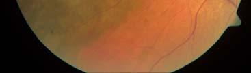
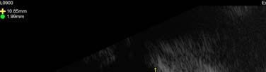
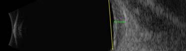
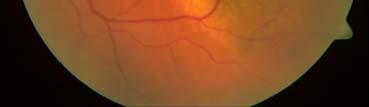
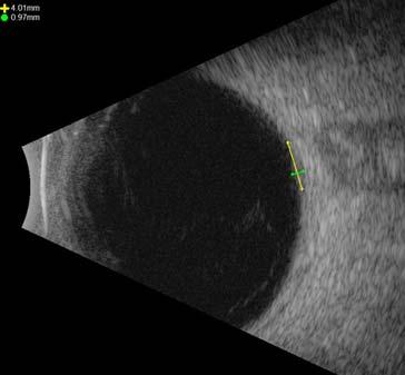
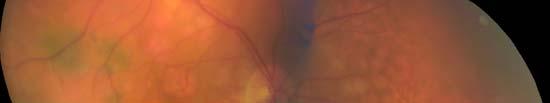
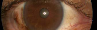
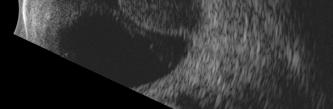
1 Dispelling Rumors about Tumors Jesse L. Berry, MD Arizona Ophthalmology Society 2017 Associate Director, Ocular Oncology Service Associate Program Director USC/CHLA, Keck School of Medicine Case 65 year old female presents with flashes 1
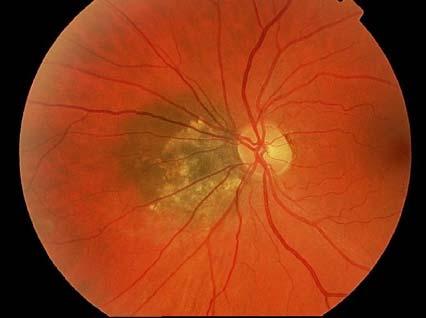
2 Diagnosis: Choroidal Melanoma 5% of all melanomas in the US most common primary IO tumor in adults 6 cases/million 1500 cases per year in US years/women=men/caucasian Diagnosis based on fundoscopy + ultrasound Rumor #1: Everything that s pigmented is a melanoma 2


3 Melanocytoma Differential diagnosis Vortex varix CHRPE Choroidal nevus Rumor #2: Everything that s pigmented and elevated must be a melanoma 3


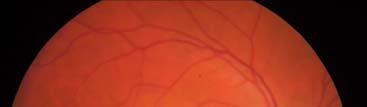
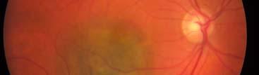
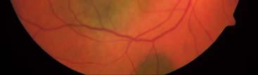
4 Case: Melanoma v. Nevus? Melanoma v. Nevus? 4


5 Melanoma v. Nevus? Melanoma v. Nevus? The only melanoma in the bunch 5
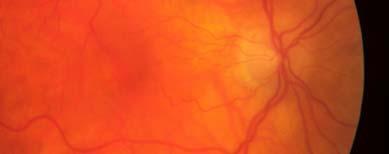
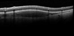
6 Diagnosis: Choroidal Nevus Benign tumors Collection of bland spindle A melanocytes The edges are defined but not sharply demarcated Dark brown or grey pigmentation Amelanotic not unusual High risk features which predispose to growth Growth may or may not be a sign of malignancy Which nevi grow? Feature Feature in Nevi that progress to Melanoma (%) Thickness > 2mm 19 2 Fluid 27 3 Symptoms 23 2 Orange Pigment 30 3 Margin <3mm to disc 13 2 Ultrasonographic Hollowness 25 3 Halo Absence 7 6 HR To Find Small Ocular Melanoma Using Hints Helpful Thickness Fluid Symptoms Orange Margin to Ultrasound Halo Thick Orange Fluids Sometimes Hale disc Hollow Melanoma Discovery Thickness Orange Pigment Fluid Symptoms Halo U/S Hollowness Disc distance 6
 Lifetime risk <1% Risk Factors Shields Combination of clinical factors If zero risk factors: 4% chance of")
7 Which nevi grow? Shields - Chance of Growth at 5 years % # of Risk Factors 27x greater risk ratio for 5 factors vs. 0 factors Growth not guarantee of malignancy Drusen are a sign of chronicity (favorable) Lifetime risk <1% Risk Factors Shields Combination of clinical factors If zero risk factors: 4% chance of growth/5 years If one risk factor: 36% chance of growth/5 years If 2 risk factors: >45% chance of growth/5 years If all risk factors: >56% chance of growth/5 years 27x greater risk ratio for 5 factors v 0 factors Growth not guarantee of malignancy Drusen are a sign of chronicity (favorable) 7


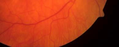
8 Rumor #3: Everything that s pigmented and elevated must be a melanoma or a high risk choroidal nevus 8
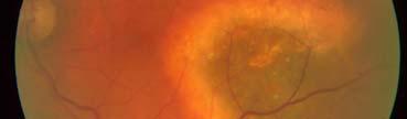
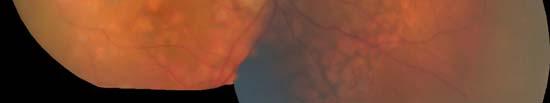
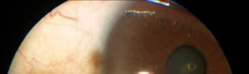
 Large choroidal and subretinal heme")

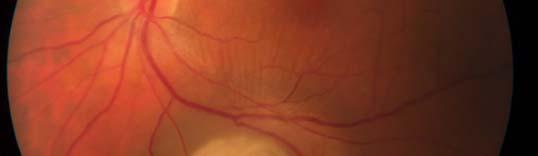
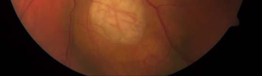
9 Diagnosis: PEHCR (peripheral exudative hemorrhagic chorioretinopathy) Large choroidal and subretinal heme Elderly, caucasian patients Associated with drusen and blood thinner use Usually no trauma Often temporal Lumpy bumpy and cystic spaces on Bscan 9


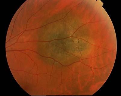
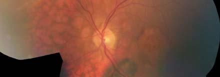
10 Case 55 yo F with known breast cancer presents with sudden loss of vision Diagnosis: Choroidal Metastases most common choroidal tumor in adults Women=Breast, lung, unknown Men=Lung, Unknown, GI Lung often preceeds the systemic diagnosis Breast rarely does Poorly circumscribed, amelanotic, associated with subretinal fluid Can be bilateral and/or multifocal (20%) 10


11 Rumor #4: Everything that s amelanotic must then be a met Choroidal metastases Choroidal hemangioma Combined hamartoma RPE Choroidal osteoma Astrocytic hamartoma 11

12 Amelanoic choroidal nevus Amelanotic melanoma CHPRE and nevus in the other eye! 12


13 case 65 yo F with known breast cancer presents for routine evaluation Known history of breast cancer with Amelanotic mass 13
14 Breast cancer and primary Amelanotic choroidal melanoma Occam s razor does not always apply Patients can have two primary cancers Rumor #5: Rules exist for a reason (but they don t always apply in Ocular Oncology ) 14


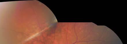
15 Case 65 yo M with a history of a nevus with painless vision loss One year ago now 15

16 Rumor #6: Everything that grows is cancer 16
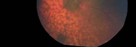
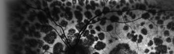
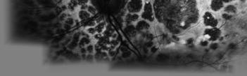
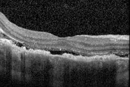
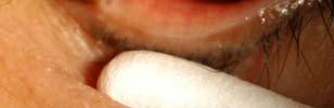
17 Diagnosis: BDUMP (Bilateral diffuse uveal melanocytic proliferation) rare paraneoplastic ocular syndrome benign hyperplasia of uveal melanocytes The GROWTH is not CANCER Painless bilateral vision loss Diffuse pigment clumping and orange pigment Subretinal fluid is common May precede diagnosis of systemic carcinoma by 3-12 months Case A 45 year old male presents with a red, painless eye 17


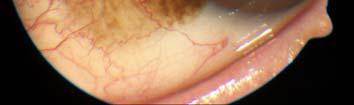
18 Diagnosis: Ocular Adnexal Lymphoma Low grade Non-Hodgkins B-cell Lymphoma 80% Extranodal marginal Zone lymphoma/mucosa associated lymphoma Often affects the orbit, lacrimal gland, lids and conjunctiva Associated with systemic disease in 30% Conjunctival involvement is most associated with systemic disease Presents as a thick, velvety salmon patch Do not confuse with OSSN! No touch with cryo 18


19 Rumor #7: Ocular adnexal lymphoma is always an external disease 19


20 Diagnosis: Uveal Lymphoma Low-grade Non-Hodgkins B-cell Lymphoma (MALT) Often affects the choroid, iris and/or ciliary body Prolonged indolent course often misdiagnosed as birdshot, white dots syndrome or VKH Key finding: yellow-white choroidal infiltrates with associated cresenteric choroidal thickening and hypofluoresence of ICG It is NOT vitreoretinal lymphoma (worse prognosis by far) 60% of patients with uveal lymphoma have OAL overlap 50% are bilateral 30% have systemic involvement Don t fall for the rumor - Dilate patients with a salmon patch 20


21 Case 56 yo M presented for evaluation of a spot, recently started timolol in the right eye only 21


22 Diagnosis: Iris Nevus Common iris tumor Concern for melanoma with thickness >1 mm (average is 2mm), distortion of iris stroma, correctopia, ectropion uveae, feather borders, angle involvement Risk for malignant conversion ~8% Risk for metastatic disease is low ~3% Other high risk features: ABCDE young Age, Blood (hyphema), inferior Clock hour, Diffuse, Ectropion uveae High pressure also a risk factor 22
23 Rumor #8: Iris nevi are no big deal Case continues Treated for recalcitrant unilateral glaucoma 23


24 case 58 yo Male from Egypt is referred for evaluation of a conjunctival nevus 24
25 Diagnosis: Primary Acquired Melanosis Painless, flat brown spot Often misdiagnosed as freckle or nevus Benign PAM with atypia precancerous lesion with ~15% risk of progression to conjunctival melanoma Conjunctival melanoma ~50% mortality at 3 years, worse with >2mm, ulceration, caruncular involvement Rumor #9: Conjunctival nevi are no big deal either 25


26 Conjunctival nevus Conjunctival concretions Primary acquired melanosis Conjunctival nevus Conjunctival melanoma Caruncular involvement Conjunctival melanoma 26

27 case 28 yo Hispanic Female presents for evaluation of decrease vision x 6 months, worse after becoming pregnant 27
28 Diagnosis: Pigmented IridoCiliary Body Mass Often a late diagnosis May cause sectoral cataract Look for a sentinel vessel (important clue!) Considered a worse prognostic feature for uveal melanoma because it is detected later Rumor #10: Ciliary body tumors are bad, bad, bad 28


29 Ciliary body adenocarcinoma Ciliary body adenocarcinoma Ciliary body lymphoma Ciliary body adenoma Ciliary body melanoma Ciliary body melanocytoma 29
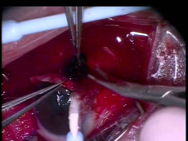

30 Case continues: Biopsy Pathology: adenoma Ciliary body adenoma 30


31 Thank you! 31
Tall, dark and.. Uh oh
 Tall, dark and.. Uh oh Jesse L. Berry, MD Arizona Ophthalmology Society 2017 Ocular Oncology Service USC Eye Institute Financial Disclosures Research Support: Bright Eyes Nautica Foundation Knights Templar
CLINICAL PEARLS IN OCULAR ONCOLOGY
 CLINICAL PEARLS IN OCULAR ONCOLOGY IRIS NEVUS - Two kinds circumscribed and diffuse - Photodocumentation important to monitor growth - Risk Factors for iris nevus growth to melanoma (ABCDEF) A Age (young),
Retina Center of Oklahoma Sam S. Dahr, M.D. Adult Intraocular Tumors
 Adult Intraocular Tumors Sam S. Dahr, M.D. Retina Center of Oklahoma www.retinacenteroklahoma.com www.rcoklahoma.com Table of Contents Posterior uveal malignant melanoma Uveal metastasis Uveal melanoma
Ocular Neoplasia What s Common? What s New? Richard R Dubielzig
 Ocular Neoplasia What s Common? What s New? Richard R Dubielzig Orbit 288 6% Tumors of the globe make up 3225 out of 6110 total neoplasms = 53%. Tumors of the conjunctiva make up 1192 out of 6110 total
Anatomic Divisions. Ocular Surface. Intraocular. Orbital. Lacrimal. Eyelid
 Anatomic Divisions Ocular Surface Intraocular Orbital Lacrimal Eyelid Ocular Surface Melanocytic Squamous Neoplasia Lymphoid Melanocytic Nevi PAM (Primary Acquired Melanosis) Ocular Melanocytosis Melanoma
Outline. Brief history and principles of ophthalmic ultrasound. Types of ocular ultrasound. Examination techniques. Types of Ultrasound
 Ultrasound and Intraocular Tumors 2015 Ophthalmic Photographers' Society Mid-Year Program Cagri G. Besirli MD, PhD Kellogg Eye Center University of Michigan Outline Brief history and principles of ophthalmic
Ocular Neoplasia CL Davis 9/08. Richard R Dubielzig
 Ocular Neoplasia CL Davis 9/08 Richard R Dubielzig 2135/5722 Canine Melanocytic Tumors Outside the Globe: 264 Conjunctival: 159 Eye Lid: 72 Skin: 33 Affecting the Globe: 1871 Anterior Uveal Melanocytoma:
ACTIVATED OR NOT? RETINAL CASE PRESENTATION Shorye Payne, MD Medical Retinal Specialist Robley Rex VA Eye Clinic
 ACTIVATED OR NOT? RETINAL CASE PRESENTATION Shorye Payne, MD Medical Retinal Specialist Robley Rex VA Eye Clinic C We anticipate that the future management of posterior uveal melanoma (PUM) will focus
Table of Contents 1 Orbit 3 2 Eyelids 7
 Table of Contents Preface, x List of abbreviations xi Glossary xii Section I Atlas 1 1 Orbit 3 Clinical signs associated with orbital neoplasia 3 Clinical signs associated with orbital cellulitis 3 Enophthalmos
A study of iris melanoma in Northern Ireland
 British Journal of Ophthalmology, 1989, 73, 591-595 A study of iris melanoma in Northern Ireland J N McGALLIARD AND P B JOHNSTON From the Department of Ophthalmology, Royal Victoria Hospital, Grosvenor
Pseudohypopyon in Retinoblastoma. Choroidal Nevus. Masquerade Syndromes. Vision pathways. Flat with uniform color
 Primary Intraocular Tumors Thomas F. Freddo, O.D., Ph.D., F.A.A.O. Professor and Former Director School of Optometry University of Waterloo Masquerade Syndromes
Pigmented lesions of the
 Pigmented lesions of the choroid and retina are commonly encountered by optometrists in everyday practice. The increasing use of retinal imaging and indirect ophthalmoscopy among community optometrists
Cancer and the Eye. Cancer factoids. Cancer factoids. Cancer factoids. Recent study. Cancer types. Clinical Professor IU School of Optometry
 DISCLOSURE STATEMENT No financial disclosures Course Title: Cancer and the Eye Lecturer: Brad Sutton, OD, FAAO Clinical Professor IU School of Optometry Can affect any tissue or organ at any age All cancers
Cancer and the Eye. Cancer and the Eye. Cancer factoids. Cancer factoids. Cancer factoids. Recent study
 Course Title: Cancer and the Eye Brad Sutton, OD, FAAO Clinical Professor IU School of Optometry Cancer and the Eye No financial disclosures. Cancer factoids Cancer factoids Can affect any tissue or organ
Periocular Malignancies
 Periocular Malignancies Andrew Gurwood, O.D., F.A.A.O., Dipl. Marc Myers, O.D., F.A.A.O. Drs. Myers and Gurwood have no financial interests to disclose. Course Description Discussion of the most common
Ocular Pathology. I. Congenital and/or developmental. A. Trisomy 21. Hypertelorism (widely spaced eyes) Keratoconus (cone shaped cornea)
 Keratoconus (cone shaped cornea)") I. Congenital and/or developmental Robbins Pathologic Basis of Disease, 6 th Ed. A. Trisomy 21 Hypertelorism (widely spaced eyes) Keratoconus (cone shaped cornea) Focal hypoplasia of iris Cataracts frequently
I. Congenital and/or developmental Robbins Pathologic Basis of Disease, 6 th Ed. A. Trisomy 21 Hypertelorism (widely spaced eyes) Keratoconus (cone shaped cornea) Focal hypoplasia of iris Cataracts frequently
FELINE DIFFUSE IRIDAL MELANOMA
 Vet Times The website for the veterinary profession https://www.vettimes.co.uk FELINE DIFFUSE IRIDAL MELANOMA Author : JAMES OLIVER Categories : Vets Date : June 3, 2013 JAMES OLIVER looks at several clinical
Intraocular tumors. Zsuzsa Récsán
 Intraocular tumors Zsuzsa Récsán Definition Uveal melanoma Primary acquired malignant neoplasm of uveal melanocytes Epidemiology: most common primary mal tu in adults Much more common in lighter-skinned
VACAVILLE DERMATOLOGY
 Connecting the Dots on those Spots NANDAN V. KAMATH, M.D. VACAVILLE DERMATOLOGY Sources All of the photos were taken with permission from the Dermnet NZ website - Dermnet New Zealand after communicating
Characteristic Ultrasonographic Findings of Choroidal Tumors
 Characteristic Ultrasonographic Findings of Choroidal Tumors Tsung-Jen Wang, Chang-Hao Yang, Shu-Lang Liao, Tzyy-Chang Ho, Jen-Shang Huang, Chang-Ping Lin, Chung-May Yang, Muh-Shy Chen and Luke Long-Kuang
J of Evolution of Med and Dent Sci/ eissn , pissn / Vol. 4/ Issue 55/ July 09, 2015 Page 9665
 RARE PRESENTATION OF BILATERAL CHOROIDAL METASTASIS FROM PRIMARY MUCO-EPIDERMOID CARCINOMA OF THE PAROTID GLAND: A G. Premalatha 1, Ramya Seetamraju 2 HOW TO CITE THIS ARTICLE: G. Premalatha, Ramya Seetamraju.
Uveal Melanoma. Protocol applies to malignant melanoma of the uvea.
 Uveal Melanoma Protocol applies to malignant melanoma of the uvea. Protocol revision date: January 2005 Based on AJCC/UICC TNM, 6 th edition Procedures Cytology (No Accompanying Checklist) Biopsy (No Accompanying
2008 Gross Ocular Pathology. Gross Pathology 2
 2008 Gross Ocular Pathology Gross Pathology 2 08rd1281 Feline T-Cell Lymphoma 08rd1300 Canine Iridociliary Adenoma Foam Cell Variant 08rd1331 Feline Feline Iridociliary Adenoma 08rd1340 Canine Retinal
Gross Ocular Pathology
 Gross Ocular Pathology 06rd0850 Feline Metastatic sarcoma 06rd0878 Feline Cryptococcosis 06rd0880 Canine Malignant Melanoma 06rd0929 Feline Conjunctivalization of the epithelium 06rd0931 Name the Species
Lumps Bumps and Magic Potions Review of Ocular Tumors" Ocular Tumors" Eyelids" Conjunctiva" Intraocular" Orbit"
 The authors have no financial interests in the materials in this presentation! Lumps Bumps and Magic Potions Review of Ocular Tumors Lumps Bumps and Magic Potions Review of Ocular Tumors Carol Shields
Financial Disclosure. I have nothing to disclose. Lifetime risk of developing cancer in U.S.*
 Brian P. Mahoney, OD, FAAO Department of Veterans Affairs Wilmington, DE bcktmahoney@msn.com Financial Disclosure I have nothing to disclose Top 10 cancers in the US 1. Skin 2. Lung 3. Prostate 4. Breast
Financial Disclosures. The Eye in Neoplastic Disease. Course Goal. We wish to acknowledge and thank: Tumor Definition
 The Eye in Neoplastic Disease Carlo J. Pelino, OD, FAAO Joseph J. Pizzimenti, OD, FAAO cpelino@salus.edu pizzimen@uiwtx.edu Financial Disclosures! Speakers have no relevant financial relationships to declare.
Pediatric Ocular Sonography
 Pediatric Ocular Sonography Cicero J Torres A Silva, MD Associate Professor of Radiology 2016 SPR Pediatric Ultrasound Course Yale University School of Medicine None Disclosures Objectives of Presentation
Familial iris melanosis -a misnomer?
 Familial iris melanosis -a misnomer? BRIAN C JOONDEPH AND MORTON F British Journal of Ophthalmology, 1989, 73, 289-293 GOLDBERG From the Department of Ophthalmology, Eye and Ear Infirmary, University of
Orbital Tumors - A Clinico Pathological Study
 Orbital Tumors - A Clinico Pathological Study Radha. J. DO, Ani Sreedhar. MS. Little Flower Hospital, Angamaly, Kerala ORIGINAL ARTICLES Abstract: Aim. To study the clinical and histopathological profiles
Desmoplastic Melanoma R/O BCC. Clinical Information. 74 y.o. man with lesion on left side of neck r/o BCC
 R/O BCC Sabine Kohler, M.D. Professor of Pathology and Dermatology Dermatopathology Service Stanford University School of Medicine Clinical Information 74 y.o. man with lesion on left side of neck r/o
Asadi-Amoli et al Adenocarcinoma of RPE Iranian Journal of Ophthalmology - Volume 19, Number 4, 2007
 Adenocarcinoma of Retinal Pigment Epithelium Clinically Diagnosed as Malignant Melanoma; A Case Report with Unsystematic Review of Literature Fahimeh Asadi-Amoli, MD, 1 Hedyeh Moradi, MD 2 Mohammad-Taher
Pathology of the skin. 2nd Department of Pathology, Semmelweis University
 Pathology of the skin 2nd Department of Pathology, Semmelweis University Histology of the skin Epidermis: Stratum corneum Stratum granulosum Stratum spinosum Stratum basale Dermis: papillary and reticular
Complicated Cataract to Intraocular Tumors, Beware of the unexpected
 Complicated Cataract to Intraocular Tumors, Beware of the unexpected Ihab Saad Othman, MD, FRCS Professor of Ophthalmology Cairo University In this part of the world: We Master Phakoemulsification 1 Intraoperative/Second
Iris-Ciliary Body Melanoma: 57-year-old female with iris lesion
 Iris-Ciliary Body Melanoma: 57-year-old female with iris lesion Gina M. Rogers, MD, Nasreen A. Syed, MD, Wallace L. M. Alward, MD, Juan Fernandez de Castro, MD, Lauren Jensen, BS Chief Complaint: Spot
COEXISTENCE OF OPTIC NERVE HEAD DRUSEN
 COEXISTENCE OF OPTIC NERVE HEAD DRUSEN AND COMBINED HAMARTOMA OF THE RETINA AND RETINAL PIGMENT EPITHELIUM IN A TAIWANESE MALE Yo-Chen Chang 1 and Rong-Kung Tsai 2,3 1 Department of Ophthalmology, Kaohsiung
Case Study. Monocular Malignant Melanoma
 Case Study Monocular Malignant Melanoma Case History A 52 year old Caucasian female presented with a number of naevi on the skin and a right ciliary body malignant melanoma twelve years ago and had an
Advances in Ocular Imaging
 Wide angle fundus imaging and Fuorescein angiography in evaluation and management of intraocular tumors Ihab Saad Othman, MD, FRCS Professor of Ophthalmology Cairo University Cairo, Egypt Advances in Ocular
Technicians & Nurses Program
 ASCRS ASOA Symposium & Congress Technicians & Nurses Program April 17-21, 2015 San Diego, California Clinical-Pathological Correlation of Common Periocular Lesions Todd Mondzelewski, MD Comprehensive Ophthalmology
Dr. Lim, maybe we should start by you telling us a little about yourself and what exactly you do.
 Support for Yale Cancer Answers comes from AstraZeneca, dedicated to providing innovative treatment options for people living with cancer. Learn more at astrazeneca-us.com Welcome to Yale Cancer Answers
Among the benign intraepithelial melanocytic proliferations, Inflamed Conjunctival Nevi. Histopathological Criteria. Resident Short Reviews
 Resident Short Reviews Inflamed conjunctival nevi (ICN) may suggest malignancy because of their rapid growth and atypical histology. The objective of this study was to characterize the diagnostic features
Proceedings of the 36th World Small Animal Veterinary Congress WSAVA
 www.ivis.org Proceedings of the 36th World Small Animal Veterinary Congress WSAVA Oct. 14-17, 2011 Jeju, Korea Next Congress: Reprinted in IVIS with the permission of WSAVA http://www.ivis.org 14(Fri)
Vitreoretinal surgical management In ocular oncology
 www.ophtalmique.ch Vitreoretinal surgical management In ocular oncology Pournaras Jean-Antoine C Vitreoretinal Surgery Unit 1. Surgical resection after proton beam therapy 2. Ocular Biopsy 3. RD in advanced
Mild NPDR. Moderate NPDR. Severe NPDR
 Diabetic retinopathy Diabetic retinopathy is the most common cause of blindness in adults aged 35-65 years-old. Hyperglycaemia is thought to cause increased retinal blood flow and abnormal metabolism in
CLINICAL SCIENCES. for less than 1% of all malignant eyelid lesions. 1-4 In a survey of
 CLINICAL SCIENCES Metastatic Tumors to the Eyelid Report of 20 Cases and Review of the Literature Carlos ianciotto, MD; Hakan Demirci, MD; Carol L. Shields, MD; Ralph C. Eagle Jr, MD; Jerry A. Shields,
Finding Melanoma. Is not easy!
 Finding Melanoma Is not easy! Finding Melanoma Victoria mean depth at diagnosis is 1.5 mm. Melanoma 1.5mm Has Stage 1B Mortality 10% Melanoma Spotting a killer! Spotting a killer Visual Clues What are
Early detection of Retinoblastoma in children. Max Mantik
 Early detection of Retinoblastoma in children Max Mantik Introduction The most common primary intraocular malignancy of childhood 10 to 15 % of cancers that occur within the first year of life Typical
Ocular Malignancies in the Elderly
 Cancer Ocular Malignancies in the Elderly E. Rand Simpson, MD, Associate Professor of Ophthalmology, University of Toronto; Director, Ocular Oncology, Princess Margaret Hospital,Toronto, ON. Larry Ulanski
An Overview of Melanoma. Harriet Kluger, M.D. Associate Professor Section of Medical Oncology Yale Cancer Center
 An Overview of Melanoma Harriet Kluger, M.D. Associate Professor Section of Medical Oncology Yale Cancer Center Melanoma Statistics Median age at presentation 45-55 55 years Incidence: 2003 54,200 cases
Benign versus Cancerous Lesions How to tell the difference FMF 2014 Christie Freeman MD, CCFP, DipPDerm, MSc
 1 Benign versus Cancerous Lesions How to tell the difference FMF 2014 Christie Freeman MD, CCFP, DipPDerm, MSc Benign lesions Seborrheic Keratoses: Warty, stuck-on Genetics and birthdays Can start in late
UVEAL MELANOMA: FROM GENETICS TO TREATMENT. Carlo J. Pelino, OD, FAAO Joseph J. Pizzimenti, OF, FAAO
 UVEAL MELANOMA: FROM GENETICS TO TREATMENT Carlo J. Pelino, OD, FAAO Joseph J. Pizzimenti, OF, FAAO Financial Disclosure Speakers have no relevant financial relationships to declare and no proprietary
Vascularized solid iris lesion in a 3 year old child: 5 years of follow up
 Fea et al. BMC Ophthalmology (2016) 16:89 DOI 10.1186/s12886-016-0267-4 CASE REPORT Open Access Vascularized solid iris lesion in a 3 year old child: 5 years of follow up Antonio Maria Fea *, Cristina
CLINICAL SCIENCES. Visual Acuity in 3422 Consecutive Eyes With Choroidal Nevus
 CLINICAL SCIENCES Visual Acuity in 3422 Consecutive Eyes With Choroidal Nevus Carol L. Shields, MD; Minoru Furuta, MD; Arman Mashayekhi, MD; Edwina L. Berman, MBBS; Jonathan D. Zahler, DO; Daniel M. Hoberman,
Malignant Melanoma Early Stage. A guide for patients
 This melanoma patient brochure is designed to help educate melanoma patients and their caregivers. It was developed under the guidance of Dr. Michael Smylie, Professor, Department of Oncology, University
Around The Globe in 60 Minutes
 Around The Globe in 60 Minutes Around the GLOBE in Sixty Minutes Basic Ocular Anatomy, Examination, and Diagnostic Techniques Introduction Focusing on canine and feline ocular anatomy and basic examination
Gene Expression Profiling has been proposed as a method of risk stratification for uveal melanoma.
 Last Review Status/Date: September 2014 Description Page: 1 of 5 Gene Expression Profiling has been proposed as a method of risk stratification for uveal melanoma. Background Uveal melanoma Uveal melanoma,
Slide 4. Slide 5. Slide 6
 Slide 1 Slide 4 Demographics El Paso Eye Care Border Healthcare-Based Grand Rounds Derek N. Cunningham, O.D. 80-90% Mexican-Americans Diabetes Hypertension Hyperlipidemia Obesity 70% uninsured High poverty
Ocular Urgencies and Emergencies
 Ocular Urgencies and Emergencies Pam Boyce, O.D., F.A.A.O. Boyce Family Eye Care, Ltd. 528 Devon Ave. Park Ridge, IL 60068 847-518-0303 Somebody s going to lose an eye Epidemiology 2.4 million ocular and
Patricia Chevez-Barrrios AAOOP-USCAP /12/2016
 Biomarkers in Ocular Melanoma Patricia Chévez-Barrios, MD Pathology and Genomic Medicine, Houston Methodist Hospital Professor of Pathology and Laboratory Medicine and Ophthalmology, Weill Cornell Medical
Bilateral Diffuse Uveal Melanocytic Proliferation (BDUMP) associated with B-cell lymphoma: report of a rare case
 associated with B-cell lymphoma: report of a rare case") Pefkianaki et al. BMC Cancer (2015) 15:23 DOI 10.1186/s12885-015-1020-8 CASE REPORT Open Access Bilateral Diffuse Uveal Melanocytic Proliferation (BDUMP) associated with B-cell lymphoma: report of a rare
Pefkianaki et al. BMC Cancer (2015) 15:23 DOI 10.1186/s12885-015-1020-8 CASE REPORT Open Access Bilateral Diffuse Uveal Melanocytic Proliferation (BDUMP) associated with B-cell lymphoma: report of a rare
Clinical characteristics
 Skin Cancer Fernando Vega, MD Seattle Healing Arts Clinical characteristics Precancerous lesions Common skin cancers ACTINIC KERATOSIS Precancerous skin lesions Actinic keratoses Dysplastic melanocytic
Therapy of melanocytic conjunctival tumors
 DOI: 10.4149/BLL_2013_093 Bratisl Lek Listy 2013; 114 (8) CLINICAL STUDY Therapy of melanocytic conjunctival tumors Halas M Jr 1, Svetlosakova Z 1, Babal P 2 Department of Ophthalmology, Comenius University,
Toby Maurer, MD University of California, San Francisco. Lifetime risk of an American developing melanoma
 Distinguishing Pigmented Skin Lesions and Melanoma Toby Maurer, MD University of California, San Francisco Epidemiology of Melanoma Lifetime risk of an American developing melanoma 1935: 1 in 1500 1980:
Benign and malignant epithelial lesions: Seborrheic keratosis: A common benign pigmented epidermal tumor occur in middle-aged or older persons more
 Benign and malignant epithelial lesions: Seborrheic keratosis: A common benign pigmented epidermal tumor occur in middle-aged or older persons more common on the trunk; but extremities, head and neck are
Toby Maurer, MD University of California, San Francisco. Lifetime risk of an American developing melanoma
 Distinguishing Pigmented Skin Lesions and Melanoma Toby Maurer, MD University of California, San Francisco Epidemiology of Melanoma Lifetime risk of an American developing melanoma 1935: 1 in 1500 1980:
Central venous occlusion
 Central venous occlusion Central venous occlusion (right eye) There are dark haemorrhages at the macula and all over the retina. Choroidal haemangioma A choroidal haemangioma has salmon pink colour. There
Dermatopathology: The tumor is composed of keratinocytes which show atypia, increase mitoses and abnormal mitoses.
 Squamous cell carcinoma (SCC): A common malignant tumor of keratinocytes arising in the epidermis, usually from a precancerous condition: 1- UV induced actinic keratosis, usually of low grade malignancy.
Referral pathways for adult ocular tumours
 Clinical Guideline Referral pathways for adult ocular tumours November 2018 18 Stephenson Way, London, NW1 2HD, T. 02037705322 contact@rcophth.ac.uk @rcophth.ac.uk The Royal College of Ophthalmologists
10 EYE EMERGENCIES. Who goes, who you better not send! Brant Slomovic, MD, FRCPC University Health Network
 10 EYE EMERGENCIES Who goes, who you better not send! Brant Slomovic, MD, FRCPC University Health Network DISCLOSURES I have none PVD CASE 1 WHAT IS A PVD? a process of aging (45-55) liquefaction of vitreous
Coagulative necrosis in a malignant melanoma of the choroid at the macula with extensive subretinal hemorrhage
 Coagulative necrosis in a malignant melanoma of the choroid at the macula with extensive subretinal hemorrhage Robert D. Yee, Robert Y. Foos, and Bradley R. Straatsma The authors present a case report
generalized neurofibromatosis
 Brit. 7. Ophthal. (I972) 56, 487 Glial hamartoma of the retina in generalized neurofibromatosis von Recklinghausen's disease L. J. MNIARTYN AND D. L. KNOX From the li'ilmer Institute, the Johns Hopkins
6/14/2012. Palpebral. Conjunctival. Bulbar. Fornix. Limbal Stem Cells
 Conjunctival Lesions: Links to Systemic Disease Thomas F. Freddo, O.D., Ph.D., F.A.A.O. Professor and Former Director School of Optometry University of Waterloo Tissues of the Conjunctiva Limbal Stem Cells
Size Overlap between Benign Melanocytic Choroidal Nevi and Choroidal Malignant Melanomas
 Size Overlap between Benign Melanocytic Choroidal Nevi and Choroidal Malignant Melanomas James J. Augsburger, Zélia M. Corrêa, Nikolaos Trichopoulos, and Adeel Shaikh PURPOSE. To estimate size overlap
BAP-oma & BEYOND MICHAEL A NOWAK, MD
 BAP-oma & BEYOND MICHAEL A NOWAK, MD CONFLICTS No conflicts with the content of this lecture BAP-oma Wiesner 2011: Families with multiple tan dome-shaped papules of head, neck, trunk, and extremities.
The many faces of extranodal lymphoma
 The many faces of extranodal lymphoma Frank Pameijer Departments of Radiology and Radiation Oncology University Medical Center Utrecht Special thanks to Ilona M Schmalfuss, MD University of Florida Gainesville,
2013/14 NHS STANDARD CONTRACT FOR OCULAR ONCOLOGY SERVICE (ADULTS AND ADOLESCENTS) D12/S(HSS)/a Ocular oncology service (Adults and Adolescents)
 D12/S(HSS)/a Ocular oncology service (Adults and Adolescents)") D12/S(HSS)/a 2013/14 NHS STANDARD CONTRACT FOR OCULAR ONCOLOGY SERVICE (ADULTS AND ADOLESCENTS) PARTICULARS, SCHEDULE 2 - THE SERVICES, A SERVICE SPECIFICATIONS Service Specification No. Service Commissioner
D12/S(HSS)/a 2013/14 NHS STANDARD CONTRACT FOR OCULAR ONCOLOGY SERVICE (ADULTS AND ADOLESCENTS) PARTICULARS, SCHEDULE 2 - THE SERVICES, A SERVICE SPECIFICATIONS Service Specification No. Service Commissioner
Five Retinal Findings Not to Miss 2019 Susan M. Malinowski, MD, FACS Retina Consultants of Michigan
 Five Retinal Findings Not to Miss 2019 Retina Consultants of Michigan I. UGH Plus Syndrome associated with PCIOL A. Definition 1. Uveitis-glaucoma-hyphema (UGH) syndrome was first described by Ellingson
IT S FUNDAMENTAL MY DEAR WATSON! A SHERLOCKIAN APPROACH TO DERMATOLOGY
 IT S FUNDAMENTAL MY DEAR WATSON! A SHERLOCKIAN APPROACH TO DERMATOLOGY Skin, Bones, and other Private Parts Symposium Dermatology Lectures by Debra Shelby, PhD, DNP, FNP-BC, FADNP, FAANP Debra Shelby,
Ocular Lecture. Sue Bednar NP Ali Atwater PA-C
 Ocular Lecture Sue Bednar NP Ali Atwater PA-C Triaging Ocular Complaints Painful Eye/Red eye +/-blurry vision +/-visual loss +/-floaters +/-fevers If any of the above findings exist, pt is likely to have
Proton Radiation Therapy of Ocular Melanoma at PSI
 Proton Radiation Therapy of Ocular Melanoma at PSI G. Goitein*, A. Schalenbourg, J. Verwey*, A. Bolsi*, C. Ares*, L. Chamot, E. Hug*, L. Zografos *Paul Scherrer Institut, 5232 Villigen PSI; Hôpital Ophtalmique,
EYE TRAUMA: INCIDENCE
 Introduction EYE TRAUMA: INCIDENCE 2.5 million eye injuries per year in U.S. 40,000 60,000 of eye injuries lead to visual loss Introduction Final visual outcome of many ocular emergencies depends on prompt,
Malignant tumors of melanocytes: Part 1. Deba P Sarma, MD., Omaha
 Malignant tumors of melanocytes: Part 1 Deba P Sarma, MD., Omaha The melanocytic tumor is one of the most difficult and confusing areas in Dematopathology. It is true that most (95%) of such lesions are
Atlas of Eyelid and Conjunctival Tumors
 Atlas of Eyelid and Conjunctival Tumors Jerry A. Shields, M.D. Director, Ocular Oncology Service Wills Eye Hospital Professor of Ophthalmology Thomas Jefferson University Philadelphia, Pennsylvania Carol
Top Pediatric Retinal Diseases you don t want to miss! Retinopathy of Prematurity (ROP) Aggressive, Posterior ROP (AP ROP)
 Aggressive, Posterior ROP (AP ROP)") Top 10 10 Pediatric Retinal Diseases you don t want to miss! Polly Quiram MD, PhD Vitreoretinal Surgery, PA Retinal Update Jan 26th, 2018 ROP Retinoblastoma Coats disease Persistent fetal vasculature Familial
Top 10 10 Pediatric Retinal Diseases you don t want to miss! Polly Quiram MD, PhD Vitreoretinal Surgery, PA Retinal Update Jan 26th, 2018 ROP Retinoblastoma Coats disease Persistent fetal vasculature Familial
Misdiagnosed Vogt-Koyanagi-Harada (VKH) disease and atypical central serous chorioretinopathy (CSC)
 disease and atypical central serous chorioretinopathy (CSC)") HPTER 12 Misdiagnosed Vogt-Koyanagi-Harada (VKH) disease and atypical central serous chorioretinopathy (S) linical Features VKH disease is a bilateral granulomatous panuveitis often associated with exudative
HPTER 12 Misdiagnosed Vogt-Koyanagi-Harada (VKH) disease and atypical central serous chorioretinopathy (S) linical Features VKH disease is a bilateral granulomatous panuveitis often associated with exudative
Cancer Reporting for Dermatologists. Florida Department of Health Florida Cancer Data System. March 9, Agenda
 Cancer Reporting for Dermatologists Florida Department of Health Florida Cancer Data System March 9, 2011 Agenda Welcome Introductions Cancer Reporting in Florida BETA Participation Expectations Review
A PRACTICAL APPROACH TO ATYPICAL MELANOCYTIC LESIONS BIJAN HAGHIGHI M.D, DIRECTOR OF DERMATOPATHOLOGY, ST. JOSEPH HOSPITAL
 A PRACTICAL APPROACH TO ATYPICAL MELANOCYTIC LESIONS BIJAN HAGHIGHI M.D, DIRECTOR OF DERMATOPATHOLOGY, ST. JOSEPH HOSPITAL OBJECTIVES Discuss current trends and changing concepts in our understanding of
11/29/2016 MACULAR MALADIES: TYPICAL & ATYPICAL CASES
 MACULAR MALADIES: TYPICAL & ATYPICAL CASES Dawn Pewitt, OD, FAAO Triad Eye Institute, Grove, OK Dpewitt@triadeye.com Disclosure Statement: No financial disclosures COPE 51218-PS Please silence all mobile
Lesion Imaging Characteristics Mass, Favoring Benign Circumscribed Margins Intramammary Lymph Node
 Lesion Imaging Characteristics Mass, Favoring Benign Circumscribed Margins Intramammary Lymph Node Oil Cyst Mass, Intermediate Concern Microlobulated Margins Obscured Margins Mass, Favoring Malignant Indistinct
Dr/ Marwa Abdellah EOS /16/2018. Dr/ Marwa Abdellah EOS When do you ask Fluorescein angiography for optic disc diseases???
 When do you ask Fluorescein angiography for optic disc diseases??? 1 NORMAL OPTIC DISC The normal optic disc on fluorescein angiography is fluorescent due to filling of vessels arising from the posterior
Comparative Ocular Pathology Rounds
 Comparative Ocular Pathology Rounds 2007 2 07RD0943 Bovine Anterior Segment Dysgenesis 07RD1072 Feline Feline post-traumatic sarcoma spindle cell variant 07rd1124 Canine Stick in the Eye 07rd1132 Feline
By Darlene Jones, Nurse. May 2017
 By Darlene Jones, Nurse May 2017 Disclosure of potential conflict of interest Darlene Jones, Nurse I have no conflict of interest Course objectives Become familiar with the different pathologies in ophthalmology
Case Rep Oncol 2012;5: DOI: /
 This is an Open Access article licensed under the terms of the Creative Commons Attribution-NonCommercial-NoDerivs 3.0 License (www.karger.com/oa-license), applicable to the online version of the article
af Diagnostic Atlas A Retinal Reference Guide Building The Retina Company
 af Diagnostic Atlas A Retinal Reference Guide Building The Retina Company af Diagnostic Atlas A Retinal Reference Guide Optos core devices produce ultra-widefield (UWF ), high resolution digital images
Melanoma Update: 8th Edition of AJCC Staging System
 Melanoma Update: 8th Edition of AJCC Staging System Rosalie Elenitsas, M.D. Professor of Dermatology Director, Dermatopathology University of Pennsylvania DISCLOSURE OF RELATIONSHIPS WITH INDUSTRY None
Tumors Of The Eye And Ocular Adnexa (Atlas Of Clinical Oncology) By Devron H. Char
 By Devron H. Char") Tumors Of The Eye And Ocular Adnexa (Atlas Of Clinical Oncology) By Devron H. Char If you are searching for a book by Devron H. Char Tumors of the Eye and Ocular Adnexa (Atlas of Clinical Oncology) in
Tumors Of The Eye And Ocular Adnexa (Atlas Of Clinical Oncology) By Devron H. Char If you are searching for a book by Devron H. Char Tumors of the Eye and Ocular Adnexa (Atlas of Clinical Oncology) in
FINALIZED SEER SINQ QUESTIONS
 0076 Source 1: WHO Class CNS Tumors pgs: 33 MP/H Rules/Histology--Brain and CNS: What is the histology code for a tumor originating in the cerebellum and extending into the fourth ventricle described as
CLINICAL SCIENCES. Acquired Tumors Arising From Congenital Hypertrophy of the Retinal Pigment Epithelium. well-known fundus condition
 CLINICAL SCIENCES Acquired Tumors Arising From Congenital Hypertrophy of the Retinal Pigment Epithelium Jerry A. Shields, MD; Carol L. Shields, MD; Arun D. Singh, MD Background: Congenital hypertrophy
Fluorescein and Indocyanine Green Videoangiography of Choroidal Melanomas
 luorescein and Indocyanine Green Videoangiography of Choroidal Melanomas Leyla S. Atmaca, igen Batioğlu and Pelin Atmaca Eye Clinic, Ankara University Medical School, Ankara, Turkey Purpose: This study