Cancer Reporting for Dermatologists. Florida Department of Health Florida Cancer Data System. March 9, Agenda
|
|
|
- Dwayne Beasley
- 5 years ago
- Views:
Transcription
1 Cancer Reporting for Dermatologists Florida Department of Health Florida Cancer Data System March 9, 2011 Agenda Welcome Introductions Cancer Reporting in Florida BETA Participation Expectations Review of Documentation Materials Registering a Physician Live Demo Creating a new IDEA USERID Live Demo Dermatology Data Acquisition Manual Introduction to Skin Cancers Inputting a Cancer Abstract Live Demo Q & A 1
2 Introductions FCDS Main Number Michael Thiry Manager Data Acquisition Carlos Alvarez Field Coordinator Steven Peace Manager Quality Control and Training Cancer Reporting in Florida FCDS is Florida's statewide cancer surveillance system. FCDS was legislatively mandated in 1978 to collect incidence data on all cases seen in Florida since The goal of FCDS is to reduce death and illness due to cancer by providing data on cancer incidence. Data are used to observe cancer trends and provide a research base for studies into the possible causes of cancer. 2
3 Cancer Reporting in Florida (cont.) Who needs to report: Any licensed practitioner in the state of Florida that practices medicine, osteopathic, chiropractic medicine, naturopathy or veterinary medicine are required to report under Florida Statue or any laboratory licensed under chapter Florida Statute 483 that diagnoses or suspects the existence of a disease of public health significance shall immediately report the fact to the Department of Health. See the Laws and Rules section of the Dermatology Data Acquisition Manual and our website at: Cancer Reporting in Florida (cont.) Physician Reporting Many physicians in the state do not know about their legislatively mandated obligation to report Due to the special skill and knowledge required to report, many are unwilling to undertake the burden DOH and FCDS Renewed effort to get all physicians to report New software to make reporting easier to eliminate any barriers customized for dermatologists and the cancers they report Anyone from the office, with minimal training by FCDS, should be able to complete the reporting 3

4 BETA Participation Expectations Work with the new system Provide feedback Relay to us any problems (call, ) Ask questions Make suggestions Review of Package Information Letter from the Department of Health The departments commitment to improve reporting Physician Personal Identifier Dermatology Data Acquisition Manual Legislation and Reporting Guidelines for reporting Reportable Cases Casefinding General Abstracting Instructions Field by field description and explanation of the input fields Instructions on registering a physician and creating an new IDEA USERID 4
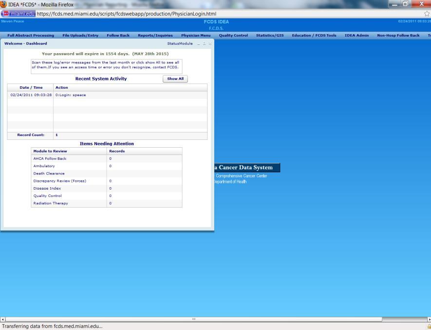
5 Getting Started Introduction to IDEA Register a Physician/Practice Create New User ID Live Demonstration FCDS IDEA FCDS IDEA is the Florida Cancer Data System Internet Data Entry and Abstracting System. Create New Facility/Practice Accounts Create New User Accounts Enter Cancer Case Abstracts Quality Control 5
6 Facility Number (Practice ID) All dermatologists are required to register in our system. You must register even if you have previously reported and already have a unique facility number for your practice. A new Facility Number (Practice ID) will be assigned and sent to you in the Physician Registration Confirmation as part of the physician/practice registration process. Retain the new Facility Number (Practice ID) for your records Use your Facility Number when communicating with FCDS. Physician Personal Identifier Physician Personal Identifier is a unique 12 character code specific to each Dermatologist identified through the Florida Department of Health. The Physician Personal Identifier can be found at the top of the letter sent to you by the Florida Department of Health announcing enhanced Dermatology Cancer Reporting through FCDS. The Physician Personal Identifier is required: to register a Physician; to create a new user account in the FCDS IDEA cancer reporting system; and to access the FCDS IDEA cancer reporting system to abstract and report cancer cases in compliance with Florida reporting statutes/rules. 6
7 Associate User to Physician/Practice Once a physician is registered, each user that will be inputting cancer abstracts for that physician must associate themselves with that physician. This can be done in 2 ways. Initial Account Setup: If you are accessing the system for the first time, you will be prompted, at the completion of the registration process, if you want to create a new user. If you then create a user, you will be automatically associated to the physician you registered. Associate User to Physician/Practice Add New User After Initial Setup: If you want to associate a user after the initial setup, sign into the IDEA system, access the Physician tab at the top of the screen and then select Physician Registration. Input the Physician Personal Identifier for the physician you want to associate to and you will be prompted to either complete their profile (if this had not already been completed) or associate yourself with this physician. 7

8 Already have an FCDS IDEA USERID? You can use your existing FCDS IDEA USERID and password to register a physician and to submit Dermatology Abstracts. You do NOT need to create a new userid to access this new system. To access the Physician Registration form, sign into IDEA, go to the Physician tab along the top of the screen and select Physician Registration. Your userid will automatically associate with any physician you register. Demo: Register a Physician/Practice 8
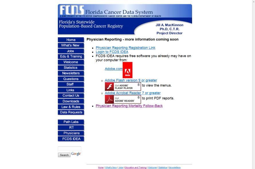
9 Demo: Register a Physician/Practice STEP 1: LOCATE THE LETTER SENT BY DOH WITH YOUR PHYSICIAN PERSONAL IDENTIFIER. If you cannot locate this letter, please contact FCDS at (305) and ask for Mike Thiry, FCDS Data Acquisition Manager. STEP 2: Go to the FCDS main web page: and go to Physicians Demo: Register a Physician/Practice Live DEMO For those of you unable to view the webcast (those participating only by phone) the next several slides will help orient you to the FCDS website, registration process, etc. The screenshots are also available in the DAM. 9

10 Demo: Register a Physician/Practice Demo: Register a Physician/Practice 10

11 Demo: Register a Physician/Practice Demo: Register a Physician/Practice 11
 the next several slides will help orient you to the FCDS website, registration process, etc. The screenshots are also available in the DAM. 12")
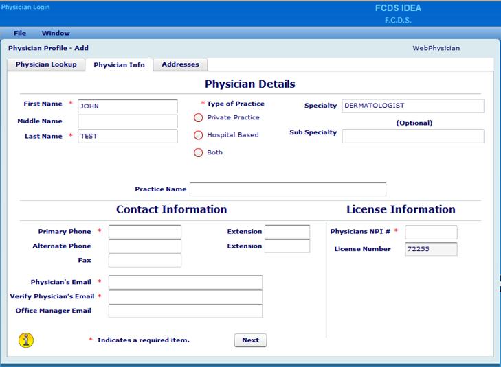
12 Demo: Register a Physician/Practice Demo: Create a New IDEA USERID Live DEMO For those of you unable to view the webcast (those participating only by phone) the next several slides will help orient you to the FCDS website, registration process, etc. The screenshots are also available in the DAM. 12

13 Demo: Create a New IDEA USERID Demo: Create a New IDEA USERID Unique USERID: - USERID s must be 5-15 alphanumeric characters - Passwords must be 8 32 alphanumeric characters USERID s cannot be duplicated. A check will be made at entry time to determine if that USERID is already in use. If so, you will be prompted to choose another USERID. 13
14 Demo: Create a new IDEA USERID Once your USERID and Password have been validated you will need to complete the remaining Profile items on the screen. All items on this screen marked with an asterisk (*) are required and must be completed. Demo: Create a New IDEA USERID Press Submit to ensure the information you entered is saved in your profile. Once completed, an is generated to the User address entered in the profile. You will need to retrieve this and click on the link embedded to complete the registration process. REQUIRED: Open the confirmation and click on the link to complete your registration. 14
15 Demo: Create a New IDEA USERID TEST YOUR LOGIN: You are now authorized to sign in to the FCDS IDEA system. Go To: Find the FCDS IDEA button down the left hand of the screen, click on it and then click on the Access the New IDEA Login Page. If you have any problems with your new user account, contract FCDS at (305) General Instructions Data Acquisition Manual IDEA Data Entry Module Live Demo 15
16 Data Acquisition Manual Data Acquisition Manual 16
.")
17 Data Acquisition Manual Section I - Guidelines for Reporting CASE ELIGIBILITY Reportable Cases Determination of whether or not a given primary neoplasm is reportable is made by reference to the histology and behavior codes of the International Classification of Diseases for Oncology, 3 rd edition (ICD-O- 3). The ICD-O-3 lists a preferred Histologic term along with synonyms, any of which applies. 17
18 Reportable Neoplasms Code Term Code Term 8247/3 Merkel Cell Carcinoma 8800/3 Sarcoma 8400/3 Sweat Gland Adenocarcinoma 8810/3 Fibrosarcoma 8410/3 Sebaceous Adenocarcinoma 8832/3 Dermatofibrosarcoma 8720/2 Melanoma In Situ 8850/3 Liposarcoma 8720/3 Melanoma Malignant 8890/3 Leiomyosarcoma 8721/3 Melanoma Nodular 9140/3 Kaposi Sarcoma 8730/3 Melanoma Amelanotic 9591/3 Non-Hodgkin Lymphoma 8742/2 Lentigo Maligna 9650/3 Hodgkin Lymphoma 8742/3 Lentigo Maligna Melanoma 9680/3 Diffuse Large B-Cell Lymphoma 8743/3 Melanoma Superficial Spreading 9700/3 Mycosis Fungoides 8772/3 Melanoma Spindle Cell 9709/3 Cutaneous T-Cell Lymphoma Not Reportable Neoplasms Basal cell and squamous cell carcinoma of non-genital skin ARE NOT REPORTED to FCDS. ICD-O-3 Code Term 8000/3 8005/3 Neoplasm, malignant, NOS of the skin 8010/3 8046/3 Epithelial carcinoma, NOS of the skin 8050/3 8084/3 Papillary and squamous cell neoplasm of the skin 8090/3 8110/3 Basal cell carcinoma of the skin 18
19 Not Reportable Cases Patients seen only in consultation for a second opinion to confirm a diagnosis or a treatment plan are not reportable. Patients in remission (no evidence of neoplasm) and not receiving primary surgical, prophylactic or adjuvant therapy are not reportable. Note: A wide excision performed as follow-up treatment for a previously excised melanoma is primary surgical treatment (first course of treatment) for melanoma of the skin and must be reported to FCDS. Casefinding Casefinding is the term used to describe the method of locating new cancer cases that meet the FCDS case reporting criteria that must be abstracted and reported. The intent of casefinding is to identify each new case of cancer that must be abstracted and reported to FCDS and to track whether or not each case has been reported. Complete casefinding is a key responsibility of each medical practice reporting cases to FCDS. 19
20 Casefinding Case identification may be accomplished utilizing unified billing system reports, medical record reviews, surgical pathology report reviews, or any combination. Most dermatology/dermatopathology practices include shave, punch, or excisional biopsy and/or wide-excision surgical resection procedures as a part of the practice. Please include a review of all anatomic (surgical) pathology reports as your primary casefinding method. Abstracting and Data Transmission Cases should be reported to FCDS within 6 months of initial diagnosis, treatment, or first patient encounter related to this neoplasm. FCDS requires that facilities (including physician practices) transmit data at least quarterly. Monthly data submission is recommended for large volume practices. FCDS encourages all physician practices to report on a routine and timely basis. 20
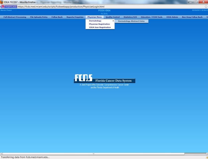
21 Abstracting and Data Transmission A Dermatology Data Entry Module has been created for dermatology practices providing a single site to register a practice, establish user account(s), and to enter cases with sufficient data to meet the FCDS reporting criteria. This module is specific to dermatology cases. Section II Abstracting Instructions It is the responsibility of the reporter/abstractor to know the content of the FCDS Data Acquisition Manual (DAM) for Dermatology Cancer Reporting. The DAM Includes explanation of each data item required for Florida Cancer Data System (FCDS) case reporting for Dermatology and Dermato-Pathology Practices Only. The DAM should be used as the primary information resource for any data item that must be coded and documented for dermatology reporting and in accordance with Florida cancer reporting rules and statutes. 21
22 Section II Abstracting Instructions Section III Register Physician/New User Includes all of the information we covered at the beginning of our webcast. Use Section III as reference when adding new physician, new practice, new user(s), etc. You must have the Physician Personal Identifier from the Florida Department of Health Letter to register a physician. Registration results in assignment of Facility/Practice ID and User Accounts associated with a Facility/Practice ID. 22

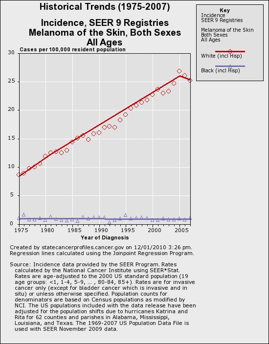
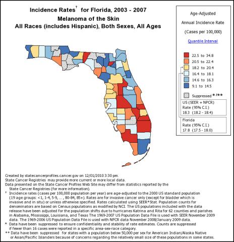
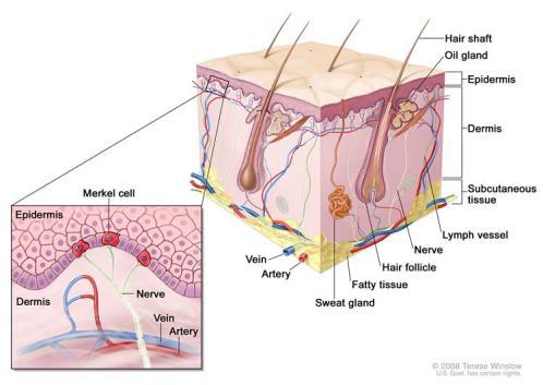
23 Introduction to Skin Cancers Source: 45 Anatomy Source:

24 Anatomy Source: 47 Skin Cancer


25 Skin Cancer Interactive Map/Rates Utility 49 Neoplasms of Skin Benign Atypical Malignant Metastatic Congenital Acquired Sun Exposure Viral Exposure Toxic Exposure Vitamin/Mineral Deficiencies 50 25

 Dermatofibrosarcoma Protuberans Source: http://www.dermis.")
26 Neoplasms of Skin Keratinocytic Tumors Basal Cell Carcinoma Squamous Cell Carcinoma Actinic Keratosis Verruca Acanthoma Melanocytic Tumors Malignant Melanoma Lentigo Maligna Congenital Melanocytic Nevus Bleu Nevi Simple Lentigo Dysplatstic nevus Spitz Nevus Source: 51 Neoplasms of Skin Neural Tumors Neuroma Merkel Cell Carcinoma PNET/Extraskeletal Ewing Sarcoma Appendageal Tumors Apocrine Tumors Eccrine Tumors Sebaceous Tumors Follicular Tumors Soft Tissue Tumors Fibroma Leimyosarcoma Vascular Tumors (hemangioma, Kaposi sarcoma) Dermatofibrosarcoma Protuberans Source:


27 Neoplasms of Skin Hematolymphoid Tumors Mastocytosis Parapsoriasis Sezary Syndrome Mycosis Fungoides Hodgkin Lymphoma Cutaneous T-cell Lymphoma Cutaneous B-cell Lymphoma Diffuse Large B-cell Lymphoma Langerhans Cell Histiocytosis CD30+ T-cell Lymphoproliferative Disorder Subcutaneous Panniculitis-like T-cell Lymphoma Source: Cutaneous Aggressive Epidermotropic CD8+ Cytotoxic T-cell Lymphoma Hydroa Vacciniforme-like Cutaneous T-cell Lymphoma 53 Signs and Symptoms Source: National Cancer Institute 54 27

28 Melanoma of the Skin Types of Melanoma Melanoma in situ Malignant melanoma, NOS Nodular melanoma Amelanotic melanoma Melanoma in nevus Lentigo maligna Lentigo maligna melanoma Superficial spreading melanoma Acral lentiginous melanoma Desmoplastic melanoma Epithelioid melanoma Spindle cell melanoma 56 28

29 Precancerous Terminology Pigmented nevi Atypical melanosis Melanocytic dysplasia Benign juvenile melanoma Dysplastic melanocytic nevi Atypical melanocytic hyperplasia Atypical melanocytic proliferation Intraepithelial melanocytic neoplasia Intraepithelial melanocytic proliferation Circumscribed precancerous melanosis Intraepithelial atypical melanocytic hyperplasia 57 How Melanoma Typically Grows Melanocyte Typical Atypical Hyperplastic Dysplastic Radial/Horizontal Growth Phase - The early pattern of growth of cutaneous malignant melanoma in which tumor cells spread laterally into the epidermis. During its horizontal phase of growth, a melanoma is normally flat. Vertical Growth Phase - The late pattern of growth of cutaneous malignant melanoma in which tumor cells spread from the epidermis into the dermis. As the vertical phase develops, the melanoma becomes thickened and raised
 Abnormal moles (atypical or dysplastic nevi) 59 Diagnostic and Staging Procedures Melafind (?")
30 Risk Factors Sun exposure, particularly during childhood Fair skin that burns easily Blistering sunburn, especially when young Previous melanoma Previous non-melanoma skin cancer Family history of melanoma Large numbers of moles (more than 100) Abnormal moles (atypical or dysplastic nevi) 59 Diagnostic and Staging Procedures Melafind (?) Shave Biopsy Punch Biopsy Excisional Biopsy Wide Local Excision Sentinel Node Biopsy 60 30


31 Prognostic Factors Source: 61 Clark s Level 62 Source: 31

32 Measured Thickness (Depth) 63 Merkel Cell Carcinoma of the Skin 32

33 Merkel Cell Carcinoma Image courtesy of Paul Nghiem, MD, PhD 65 Merkel Cell Carcinoma Source:

34 Incidence by Age M F Image courtesy of Paul Nghiem, MD, PhD 67 Diagnostic and Staging Procedures Shave Biopsy Punch Biopsy Excisional Biopsy Wide Local Excision Sentinel Node Biopsy Special Stains Confirm the Diagnosis 68 34


35 Prognostic Factors Location Depth of invasion Measured thickness Lymph node involvement Age and general health (particularly immune status) Initial diagnosis or recurrence 69 Mycosis Fungoides


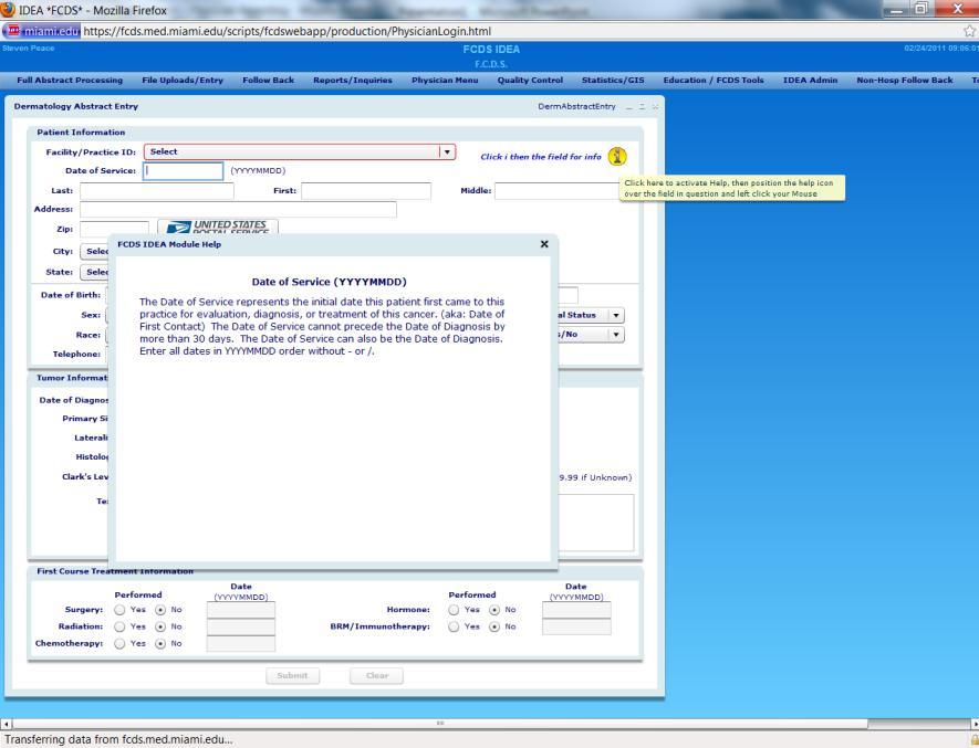
36 Kaposi Sarcoma Source: Source: 71 Entering Data The Cancer Abstract Live DEMO Use DAM and System Prompts For those of you unable to view the webcast (those participating only by phone) the next several slides will help orient you to the FCDS website, registration process, etc. The screenshots are also available in the DAM. 36
37 37
38 38
39 39
40 40
41 Q & A Questions? 41
Dermatology Cancer Reporting
 FLORIDA CANCER DATA SYSTEM Dermatology Cancer Reporting Data Acquisition Manual 5/17/2011 A joint project of the Sylvester Comprehensive Cancer Center and the Florida Department of Health. Table of Contents
Dermatology for the PCP Deanna G. Brown, MD, FAAD Susong Dermatology Consulting Staff at CHI Memorial
 Dermatology for the PCP Deanna G. Brown, MD, FAAD Susong Dermatology Consulting Staff at CHI Memorial Cutaneous Oncology for the PCP Deanna G. Brown, MD, FAAD Susong Dermatology Consulting Staff at CHI
IT S FUNDAMENTAL MY DEAR WATSON! A SHERLOCKIAN APPROACH TO DERMATOLOGY
 IT S FUNDAMENTAL MY DEAR WATSON! A SHERLOCKIAN APPROACH TO DERMATOLOGY Skin, Bones, and other Private Parts Symposium Dermatology Lectures by Debra Shelby, PhD, DNP, FNP-BC, FADNP, FAANP Debra Shelby,
David B. Troxel, MD. Common Medicolegal Situations: Misdiagnosis of Melanoma
 Common Medicolegal Situations: Misdiagnosis of Melanoma David B. Troxel, MD Medical Director, The Doctors Company, Napa, California Clinical Professor Emeritus, University of California at Berkeley Past
Springer Healthcare. Staging and Diagnosing Cutaneous Melanoma. Concise Reference. Dirk Schadendorf, Corinna Kochs, Elisabeth Livingstone
 Concise Reference Staging and Diagnosing Cutaneous Melanoma Dirk Schadendorf, Corinna Kochs, Elisabeth Livingstone Extracted from Handbook of Cutaneous Melanoma: A Guide to Diagnosis and Treatment Published
Identifying Skin Cancer. Mary S. Stone MD Professor of Dermatology and Pathology University of Iowa Carver College of Medicine March, 2018
 Identifying Skin Cancer Mary S. Stone MD Professor of Dermatology and Pathology University of Iowa Carver College of Medicine March, 2018 American Cancer Society web site Skin Cancer Melanoma Non-Melanoma
DERMATOLOGY ROTATION: COMPETENCY-BASED GOALS AND OBJECTIVES
 UNC DIVISION OF PLASTIC AND RECONSTRUCTIVE SURGERY DERMATOLOGY ROTATION: COMPETENCY-BASED GOALS AND OBJECTIVES MEDICAL KNOWLEDGE A. Anatomy/Physiology/Embryology Goal: The resident will have knowledge
Benign versus Cancerous Lesions How to tell the difference FMF 2014 Christie Freeman MD, CCFP, DipPDerm, MSc
 1 Benign versus Cancerous Lesions How to tell the difference FMF 2014 Christie Freeman MD, CCFP, DipPDerm, MSc Benign lesions Seborrheic Keratoses: Warty, stuck-on Genetics and birthdays Can start in late
Dermatopathology: The tumor is composed of keratinocytes which show atypia, increase mitoses and abnormal mitoses.
 Squamous cell carcinoma (SCC): A common malignant tumor of keratinocytes arising in the epidermis, usually from a precancerous condition: 1- UV induced actinic keratosis, usually of low grade malignancy.
SKIN CANCER. Most common cancer diagnosis 40% of all cancers
 SKIN CANCER Most common cancer diagnosis 40% of all cancers OBJECTIVES Review common and uncommon cancers of the skin. Special emphasis on melanoma and dysplastic nevus Review pathology/tnm/staging, which
Malignant Melanoma Early Stage. A guide for patients
 This melanoma patient brochure is designed to help educate melanoma patients and their caregivers. It was developed under the guidance of Dr. Michael Smylie, Professor, Department of Oncology, University
Pathology of the skin. 2nd Department of Pathology, Semmelweis University
 Pathology of the skin 2nd Department of Pathology, Semmelweis University Histology of the skin Epidermis: Stratum corneum Stratum granulosum Stratum spinosum Stratum basale Dermis: papillary and reticular
Dermatopathology. Dr. Rafael Botella Estrada. Hospital La Fe de Valencia
 Dermatopathology Dr. Rafael Botella Estrada. Hospital La Fe de Valencia Melanoma and mimics Dr. Martin Mihm Malignant lesions result from the accumulation of mutations Class I lesions (benign) Class II
Clinical characteristics
 Skin Cancer Fernando Vega, MD Seattle Healing Arts Clinical characteristics Precancerous lesions Common skin cancers ACTINIC KERATOSIS Precancerous skin lesions Actinic keratoses Dysplastic melanocytic
A PRACTICAL APPROACH TO ATYPICAL MELANOCYTIC LESIONS BIJAN HAGHIGHI M.D, DIRECTOR OF DERMATOPATHOLOGY, ST. JOSEPH HOSPITAL
 A PRACTICAL APPROACH TO ATYPICAL MELANOCYTIC LESIONS BIJAN HAGHIGHI M.D, DIRECTOR OF DERMATOPATHOLOGY, ST. JOSEPH HOSPITAL OBJECTIVES Discuss current trends and changing concepts in our understanding of
Cutaneous Malignancies: A Primer COPYRIGHT. Marissa Heller, M.D.
 Cutaneous Malignancies: A Primer Marissa Heller, M.D. Associate Director of Dermatologic Surgery Department of Dermatology Beth Israel Deaconess Medical Center December 10, 2016 Skin Cancer Non-melanoma
VACAVILLE DERMATOLOGY
 Connecting the Dots on those Spots NANDAN V. KAMATH, M.D. VACAVILLE DERMATOLOGY Sources All of the photos were taken with permission from the Dermnet NZ website - Dermnet New Zealand after communicating
An Overview of Melanoma. Harriet Kluger, M.D. Associate Professor Section of Medical Oncology Yale Cancer Center
 An Overview of Melanoma Harriet Kluger, M.D. Associate Professor Section of Medical Oncology Yale Cancer Center Melanoma Statistics Median age at presentation 45-55 55 years Incidence: 2003 54,200 cases
PATHOLOGY OF THE SKIN 2. Tumours of the skin
 PATHOLOGY OF THE SKIN 2. Tumours of the skin Máirín E. McMenamin MB MRCPI FRCPath Dip (Dermatopathol) RCPath St. James s Hospital and University of Dublin, Trinity College Tumour (Neoplasia) Benign or
Protocol applies to melanoma of cutaneous surfaces only.
 Melanoma of the Skin Protocol applies to melanoma of cutaneous surfaces only. Procedures Biopsy (No Accompanying Checklist) Excision Re-excision Protocol revision date: January 2005 Based on AJCC/UICC
VULVAR CARCINOMA. Page 1 of 5
 VULVAR CARCINOMA EXAMPLE OF A VULVAR CARCINOMA USING PROPOSED TEMPLATE Case: Invasive squamous cell carcinoma arising in D-VIN Tumor in left labia major Left partial vaginectomy and sentinel lymph node
Atlas of Eyelid and Conjunctival Tumors
 Atlas of Eyelid and Conjunctival Tumors Jerry A. Shields, M.D. Director, Ocular Oncology Service Wills Eye Hospital Professor of Ophthalmology Thomas Jefferson University Philadelphia, Pennsylvania Carol
Subject Index. Dry desquamation, see Skin reactions, radiotherapy
 Subject Index Actinic keratosis disseminated disease 42 surgical excision 42 AIDS, see Kaposi s sarcoma Amifostine, skin reaction prophylaxis 111 Basal cell carcinoma, superficial X-ray therapy Bowen s
NAACCR Webinar Series 1
 Collecting Cancer Data: Skin Malignancies 2/4/2010 NAACCR 2009 2010 Webinar Series Questions Please use the Q&A panel to submit your questions Send questions to All Panelist Collecting Cancer Data: Skin
Contrast with Australian Guidelines A/Pr Pascale Guitera,
 Contrast with Australian Guidelines A/Pr Pascale Guitera, Dermatologist, Sydney University NO CONFLICT OF INTEREST Sydney Melanoma Diagnostic Centre, RPAH 2011 2008 225 pages 16 pages http://www.cancer.org.au/file/healthprofessionals/clinica
Benign and malignant epithelial lesions: Seborrheic keratosis: A common benign pigmented epidermal tumor occur in middle-aged or older persons more
 Benign and malignant epithelial lesions: Seborrheic keratosis: A common benign pigmented epidermal tumor occur in middle-aged or older persons more common on the trunk; but extremities, head and neck are
We are IntechOpen, the world s leading publisher of Open Access books Built by scientists, for scientists. International authors and editors
 We are IntechOpen, the world s leading publisher of Open Access books Built by scientists, for scientists 3,500 108,000 1.7 M Open access books available International authors and editors Downloads Our
Malignant tumors of melanocytes : Part 3. Deba P Sarma, MD., Omaha
 Malignant tumors of melanocytes : Part 3 Deba P Sarma, MD., Omaha Let s go over one case of melanoma using the following worksheet. Of the various essential information that needs to be included in the
Periocular Malignancies
 Periocular Malignancies Andrew Gurwood, O.D., F.A.A.O., Dipl. Marc Myers, O.D., F.A.A.O. Drs. Myers and Gurwood have no financial interests to disclose. Course Description Discussion of the most common
NAACCR Hospital Registry Webinar Series
 NAACCR Hospital Registry Webinar Series October 4, 2007 Abstracting Melanoma Cancer Incidence and Treatment Data Image source: commons.wikimedia.org/wiki/image.melanoma.jpg Sites include Melanoma Skin
Malignant tumors of melanocytes: Part 1. Deba P Sarma, MD., Omaha
 Malignant tumors of melanocytes: Part 1 Deba P Sarma, MD., Omaha The melanocytic tumor is one of the most difficult and confusing areas in Dematopathology. It is true that most (95%) of such lesions are
Quality ID #263: Preoperative Diagnosis of Breast Cancer National Quality Strategy Domain: Effective Clinical Care
 Quality ID #263: Preoperative Diagnosis of Breast Cancer National Quality Strategy Domain: Effective Clinical Care 2018 OPTIONS FOR INDIVIDUAL MEASURES: REGISTRY ONLY MEASURE TYPE: Process DESCRIPTION:
Desmoplastic Melanoma R/O BCC. Clinical Information. 74 y.o. man with lesion on left side of neck r/o BCC
 R/O BCC Sabine Kohler, M.D. Professor of Pathology and Dermatology Dermatopathology Service Stanford University School of Medicine Clinical Information 74 y.o. man with lesion on left side of neck r/o
 Page 1 of 15 Title Authored By Course No Contact Hours 2 Skin Cancer the Real Picture for Early Detection and Treatment Cheryl Sommer RN, MSN, ARNP SC120604 Purpose The purpose of this course is to provide
APPENDIX I. Free-Standing Radiation Therapy Centers Cancer Case Identification Program
 APPENDIX I Free-Standing Radiation Therapy Centers Cancer Case Identification Program I-1 Sending Radiation Therapy data to FCDS Beginning January 1, 2003, all Florida Radiation Therapy Centers must send
Pathology. Skin Tumor. Bayan N. Mohammad 15/10/2015. Mohammad al-orjani. Page 0 of 23
 #7 35 Pathology Skin Tumor Bayan N. Mohammad 15/10/2015 Mohammad al-orjani Page 0 of 23 بسم هللا الرحمن الرحيم GREETINGS This lecture is about skin tumors, all the slides are included and every slide will
Format Of ICD-O Terms In Numerical List Each topographic and morphologic term appears only once The first listed term in Bold Type is the Preferred Te
 Florida Cancer Data System International Classification of Diseases for Oncology ICD-O-3 1 Basic Concepts Primary Site/Topography Histology/Morphology Behavior Grade/Immunophenotype 2 ICD-O 3 Structure/Format
Skin Cancer. 5 Warning Signs. American Osteopathic College of Occupational and Preventive Medicine OMED 2012, San Diego, Monday, October 8, 2012 C-1
 Skin Cancer AMERICAN OSTEOPATHIC COLLEGE OF OCCUPATIONAL & PREVENTIVE MEDICINE OMED 2012 October 8, 2012 E. Robert Wanat II, D.O., M.P.H. Learning Objectives: Identify the 3 Basic Types of Skin Cancer
Multiple Primary Melanoma in a Thai Male: A Case Report
 Case Report Multiple Primary Melanoma in a Thai Male: A Case Report J Med Assoc Thai 2014; 97 (Suppl. 2): S234-S238 Full text. e-journal: http://www.jmatonline.com Kittisak Payapvipapong MD*, Pinyapat
Simulators of melanoma
 Simulators of melanoma Philip E. LeBoit, M.D. Depts. of Pathology and Dermatology University of California, San Francisco Simulators of melanoma Simulators of melanoma in situ Melanocytic Non-melanocytic
WHAT DOES THE PATHOLOGY REPORT MEAN?
 Melanoma WHAT IS MELANOMA? Melanoma is a type of cancer that affects cells called melanocytes. These cells are found mainly in skin but also in the lining of other areas such as nose and rectum, and also
Principles of Anatomy and Physiology
 Principles of Anatomy and Physiology 14 th Edition CHAPTER 5 The Integumentary System Introduction The organs of the integumentary system include the skin and its accessory structures including hair, nails,
Atypical Nevi When to Re-excise. Catherine Barry, DO Dermatopathologist
 Atypical Nevi When to Re-excise Catherine Barry, DO Dermatopathologist Why talk about skin cancer? Because it s the most common type of cancer! Non-melanoma Skin Cancers Basal Cell Carcinoma Squamous Cell
What s New in 2012 FCDS Annual Meeting Review
 What s New in 2012 FCDS Annual Meeting Review FCDS Educational Webcast Series August 16, 2012 FCDS Staff Steven Peace, CTR 2012 FORDS Cancer Program Standards CSv02.04 1 What is Cancer / What is Reportable
Basal cell carcinoma 5/28/2011
 Goal of this Presentation A practical approach to the diagnosis of cutaneous carcinomas and their mimics Thaddeus Mully, MD University of California San Francisco To review common non-melanoma skin cancers
Toby Maurer, MD University of California, San Francisco. Lifetime risk of an American developing melanoma
 Distinguishing Pigmented Skin Lesions and Melanoma Toby Maurer, MD University of California, San Francisco Epidemiology of Melanoma Lifetime risk of an American developing melanoma 1935: 1 in 1500 1980:
المركب النموذج--- سبيتز وحمة = Type Spitz's Nevus, Compound SPITZ NEVUS 1 / 7
 SPITZ NEVUS 1 / 7 Epidemiology An annual incidence rate of 1.4 cases of Spitz nevus per 100,000 individuals has been estimated in Australia, compared with 25.4 per 100,000 individuals for cutaneous melanoma
Toby Maurer, MD University of California, San Francisco. Lifetime risk of an American developing melanoma
 Distinguishing Pigmented Skin Lesions and Melanoma Toby Maurer, MD University of California, San Francisco Epidemiology of Melanoma Lifetime risk of an American developing melanoma 1935: 1 in 1500 1980:
Primary Cutaneous Melanoma Pathology Reporting Proforma DD MM YYYY. *Tumour site. *Specimen laterality. *Specimen type
 Primary Cutaneous Melanoma Pathology Reporting Proforma Includes the International Collaboration on Cancer reporting dataset denoted by * Family name Given name(s) Date of birth DD MM YYYY Sex Male Female
Skin SSG (Anglia East & Anglia West)
") Skin SSG (Anglia East & Anglia West) Author: Dr Jennifer Garioch, Consultant Dermatologist Dr Pamela Todd, Consultant Dermatologist Approved by: Anglia Cancer Network Skin NSSG Approved on: Reviewed and
Skin SSG (Anglia East & Anglia West) Author: Dr Jennifer Garioch, Consultant Dermatologist Dr Pamela Todd, Consultant Dermatologist Approved by: Anglia Cancer Network Skin NSSG Approved on: Reviewed and
CASEFINDING. KCR Abstractor s Training
 CASEFINDING KCR Abstractor s Training 1 Introduction Casefinding Definition Purpose Methods Sources vs Resources Reportable Cancer Conditions Non-Reportable Conditions Ambiguous Terminology 2 https://www.google.com/search?q=puzzle+pieces&client=firefox-a&hs=mdc&rls=org.mozilla
Dr. Brent Doolan, BSc MBBS MPH
 Impact of partial biopsies on the need for complete excisional surgery in the management of cutaneous melanomas: A multi-centre review Dr. Brent Doolan, BSc MBBS MPH Peter MacCallum Cancer Centre, Melbourne
Dermatological Manifestations in the Elderly. Sanjay Siddha Staff Dermatologist UHN & MSH
 Dermatological Manifestations in the Elderly Sanjay Siddha Staff Dermatologist UHN & MSH Disclosure No actual or potential conflicts of interest or commercial relationships to declare Objectives Recognize
Cutaneous Adnexal Tumors
 Cutaneous Adnexal Tumors Lesions with Predominant Follicular Differentiation Special Emphasis on Basal Cell Carcinoma 2014-04-01 Prof. Dr. med. Katharina Glatz Pathologie Cutaneous Adnexal Tumors Hair
NAACCR Webinar Series 1
 Collecting Cancer Data: Melanoma 2013 2014 NAACCR Webinar Series April 3, 2014 Q&A Please submit all questions concerning webinar content through the Q&A panel. Reminder: If you have participants watching
NPQR Quality Payment Program (QPP) Measures 21_18247_LS.
 Measures 21_18247_LS.") NPQR Quality Payment Program (QPP) Measures 21_18247_LS MEASURE ID: QPP 99 MEASURE TITLE: Breast Cancer Resection Pathology Reporting pt Category (Primary Tumor) and pn Category (Regional Lymph Nodes)
NPQR Quality Payment Program (QPP) Measures 21_18247_LS MEASURE ID: QPP 99 MEASURE TITLE: Breast Cancer Resection Pathology Reporting pt Category (Primary Tumor) and pn Category (Regional Lymph Nodes)
Epithelial Cancer- NMSC & Melanoma
 Epithelial Cancer- NMSC & Melanoma David Chin MB, BCh, BAO, LRCP, LRCS (Ireland) MCh(MD), PhD (UQ), FRCS, FRACS (Plast) Plastic & Reconstructive Surgeon Visiting Scientist Melanoma Genomic Group & Drug
Comprehensive Cancer Cover
 Comprehensive Cancer Cover Tech Spec Comprehensive Cancer Cover provides the life insured with cover for the diagnosis and treatment of defined malignant tumours. These tumours must be characterised either
Epidemiology. Objectives 8/28/2017
 Case based Discussion of Head and Neck Melanoma: Review of Epidemiology, Risk Factors, Identification, Treatments and Prevention Jacqueline M. Doucette MS FNP-C Objectives Define and identify melanoma
4 Skin and Body Membranes Study Guide
 Name: SKIN AND BODY MEMBRANES: 4 Skin and Body Membranes Study Guide Period: Body membranes, which cover body surfaces, line its cavities, and form protective sheets around organs, fall into two major
LUMPS AND BUMPS: AN ORGANIZED APPROACH TO DIAGNOSIS AND MANAGEMENT
 LUMPS AND BUMPS: AN ORGANIZED APPROACH TO DIAGNOSIS AND MANAGEMENT Tammy P. Than, M.S., O.D., F.A.A.O. The University of Alabama at Birmingham / School of Optometry 1716 University Blvd. Birmingham, AL
Genetic Testing: When should it be ordered? Julie Schloemer, MD Dermatology
 Genetic Testing: When should it be ordered? Julie Schloemer, MD Dermatology Outline Germline testing CDKN2A BRCA2 BAP1 Somatic testing Gene expression profiling (GEP) BRAF Germline vs Somatic testing
Histopathology: skin pathology
 Histopathology: skin pathology These presentations are to help you identify, and to test yourself on identifying, basic histopathological features. They do not contain the additional factual information
Histopathology of Melanoma
 THE YALE JOURNAL OF BIOLOGY AND MEDICINE 48, 409-416 (1975) Histopathology of Melanoma G. J. WALKER SMITH Department ofpathology, Yale University School ofmedicine, 333 Cedar Street, New Haven, Connecticut
Gross Appearance & Histology of Skin Cancer. Kyle Mannion M.D. January 21, 2005
 Gross Appearance & Histology of Skin Cancer Kyle Mannion M.D. January 21, 2005 Actinic Keratosis 5-20% will develop squamous/basal cell ca Almost solely from solar damage Usually develop during 4 th decade
Associate Clinical Professor of Dermatology MUSC
 Re-excision of Moderately Dysplastic Nevi: Should we or shouldn t we? John C. Maize, Jr, M.D. Dermatologist and Dermatopathologist Trident Dermatology, Charleston SC Associate Clinical Professor of Dermatology
Skin Cancer of the Nose: Common and Uncommon
 Skin Cancer of the Nose: Common and Uncommon Mark Russell, M.D. Associate Professor of Dermatology, Otolaryngology, and Pathology University of Virginia Objectives Review clinical presentations of select
Outline. How to Use the AJCC Cancer Staging Manual, 7 th ed. 7/9/2015 FCDS ANNUAL CONFERENCE ST PETERSBURG, FLORIDA JULY 30, 2015.
 1 How to Use the AJCC Cancer Staging Manual, 7 th ed. FCDS ANNUAL CONFERENCE ST PETERSBURG, FLORIDA JULY 30, 2015 Steven Peace, CTR Outline 2 History, Purpose and Background Purchase and Ordering Information
Not For Sale. Alternative Billing Codes (ABC Codes) Clinical Care Classification (CCC) System
 Clinical Care Classification (CCC) System") Alternate Health Care Coding Systems In addition to the ICD-9-CM, ICD-10-CM, ICD-10-PCS, HCPCS level II national, and CPT coding systems, health care providers routinely classify diagnoses and procedures
Alternate Health Care Coding Systems In addition to the ICD-9-CM, ICD-10-CM, ICD-10-PCS, HCPCS level II national, and CPT coding systems, health care providers routinely classify diagnoses and procedures
Chapter 3. Neoplasms. Copyright 2015 Cengage Learning.
 Chapter 3 Neoplasms Terminology Related to Neoplasms and Tumors Neoplasm New growth Tumor Swelling or neoplasm Leukemia Malignant disease of bone marrow Hematoma Bruise or contusion Classification of Neoplasms
Setting the stage for change: upgrading the physician cancer case reporting application in New York
 Setting the stage for change: upgrading the physician cancer case reporting application in New York April Austin New York State Cancer Registry (NYSCR) July 12, 2018 June 13, 2018 Aerial view of Thousand
Melanoma. Kaushik Mukherjee MD A. Scott Pearson MD
 Melanoma Kaushik Mukherjee MD A. Scott Pearson MD Disclosures You still have to study Not all inclusive No Western blots Extensive use of Google Image Search and Sabiston Melanoma Basics 8 th most common
Lid Lesions: Relax or Refer
 Lid Lesions: Relax or Refer Blair Lonsberry, MS, OD, MEd., FAAO Professor of Optometry Pacific University College of Optometry blonsberry@pacificu.edu Agenda Benign vs. Malignant lesions Benign Eyelid
Common Benign Lesions and Skin Cancers. 22nd May 2015 Dr Mark Foley
 Common Benign Lesions and Skin Cancers 22nd May 2015 Dr Mark Foley Thank you for downloading this file. This intended to supplement the presentation given at the NZ Wound Care Conference, it is not intended
Case Scenario 1 Worksheet. Primary Site C44.4 Morphology 8743/3 Laterality 0 Stage/ Prognostic Factors
 CASE SCENARIO 1 9/10/13 HISTORY: Patient is a 67-year-old white male and presents with lesion located 4-5cm above his right ear. The lesion has been present for years. No lymphadenopathy. 9/10/13 anterior
Melanoma Underwriting Presented at 2018 AHOU Conference. Hank George FALU
 Melanoma Underwriting Presented at 2018 AHOU Conference Hank George FALU MELANOMA EPIDEMIOLOGY 70-80,000 American cases annually Majority are in situ or thin > 20% are diagnosed age 45 8-9,000 melanoma
OSCaR UPDATE. Oregon State Cancer Registry
 Summer 2007 OSCaR UPDATE Oregon State Cancer Registry Volume 8, Quarter 3 Manager s Update Donald Shipley, MS As a very busy summer draws to an end and we prepare for an equally busy autumn, I d like to
Cutaneous Melanoma: Epidemiology (USA) The Sentinel Node in Head and Neck Melanoma. Cutaneous Melanoma: Epidemiology (USA)
 The Sentinel Node in Head and Neck Melanoma. Cutaneous Melanoma: Epidemiology (USA)") The Sentinel Node in Head and Neck Melanoma Cutaneous Melanoma: Epidemiology (USA) 6 th leading cause of cancer among men and women 68,720 new cases of invasive melanoma in 2009 8,650 deaths from melanoma
The Sentinel Node in Head and Neck Melanoma Cutaneous Melanoma: Epidemiology (USA) 6 th leading cause of cancer among men and women 68,720 new cases of invasive melanoma in 2009 8,650 deaths from melanoma
أملس عضلي غرن = Leiomyosarcoma. Leiomyosarcoma 1 / 5
 Leiomyosarcoma 1 / 5 EPIDEMIOLOGY Exact incidence is unknown, but older studies suggest that leiomyosarcomas comprise approximately 3 percent of soft-tissue sarcomas. Superficial leiomyosarcoma occurs
Dermoscopy: Recognizing Top Five Common In- Office Diagnoses
 Dermoscopy: Recognizing Top Five Common In- Office Diagnoses Vu A. Ngo, DO Department of Family Medicine and Dermatology Choctaw Nation Health Services Authority Learning Objectives Introduction to dermoscopy
Maligna Melanoma and Atypical Fibroxanthoma: An Unusual Collision Tumour G Türkcü 1, A Keleş 1, U Alabalık 1, D Uçmak 2, H Büyükbayram 1 ABSTRACT
 Maligna Melanoma and Atypical Fibroxanthoma: An Unusual Collision Tumour G Türkcü 1, A Keleş 1, U Alabalık 1, D Uçmak 2, H Büyükbayram 1 ABSTRACT Two different neoplasia in the same biopsy material called
Incidence and Trends of Cutaneous Malignancies in the Netherlands,
 ORIGINAL ARTICLE Incidence and Trends of Cutaneous Malignancies in the Netherlands, 1989 2005 Cynthia Holterhues 1, Esther de Vries 1,2, Marieke W. Louwman 3, Senada Koljenović 4 and Tamar Nijsten 1 Epidemiology
Benign vs. Cancer. Oculofacial Biopsy. Evolution of skin cancer. Richard E. Castillo, OD, DO
 Oculofacial Biopsy Richard E. Castillo, OD, DO Benign vs. Cancer Evolution of skin cancer Metaplasia Dysplasia Carcinoma-in-situ Invasive carcinoma Intravasation Overview Preoperative Planning Choosing
Melanoma and Dermoscopy. Disclosure Statement: ABCDE's of melanoma. Co-President, Usatine Media
 Melanoma and Dermoscopy Richard P. Usatine, MD, FAAFP Professor, Family and Community Medicine Professor, Dermatology and Cutaneous Surgery Medical Director, University Skin Clinic University of Texas
BAP-oma & BEYOND MICHAEL A NOWAK, MD
 BAP-oma & BEYOND MICHAEL A NOWAK, MD CONFLICTS No conflicts with the content of this lecture BAP-oma Wiesner 2011: Families with multiple tan dome-shaped papules of head, neck, trunk, and extremities.
Diploma Examination. Dermatopathology: First paper. Tuesday 20 March Candidates must answer FOUR questions. Time allowed: 3 hours
 Dermatopathology: First paper Tuesday 20 March 2018 Candidates must answer FOUR questions Time allowed: 3 hours 1. Give an account of the genetic aberrations encountered in Spitzoid neoplasms and how these
Learning Objectives. Tanning. The Skin. Classic Features. Sun Reactive Skin Type Classification. Skin Cancers: Preventing, Screening and Treating
 Learning Objectives Skin Cancers: Preventing, Screening and Treating Robert A. Baldor, MD, FAAFP Professor, Family Medicine & Community Health University of Massachusetts Medical School Distinguish the
Directly Coded Summary Stage Melanoma
 Directly Coded Summary Stage Melanoma National Center for Chronic Disease Prevention and Health Promotion Division of Cancer Prevention and Control, National Program of Cancer Registries Directly Coded
Appendageal skin tumors
 Appendageal skin tumors Ibrahim Khalifeh, M.D. Associate Professor Department of Pathology American University of Beirut Medical Center Beirut, Lebanon Appendageal tumors Neoplasms whose differentiation
CODING TUMOUR MORPHOLOGY. Otto Visser
 CODING TUMOUR MORPHOLOGY Otto Visser INTRODUCTION The morphology describes the tissue of the tumour closest to normal tissue Well differentiated tumours are closest to normal Undifferentiated tumours show
Department of Dermatology, Queen Margaret & Victoria Hospitals
 Department of Dermatology, Queen Margaret & Victoria Hospitals Management of primary skin cancer A copy of these local guidelines, national guidelines, information leaflets and other useful information
Overview FLORIDA CANCER DATA SYSTEM
 Florida Cancer Data System 1 Florida Cancer Data System 2 FCDS is the legislatively mandated, population-based central cancer registry for the state of Florida. Data in the FCDS are collected from patient
Technicians & Nurses Program
 ASCRS ASOA Symposium & Congress Technicians & Nurses Program May 6-10, 2016 New Orleans Evaluation and Treatment of Eyelid Malignancies Richard C. Allen MD PhD FACS Professor Section of Ophthalmology Dept.
Glenn D. Goldman, MD. University of Vermont Medical Center. University of Vermont College of Medicine
 Glenn D. Goldman, MD University of Vermont Medical Center University of Vermont College of Medicine Recognize and identify the main types of skin cancer and their precursors Identify and understand new
2018 Updates to National Standards
 2018 Updates to National Standards 1 F C D S A N N U A L C O N F E R E N C E O R L A N D O, F L O R I D A 7 / 2 6 / 2 0 1 7 S T E V E N P E A C E, C T R ICD-O-3 New Histology Codes ICD-O-3 Behavior Changes
1. Opdivo + Ipilumimab is now the first line therapy for metastatic melanoma.
 Melanoma UpToDate: Introduction: Risk Factors: 1. Opdivo + Ipilumimab is now the first line therapy for metastatic melanoma. Median age = 50 yrs Incidence is rising - Sun exposure: UVB (290-320nm) > UVA
Doctors of Optometry Course Notes
 Doctors of Optometry Course Notes OD19 1CE COPE: 43871-AS Eyelid Lumps and Bumps Sunday, February 26, 2017 2:40 pm 3:30 pm Regency C 3 rd Floor Presenter: Blair Lonsberry, OD, FAAO Dr. Lonsberry is a Full
Melanoma: The Basics. What is a melanocyte?
 Melanoma: The Basics What is a melanocyte? A melanocyte is a normal cell, found in the skin, which produces melanin. Melanin is a black or dark brown pigment that is seen in the skin, hair, and parts of