points causes features therapy collaborative ocular melanoma study prognosis Choroid Nevus
|
|
|
- Alexia Holland
- 5 years ago
- Views:
Transcription


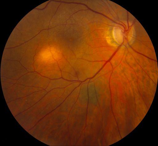
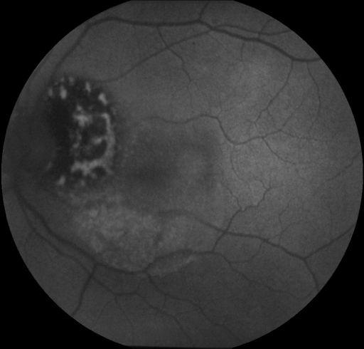
1 The Game of Uveal Melanoma Carol Shields MD Oncology Service Wills Eye Hospital Philadelphia PA USA Oncology Service Wills Eye Institute points causes features therapy collaborative ocular melanoma study prognosis Causes of uveal melanoma host factors nevus melanocytosis light eye color fair skin color inability to tan environmental factors arc welding chronic sunlight exposure few reports white population 7% features pigmented 90% thickness <2mm drusen RPE atrophy questions answers do the features change over time can nevus affect vision can nevus enlarge rate of transformation into melanoma 1/8000 Clinical Spectrum of Choroidal Nevi Based on Age at Presentation in 3422 Consecutive Eyes Carol L. Shields, MD, Minoru Furuta, MD, Arman Mashayekhi, MD, Edwina L. Berman, MBBS, Jonathan D. Zahler, DO, Daniel M. Hoberman, BS, Diep H. Dinh, BS, Jerry A. Shields, MD Purpose: To evaluate the clinical features of choroidal nevi based on patient age at presentation and to investigate features of the nevi that are predictive of patient symptoms. Design: Observational case series. Participants: Three thousand four hundred twenty-two consecutive eyes of 3187 patients. Methods: Retrospective clinic-based study of clinical features at referral. Cox proportional hazards regressions were used for evaluation of factors predictive of patient symptoms. Main Outcome Measures: Nevus features as related to patient age group at diagnosis (young [ 20 years], mid-adult [21 50 years], older adult [ 50 years]) and factors predictive of patient symptoms secondary to the nevus. Results: Of the 3422 eyes with choroidal nevus, 63 (2%) were in young patients, 795 (23%) in mid-adults, and 2564 (75%) in older adults. The following factors showed no substantial increase or decrease by age category (young, mid-adult, older adult) at presentation: symptoms (14%, 12%, 13%), mean nevus base (5.6, 4.7, 5.2 mm), intrinsic nevus pigmentation (89%, 74%, 77%), related subretinal fluid (SRF) (11%, 15%, 9%), overlying orange pigment (6%, 10%, 6%), retinal pigment epithelial hyperplasia (0%, 9%, 7%), and retinal pigment epithelial atrophy (2%, 13%, 10%). The following factors statistically increased with age category: multiple nevi per eye (2%, 8%, 10%) (P ), mean nevus thickness (1.2, 1.5, 1.6 mm) (P ), and overlying drusen (11%, 40%, 58%) (P ). Using multivariate analysis of the entire group, factors predictive of any symptom included nonpigmented nevus (P 0.001), location 3 mm to foveola (P 0.001), subfoveolar fluid (P 0.002), any SRF (P 0.02), and subfoveolar nevus (P 0.027). Conclusions: Choroidal nevi show similar clinical features regardless of age of presentation, with the exception of increasing number of nevi per eye, slightly increasing thickness, and increasing drusen in adults versus younger patients. Symptomatic nevi are more likely to be nonpigmented, beneath the foveola, and with subfoveolar fluid. Ophthalmology 2008;115: by the American Academy of Ophthalmology. groups young 0-20 yrs mid adult yrs older adult >50 yrs thick >1per eye drusen 1.2 2% 11% 1.5 8% 40% % 58% Young Mid Adult p<0.0001


 lines at 5, 10, and 15 years occurred in less than 1%, 1%, and 2% of eyes with")
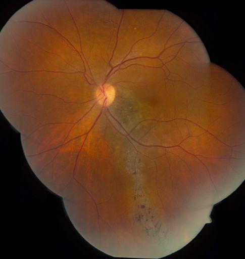
2 groups Older Adult subfoveal extrafoveal initial final 20/20 20/30 20/20 20/25 vision loss @20 yrs 15% 20% 26% <1% 1% 2% questions answers do the features change over time can nevus affect vision can nevus enlarge rate of transformation into melanoma 1/8000 questions answers do the features change over time can nevus affect vision can nevus enlarge rate of transformation into melanoma 1/8000 CLINICAL SCIENCES Visual Acuity in 3422 Consecutive Eyes With Choroidal Nevus Carol L. Shields, MD; Minoru Furuta, MD; Arman Mashayekhi, MD; Edwina L. Berman, MBBS; Jonathan D. Zahler, DO; Daniel M. Hoberman, BS; Diep H. Dinh, BS; Jerry A. Shields, MD Objective: To evaluate visual acuity in eyes with choroidal nevus. Design: This was an observational case series. Of 3422 consecutive eyes with choroidal nevus, vision loss at 15 years occurred in 2% of eyes with extrafoveolar nevus and in 26% of eyes with subfoveolar nevus, particularly those with overlying retinal pigment epithelial detachment and foveal edema. A retrospective medical record review was conducted, with evaluation of visual acuity at presentation and at final examination. The main outcome measure was visual acuity. Results: The median visual acuity at presentation was 20/20 for eyes with either extrafoveolar or subfoveolar choroidal nevus. Using Kaplan-Meier estimates, vision loss of 3 or more logarithm of the minimum angle of resolution (logmar) lines at 5, 10, and 15 years occurred in less than 1%, 1%, and 2% of eyes with extrafoveolar nevus compared with 15%, 20%, and C26% of eyes with subfoveolar choroidal nevus, respectively. By multivariate analysis, factors predictive of visual loss of 3 or more log- MAR lines included subfoveolar nevus location (relative risk [RR], 15.52), juxtapapillary nevus location (RR, 4.52), initial visual acuity of 20/50 or worse (RR, 15.40), overlying retinal pigment epithelial detachment (RR, 22.16), and foveal edema (RR, 9.02). Factors predictive of poor final visual acuity of 20/200 or worse included subfoveolar nevus location (RR, 11.32), overlying orange pigment (RR, 3.68), overlying retinal pigment epithelial detachment (RR, 12.80), and foveal edema (RR, 18.72). Conclusion: Mild vision loss over many years should be anticipated in patients with subfoveolar choroidal nevus, particularly those with overlying retinal pigment epithelial detachment, orange pigment, and foveal edema. Arch Ophthalmol. 2007;125(11): why does subfoveal nevus have vision loss? growth is a presumed indicator of malignant potential Slow Enlargement of Choroidal Nevi: A Long-Term Follow-Up Study Enlargement growth is not an unequivocal indicator of malignancy Arman Mashayekhi, MD, Sophia Siu, BS, Carol L. Shields, MD, Jerry A. Shields, MD Purpose: Choroidal nevi are generally considered to be stable lesions, and growth of a choroidal nevus is usually believed to be a sign of malignant transformation. We performed this study to determine whether choroidal nevi enlarge over a long period of follow-up without undergoing malignant transformation. Design: Retrospective observational case series. Participants: A total of 278 patients with 284 nevi who had at least 7 years of photographic follow-up without clinical signs of transformation into melanoma were included in the study. Methods: Data on demographic and clinical information were extracted from patients charts. Detailed fundus drawings and color fundus photographs were reviewed and compared for evidence of enlargement. Main Outcome Measures: Nevus enlargement without clinical evidence of transformation into melanoma. Results: Of the 278 patients, 69% were female and more than 99% were White with a median age at presentation of 57 years (range, 4 87 years). The largest nevus basal diameter was a median of 5 mm (range, mm), and the median thickness was 1.5 mm (range, mm). Only 14 nevi (5%) had subretinal fluid outside the nevus, and 6% showed overlying orange pigment. Overlying retinal pigment epithelial alterations included drusen (61%), atrophy (6%), hyperplasia (10%), and fibrous metaplasia (6%). Of 284 nevi, 31% showed slight enlargement over a mean follow-up of 15 years. The median increase in diameter was 1 mm (mean, 0.9 mm; range, mm), and the median rate of enlargement was 0.06 mm/yr (mean, 0.06 mm/yr; range, mm/yr). None of the lesions that enlarged developed new risk factors that are generally associated with malignant transformation. Frequency of enlargement was 54% in patients aged less than 40 years and 19% in patients aged more than 60 years. On multivariate analysis, younger patient age was the only factor predictive of nevus enlargement (P 0.001). Conclusions: With long-term follow up, 31% of choroidal nevi showed slight enlargement without clinical evidence of transformation into melanoma. The frequency of enlargement was inversely related to patient age. Financial Disclosure(s): The author(s) have no proprietary or commercial interest in any materials discussed in this article. Ophthalmology 2011;118: by the American Academy of Ophthalmology. 16 yowm enlargement over 10 years


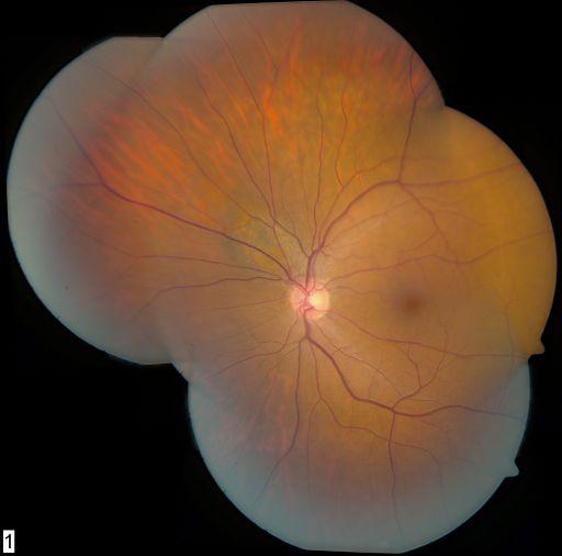
3 Enlargement Enlargement Melanoma questions answers do the features change over time can nevus affect vision can nevus enlarge rate of transformation into melanoma 1/8000 enlargement over 10 years 3 mm growth in 2 years Nevus Growth to Melanoma Nevus Growth to Melanoma Mathematical estimates Ganley 1/4500 Singh et al 1/8845 annual rate cumulative rate Kivela et al 1/ if we live to 80 yrs Population based study - Blue Mtn Eye Study Clinic based study - Shields et al Nevus Growth to Melanoma Risk factors low risk high risk Nevus Growth to Melanoma most important paper from our practice published 1995 published 2009 Nevus Growth to Melanoma Th > 2 mm Fluid Symptoms Orange pigment Margin at disc To Find Small Ocular Melanoma Using Helpful Hints Daily US Hollow Halo absent Drusen absent Nevus Growth to Melanoma Th > 2 mm Fluid - Each factor adds relative risk of ~ x greater risk Symptoms Orange pigment Margin at disc US Hollow Halo absent Drusen absent


4 Tests OCT EDI OCT Autofluorescence pigmented lesion no transmission poor posterior border compression of choroidal vessels Ultrasound Fluorescein Angiography Nevus vs Melanoma Nevus vs Melanoma thicker subretinal fluid shaggy photoreceptors p=0.001 important difference is shaggy photoreceptors edi oct of choroidal nevus versus melanomaimportant difference is shaggy photoreceptors - elongated shaggy photoreceptors Autofluorescence Autofluorescence nevus nevus melanoma melanoma Causes of uveal melanoma host factors nevus melanocytosis light eye color fair skin color inability to tan environmental factors arc welding chronic sunlight exposure


5 Melanocytosis! 1/400 risk Causes for uveal melanoma Melanocytosis! 1/400 risk Causes for uveal melanoma at risk for melanoma at risk for melanoma at risk for melanoma points causes features therapy collaborative ocular melanoma study prognosis color melanotic 85% amelanotic 15% size base 11 mm thickness 5.5 mm


6 Quadrant uveal melanoma in 8033 eyes Nasal 21% Superior 22% Macula 4% Temporal 28%! 2.6 mm!! 4.3 mm!! 7.0 mm points causes features therapy collaborative ocular melanoma study prognosis Diffuse 3% Inferior 22% Treatment Uveal Melanoma Observation Laser Thermotherapy Plaque radiotherapy Charged particle radiotherapy Resection Enucleation Exenteration Systemic therapies Treatment Uveal Melanoma Depends Tumor size Tumor location Status of opposite eye Patient age Patient health Patient desires and fears Basically Small Medium Large Treatment Uveal Melanoma 0-3mm 3-8mm >8mm TTT Radiotherapy Radiotherapy Resection Enucleation Radiotherapy Enucleation Plaque Radiotherapy Melanoma Plaque Radiotherapy Melanoma Radiation parameters Apex 8000 cgy Base cgy custom curvilinear standard custom notch custom round


7 Radiation Team Radiation Lead Container Radiation oncologist Radiation physicist Plaque transportation Prevent exposure Ocular Oncologists All personnel Radiation Device Surgical placement of radiation plaque Every person in the room wears radiation badge Exposure measurement Dummy plaque Radioactive plaque Radioactive Plaque Radioactive Plaque Radioactive Plaque Stored on hospital tray Betadiene Sterile metal container face down Rinsed Rinsed Seed count Sutures already in sclera Tie down Precisely localize intraocular tumor with transillumination or scleral depression Rotate eye with muscle sutures Long sutures to find plaque for removal


8 Radioactive Plaque Conjunctival closure with 7-0 vicryl Maxitrol and Atropine ointments Tarsorrhaphy suture Patch Plaque Radiotherapy Choroid Melanoma Plaque Radiotherapy Iris Melanoma Plaque Radiotherapy Follow up Eye Maxitrol tid 3-6 wks Atropine qhs 3-6 wks Recheck and measure 4 months Eye exam q 4 months then q 6 months for life Systemic Twice yearly Physical examination (liver and lung) Liver function tests Once yearly Chest xray MRI abdomen Plaque Small Melanoma Plaque Small Melanoma Plaque TTT small melanoma before!! after 3.0 mm!! 1.3 mm Plaque Medium Melanoma thickness 5 mm!!!!! 2 mm Plaque Large Melanoma thickness 9 mm!!!!! 2 mm Plaque Large Melanoma thickness 10 mm!!!!! 2 mm Oncology Service Wills Eye Institute


.")

 and median thickness was 3.5 mm (range, 0.5 14.8).")
9 Juxtapapillary choroidal melanoma Magnitude of this problem of >10,000 uveal melanoma treated over 30 years n=650 juxtapapillary melanoma 7% of all uveal melanoma Juxtapapillary choroidal melanoma Requirements include knowledge and precision in Tumor measurement Plaque design Plaque placement 8 x 6 x 2.9 mm require 12 mm radiation field situate on 15 mm notched plaque post distribution precision in placement Juxtapapillary choroidal melanoma Plaque radiotherapy Juxtapapillary n=650 Epipapillary! Circumpapillary Plaque Radiotherapy for Juxtapapillary Choroidal Melanoma Tumor Control in 650 Consecutive Cases Mandeep S. Sagoo, MB, PhD, 1 Carol L. Shields, MD, 1 Arman Mashayekhi, MD, 1 Jorge Freire, MD, 2 Jacqueline Emrich, PhD, 2 Jay Reiff, PhD, 2 Lydia Komarnicky, MD, 2 Jerry A. Shields, MD 1 Purpose: To evaluate treatment of juxtapapillary choroidal melanoma with plaque radiotherapy and to investigate the role of supplemental transpupillary thermotherapy (TTT). Design: Retrospective, comparative case series. Participants: We included 650 consecutive eyes with juxtapapillary choroidal melanoma within 1 mm of the optic disc. Methods: Eyes with juxtapapillary choroidal melanoma receiving plaque radiotherapy over a 31-year period from October 1974 to November 2005 were included in the study. The TTT and no TTT groups were analyzed separately and compared. Main Outcome Measures: Local tumor control, metastasis, and tumor-related mortality. Results: The median basal tumor diameter was 10 mm (range, ) and median thickness was 3.5 mm (range, ). In 481 eyes (74%), the tumor was directly adjacent to the optic disc and in 169 eyes (26%) the posterior tumor margin was between 0.1 and 1.0 mm from the optic disc. The circumpapillary extent of the tumor was 4 clock-hours in 321 eyes (50%), 4 8 clock-hours in 250 eyes (38%), and 8 clock-hours in 79 eyes (12%). Plaque radiotherapy using iodine-125 in 616 eyes (95%), cobalt-60 in 19 eyes (3%), iridium-192 in 12 eyes (2%), and ruthenium-106 in 3 eyes ( 1%) delivered a median radiation dose of 8000 cgy (range, ) to the tumor apex and adjunctive TTT was used in 307 eyes (56%). Kaplan-Meier estimates for tumor recurrence, metastasis, and death were 14%, 11%, and 4% at 5 years and 21%, 24%, and 9% at 10 years, respectively. Eyes treated with additional TTT showed slight (statistically nonsignificant) reduction in recurrence and metastasis. Using multivariable analysis, factors predictive of tumor recurrence included foveolar tumor requiring TTT (hazard ratio, 5.07; P 0.001) and greater tumor thickness (hazard ratio, 1.29 per mm increase; P 0.001). Factors predictive of metastasis included greater tumor base (hazard ratio, 1.21 per mm increase; P 0.001) and increasing intraocular pressure (hazard ratio, 1.11 per mmhg increase; P 0.020). Conclusions: Plaque radiotherapy for juxtapapillary melanoma provides local tumor control in approximately 80% of eyes at 10 years. In subjects who received TTT, there was slight but nonsignificant improved local tumor control and lower metastatic rate. Financial Disclosure(s): The authors have no proprietary or commercial interest in any of the materials discussed in this article. Ophthalmology 2011;118: by the American Academy of Ophthalmology. points causes features therapy collaborative ocular melanoma study prognosis Collaborative Ocular Melanoma Study Collaborative Ocular Melanoma Study what should you remember? 1. medium melanoma 3-8 mm thickness plaque provides same prognosis as enucleation 2. large melanoma >8 mm thickness no need to irradiate eye before enucleation neither proved that therapy prevents metastasis early detection may be the best way to minimize metastasis points causes features therapy collaborative ocular melanoma study prognosis Prognosis of Uveal Melanoma melanoma is a deadly eye cancer


10 (REPRINTED) ARCH OPHTHALMOL / VOL 127 (NO. 8), AUG Prognosis of Uveal Melanoma old literature several reports on prognosis factors related to prognosis clinical histopathology cytology genetic Prognosis of Uveal Melanoma 2003 Kujala, Makitie, Kivela Metastasis n=289 5 yrs yrs yrs yrs 52% Prognosis of Uveal Melanoma 2003 Kujala, Makitie, Kivela If death from melanoma 62% by 5 yrs 90% by 10 yrs 98% by 25 yrs 100% by 35 yrs if metastasis, most picked up by 15 years following eye diagnosis Prognosis of Uveal Melanoma 2009 Shields CL, Furuta, Thangappan, et al. n=8033 Metastasis Practical millimeter by millimeter basis CLINICAL SCIENCES Metastasis of Uveal Melanoma Millimeter-by-Millimeter in 8033 Consecutive Eyes Carol L. Shields, MD; Minoru Furuta, MD; Archana Thangappan, MD; Saya Nagori, MD; Arman Mashayekhi, MD; David R. Lally, MD; Cecilia C. Kelly, MD; Danielle S. Rudich, MD; Anand V. Nagori, MD; Oojwala A. Wakade, MD; Sonul Mehta, MD; Lauren Forte, BS; Andrew Long, BS; Elaina F. Dellacava, MD; Bonnie Kaplan, MD; Jerry A. Shields, MD Objective: To determine the rate of metastasis of uveal melanoma on the basis of tumor thickness in millimeters. Methods: Retrospective medical record review. Results: The mean (median) patient age was 58 (59) years. A total of 8033 eyes were examined. Of the 285 eyes with iris melanoma, the mean tumor thickness was 2.7 mm and metastasis occurred in 0.5%, 4%, and 7% at 3, 5, and 10 years, respectively. Of the 492 eyes with ciliary body melanoma, the mean tumor thickness was 6.6 mm and metastasis occurred in 12%, 19%, and 33% at 3, 5, and 10 years, respectively. Of the 7256 eyes with choroidal melanoma, the mean tumor thickness was 5.5 mm and metastasis occurred in 8%, 15%, and 25% at 3, 5, and 10 years, respectively. For all uveal melanoma, metastasis at 5, 10, and 20 years was 6%, 12%, and 20% for small melanoma (0-3.0 mm thickness), 14%, 26%, and Author Affiliations: Ocular Oncology Service, Wills Eye Institute, Thomas Jefferson University, Philadelphia, Pennsylvania. 37% for medium melanoma ( mm), and 35%, 49%, and 67% for large melanoma ( 8.0 mm). More specifically, metastasis per millimeter increment at 10 years was 6% (0-1.0 mm thickness), 12% ( mm), 12% ( mm), 16% ( mm), 27% ( mm), 28% ( mm), 29% ( mm), 41% ( mm), 50% ( mm), 44% ( mm), and 51% ( 10.0 mm). Clinical factors predictive of metastasis by multivariate analysis included increasing patient age, ciliary body location, increasing tumor diameter, increasing tumor thickness, having a brown tumor, and the presence of subretinal fluid, intraocular hemorrhage, or extraocular extension. Conclusion: Increasing millimeter thickness of uveal melanoma is associated with increasing risk for metastasis. Arch Ophthalmol. 2009;127(8): IN 1962, PAUL ET AL 1 FROM THE Armed Forces Institute of Pathology reported the demographic data and prognosis of 3852 patients with uveal melanoma, the largest collection of patients with intraocular melanoma then. Their data revealed the following information: mean age at diagnosis of 55 years, approximately 54% male, and less than 1% African American. On the basis of the follow-up of 2652 cases, mortality rate by actuarial method was 29% at 5 years, 40% at 10 years, and 46% at 15 years, with a median survival of 15 years. Ten-year mortality was lower in younger patients (aged years) at 26% vs older patients (aged 70 years) at 51%. In 1992, Diener-West et al 2 provided a meta-analysis of 8 published articles that further refined our understanding of uveal melanoma prognosis by general tumor size. The combined weighted estimate of 5-year mortality was 16% for small tumors, 32% for medium tumors, and 53% for large tumors. Later, the Collaborative Ocular Melanoma Study disclosed melanoma-related mortality at 10 years to be 17% to 18% for medium melanoma and 40% to 45% for large melanoma. 3-6 Uveal melanoma prognosis has been shown to be dependent on several clinical factors including tumor location in the ciliary body, large tumor size, diffuse (flat) configuration, and extraocular extension as well as histopathologic and cytogenetic factors including epithelioid cell type, increased mitotic activity, infiltrating lymphocytes, tumor vascular networks, and chromosomal mutations including monosomy 3 and 8q addition. 7,8 In several articles, tumor size has been identified as one of the key clinical features predictive of metastasis. 9,10 Furthermore, increasing tumor thickness, from small to medium to large, has been correlated with increasing risk for metastasis, but the exact relationship per millimeter of tumor thickness has not been previously addressed, to our knowledge. In this analysis, we evaluate a large cohort of 8033 patients observed long-term for melanoma-related Practical method for estimating metastatic risk Downloaded from at Thomas Jefferson University, on August 13, American Medical Association. All rights reserved. Melanoma Prognosis tumor thickness ajcc classification age melanocytosis configuration genetics - most powerful Prognosis of Uveal Melanoma doctor, what is my prognosis? individualize prognosis based on data in office at first visit CLINICAL SCIENCES Metastasis of Uveal Melanoma Millimeter-by-Millimeter in 8033 Consecutive Eyes Carol L. Shields, MD; Minoru Furuta, MD; Archana Thangappan, MD; Saya Nagori, MD; Arman Mashayekhi, MD; David R. Lally, MD; Cecilia C. Kelly, MD; Danielle S. Rudich, MD; Anand 8033 V. Nagori, MD; eyes Oojwala A. Wakade, MD; Sonul Mehta, MD; Lauren Forte, BS; Andrew Long, BS; Elaina F. Dellacava, MD; Bonnie Kaplan, MD; Jerry A. Shields, MD each mm adds 5% risk for mets 2 mm emphasize (x5%) = early 10% detection 4 mm small (x5%) melanoma = 20% 8 mm reduce (x5%) risk = for 40% mets Objective: To determine the rate of metastasis of uveal melanoma on the basis of tumor thickness in millimeters. Methods: Retrospective medical record review. Results: The mean (median) patient age was 58 (59) years. A total of 8033 eyes were examined. Of the 285 eyes 10mm with iris melanoma, (x5%) the mean tumor = thickness 50% was 2.7 mm and metastasis occurred in 0.5%, 4%, and 7% at 3, 5, and 10 years, respectively. Of the 492 eyes with ciliary body melanoma, the mean tumor thickness was 6.6 mm and metastasis occurred in 12%, 19%, and 33% at 3, 5, and 10 years, respectively. Of the 7256 eyes with choroidal melanoma, the mean tumor thickness was 5.5 mm and metastasis occurred in 8%, 15%, and 25% at 3, 5, and 10 years, respectively. For all uveal melanoma, metastasis at 5, 10, and 20 years was 6%, 12%, and 20% for small melanoma (0-3.0 mm thickness), 14%, 26%, and IN 1962, PAUL ET AL 1 FROM THE Armed Forces Institute of Pathology reported the demographic 37% for medium melanoma ( mm), and 35%, 49%, and 67% for large melanoma ( 8.0 mm). More specifically, metastasis per millimeter increment at 10 years was 6% (0-1.0 mm thickness), 12% ( mm), 12% ( mm), 16% ( mm), 27% ( mm), 28% ( mm), 29% ( mm), 41% ( mm), 50% ( mm), 44% ( mm), and 51% ( 10.0 mm). Clinical factors predictive of metastasis by multivariate analysis included increasing patient age, ciliary body location, increasing tumor diameter, increasing tumor thickness, having a brown tumor, and the presence of subretinal fluid, intraocular hemorrhage, or extraocular extension. Conclusion: Increasing millimeter thickness of uveal melanoma is associated with increasing risk for metastasis. Arch Ophthalmol. 2009;127(8): closed melanoma-related mortality at 10 years to be 17% to 18% for medium melanoma and 40% to 45% for large mela- Choroid Melanoma > >50


11 Melanoma Prognosis >50 tumor thickness ajcc classification age melanocytosis configuration genetics - most powerful AJCC defined by base and thickness Melanoma Prognosis tumor thickness ajcc classification age melanocytosis configuration genetics - most powerful Melanoma Prognosis Choroid melanoma in ocular melanocytosis children have slightly better pronosis than adults tumor thickness ajcc classification age melanocytosis configuration genetics - most powerful melanoma arising from ocular melanocytosis has 2x higher risk for metastasis than those without
 melanoma could be worse diffuse melanoma carries 2x risk for metastasis compared to")

.")

 and occurrence of melanoma metastasis.")
, and 24.0% for complete monosomy 3.")
12 Melanoma Prognosis tumor thickness ajcc classification age melanocytosis configuration genetics - most powerful AJCC defined by base and thickness diffuse melanoma flat 18x18x Diffuse Melanoma special variant of flat melanoma <3 mm thickness thin melanoma is better thin (flat) melanoma could be worse diffuse melanoma carries 2x risk for metastasis compared to non-diffuse if <3 mm: flat (diffuse) melanoma has 2x higher risk for metastasis than non-diffuse Melanoma Prognosis tumor thickness ajcc classification age melanocytosis configuration genetics - most powerful Oncology Service Wills Eye Prognosis of Uveal Melanoma in 500 Cases Using Genetic Testing of Fine-Needle Aspiration Biopsy Specimens Carol L. Shields, MD, 1 Arupa Ganguly, PhD, 2 Carlos G. Bianciotto, MD, 1 Kiran Turaka, MD, 1 Ali Tavallali, MD, 1 Jerry A. Shields, MD 1 questions Purpose: To determine the relationship between monosomy 3 and incidence of metastasis after genetic testing of uveal melanoma using fine-needle aspiration biopsy (FNAB). Design: how Noncomparative do we retrospective do case itseries. Participants: Five hundred patients. Methods: Fine-needle aspiration biopsy was performed intraoperatively immediately before plaque radiotherapy. what The specimenare underwenthe geneticresults analysis using DNA amplification and microsatellite assay. Systemic follow-up was obtained regarding melanoma-related metastasis. Main Outcome Measures: Presence of chromosome 3 monosomy (loss of heterozygosity) and occurrence of melanoma metastasis. Results: Disomy 3 was found in 241 melanomas (48%), partial monosomy 3 was found in 133 melanomas (27%), and complete monosomy 3 was found in 126 melanomas (25%). The cumulative probability for metastasis by 3 years was 2.6% for disomy 3, 5.3% for partial monosomy 3 (equivocal monosomy 3), and 24.0% for complete monosomy 3. At 3 years, for tumors with disomy 3, the cumulative probability of metastasis was 0% for small (0 3 mm thickness), 1.4% for medium (3.1 8 mm thickness), and 23.1% for large ( 8 mm thickness) melanomas. At 3 years, for tumors with partial monosomy 3, the cumulative probability of metastasis was 4.5% for small, 6.9% for medium, and [insufficient numbers] for large melanomas. At 3 years, for tumors with complete monosomy 3, the cumulative probability of metastasis was 0% for small, 24.4% for medium, and 57.5% for large melanomas. The most important factors predictive of partial or complete monosomy 3 included increasing tumor thickness (P 0.001) and increasing distance to optic disc (P 0.002). Conclusions: According to FNAB results, patients with uveal melanoma demonstrating complete monosomy 3 have substantially poorer prognosis at 3 years than those with partial monosomy 3 or disomy 3. Patients with partial monosomy 3 do not significantly differ in outcome from those with disomy 3. Financial Disclosure(s): The author(s) have no proprietary or commercial interest in any materials discussed in this article. Ophthalmology 2011;118: by the American Academy of Ophthalmology.


13 after FNAB plaque applied microarray analysis monosomy 3 correlated with increasing thickness peripheral location monosomy 3 small mm 25% med mm 30% large mm 50% MLPA analysis Mortality at 10 years disomy 3 0% monosomy 3 55% monosomy 3, 8q gain 71% GEP gene expression profiling 2 classes correlate with survival at 8 years class 1 95% class 2 31% points causes features therapy collaborative ocular melanoma study prognosis The Game of Uveal Melanoma Carol Shields MD Oncology Service Wills Eye Hospital Philadelphia PA USA Oncology Service Wills Eye Institute
CLINICAL SCIENCES. Visual Acuity in 3422 Consecutive Eyes With Choroidal Nevus
 CLINICAL SCIENCES Visual Acuity in 3422 Consecutive Eyes With Choroidal Nevus Carol L. Shields, MD; Minoru Furuta, MD; Arman Mashayekhi, MD; Edwina L. Berman, MBBS; Jonathan D. Zahler, DO; Daniel M. Hoberman,
ACTIVATED OR NOT? RETINAL CASE PRESENTATION Shorye Payne, MD Medical Retinal Specialist Robley Rex VA Eye Clinic
 ACTIVATED OR NOT? RETINAL CASE PRESENTATION Shorye Payne, MD Medical Retinal Specialist Robley Rex VA Eye Clinic C We anticipate that the future management of posterior uveal melanoma (PUM) will focus
Tall, dark and.. Uh oh
 Tall, dark and.. Uh oh Jesse L. Berry, MD Arizona Ophthalmology Society 2017 Ocular Oncology Service USC Eye Institute Financial Disclosures Research Support: Bright Eyes Nautica Foundation Knights Templar
Retina Center of Oklahoma Sam S. Dahr, M.D. Adult Intraocular Tumors
 Adult Intraocular Tumors Sam S. Dahr, M.D. Retina Center of Oklahoma www.retinacenteroklahoma.com www.rcoklahoma.com Table of Contents Posterior uveal malignant melanoma Uveal metastasis Uveal melanoma
Gene Expression Profiling has been proposed as a method of risk stratification for uveal melanoma.
 Last Review Status/Date: September 2014 Description Page: 1 of 5 Gene Expression Profiling has been proposed as a method of risk stratification for uveal melanoma. Background Uveal melanoma Uveal melanoma,
Transvitreal Fine Needle Aspiration Biopsy of Choroidal Melanoma via Pars Plana Vitrectomy
 Surgical Technique Is pars plana vitrectomy a safe method for performing fine needle aspiration biopsy of choroidal melanoma? What are the rates of complications? Clinical Characteristics Do tumor thickness
Uveal Melanoma. Protocol applies to malignant melanoma of the uvea.
 Uveal Melanoma Protocol applies to malignant melanoma of the uvea. Protocol revision date: January 2005 Based on AJCC/UICC TNM, 6 th edition Procedures Cytology (No Accompanying Checklist) Biopsy (No Accompanying
Continuing Medical Education: Primary photodynamic therapy with verteporfin for small pigmented posterior pole choroidal melanoma
 (2017) 31, 519 528 2017 Macmillan Publishers Limited, part of Springer Nature. All rights reserved 0950-222X/17 www.nature.com/eye Continuing Medical Education: Primary photodynamic therapy with verteporfin
Dispelling Rumors about Tumors. Case
 Dispelling Rumors about Tumors Jesse L. Berry, MD Arizona Ophthalmology Society 2017 Associate Director, Ocular Oncology Service Associate Program Director USC/CHLA, Keck School of Medicine Case 65 year
Management and Outcome of Uveal Melanoma in a Single Tertiary Cancer Center in Jordan
 Original Article doi:./tjpath.. Management and Outcome of Uveal Melanoma in a Single Tertiary Cancer Center in Jordan Ahmed Zewar, Ibrahim Nawaiseh, Imad Jaradat, Jakub Khzouz, Khaleel AlRawashdeh, Ghadeer
Proton Radiation Therapy of Ocular Melanoma at PSI
 Proton Radiation Therapy of Ocular Melanoma at PSI G. Goitein*, A. Schalenbourg, J. Verwey*, A. Bolsi*, C. Ares*, L. Chamot, E. Hug*, L. Zografos *Paul Scherrer Institut, 5232 Villigen PSI; Hôpital Ophtalmique,
Pseudohypopyon in Retinoblastoma. Choroidal Nevus. Masquerade Syndromes. Vision pathways. Flat with uniform color
 Primary Intraocular Tumors Thomas F. Freddo, O.D., Ph.D., F.A.A.O. Professor and Former Director School of Optometry University of Waterloo Masquerade Syndromes
CLINICAL PEARLS IN OCULAR ONCOLOGY
 CLINICAL PEARLS IN OCULAR ONCOLOGY IRIS NEVUS - Two kinds circumscribed and diffuse - Photodocumentation important to monitor growth - Risk Factors for iris nevus growth to melanoma (ABCDEF) A Age (young),
Indocyanine Green-Enhanced Transpupillary Thermotherapy for Retinoblastoma: Analysis of 42 Tumors
 Indocyanine Green-Enhanced Transpupillary Thermotherapy for Retinoblastoma: Analysis of 42 Tumors Murat Hasanreisoglu, MD; Jarin Saktanasate, MD; Rachel Schwendeman, NR-CMA; Jerry A. Shields, MD; Carol
Advances in Ocular Imaging
 Wide angle fundus imaging and Fuorescein angiography in evaluation and management of intraocular tumors Ihab Saad Othman, MD, FRCS Professor of Ophthalmology Cairo University Cairo, Egypt Advances in Ocular
UCSF Uveal Melanoma Program: Outcomes with Proton Beam Radiation Therapy Kavita K. Mishra, M.D., M.P.H. UCSF Comprehensive Cancer Center
 Disclosures UCSF Uveal Melanoma Program: Outcomes with Proton Beam Radiation Therapy No disclosures Kavita K. Mishra, M.D., M.P.H. UCSF Comprehensive Cancer Center UCSF Uveal Melanoma Program: Ocular Melanoma
Vitreoretinal surgical management In ocular oncology
 www.ophtalmique.ch Vitreoretinal surgical management In ocular oncology Pournaras Jean-Antoine C Vitreoretinal Surgery Unit 1. Surgical resection after proton beam therapy 2. Ocular Biopsy 3. RD in advanced
Outline. Brief history and principles of ophthalmic ultrasound. Types of ocular ultrasound. Examination techniques. Types of Ultrasound
 Ultrasound and Intraocular Tumors 2015 Ophthalmic Photographers' Society Mid-Year Program Cagri G. Besirli MD, PhD Kellogg Eye Center University of Michigan Outline Brief history and principles of ophthalmic
Characteristic Ultrasonographic Findings of Choroidal Tumors
 Characteristic Ultrasonographic Findings of Choroidal Tumors Tsung-Jen Wang, Chang-Hao Yang, Shu-Lang Liao, Tzyy-Chang Ho, Jen-Shang Huang, Chang-Ping Lin, Chung-May Yang, Muh-Shy Chen and Luke Long-Kuang
Pigmented lesions of the
 Pigmented lesions of the choroid and retina are commonly encountered by optometrists in everyday practice. The increasing use of retinal imaging and indirect ophthalmoscopy among community optometrists
Financial Disclosures. The Eye in Neoplastic Disease. Course Goal. We wish to acknowledge and thank: Tumor Definition
 The Eye in Neoplastic Disease Carlo J. Pelino, OD, FAAO Joseph J. Pizzimenti, OD, FAAO cpelino@salus.edu pizzimen@uiwtx.edu Financial Disclosures! Speakers have no relevant financial relationships to declare.
Case Study. Monocular Malignant Melanoma
 Case Study Monocular Malignant Melanoma Case History A 52 year old Caucasian female presented with a number of naevi on the skin and a right ciliary body malignant melanoma twelve years ago and had an
Continuing Medical Education
 Continuing Medical Education The Department of Radiation Oncology offers free Continuing Medical Education credit to readers who read the designated CME article and successfully complete a follow-up test
Metastasis of choroidal melanoma to the contralateral
 British Journal of Ophthalmology, 1988, 72, 456-460 Metastasis of choroidal melanoma to the contralateral choroid, orbit, and eyelid* JERRY A SHIELDS,' CAROL L SHIELDS,' ERIC P SHAKIN,' AND LARRY E KOBETZ2
CLINICAL SCIENCES. with thermotherapy or cryotherapy is an important
 CLINICAL SCIENCES Macular Retinoblastoma Managed With Chemoreduction Analysis of Tumor Control With or Without Adjuvant Thermotherapy in 68 Tumors Carol L. Shields, MD; Arman Mashayekhi, MD; Jacqueline
Gene Expression Profiling for Melanoma
 Medical Policy Manual Genetic Testing, Policy No. 29 Gene Expression Profiling for Melanoma Next Review: April 2019 Last Review: April 2018 Effective: June 1, 2018 IMPORTANT REMINDER Medical Policies are
Ruthenium-106 plaque brachytherapy in the primary management of ocular medulloepithelioma
 Poon, DS; Reich, E; Smith, VM; Kingston, J; Reddy, MA; Hungerford, JL; Sagoo, MS; (2015) Ruthenium-106 Plaque Brachytherapy in the Primary Management of Ocular Medulloepithelioma. Ophthalmology, 122 (9)
Research Article Outcomes and Control Rates for I-125 Plaque Brachytherapy for Uveal Melanoma: A Community-Based Institutional Experience
 ISRN Ophthalmology, Article ID 95975, 7 pages http://dx.doi.org/1.1155/214/95975 Research Article Outcomes and Control Rates for I-125 Plaque Brachytherapy for Uveal Melanoma: A Community-Based Institutional
Uveal melanoma (UM) is a deadly tumor associated with loss. Genomic Profile of 320 Uveal Melanoma Cases: Chromosome 8p-Loss and Metastatic Outcome
 is a deadly tumor associated with loss. Genomic Profile of 320 Uveal Melanoma Cases: Chromosome 8p-Loss and Metastatic Outcome") Genetics Genomic Profile of 320 Uveal Melanoma Cases: Chromosome 8p-Loss and Metastatic Outcome Kathryn G. Ewens, 1 Peter A. Kanetsky, 2 Jennifer Richards-Yutz, 1 Saad Al-Dahmash, 3 Maria Carla De Luca,
Genetics Genomic Profile of 320 Uveal Melanoma Cases: Chromosome 8p-Loss and Metastatic Outcome Kathryn G. Ewens, 1 Peter A. Kanetsky, 2 Jennifer Richards-Yutz, 1 Saad Al-Dahmash, 3 Maria Carla De Luca,
Factory loaded, sterilized, ready to implant plaques:!
 in partnership with Factory loaded, sterilized, ready to implant plaques: Eye Physics plaques. 2 nd generation plaques (cast in 18K gold from hand carved wax prototypes). 3 rd generation plaques (cast
DIAGNOSTIC TRANSVITREAL FINE-NEEDLE ASPIRATION BIOPSY OF SMALL MELANOCYTIC CHOROIDAL TUMORS IN NEVUS VERSUS MELANOMA CATEGORY
 DIAGNOSTIC TRANSVITREAL FINE-NEEDLE ASPIRATION BIOPSY OF SMALL MELANOCYTIC CHOROIDAL TUMORS IN NEVUS VERSUS MELANOMA CATEGORY BY James J. Augsburger, MD, Zélia M. Corrêa, MD (BY INVITATION), Susan Schneider,
Complicated Cataract to Intraocular Tumors, Beware of the unexpected
 Complicated Cataract to Intraocular Tumors, Beware of the unexpected Ihab Saad Othman, MD, FRCS Professor of Ophthalmology Cairo University In this part of the world: We Master Phakoemulsification 1 Intraoperative/Second
Can Protons replace Eye Brachytherapy? 1 Department of Radiation Oncology
 Can Protons replace Eye Brachytherapy? Richard Pötter 1,2, Roman Dunavölgyi 3, Karin Dieckmann 1, Dietmar Georg 1,2 1 Department of Radiation Oncology 2 Christian Doppler Laboratory for Medical Radiation
Coagulative necrosis in a malignant melanoma of the choroid at the macula with extensive subretinal hemorrhage
 Coagulative necrosis in a malignant melanoma of the choroid at the macula with extensive subretinal hemorrhage Robert D. Yee, Robert Y. Foos, and Bradley R. Straatsma The authors present a case report
OCT Angiography in Primary Eye Care
 OCT Angiography in Primary Eye Care An Image Interpretation Primer Julie Rodman, OD, MS, FAAO and Nadia Waheed, MD, MPH Table of Contents Diabetic Retinopathy 3-6 Choroidal Neovascularization 7-9 Central
Asadi-Amoli et al Adenocarcinoma of RPE Iranian Journal of Ophthalmology - Volume 19, Number 4, 2007
 Adenocarcinoma of Retinal Pigment Epithelium Clinically Diagnosed as Malignant Melanoma; A Case Report with Unsystematic Review of Literature Fahimeh Asadi-Amoli, MD, 1 Hedyeh Moradi, MD 2 Mohammad-Taher
A study of iris melanoma in Northern Ireland
 British Journal of Ophthalmology, 1989, 73, 591-595 A study of iris melanoma in Northern Ireland J N McGALLIARD AND P B JOHNSTON From the Department of Ophthalmology, Royal Victoria Hospital, Grosvenor
Michael P. Blair, MD Retina Consultants, Ltd Libertyville/Des Plaines, Illinois Clinical Associate University of Chicago 17 October 2015
 Michael P. Blair, MD Retina Consultants, Ltd Libertyville/Des Plaines, Illinois Clinical Associate University of Chicago 17 October 2015 So What Parts of the Eye Retina are Affected by VHL Neural tissue
Uveal Melanoma: Current Trends in Diagnosis and Management
 DOI: 10.4274/tjo.37431 Turk J Ophthalmol 2016;46:123-137 Review Uveal Melanoma: Current Trends in Diagnosis and Management Berçin Tarlan*, Hayyam Kıratlı** *Private Practice, Ophthalmology, Ankara, Turkey
The Egyptian Journal of Hospital Medicine (October 2018) Vol. 73 (9), Page
 Vol. 73 (9), Page") The Egyptian Journal of Hospital Medicine (October 2018) Vol. 73 (9), Page 7412-7417 Mohammad Ahmad Wahdan 1, Abd Allah El Hussainy Shaleel 1, Hossam El Dein Ahmed El Zomor 2, Hossam El Din Hassan El Sayed
The Egyptian Journal of Hospital Medicine (October 2018) Vol. 73 (9), Page 7412-7417 Mohammad Ahmad Wahdan 1, Abd Allah El Hussainy Shaleel 1, Hossam El Dein Ahmed El Zomor 2, Hossam El Din Hassan El Sayed
MEDICAL POLICY SUBJECT: TRANSPUPILLARY THERMOTHERAPY. POLICY NUMBER: CATEGORY: Technology Assessment
 MEDICAL POLICY SUBJECT: TRANSPUPILLARY EDITED DATE: 08/20/15, 08/18/16, 08/17/17 PAGE: 1 OF: 6 If a product excludes coverage for a service, it is not covered, and medical policy criteria do not apply.
Visual Acuity, Contrast Sensitivity and Color Vision Three Years After Iodine-125 Brachytherapy for Choroidal and Ciliary Body Melanoma
 Send Orders for Reprints to reprints@benthamscience.ae The Open Ophthalmology Journal, 2015, 9, 131-135 131 Open Access Visual Acuity, Contrast Sensitivity and Color Vision Three Years After Iodine-125
J of Evolution of Med and Dent Sci/ eissn , pissn / Vol. 4/ Issue 55/ July 09, 2015 Page 9665
 RARE PRESENTATION OF BILATERAL CHOROIDAL METASTASIS FROM PRIMARY MUCO-EPIDERMOID CARCINOMA OF THE PAROTID GLAND: A G. Premalatha 1, Ramya Seetamraju 2 HOW TO CITE THIS ARTICLE: G. Premalatha, Ramya Seetamraju.
CLINICAL SCIENCES. Acquired Tumors Arising From Congenital Hypertrophy of the Retinal Pigment Epithelium. well-known fundus condition
 CLINICAL SCIENCES Acquired Tumors Arising From Congenital Hypertrophy of the Retinal Pigment Epithelium Jerry A. Shields, MD; Carol L. Shields, MD; Arun D. Singh, MD Background: Congenital hypertrophy
Lumps Bumps and Magic Potions Review of Ocular Tumors" Ocular Tumors" Eyelids" Conjunctiva" Intraocular" Orbit"
 The authors have no financial interests in the materials in this presentation! Lumps Bumps and Magic Potions Review of Ocular Tumors Lumps Bumps and Magic Potions Review of Ocular Tumors Carol Shields
Fluorescein and Indocyanine Green Videoangiography of Choroidal Melanomas
 luorescein and Indocyanine Green Videoangiography of Choroidal Melanomas Leyla S. Atmaca, igen Batioğlu and Pelin Atmaca Eye Clinic, Ankara University Medical School, Ankara, Turkey Purpose: This study
M ALIGNANT MELANOMA OF THE UVEA STAGING FORM
 CLINICAL Extent of disease before any treatment y clinical staging completed after neoadjuvant therapy but before subsequent surgery TX T0 a a a d b c d a TUMOR SIZE: S TAGE C ATEGORY D EFINITIONS LATERALITY:
Carlo Mosci. Ocular Oncology Service Galliera Hospital Genova Italy (www.galliera.it)
") RADIATION INDUCED Carlo Mosci Ocular Oncology Service Galliera Hospital Genova Italy (www.galliera.it) "Working Day - Radiation Side Effects" carlo.mosci@galliera.it RADIATION INDUCED Different treatment
RADIATION INDUCED Carlo Mosci Ocular Oncology Service Galliera Hospital Genova Italy (www.galliera.it) "Working Day - Radiation Side Effects" carlo.mosci@galliera.it RADIATION INDUCED Different treatment
V.M.L. COHEN, S. DINAKARAN, M.A. PARSONS, I.G. RENNIE
 Transvitreal fine needle aspiration biopsy: the V.M.L. COHEN, S. DINAKARAN, M.A. PARSONS, I.G. RENNIE influence of intraocular lesion size on diagnostic biopsy resu It Abstract Purpose To detennine the
A practical approach to estimating optic disc dose and macula dose without treatment planning in ocular brachytherapy using 125 I COMS plaques
 Lee et al. Radiation Oncology (2018) 13:221 https://doi.org/10.1186/s13014-018-1166-z RESEARCH Open Access A practical approach to estimating optic disc dose and macula dose without treatment planning
Financial Disclosures
 Retinoblastoma Management: Update Jesse L. Berry, MD Associate Director, Ocular Oncology Service Associate Program Director USC/CHLA, Keck School of Medicine Financial Disclosures Research Support: Bright
Iris-Ciliary Body Melanoma: 57-year-old female with iris lesion
 Iris-Ciliary Body Melanoma: 57-year-old female with iris lesion Gina M. Rogers, MD, Nasreen A. Syed, MD, Wallace L. M. Alward, MD, Juan Fernandez de Castro, MD, Lauren Jensen, BS Chief Complaint: Spot
Photocoagulation of disciform macular lesions
 British Journal of Ophthalmology, 1979, 63, 669-673 Photocoagulation of disciform macular lesions with krypton laser A. C. BIRD AND R. H. B. GREY From the Institute of Ophthalmology, Moorfields Eye Hospital,
The Development and Early Diagnosis of Primary and Disseminated Uveal Melanoma
 Department of Ophthalmology University of Helsinki Helsinki, Finland The Development and Early Diagnosis of Primary and Disseminated Uveal Melanoma By Sebastian Eskelin Academic Dissertation To be publicly
HHS Public Access Author manuscript Ophthalmic Surg Lasers Imaging Retina. Author manuscript; available in PMC 2016 January 14.
 High-Speed Ultrahigh-Resolution OCT of Bruch s Membrane in Membranoproliferative Glomerulonephritis Type 2 Mehreen Adhi, MD, Sarah P. Read, MD, PhD, Jonathan J. Liu, PhD, James G. Fujimoto, PhD, and Jay
Tiffany L. Kruger, D.O. Children s Hospital of Michigan Wayne State University/Kresge Eye Institute
 Pediatric Cases Nt Not To Be Missed Tiffany L. Kruger, D.O. Pediatric Ophthalmology Fellow Children s Hospital of Michigan Wayne State University/Kresge Eye Institute Case Presentation CC: Left eye turns
Choroidal Melanoma: from diagnosis to treatment
 Choroidal Melanoma: from diagnosis to treatment Poster No.: R-025 Congress: RANZCR-AOCR 2012 Type: Educational Exhibit Authors: C. Mandel, N. Bergen, C. Phillips Keywords: Eyes, Oncology, Neuroradiology
M alignant melanoma of the uvea causes clinical metastases
 333 CLINICAL SCIENCE Mode of presentation and time to treatment of uveal melanoma in Finland S Eskelin, T Kivelä... See end of article for authors affiliations... Correspondence to: Sebastian Eskelin,
Routes of Extraocular Extension of Uveal Melanoma
 Routes of Extraocular Extension of Uveal Melanoma Risk Factors and Influence on Survival Probability Sarah E. Coupland, MBBS, PhD, 1 Ian Campbell, MD, FRCS, 2 Bertil Damato, MD, PhD 3 Purpose: To correlate
SITE OF ELECTIVE: OCULAR ONCOLOGY DEPARTMENT, WILLS EYE INSTITUTE, PHILADELPHIA, PENNSYLVANNIA, USA
 TEJAL MAGAN FINAL YEAR MEDICAL ELECTIVE REPORT 2015 SITE OF ELECTIVE: OCULAR ONCOLOGY DEPARTMENT, WILLS EYE INSTITUTE, PHILADELPHIA, PENNSYLVANNIA, USA FUNDING: ROYAL COLLEGE OF OPHTHALMOLOGISTS PATRICK
DOME SHAPED MACULOPATHY. Ιωάννης Ν. Βαγγελόπουλος Χειρ. Οφθαλμίατρος - Βόλος
 DOME SHAPED MACULOPATHY Ιωάννης Ν. Βαγγελόπουλος Χειρ. Οφθαλμίατρος - Βόλος DOME SHAPED MACULOPATHY-DEFINITIONS The entity Dome Shaped Macula ( DSM ) was first described by Gaucher and associates in 2008
Retinoblastoma. Protocol applies to retinoblastoma only.
 Retinoblastoma Protocol applies to retinoblastoma only. Protocol revision date: January 2005 Based on AJCC/UICC TNM, 6 th edition Procedures Cytology (No Accompanying Checklist) Biopsy (No Accompanying
Long-Term Survivors with Metastatic Uveal Melanoma
 The Open Ophthalmology Journal, 2012, 6, 49-53 49 Long-Term Survivors with Metastatic Uveal Melanoma Open Access Dominic M. Buzzacco,1, Mohamed H. Abdel-Rahman,1,2, Stanley Park 1, Frederick Davidorf 1,
Funduscopic Interpretation Understanding the Fundus: is that normal?
 Funduscopic Interpretation Understanding the Fundus: is that normal? Gillian McLellan BVMS PhD DVOphthal DECVO DACVO MRCVS With thanks to Christine Heinrich and all who contributed images Fundus Retina
Cancer and the Eye. Cancer and the Eye. Cancer factoids. Cancer factoids. Cancer factoids. Recent study
 Course Title: Cancer and the Eye Brad Sutton, OD, FAAO Clinical Professor IU School of Optometry Cancer and the Eye No financial disclosures. Cancer factoids Cancer factoids Can affect any tissue or organ
ABOUT 50% OF PATIENTS
 CLINICAL SCIENCES Translating Uveal Melanoma Cytogenetics Into Clinical Care Bertil Damato, MD, PhD, FRCOphth; Sarah E. Coupland, MBBS, PhD, FRCPath Objective: To report our experience in translating uveal
Clinically Significant Macular Edema (CSME)
") Clinically Significant Macular Edema (CSME) 1 Clinically Significant Macular Edema (CSME) Sadrina T. Shaw OMT I Student July 26, 2014 Advisor: Dr. Uwaydat Clinically Significant Macular Edema (CSME) 2
Clinically Significant Macular Edema (CSME) 1 Clinically Significant Macular Edema (CSME) Sadrina T. Shaw OMT I Student July 26, 2014 Advisor: Dr. Uwaydat Clinically Significant Macular Edema (CSME) 2
radiotherapy Treatment of non-resectable malignant iris tumours with custom designed plaque n551 cgy ORIGINAL ARTICLES - Clinical science
 306 British Journal of Ophthalmology 1995; 79: 306-312 ORIGINAL ARTICLES - Clinical science Ocular Oncology Service, Wills Eye Hospital, Thomas Jefferson University, Philadelphia, USA C L Shields J A Shields
Neuropathy (NAION) and Avastin. Clinical Assembly of the AOCOO-HNS Foundation May 9, 2013
 and Avastin. Clinical Assembly of the AOCOO-HNS Foundation May 9, 2013") Non Arteritic Ischemic Optic Neuropathy (NAION) and Avastin Shalom Kelman, MD Clinical Assembly of the AOCOO-HNS Foundation May 9, 2013 Anterior Ischemic Optic Neuropathy Acute, painless, visual loss,
Non Arteritic Ischemic Optic Neuropathy (NAION) and Avastin Shalom Kelman, MD Clinical Assembly of the AOCOO-HNS Foundation May 9, 2013 Anterior Ischemic Optic Neuropathy Acute, painless, visual loss,
Protocol for the Examination of Specimens From Patients With Uveal Melanoma
 Protocol for the Examination of Specimens From Patients With Uveal Melanoma Version: Protocol Posting Date: June 2017 Includes ptnm requirements from the 8 th Edition, AJCC Staging Manual For accreditation
We are IntechOpen, the world s leading publisher of Open Access books Built by scientists, for scientists. International authors and editors
 We are IntechOpen, the world s leading publisher of Open Access books Built by scientists, for scientists 3,900 116,000 120M Open access books available International authors and editors Downloads Our
Progression of Cutaneous Vitiligo in a Patient with Large Posterior Choroidal Melanoma: A Case Report
 Received: November 27, 2014 Accepted after revision: January 28, 2015 Published online: April 1, 2015 2296 4681/15/0014 0241$39.50/0 Case Series and Brief Reports Progression of Cutaneous Vitiligo in a
Cyberknife Radiosurgery for Uveal Melanoma
 Cyberknife Radiosurgery for Uveal Melanoma Kirsten Eibl, Alexander Muacevic, Anselm Kampik Cyberknife Center Munich-Großhadern in cooperation with the University Hospital of the University Munich Uveal
Optical Coherence Tomography (OCT) in Uveitis Piergiorgio Neri, BMedSc, MD, PhD Head Ocular Immunology Unit
 in Uveitis Piergiorgio Neri, BMedSc, MD, PhD Head Ocular Immunology Unit") The Eye Clinic Polytechnic University of Marche Head: Prof Alfonso Giovannini November, 1991 Optical Coherence Tomography (OCT) in Uveitis Piergiorgio Neri, BMedSc, MD, PhD Head Ocular Immunology Unit
The Eye Clinic Polytechnic University of Marche Head: Prof Alfonso Giovannini November, 1991 Optical Coherence Tomography (OCT) in Uveitis Piergiorgio Neri, BMedSc, MD, PhD Head Ocular Immunology Unit
OCT Assessment of the Vitreoretinal Relationship in CSME
 December 2007 Sonia Rani John et al. - IFIS 375 ORIGINAL ARTICLE OCT Assessment of the Vitreoretinal Relationship in CSME Dr. Manoj S. DNB FRCS, Dr. Unnikrishnan Nair MS DO FRCS, Dr. Gargi Sathish MS Introduction
Gender Differences in Clinical Presentation and Prognosis of Uveal Melanoma METHODS. Patients
 Anatomy and Pathology Gender Differences in Clinical Presentation and Prognosis of Uveal Melanoma Ofira Zloto, Jacob Pe er, and Shahar Frenkel PURPOSE. We examined the clinical differences in manifestation
CLINICAL SCIENCES. Neoplasms of the Retinal Pigment Epithelium. The 1998 Albert Ruedemann, Sr, Memorial Lecture, Part 2
 CLINICAL SCIENCES Neoplasms of the Retinal Pigment Epithelium The 1998 Albert Ruedemann, Sr, Memorial Lecture, Part 2 Jerry A. Shields, MD; Carol L. Shields, MD; Kaan Gündüz, MD; Ralph C. Eagle, Jr, MD
Ruthenium-106 Brachytherapy with or without Additional Local Therapy Shows Favorable Outcome for Variable-Sized Choroidal Melanomas in Korean Patients
 pissn 1598-2998, eissn 25-9256 Cancer Res Treat. 218;5(1):138-147 Original Article https://doi.org/1.4143/crt.216.391 Open Access Ruthenium-16 Brachytherapy with or without Additional Local Therapy Shows
Mitsuko Yuzawa,* Takako Isomae,* Ryuzaburo Mori,* Hiroyuki Shimada* and Izumi Utsunomiya
 Surgical Excision Versus Laser Photocoagulation for Subfoveal Choroidal Neovascular Membrane with Age-related Macular Degeneration: Comparison of Visual Outcomes Mitsuko Yuzawa,* Takako Isomae,* Ryuzaburo
Case Report Complete Disappearance of Choroidal Metastasis from Lung Adenocarcinoma Treated with Bevacizumab and Chemotherapy
 Case Reports in Ophthalmological Medicine Volume 2015, Article ID 142408, 4 pages http://dx.doi.org/10.1155/2015/142408 Case Report Complete Disappearance of Choroidal Metastasis from Lung Adenocarcinoma
10 EYE EMERGENCIES. Who goes, who you better not send! Brant Slomovic, MD, FRCPC University Health Network
 10 EYE EMERGENCIES Who goes, who you better not send! Brant Slomovic, MD, FRCPC University Health Network DISCLOSURES I have none PVD CASE 1 WHAT IS A PVD? a process of aging (45-55) liquefaction of vitreous
Cirrus TM HD-OCT. Details defi ne your decisions
 Cirrus TM HD-OCT Details defi ne your decisions 2 With high-defi nition OCT Carl Zeiss Meditec takes you beyond standard spectral domain Built on 10 years experience at the vanguard of innovation, Carl
Plaque Radiotherapy in Recurrent or Incomplete- Excised Conjunctival Squamous Cell Carcinoma and Melanoma
 Plaque Radiotherapy in Recurrent or Incomplete- Excised Conjunctival Squamous Cell Carcinoma and Melanoma Masood Naseripour, MD 1 Mohsen Bahmani-Kashkouli, MD 1 Ramin Jaberi, MS 2 Gholam-Hossein Aghaee,
CHEMOREDUCTION FOR RETINOBLASTOMA: ANALYSIS OF TUMOR CONTROL AND RISKS FOR RECURRENCE IN 457 TUMORS
 CHEMOREDUCTION FOR RETINOBLASTOMA: ANALYSIS OF TUMOR CONTROL AND RISKS FOR RECURRENCE IN 457 TUMORS BY Carol L. Shields MD,* Arman Mashayekhi MD, Jacqueline Cater PhD, Abdallah Shelil MD, Anna T. Meadows
EVIDENCE BASED MANAGEMENT FOR Retinoblastoma
 CLINICAL EVALUATION & STAGING EVIDENCE BASED MANAGEMENT FOR Retinoblastoma Symptoms & Signs : White eye reflex, squint, diminished vision, red eye, proptosis. History - Family history of retinoblastoma
Melanoma-Back to Basics I Thought I Knew Ya! Paul K. Shitabata, M.D. Dermatopathologist APMG
 Melanoma-Back to Basics I Thought I Knew Ya! Paul K. Shitabata, M.D. Dermatopathologist APMG At tumor board, a surgeon insists that all level II melanomas are invasive since they have broken through the
Dr. Lim, maybe we should start by you telling us a little about yourself and what exactly you do.
 Support for Yale Cancer Answers comes from AstraZeneca, dedicated to providing innovative treatment options for people living with cancer. Learn more at astrazeneca-us.com Welcome to Yale Cancer Answers
Ciliary Body Metastasis Masquerading as Scleritis. Brian J. Lee, MD 1. Careen Y. Lowder, MD, PhD 1. Charles Biscotti, MD 2. Lynn Schoenfield, MD 2
 Ciliary Body Metastasis Masquerading as Scleritis Brian J. Lee, MD 1 Careen Y. Lowder, MD, PhD 1 Charles Biscotti, MD 2 Lynn Schoenfield, MD 2 Arun D. Singh, MD 1 Cole Eye Institute 1 and Department of
Cancer and the Eye. Cancer factoids. Cancer factoids. Cancer factoids. Recent study. Cancer types. Clinical Professor IU School of Optometry
 DISCLOSURE STATEMENT No financial disclosures Course Title: Cancer and the Eye Lecturer: Brad Sutton, OD, FAAO Clinical Professor IU School of Optometry Can affect any tissue or organ at any age All cancers
Surveillance following treatment of primary ocular melanoma
 Surveillance following treatment of primary ocular melanoma Introduction 50% of UM patients relapse with predominantly liver metastases Risk of metastatic disease can be predicted relatively accurately
This protocol is intended to assist pathologists in providing
 Protocol for the Examination of Specimens From Patients With Uveal Melanoma A Basis for Checklists Daniel Albert, MD; Nasreen Syed, MD; for the Members of the Cancer Committee, College of American Pathologists
CHOROIDAL NEVI ARE COMmonly
 EPIDEMIOLOGY SECTION EDITOR: LESLIE HYMAN, PhD Prevalence and Characteristics of Choroidal Nevi in an Asian vs White Population Ching Hui Ng, MBBS; Jie Jin Wang, MMed, PhD; Paul Mitchell, MD, PhD; F. M.
Andrew J. Barkmeier, MD; Benjamin P. Nicholson, MA; Levent Akduman, MD
 c l i n i c a l s c i e n c e Effectiveness of Laser Photocoagulation in Clinically Significant Macular Edema With Focal Versus Diffuse Parafoveal Thickening on Optical Coherence Tomography Andrew J. Barkmeier,
Choroidal Melanoma and Disclosing Bad News: a Teaching Case Report 1
 a Teaching Case Report 1 By: Danielle L. Weiler, OD, FAAO, and Tina R. Porzukowiak, OD, FAAO Background Although choroidal melanoma is the most common primary intraocular tumor, the incidence is rare with
VERTEPORFIN IN PHOTODYNAMIC THERAPY STUDY GROUP
 Verteporfin Therapy of Subfoveal Choroidal Neovascularization in Age-related Macular Degeneration: Two-year Results of a Randomized Clinical Trial Including Lesions With Occult With No Classic Choroidal
Clinical Study Choroidal Thickness in Eyes with Unilateral Ocular Ischemic Syndrome
 Hindawi Publishing Corporation Journal of Ophthalmology Volume 215, Article ID 62372, 5 pages http://dx.doi.org/1.1155/215/62372 Clinical Study Choroidal Thickness in Eyes with Unilateral Ocular Ischemic
ZEISS AngioPlex OCT Angiography. Clinical Case Reports
 Clinical Case Reports Proliferative Diabetic Retinopathy (PDR) Case Report 969 PROLIFERATIVE DIABETIC RETINOPATHY 1 1-year-old diabetic female presents for follow-up of proliferative diabetic retinopathy
CLINICAL SCIENCES. for less than 1% of all malignant eyelid lesions. 1-4 In a survey of
 CLINICAL SCIENCES Metastatic Tumors to the Eyelid Report of 20 Cases and Review of the Literature Carlos ianciotto, MD; Hakan Demirci, MD; Carol L. Shields, MD; Ralph C. Eagle Jr, MD; Jerry A. Shields,
CLINICAL SCIENCES. Pretreatment Characteristics and Response to Plaque Radiation Therapy
 CLINICAL SCIENCES Subfoveal Choroidal Melanoma Pretreatment Characteristics and Response to Plaque Radiation Therapy Hadas Newman, MD; Kimberly J. Chin, OD; Paul T. Finger, MD Objective: To evaluate the
COEXISTENCE OF OPTIC NERVE HEAD DRUSEN
 COEXISTENCE OF OPTIC NERVE HEAD DRUSEN AND COMBINED HAMARTOMA OF THE RETINA AND RETINAL PIGMENT EPITHELIUM IN A TAIWANESE MALE Yo-Chen Chang 1 and Rong-Kung Tsai 2,3 1 Department of Ophthalmology, Kaohsiung