ZEISS AngioPlex OCT Angiography. Clinical Case Reports
|
|
|
- Monica Park
- 5 years ago
- Views:
Transcription

1 Clinical Case Reports
 0/0. Most recent panretinal laser treatment three years prior.")

![Detailed analysis of red-free fundus photograph [image ] confirms presence of persistent NVD, not noticed during initial fundus examination.](/docs-images/82/86258888/images/2-2.jpg "OCT B-scan confirms that the NVD is located at the level of the posterior vitreous cortex, internal to the internal limiting membrane [image 5].")

 along temporal arcades; and capillary nonperfusion of nasal macula explains thinning")
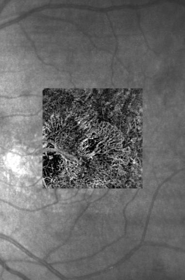
2 Proliferative Diabetic Retinopathy (PDR) Case Report 969 PROLIFERATIVE DIABETIC RETINOPATHY 1 1-year-old diabetic female presents for follow-up of proliferative diabetic retinopathy OD. Visual acuity (VA) 0/0. Most recent panretinal laser treatment three years prior. 5 AngioPlex OCT Angiography (OCT-A) reveals persistent flow in neovascularization of optic disc (NVD) and neovascularization elsewhere (NVE) unsuspected in initial evaluation [image 1]. Detailed analysis of red-free fundus photograph [image ] confirms presence of persistent NVD, not noticed during initial fundus examination. OCT B-scan confirms that the NVD is located at the level of the posterior vitreous cortex, internal to the internal limiting membrane [image 5]. Patient referred to retinologist for evaluation of chronic, possibly indolent, neovascularization to see if additional panretinal laser OD is necessary. 1 AngioPlex OCT Angiography (overlaid onto OCT fundus image): Prominent neovascularization of optic disc (NVD) growing along posterior vitreous cortex; neovascularization elsewhere (with persistent flow) along temporal arcades; and capillary nonperfusion of nasal macula explains thinning and loss of retinal segmentation in nasal macula OD. Fundus Photography (Color): OD, previous panretinal laser treatment; neovascularization along temporal arcades appear fibrotic and regressed. OCT Imaging: Thinning and loss of retinal segmentation in nasal macula OD. See summary. 5 See summary.

: Severe optic disc neovascularization and retinal capillary nonperfusion.")


![treatment). [image 5] Panretinal laser photocoagulation OD was performed.](/docs-images/82/86258888/images/3-4.jpg "5 5 Fluorescein Angiography: Old central retinal vein occlusion with severe macular and midperipheral capillary nonperfusion.")
3 Central retinal vein occlusion with optic disc neovascularization Case Report 97 CENTRAL RETINAL VEIN OCCLUSION 1 50-year-old male patient received two anti-vegf injections OD for cystoid macular edema due to central retinal vein occlusion. Patient was lost to follow-up for 8 months, and presents for evaluation of decreased vision of count fingers at one foot, OD 1 AngioPlex OCT Angiography (6, x AngioPlex images montaged together and overlaid onto FA): Severe optic disc neovascularization and retinal capillary nonperfusion. AngioPlex OCT angiography confirms macular ischemia as the cause of the macular atrophy and poor VA OD. The capillary non-perfusion and the optic disc neovascularization is better seen with OCT-A, compared with fluorescein angiography. OCT-A also has depth-encoded information with an axial resolution of 5 microns, which is relatively lacking in fluorescein angiography, proving that the optic disc neovascularization lies in a plane internal to the optic disc (which differentiates it from optic disc collateral vessels that do not require treatment). [image 5] Panretinal laser photocoagulation OD was performed. 5 5 Fluorescein Angiography: Old central retinal vein occlusion with severe macular and midperipheral capillary nonperfusion. Severe optic disc neovascularization is present. Fundus Photography: Old central retinal vein occlusion with scattered intraretinal hemorrhages in all four quadrants associated with severe optic disc neovascularization. OCT Imaging: Thinned macula with loss of retinal segmentation. See summary.


4 Proliferative diabetic retinopathy with macular edema Case Report 8905 PROLIFERATIVE DIABETIC RETINOPATHY 1 9-year-old male diabetic patient presents for anti- VEGF injection #7 for diabetic macular edema OS. Last anti-vegf injection OS was months prior, and last panretinal laser photocoagulation OS was 8 months prior. The small NVE temporal to the macula OS was difficult to visualize clinically, but easily seen with OCT-A. The treating retinologist elected to treat both the small NVE and the persistent central macular edema with increased frequency of anti-vegf injections OS, to every 5 weeks. Additional retinal photocoagulation may be required in the future. 1 Fundus Photography: Persistent central diabetic macular edema OS. Possible neovascularization temporal to the macula. OCT Imaging: Persistent diabetic macular edema with central macular thickening. Hyperreflective lesion is seen internal to the internal limiting membrane temporal to the macula, suggestive of neovascularization elsewhere (NVE). Fluorescein Angiography: Persistent fluorescein leakage within macula OS. Neovascularization elsewhere (NVE) temporal to macula, within small area of untreated capillary non-perfusion, OS. AngioPlex OCT Angiography (overlaid onto OCT fundus image): NVE with surrounding area of capillary non-perfusion temporal to macula OS is well visualized.
 for 6 years and 7 months, presents for 57 th intravitreal anti-vegf OS. Visual acuity (VA) 0/80 OS.")


5 Neovascular age-related macular degeneration Case Report 7 NEOVASCULAR (WET) AMD 1 69-year-old male patient, who has been treated with anti-vegf injections OS for wet age-related macular degeneration (AMD) for 6 years and 7 months, presents for 57 th intravitreal anti-vegf OS. Visual acuity (VA) 0/80 OS. Most recent intravitreal anti-vegf OS was 7 weeks prior, and first and only photodynamic therapy OS 8 months prior. OCT-A provides excellent visualization of cause of persistent macular exudation OS: a mature type 1 choroidal neovascular membrane. The frequency of intravitreal anti-vegf inejctions was increased to every five weeks to address the persistent intraretinal fluid. 1 Fundus Photography: Wet AMD with fibrotic appearing subfoveal choroidal neovascular membrane OS. Some atrophy inferonasal to the foveal center associated with mild pigmentation. OCT Imaging: Persistent intraretinal fluid, with severe disruption of ellipsoid zone under center of fovea OS. Fibrotic, probably type 1, neovascular membrane is seen between Bruch s membrane and neurosensory retina. RPE transillumination defect is seen near the center of the fovea, consistent with geographic atrophy. AngioPlex OCT Angiography (overlaid onto OCT fundus image): Large, persistent choroidal neovascular membrane with large feeder vessel and tangled network, under center of fovea OS.
 OD 0/0, consistent with - nuclear sclerotic cataract OD.")


6 Branch retinal artery occlusion Case Report 6605 BRANCH RETINAL ARTERY OCCLUSION 1 81-year-old male patient with history of systemic hypertension, coronary artery disease, and JAK mutation positive thrombocytosis presents with -day history of sudden onset of visual field loss above fixation OD. Visual acuity (VA) OD 0/0, consistent with - nuclear sclerotic cataract OD. Repeat fundus examination directed by OCT-A revealed the presence of an 100 X 70 -micron white embolus in the inferotemporal branch retinal arteriole, mm inferior to the foveal center OD. Fundus photography revealed the presence of a wedge-shaped area of retinal infarction distal to the embolus [image ], corresponding to the loss of retinal capillary perfusion demonstrated on OCT-A. Further questioning revealed a history of angiographically proven severe internal carotid stenosis, not amenable to surgical or radiologic endovascular intervention. Detailed analysis of the B-scan OCT confirms the presence of a homogenous hyper-reflective oval embolus located in the inferotemporal branch retinal arteriole. The patient was continued on Plavix (clopidogrel bisulfate) orally. 1 Fundus Photography: Initial fundus examination revealed no obvious cause of superior visual field loss OD. OCT Imaging: Normal central macular OCT OD. Repeat OCT inferior to macula, guided by OCT angiogram, revealed the presence of an homogeneous hyper-reflective oval embolus located in the inferotemporal branch retinal arteriole. AngioPlex OCT Angiography: Inferotemporal branch retinal arteriolar occlusion OD with severe capillary non-perfusion corresponding to visual field defect OD. See summary.
 0/80 OD.")


7 Subretinal neovascular membrane Case Report 5990 SUBRETINAL NEOVASCULAR MEMBRANE 1 51-year-old male patient with history of hypertension presents with 1-month history of metamorphopsia OD, Visual acuity (VA) 0/80 OD. 5 1 AngioPlex OCT Angiography: Extrafoveal subretinal neovascular membrane, located between RPE and neurosensory retina, is well visualized. Fundus Photography: Membrane surrounded by small rim of subretinal hemorrhage superonasal to foveal center OD. OCT Imaging: Type II neovascular membrane with subretinal fluid under central and superior portions of anatomic fovea. Fluorescein Angiography: Leakage from extrafoveal choroidal neovascular membrane surrounded by small rim of hypofluorescent hemorrhage OD. 5 See summary. OCT angiography is sufficient to guide treatment, and the fluorescein angiography may no longer be necessary in this case. OCT B-scan demonstrates precise axial location of the choroidal neovascular membrane, information that is more difficult to obtain from fluorescein angiography. [image 5] The patient was treated with thermal laser ablation of the extrafoveal choroidal neovascular membrane OD combined with intravitreal anti-vegf injections OD.
 0/50 OD and 0/00 OS.")


8 Adult-onset foveomacular vitelliform dystrophy (AOFVD) Case Report 17 AOFVD 1 85-year-old male patient presents for evaluation of decreased vision OU for many years. Right eye is pseudophakic and left eye has a moderate cataract. Visual acuity (VA) 0/50 OD and 0/00 OS. 1 Fundus Photography: Bilateral, subfoveal, slightly elevated yellow lesion at the level of the RPE, more severe in OD. OCT Imaging: Subfoveal RPE detachments OU with nonhomogeneous reflectivity of AOFPED material, without subretinal or intraretinal fluid. Severe disruption of subfoveal ellipsoid zone, OU. AngioPlex OCT Angiography: OCT-A reveals detailed anatomy of perifoveal capillary network, but no evidence of choroidal neovascular membrane. The typical AOFVD appearance of both maculas and the stability of bilateral lesions for 6 years, combined with absence of choroidal membrane with OCT-A reassured the retinologist that observation alone was required. Fluorescein angiography was not performed. Cataract surgery may be considered for the left eye in the future; patient was informed that the final post-cataract surgery VA will be limited by the pre-existing AOFVD OS.
 0/0 OS.")

 600-microns round lesion with central hyperpigmentation just superonasal to foveal center.")
9 Central serous chorioretinopathy Case Report 599 CENTRAL SEROUS CHORIORETINOPATHY 1 6-year-old female patient with 1-day history of metamorphopsia; visual acuity (VA) 0/0 OS. 1 Fluorescein Angiograhy: Hyperfluorescent staining of 600-micron lesion with central hypofluorescence. OCT Imaging: Small RPE detachment just superonasal to foveal center. OCT Angiography: No abnormal vessels seen at the level of choroid or RPE. Fundus Photography: (not shown) 600-microns round lesion with central hyperpigmentation just superonasal to foveal center. Forme fruste of central serous retinopathy. The absence of choroidal neovascular membrane on OCT-A excludes diagnosis of idiopathic wet macular degeneration. Good visual prognosis. Observation alone was recommended..
 0/0 OD.")


: OCT-A elegantly demonstrates prominent collateral vessels draining into the")
10 Branch retinal vein occlusion Case Report 666 BRANCH RETINAL VEIN OCCLUSION 1 66-year-old female with history of diabetes and hypertension, presented for evaluation of possible clinically significant diabetic macular edema. Visual acuity (VA) 0/0 OD. 1 Fundus Photography: Intraretinal hemorrhage associated with edema, surrounded by circinate lipid deposition just superior to foveal center. OCT Imaging: Retinal thickening and intraretinal cysts in superior part of anatomic macula. Hyperreflective lipid mainly in outer plexiform layer. Fluorescein Angiography: Superior macular branch retinal vein occlusion with prominent collaterals coursing through just superior to center of fovea, draining into retinal venule. AngioPlex OCT Angiography (overlaid onto OCT fundus image): OCT-A elegantly demonstrates prominent collateral vessels draining into the inferotemporal venule. OCT-A is sufficient to distinguish a macular branch retinal vein occlusion from diabetic macular edema. The vascular detail on OCT-A is sufficient to permit grid laser treatment to the area of swollen macula.


![After laser treatment, OCT reveals fibrotic remnant of NVE in pre-retinal location [image 5], and](/docs-images/82/86258888/images/11-2.jpg "OCT-A demonstrates dramatic decrease in flow in NVE [image 6].")


 disc diameters inferotemporal to the")
11 Proliferative diabetic retinopathy Case Report 5918 PROLIFERATIVE DIABETIC RETINOPATHY 1 8-year-old male patient with 6 year history of type 1 diabetes examined for evaluation of 10-day history of vitreous hemorrhage. Visual acuity (VA) 0/5 OS. 5 6 Patient underwent panretinal laser photocoagulation OS, resulting in regression of NVE. After laser treatment, OCT reveals fibrotic remnant of NVE in pre-retinal location [image 5], and OCT-A demonstrates dramatic decrease in flow in NVE [image 6]. 1 Fundus Photography: Mild central vitreous hemorrhage and moderately ischemic retina in all four quadrants, with scattered mid peripheral hemorrhages. Cotton wool spots nasal to optic disc and along the inferotemporal arcade. OCT Imaging: OCT confirmed pre-retinal location of NVE. Fluorescein Angiography: Proliferative diabetic retinopathy with subtle optic disc neovascularization and neovascularization elsewhere (NVE) disc diameters inferotemporal to the foveal center. AngioPlex OCT Angiography (overlaid onto OCT fundus image): NVE is well visualized, and can be seen growing through the internal limiting membrane and along the posterior vitreous cortex. 5 See summary. 6 See summary.
12 US_1_150_00I Printed in USA CZ-XI/015 The contents of this case report document may differ from the current status of approval of the product in your country. Please contact your regional representative for more information. Subject to change in design and scope of delivery and as a result of ongoing technical development. AngioPlex is either a trademark or registered trademark of Carl Zeiss Meditec, Inc. in the United States and/or other countries. 015 by Carl Zeiss Meditec, Inc. All copyrights reserved. 097 Carl Zeiss Meditec AG Goeschwitzer Strasse Jena Germany Carl Zeiss Meditec, Inc Hacienda Drive Dublin, CA 9568 USA
OCT Angiography in Primary Eye Care
 OCT Angiography in Primary Eye Care An Image Interpretation Primer Julie Rodman, OD, MS, FAAO and Nadia Waheed, MD, MPH Table of Contents Diabetic Retinopathy 3-6 Choroidal Neovascularization 7-9 Central
PART 1: GENERAL RETINAL ANATOMY
 PART 1: GENERAL RETINAL ANATOMY General Anatomy At Ora Serrata At Optic Nerve Head Fundoscopic View Of Normal Retina What Is So Special About Diabetic Retinopathy? The WHO definition of blindness is
Optical Coherence Tomography in Diabetic Retinopathy. Mrs Samantha Mann Consultant Ophthalmologist Clinical Lead of SEL-DESP
 Optical Coherence Tomography in Diabetic Retinopathy Mrs Samantha Mann Consultant Ophthalmologist Clinical Lead of SEL-DESP Content OCT imaging Retinal layers OCT features in Diabetes Some NON DR features
Deeper visualizations for intervening with confidence.
 CIRRUS OCT with AngioPlex from ZEISS Making the revolutionary routine New vascular quantification Deeper visualizations for intervening with confidence. CIRRUS OCT with AngioPlex from ZEISS can be a much
ZEISS AngioPlex OCT Angiography Making the revolutionary, routine.
 ZEISS AngioPlex OCT Angiography Making the revolutionary, routine. The moment that revolutionary insight becomes routine. // OCT ANGIOGRAPHY MADE BY ZEISS CIRRUS with AngioPlex creates a new era in both
Retina Conference. Janelle Fassbender, MD, PhD University of Louisville Department of Ophthalmology and Visual Sciences 09/04/2014
 Retina Conference Janelle Fassbender, MD, PhD University of Louisville Department of Ophthalmology and Visual Sciences 09/04/2014 Subjective CC/HPI: 64 year old Caucasian female referred by outside ophthalmologist
Incorporating OCT Angiography Into Patient Care
 Incorporating OCT Angiography Into Patient Care Beth A. Steele, OD, FAAO OCT A: Introduction Isolates microvascular circulation from OCT image data Axial resolution = 5 microns (i.e. fine capillaries visible)
The Quick Guide to OCT Mastery 50 Real Cases with Expert Analysis
 OPTICAL COHERENCE TOMOGRAPHY The Quick Guide to OCT Mastery 50 Real Cases with Expert Analysis VOL 1 Sanjay Sharma, MD, FRCS, MSc (Epid), MBA Ophthalmologist, Epidemiologist Queen s University, Canada
OCT Angiography The Next Frontier
 Choroid Retina avascular 5/13/2017 OCT Angiography The Next Frontier Pierce Kenworthy OD, FAAO June 9, 2017 OCT Angiography (OCTA) 2016 Non-invasive, motion contrast imaging Represents erythrocyte movement
EyePACS Grading System (Part 2): Detecting Presence and Severity of Background (Non-Proliferative) Diabetic Retinopathy Lesion
: Detecting Presence and Severity of Background (Non-Proliferative) Diabetic Retinopathy Lesion") EyePACS Grading System (Part 2): Detecting Presence and Severity of Background (Non-Proliferative) Diabetic Retinopathy Lesion George Bresnick MD MPA Jorge Cuadros OD PhD Anatomy of the eye: 3 Normal Retina
EyePACS Grading System (Part 2): Detecting Presence and Severity of Background (Non-Proliferative) Diabetic Retinopathy Lesion George Bresnick MD MPA Jorge Cuadros OD PhD Anatomy of the eye: 3 Normal Retina
Diabetic Retinopathy
 Diabetic Retinopathy Diabetes can be classified into type 1 diabetes mellitus and type 2 diabetes mellitus, formerly known as insulin-dependent diabetes mellitus, and non-insulin diabetes mellitus, respectively.
Jay M. Haynie, O.D.; F.A.A.O. Olympia Tacoma Renton Kennewick Washington
 Jay M. Haynie, O.D.; F.A.A.O. Olympia Tacoma Renton Kennewick Washington I Jay M. Haynie, OD, FAAO have received honoraria from the following companies: Reichert Technologies Notal Vision Carl Zeiss Meditec
Michael P. Blair, MD Retina Consultants, Ltd Libertyville/Des Plaines, Illinois Clinical Associate University of Chicago 17 October 2015
 Michael P. Blair, MD Retina Consultants, Ltd Libertyville/Des Plaines, Illinois Clinical Associate University of Chicago 17 October 2015 So What Parts of the Eye Retina are Affected by VHL Neural tissue
Diabetic Retinopathy. Barry Emara MD FRCS(C) Giovanni Caboto Club October 3, 2012
 Giovanni Caboto Club October 3, 2012") Diabetic Retinopathy Barry Emara MD FRCS(C) Giovanni Caboto Club October 3, 2012 Outline Statistics Anatomy Categories Assessment Management Risk factors What do you need to do? Objectives Summarize the
Diabetic Retinopathy Barry Emara MD FRCS(C) Giovanni Caboto Club October 3, 2012 Outline Statistics Anatomy Categories Assessment Management Risk factors What do you need to do? Objectives Summarize the
DOME SHAPED MACULOPATHY. Ιωάννης Ν. Βαγγελόπουλος Χειρ. Οφθαλμίατρος - Βόλος
 DOME SHAPED MACULOPATHY Ιωάννης Ν. Βαγγελόπουλος Χειρ. Οφθαλμίατρος - Βόλος DOME SHAPED MACULOPATHY-DEFINITIONS The entity Dome Shaped Macula ( DSM ) was first described by Gaucher and associates in 2008
Fluorescein Angiography
 Last revision: October 2011 by Luis Arias Fluorescein Angiography Authors: Luis Arias, MD Hospital Universitari de Bellvitge - University of Barcelona. Spain Jordi Monés, MD Institut de la Màcula i de
Clinical Case Presentation. Branch Retinal Vein Occlusion. Sarita M. Registered Nurse Whangarei Base Hospital
 Clinical Case Presentation on Branch Retinal Vein Occlusion Sarita M. Registered Nurse Whangarei Base Hospital Introduction Case Study Pathogenesis Clinical Features Investigations Treatment Follow-up
ADULT-ONSET FOVEOMACULAR VITELLIFORM DYSTROPHY. By: Chris Munnerlyn, OMT Student University of Arkansas for Medical Sciences
 ADULT-ONSET FOVEOMACULAR VITELLIFORM DYSTROPHY By: Chris Munnerlyn, OMT Student University of Arkansas for Medical Sciences ADULT-ONSET FOVEOMACULAR VITELLIFORM DYSTROPHY (AOFVD) AOFVD is a condition that
Optical Coherence Tomograpic Features in Idiopathic Retinitis, Vasculitis, Aneurysms and Neuroretinitis (IRVAN)
") Columbia International Publishing Journal of Ophthalmic Research (2014) Research Article Optical Coherence Tomograpic Features in Idiopathic Retinitis, Vasculitis, Aneurysms and Neuroretinitis (IRVAN)
Columbia International Publishing Journal of Ophthalmic Research (2014) Research Article Optical Coherence Tomograpic Features in Idiopathic Retinitis, Vasculitis, Aneurysms and Neuroretinitis (IRVAN)
EyePACS Grading System (Part 3): Detecting Proliferative (Neovascular) Diabetic Retinopathy. George Bresnick MD MPA Jorge Cuadros OD PhD
: Detecting Proliferative (Neovascular) Diabetic Retinopathy. George Bresnick MD MPA Jorge Cuadros OD PhD") EyePACS Grading System (Part 3): Detecting Proliferative (Neovascular) Diabetic Retinopathy George Bresnick MD MPA Jorge Cuadros OD PhD Anatomy of the eye: 3 Normal Retina Retinal Arcades Macula Optic
EyePACS Grading System (Part 3): Detecting Proliferative (Neovascular) Diabetic Retinopathy George Bresnick MD MPA Jorge Cuadros OD PhD Anatomy of the eye: 3 Normal Retina Retinal Arcades Macula Optic
OCT Assessment of the Vitreoretinal Relationship in CSME
 December 2007 Sonia Rani John et al. - IFIS 375 ORIGINAL ARTICLE OCT Assessment of the Vitreoretinal Relationship in CSME Dr. Manoj S. DNB FRCS, Dr. Unnikrishnan Nair MS DO FRCS, Dr. Gargi Sathish MS Introduction
Diabetic Retinopatathy
 Diabetic Retinopatathy Jay M. Haynie, OD, FAAO Financial Disclosure I have received honoraria or am on the advisory board for the following companies: Carl Zeiss Meditec Arctic DX Macula Risk Advanced
Marcus Gonzales, OD, FAAO Cedar Springs Eye Clinic
 Marcus Gonzales, OD, FAAO Cedar Springs Eye Clinic 25.6 million adults 11.3% of the adult population 10.9 million adults 65 years and older 26.9% of this age population 79 million people are Pre-diabetic!!
Disease-Specific Fluorescein Angiography
 Ruth E. Picchiottino, CRA Disease-Specific Fluorescein Angiography 15 Disease-Specific Fluorescein Angiography Recommendations for tailoring retinal fluorescein angiography to diabetic retinopathy, macular
Clinically Significant Macular Edema (CSME)
") Clinically Significant Macular Edema (CSME) 1 Clinically Significant Macular Edema (CSME) Sadrina T. Shaw OMT I Student July 26, 2014 Advisor: Dr. Uwaydat Clinically Significant Macular Edema (CSME) 2
Clinically Significant Macular Edema (CSME) 1 Clinically Significant Macular Edema (CSME) Sadrina T. Shaw OMT I Student July 26, 2014 Advisor: Dr. Uwaydat Clinically Significant Macular Edema (CSME) 2
Retinal Vein Occlusion
 Retinal Update 2018 Retinal Vein Occlusion Case Presentations to Myself Branch Vein Occlusion What medical evaluation do you recommend for this 72 year old patient? Is there anything you ask of your medical
ATLAS OF OCT. Retinal Anatomy in Health & Pathology by Neal A. Adams, MD. Provided to you by:
 ATLAS OF OCT Retinal Anatomy in Health & Pathology by Neal A. Adams, MD Provided to you by: Atlas of OCT The OCT Atlas is written by Neal A. Adams, MD, and produced by Heidelberg Engineering, Inc. to help
OCT Interpretation in Retinal Disease
 OCT Interpretation in Retinal Disease Jay M. Haynie, OD, FAAO Financial Disclosure I have received honoraria or am on the advisory board for the following companies: Carl Zeiss Meditec Advanced Ocular
Dr/ Marwa Abdellah EOS /16/2018. Dr/ Marwa Abdellah EOS When do you ask Fluorescein angiography for optic disc diseases???
 When do you ask Fluorescein angiography for optic disc diseases??? 1 NORMAL OPTIC DISC The normal optic disc on fluorescein angiography is fluorescent due to filling of vessels arising from the posterior
DIABETIC RETINOPATHY
 DIABETIC RETINOPATHY C. L. B. Canny, MD FRCSC Diabetic retinopathy is the most serious eye manifestation of diabetes and is responsible for most of the blindness caused by diabetes. Diabetic retinopathy
The Diabetic Retinopathy Clinical Research Network. Management of DME in Eyes with PDR
 The Diabetic Retinopathy Clinical Research Network Management of DME in Eyes with PDR 1 What Has Been Learned? Diabetic Retinopathy Treatment Protocol F: Results suggest that clinically meaningful differences
Leo Semes, OD, FAAO UAB Optometry
 Leo Semes, OD, FAAO UAB Optometry Safe; inert Has long track record - over 45 years Mixes with plasma and highlights blood vessel compromise Using specific exciting (490 nm)and absorption (510 nm) filters
The Human Eye. Cornea Iris. Pupil. Lens. Retina
 The Retina Thin layer of light-sensitive tissue at the back of the eye (the film of the camera). Light rays are focused on the retina then transmitted to the brain. The macula is the very small area in
Spontaneous Large Serous Retinal Pigment Epithelial Tear
 This is an Open Access article licensed under the terms of the Creative Commons Attribution-NonCommercial-NoDerivs 3.0 License (www.karger.com/oa-license), applicable to the online version of the article
Fundus Autofluorescence. Jonathan A. Micieli, MD Valérie Biousse, MD
 Fundus Autofluorescence Jonathan A. Micieli, MD Valérie Biousse, MD The retinal pigment epithelium (RPE) has many important functions including phagocytosis of the photoreceptor outer segments Cone Rod
ZEISS AngioPlex OCT Angiography Overview ZEISS OCT Angiography
 ZEISS AngioPlex OCT Angiography Overview ZEISS OCT Angiography California, ZEISS AngioPlex Ultra-clear visualization of microvascular blood flow using non-invasive OCT angiography 2 AngioPlex OCT Angiography
Ocular imaging in acquired retinopathy with multiple myeloma
 Ocular imaging in acquired retinopathy with multiple myeloma ABDELRAHMAN GABER SALMAN MD- FRCS (GLASG)- MRCS (ED) ASSOCIATE PROFESSOR AIN SHAMS UNIVERSITY EVRS 2015 Immunogammopathies Immunogammopathies
연령연관황반변성에서망막혈관종성증식과동반된망막색소상피박리의임상양상과일차적인광역학치료의결과
 연령연관황반변성에서망막혈관종성증식과동반된망막색소상피박리의임상양상과일차적인광역학치료의결과 40 Table. Clinical characteristics and results of patients undergoing photodynamic therapy for retinal angiomatous proliferation Patients No. Age/ sex Eye
THE ROLE OF anti-vegf IN DIABETIC RETINOPATHY AND AGE RELATED MACULAR DEGENERATION
 THE ROLE OF anti-vegf IN DIABETIC RETINOPATHY AND AGE RELATED MACULAR DEGENERATION MOESTIDJAB DEPARTMENT OF OPHTHALMOLOGY SCHOOL OF MEDICINE AIRLANGGA UNIVERSITY DR SOETOMO HOSPITAL SURABAYA INTRODUCTION
10/17/2017. FDA Approved. Zeiss AngioPlex TM Optovue AngioVue TM
 Images retinal microvasculature without dye injection Displays structure and function from a single imaging system Standard of Care-2011 DFE, Fundus Photos, VF 10-2, SD-OCT, FAF, or mferg 2016-AAO Baseline
Retinal Complications of Obstructive Sleep Apnea A Growing Concern!
 Retinal Complications of Obstructive Sleep Apnea A Growing Concern! Jay M. Haynie, OD, FAAO Financial Disclosure I have received honoraria or am on the advisory board for the following companies: Carl
HHS Public Access Author manuscript Ophthalmic Surg Lasers Imaging Retina. Author manuscript; available in PMC 2016 January 14.
 High-Speed Ultrahigh-Resolution OCT of Bruch s Membrane in Membranoproliferative Glomerulonephritis Type 2 Mehreen Adhi, MD, Sarah P. Read, MD, PhD, Jonathan J. Liu, PhD, James G. Fujimoto, PhD, and Jay
Cirrus TM HD-OCT. Details define your decisions
 Cirrus TM HD-OCT Details define your decisions 2 With high-definition OCT Carl Zeiss Meditec takes you beyond standard spectral domain Built on 10 years experience at the vanguard of innovation, Carl Zeiss
 measure of your overall performance. An isolated glucose test is helpful to let you know what your sugar level is at one moment, but it doesn t tell you whether or not your diabetes is under adequate control
OCCLUSIVE VASCULAR DISORDERS OF THE RETINA
 OCCLUSIVE VASCULAR DISORDERS OF THE RETINA Learning outcomes By the end of this lecture the students would be able to Classify occlusive vascular disorders (OVD) of the retina. Correlate the clinical features
R&M Solutions
 Mohamed Hosny El-Bradey, MD., Assistant Professor of Ophthalmology, Tanta University. Wael El Haig, MD., Professor of Ophthalmology. Zagazeeg University. 1 Myopic CNV is considered the most common vision
Is OCT-A Needed As An Investigative Tool During The Management Of Diabetic Macular Edema
 Is OCT-A Needed As An Investigative Tool During The Management Of Diabetic Macular Edema Ayman M Khattab MD, FRCS Professor of Ophthalmology Cairo University Diabetic Macular Edema (DME) Diabetic macular
Retinal Vein Occlusion (RVO) Treatment pathway- Northeast England. Retinal Vein Occlusion (RVO) with Macular oedema (MO)
 Treatment pathway- Northeast England. Retinal Vein Occlusion (RVO) with Macular oedema (MO)") Retinal Vein Occlusion (RVO) Treatment pathway- Northeast England (Royal Victoria Infirmary, Sunderland Eye Infirmary, James Cook University Hospital, Darlington Memorial Hospital, University Hospital
Retinal Vein Occlusion (RVO) Treatment pathway- Northeast England (Royal Victoria Infirmary, Sunderland Eye Infirmary, James Cook University Hospital, Darlington Memorial Hospital, University Hospital
Clinical Study Choroidal Thickness in Eyes with Unilateral Ocular Ischemic Syndrome
 Hindawi Publishing Corporation Journal of Ophthalmology Volume 215, Article ID 62372, 5 pages http://dx.doi.org/1.1155/215/62372 Clinical Study Choroidal Thickness in Eyes with Unilateral Ocular Ischemic
Neovascular Glaucoma Associated with Cilioretinal Artery Occlusion Combined with Perfused Central Retinal Vein Occlusion
 Neovascular Glaucoma Associated with Cilioretinal Artery Occlusion Combined with Perfused Central Retinal Vein Occlusion Man-Seong Seo,* Jae-Moon Woo* and Jeong-Jin Seo *Department of Ophthalmology, Chonnam
Diabetic Retinopathy A Presentation for the Public
 Diabetic Retinopathy A Presentation for the Public Ray M. Balyeat, MD The Eye Institute Tulsa, Oklahoma The Healthy Eye Light rays enter the eye through the cornea, pupil and lens. These light rays are
PRIMUS 200 from ZEISS The essential OCT
 PRIMUS 200 from ZEISS The essential OCT Seeing beyond the surface. ZEISS PRIMUS 200 // INNOVATION MADE BY ZEISS Clear Visualization. Advanced Technology. Reliability. Essential elements of your first OCT.
Venous Occlusive Diseases
 Venous Occlusive Diseases Bruce R. Saran, MD Adjunct Assistant Clinical Professor of Medicine Scheie Eye Institute University of Pennsylvania School of Medicine Philadelphia, PA -a division of: RVO Demographics
Posterior Segment Update
 Posterior Segment Update Featured Speaker: Dr. Kyle Cheatham, FAAO, DIP ABO DISCLOSURE STATEMENT We have no direct financial or proprietary interest in any companies, products or services mentioned in
OPTIC DISC PIT Pathogenesis and Management OPTIC DISC PIT
 OPTIC DISC PIT Pathogenesis and Management Abdel-Latif Siam Ain Shams University Cairo Egypt OPTIC DISC PIT Congenital pit is an atypical coloboma usually located on the temporal edge of the disc, associated
Optical Coherence Tomography: Pearls for the Anterior Segment Surgeon Basic Science Michael Stewart, M.D.
 Optical Coherence Tomography: Pearls for the Anterior Segment Surgeon Basic Science Michael Stewart, M.D. Disclosure OCT Optical Coherence Tomography No relevant financial relationships I will refer to
PLEX Elite 9000 from ZEISS Swept-Source OCT
 PLEX Elite 9000 from ZEISS Swept-Source OCT Uncovering the undiscovered. ZEISS PLEX Elite 9000 // INNOVATION MADE BY ZEISS 2 Ultra-wide angiography En face montage Image courtesy of Prof. G. Querques,
Diabetic Management beyond traditional risk factors and LDL-C control: Can we improve macro and microvascular risks?
 Retinopathy Diabetes has a negative effect on eyes in many ways, increasing the risk of cataracts for example, but the most common and serious ocular complication of diabetes is retinopathy. Diabetic retinopathy
Macular Morphology and Visual Acuity in the Comparison of Age-related Macular Degeneration Treatments Trials
 Macular Morphology and Visual Acuity in the Comparison of Age-related Macular Degeneration Treatments Trials Glenn J. Jaffe, MD, 1 Daniel F. Martin, MD, 2 Cynthia A. Toth, MD, 1 Ebenezer Daniel, MPH, PhD,
ANSWERING THE WHY? Clinicians discuss the latest imaging technologies for retina practice BY PETER K. KAISER, MD
 Insert to March 2018 Sponsored by MULTI-MODALITY IMAGING: LATEST EVOLUTIONS IN OCTA AND UWF As the array of safe and efficacious medical and surgical options for retinal diseases expands, so does the need
Vascular Disease Ocular Manifestations of Systemic Hypertension
 Vascular Disease Ocular Manifestations of Systemic Hypertension Maynard L. Pohl, OD, FAAO Pacific Cataract & Laser Institute 10500 NE 8 th Street, Suite 1650 Bellevue, WA 98004 USA 425-462-7664 Cerebrovascular
Cirrus TM HD-OCT. Details defi ne your decisions
 Cirrus TM HD-OCT Details defi ne your decisions 2 With high-defi nition OCT Carl Zeiss Meditec takes you beyond standard spectral domain Built on 10 years experience at the vanguard of innovation, Carl
Amber Priority. Image Library
 Amber Priority Image Library Amber flag Diabetic Maculopathy (M1) Pre-proliferative Diabetic Retinopathy (R2) Old, treated and now inactive DR (R1/M0/P1or R0/M0/P1) Where only partial or incomplete images
Stabilization of visual acuity with photodynamic therapy in eyes with chorioretinal anastomoses
 Graefe s Arch Clin Exp Ophthalmol (2004) 242:368 376 CLINICAL INVESTIGATION DOI 10.1007/s00417-003-0844-0 Rufino M. Silva José R. Faria de Abreu António Travassos José G. Cunha-Vaz Stabilization of visual
Ganglion cell analysis by optical coherence tomography (OCT) Jonathan A. Micieli, MD Valérie Biousse, MD
 Jonathan A. Micieli, MD Valérie Biousse, MD") Ganglion cell analysis by optical coherence tomography (OCT) Jonathan A. Micieli, MD Valérie Biousse, MD Figure 1. Normal OCT of the macula (cross section through the line indicated on the fundus photo)
Ganglion cell analysis by optical coherence tomography (OCT) Jonathan A. Micieli, MD Valérie Biousse, MD Figure 1. Normal OCT of the macula (cross section through the line indicated on the fundus photo)
Diagnosis and treatment of diabetic retinopathy. Blake Cooper MD Ophthalmologist Vitreoretinal Surgeon Retina Associates Kansas City
 Diagnosis and treatment of diabetic retinopathy Blake Cooper MD Ophthalmologist Vitreoretinal Surgeon Retina Associates Kansas City Disclosures Consulted for Novo Nordisk 2017,2018. Will be discussing
The College of Optometrists - Learning outcomes for the Professional Certificate in Medical Retina
 Learning outcomes for the Professional Certificate in Medical Retina, incorporating diabetic retinopathy screening and age related macular degeneration The professional certificate is a prerequisite to
IQ 532 Micropulse Green Laser treatment for Refractory Chronic Central Serous Retinopathy
 Cronicon OPEN ACCESS EC OPHTHALMOLOGY Case Report IQ 532 Micropulse Green Laser treatment for Refractory Chronic Central Serous Retinopathy Fawwaz Al Mamoori* Medical Retina Department, Eye Specialty Hospital,
ROLE OF LASER PHOTOCOAGULATION VERSUS INTRAVITREAL TRIAMCINOLONE ACETONIDE IN ANGIOGRAPHIC MACULAR EDEMA IN DIABETES MELLITUS
 ORIGINAL ARTICLE ROLE OF LASER PHOTOCOAGULATION VERSUS INTRAVITREAL TRIAMCINOLONE ACETONIDE IN ANGIOGRAPHIC MACULAR EDEMA IN DIABETES MELLITUS Aggarwal Somesh VP 1, Shah Sonali N 2, Bharwada Rekha M 3,
OCT-Angiography Clinical Cases. OCT-Angiography Clinical Cases
 OCT-Angiography Clinical Cases OCT-Angiography Clinical Cases NIDEK RS-3000 Advance AngioScan Daniela Bacherini Andrea Sodi Stanislao Rizzo CONTENTS Page Authors 3 Introduction 4 Case 1 Case 2 Case 3 Case
Diabetes and Eye Health more than meets the eye Vision Initiative - in association with PSA
 Diabetes and Eye Health more than meets the eye Vision Initiative - in association with PSA Vision 2020 Australia Vision Initiative RANZCO & OAA (Vic) Proud members of Vision 2020 Australia Outline Vision
OCT Interpretation. Financial Disclosure. Jay M. Haynie, OD, FAAO. OCT Image Layers 7/21/2014
 OCT Interpretation Jay M. Haynie, OD, FAAO Financial Disclosure I have received honoraria or am on the advisory board for the following companies: Olympia Tacoma Renton Kennewick - Washington Carl Zeiss
Diabetic Retinopathy Screening in Hong Kong. Dr. Rita Gangwani M.S, FRCS (Ophth), FCOphth(HK), FHKAM Eye Institute, The University of Hong Kong
, FCOphth(HK), FHKAM Eye Institute, The University of Hong Kong") Diabetic Retinopathy Screening in Hong Kong Dr. Rita Gangwani M.S, FRCS (Ophth), FCOphth(HK), FHKAM Eye Institute, The University of Hong Kong Co-Investigators Prof. David Wong Prof. Sarah McGhee Dr. Wico
Diabetic Retinopathy Screening in Hong Kong Dr. Rita Gangwani M.S, FRCS (Ophth), FCOphth(HK), FHKAM Eye Institute, The University of Hong Kong Co-Investigators Prof. David Wong Prof. Sarah McGhee Dr. Wico
Building The Retina Company
 Building The Retina Company Optos devices produce ultra-widefield (UWF ), high resolution images (optomap ) of approximately 82% (200 ) of the retina. A single optomap can document the retina from the
World Sight Day Case Studies. Mark Frost Screening Manager South East London DESP
 World Sight Day 2015 Case Studies Mark Frost Screening Manager South East London DESP Introduction All of the following cases have been identified in our screening programme over the last 3 years. The
Bilateral Elevated Macular Lesions
 Challenging Case Bilateral Elevated Macular Lesions Section Editor: Alireza Ramezani, MD Case presentation A 65-year-old woman presented with decreased vision in both eyes of 2 months duration. She reported
Moving forward with a different perspective
 Moving forward with a different perspective The Leader In Vision Diagnostics Offers A New Perspective Marco has served the eyecare community by offering exceptional lane products and automated high tech
8/6/17. Disclosures Aerie Pharmaceuticals Alcon BioTissue Diopsys Optovue Shire
 Nathan Lighthizer, O.D., F.A.A.O. Associate Professor Assistant Dean for Clinical Care Director of Continuing Education Chief of Specialty Care Clinics Oklahoma College of Optometry Tahlequah, OK lighthiz@nsuok.edu
Diabetic Retinopathy
 Diabetic Retinopathy Diabetes mellitus is one of the leading causes of irreversible blindness worldwide. In the United States, it is the most common cause of blindness in people younger than 65 years.
FRANZCO, MD, MBBS. Royal Darwin Hospital
 Diabetes and Eye By Dr. Nishantha Wijesinghe FRANZCO, MD, MBBS Consultant Ophthalmologist Royal Darwin Hospital 98% of Diabetics do not need to suffer from severe visual loss Yet Diabetic eye disease is
11/29/2016 MACULAR MALADIES: TYPICAL & ATYPICAL CASES
 MACULAR MALADIES: TYPICAL & ATYPICAL CASES Dawn Pewitt, OD, FAAO Triad Eye Institute, Grove, OK Dpewitt@triadeye.com Disclosure Statement: No financial disclosures COPE 51218-PS Please silence all mobile
CENTRAL SEROUS CHORIORETINOPATHY (CSC) IS
 IS") Association Between the Efficacy of Half-Dose Photodynamic Therapy With Indocyanine Green Angiography and Optical Coherence Tomography Findings in the Treatment of Central Serous Chorioretinopathy MASSIMO
Association Between the Efficacy of Half-Dose Photodynamic Therapy With Indocyanine Green Angiography and Optical Coherence Tomography Findings in the Treatment of Central Serous Chorioretinopathy MASSIMO
Acquired vitelliform detachment in patients with subretinal drusenoid deposits (reticular pseudodrusen)
") Zurich Open Repository and Archive University of Zurich Main Library Strickhofstrasse 39 CH-8057 Zurich www.zora.uzh.ch Year: 2011 Acquired vitelliform detachment in patients with subretinal drusenoid
Zurich Open Repository and Archive University of Zurich Main Library Strickhofstrasse 39 CH-8057 Zurich www.zora.uzh.ch Year: 2011 Acquired vitelliform detachment in patients with subretinal drusenoid
Year 2 MBChB Clinical Skills Session Ophthalmoscopy. Reviewed & ratified by: Mr M Batterbury Consultant Ophthalmologist
 Year 2 MBChB Clinical Skills Session Ophthalmoscopy Reviewed & ratified by: o Mr M Batterbury Consultant Ophthalmologist Learning objectives o To understand the anatomy and physiology of the external and
Mark Dunbar: Disclosure
 Important Things to Understand About OCT Mark T. Dunbar, O.D., F.A.A.O. Bascom Palmer Eye Institute University of Miami, School of Medicine Mark Dunbar: Disclosure Optometry Advisory Board for: Allergan
Thinking Beyond Wet Age-Related Macular Degeneration
 Ophthalmic Deliberations ISSN 0972-0200 Thinking Beyond Wet Age-Related Macular Degeneration Neha Goel, Atul Kumar, Mahesh P Shanmugam, Muna Bhende, Raja Narayanan, Hidetaka Matsumoto Atul Kumar MD, FAMS
Mild NPDR. Moderate NPDR. Severe NPDR
 Diabetic retinopathy Diabetic retinopathy is the most common cause of blindness in adults aged 35-65 years-old. Hyperglycaemia is thought to cause increased retinal blood flow and abnormal metabolism in
Medical Retina 2011 Nicholas Lee
 Medical Retina 2011 Nicholas Lee 1 Diabetic Retinopathy Epidemiology 1000 registered blind each year 2% diabetics registered as blind (8% of all Blind Registrations) 42% with Mild Background DR will progress
Key words: Choroidal neovascularisation, Laser coagulation, Retinal imaging
 420 Mini Review The Role of Optical Coherence Tomography (OCT) in the Diagnosis and Management of Retinal Angiomatous Proliferation (RAP) in Patients with Age-related Macular Degeneration Antonio Polito,
FA Conference. Lara Rosenwasser Newman, M.D. 10/2/14 University of Louisville Department of Ophthalmology and Visual Sciences
 FA Conference Lara Rosenwasser Newman, M.D. 10/2/14 University of Louisville Department of Ophthalmology and Visual Sciences Patient Presentation CC: (sent by optometrist) Blurry/foggy vision HPI: 62 yo
The use of a high-intensity laser to create an anastomotic
 Case Report 866 Laser Chorioretinal Venous Anastomosis for Progressive Nonischemic Central Retinal Vein Occlusion Chih-Hsin Chen, MD; Chien-Hsiung Lai 1, MD; Hsi-Kung Kuo, MD The use of high or medium-intensity
Tuberous sclerosis presenting as atypical aggressive retinal astrocytoma with proliferative retinopathy and vitreous haemorrhage
 Case Report Brunei Int Med J. 2015; 11 (1): 49-53 Tuberous sclerosis presenting as atypical aggressive retinal astrocytoma with proliferative retinopathy and vitreous haemorrhage Pui Ling TANG and Mae-Lynn
Misdiagnosed Vogt-Koyanagi-Harada (VKH) disease and atypical central serous chorioretinopathy (CSC)
 disease and atypical central serous chorioretinopathy (CSC)") HPTER 12 Misdiagnosed Vogt-Koyanagi-Harada (VKH) disease and atypical central serous chorioretinopathy (S) linical Features VKH disease is a bilateral granulomatous panuveitis often associated with exudative
HPTER 12 Misdiagnosed Vogt-Koyanagi-Harada (VKH) disease and atypical central serous chorioretinopathy (S) linical Features VKH disease is a bilateral granulomatous panuveitis often associated with exudative
CHAPTER 13 CLINICAL CASES INTRODUCTION
 2 CHAPTER 3 CLINICAL CASES INTRODUCTION The previous chapters of this book have systematically presented various aspects of visual field testing and is now put into a clinical context. In this chapter,
International Journal of Health Sciences and Research ISSN:
 International Journal of Health Sciences and Research www.ijhsr.org ISSN: 2249-9571 Original Research Article A Multivariate Analysis of Intravitreal Injection of Anti-VEGF Bevacizumab in the Treatment
Why Is Imaging Critical in My Uveitis Practice?
 Why Is Imaging Critical in My Uveitis Practice? Dilraj S. Grewal, MD Developed in collaboration Imaging Is the Backbone of Uveitis Workup and Monitoring Treatment Response FP FAF B- scan Multimodal Imaging
The Era of anti- - - VEGF Kirk L. Halvorson, OD
 The Era of anti- - - VEGF Kirk L. Halvorson, OD Introduction: Anti- - - Vascular Endothelial Growth Factor (Anti- - - VEGF) medication is a relatively a new line of medications used in treating a variety
EFFICACY OF ANTI-VASCULAR ENDOTHELIAL GROWTH FACTOR AGENTS IN RETINAL DISORDER FOR BETTER VISUAL ACUITY
 EFFICACY OF ANTI-VASCULAR ENDOTHELIAL GROWTH FACTOR AGENTS IN RETINAL DISORDER FOR BETTER VISUAL ACUITY Diwakar chaudhary *1, 2, Hu shuqiong, Long Yuan and Xiong kun 1 Yangtze University, 1 Nanhuan Road
Documentation, Codebook, and Frequencies
 Documentation, Codebook, and Frequencies Ophthalmology Retinal Imaging Examination Survey Years: 2005 to 2006 SAS Transport File: OPXRET_D.XPT December 2008 NHANES 2005 2006 Data Documentation Exam Component:
PRIMUS 200 from ZEISS The essential OCT
 EN 00_00I The contents of the brochure may differ from the current status of approval of the product in your country. Please contact your regional representative for more information. Subject to change