Enterography : A New Paradigm in Small Bowel Imaging
|
|
|
- Amos Townsend
- 5 years ago
- Views:
Transcription


1 Massachusetts General Hospital Harvard Medical School CT Enterography : A New Paradigm in Small Bowel Imaging Dushyant Sahani, M.D Director of CT Associate Professor Department of Radiology Massachusetts General Hospital Harvard Medical School
2 Small Bowel Evaluation Techniques Conventional techniques SBFT, Enteroclysis


3 Small Bowel Evaluation Techniques Conventional techniques SBFT, Enteroclysis and Endoscopy Newer techniques Capsule Endoscopy


4 Capsule Endoscopy
5 In the last decade, with significant advances in technology there has been a paradigm shift in imaging evaluation of gastro-intestinal tract (GIT) SIGNIFICANCE ADVANCES IN GASTROINTESTINAL IMAGING Success has become attainable & practical due to advent if of multidetector CT (MDCT) with administration of oral contrast media (OCM) MDCT -High resolution -Isotropic images -Volume rendering -Multiple planes OCM -Adequate distention -Luminal, mural & extra-luminal evaluation - Seeing through the bowel loops
6 CONTRAST MEDIA / AGENTS POSITIVE / WHITE (POCM) Barium Iodine NEGATIVE / BLACK Air Fat NEUTRAL / GRAY (NOCM) Water / Milk / Juice Low density barium (VoLumen) Polyethylene glycol (PGE) Lactulose Simethicone coated cellulose Locust bean gum - mannitol commercial availability in U.S.A. for CT? Targeted use of OCM allows optimal evaluation of pathology.


 Adequate distention is important")

7 EFFECT ON MOTILITY & Normal Collapsed Bowel DISTENTION Water soluble - Iodinated VoLumen Distention Click to view Click to view Click to view Water Barium 2% Symbolic Motility Waveform Click to view Click to view Motility increased due to larger volume consumed rapidly & sorbitol content (osmotic) Adequate distention is important for detection of pathology. Effect on motility needs to be considered for optimizing the time to scan.


8 CT Oral Contrast Iodine Agents Positive Contrast Agents Diluted Iodinated/barium 2-5% Used in CT because of experience with GI studies Benefits Unequivocal identification of bowel loop if opacified Extravasation identification Barium


9 Oral Contrast Agents (OCA) Positive OCA Rely on structural changes in the bowel Pseudolesions due to poor mixing May mask subtle pathology in the wall Neutral OCA Better assessment of the enhancing bowel wall Do not interfere with 3D volume sets


10 TI Crohn s Positive Oral Contrast


11 Barium VoLumen


12 GRAFT VERSUS HOST DISEASE OCM - PEARLS STRICTURES POCM NOCM Graft versus host disease the abnormal enhancing mucosa and extent of involvement is better appreciated with use of NOCM Multiple small bowel strictures with mucosal & mural enhancement suggesting active CD


13 Small Bowel Lesions: Positive Vs. Neutral CM CT CT

14 NEUTRAL (GRAY) OCM WATER - PROTOCOLS UPPER ABDOMEN SCAN <30 min 1 glass = 450 ml min On scanner table ABDOMEN & PELVIS min min On scanner table SCAN <40


15 NEUTRAL (GRAY) OCM WATER Allows assessment of luminal pathology & bowel wall Excellent for upper GIT if scanned at appropriate time GIST Adequately distended 2 nd part of duodenum with visible mural details (small arrows) Ampullary mass lesion invading the duodenum Large gastrointestinal stromal tumor (GIST) with mass effect on stomach NOCM High quality curved reformats of the pancreas due to neutral background POCM


16 NEUTRAL (GRAY) OCM WATER - PERILS Transits rapidly, absorbed distally, preventing adequate distention Suboptimal distention obscures, masks or mimics pathology Duodenum COLLAPSED SEGMENTS Sub-optimal distention of segments of small bowel Rapid transit of water and inappropriate scan delay resulting in collapsed duodenum which cannot be separated from pancreas
17 Neutral CT OCA PEG Methylcellulose Lactulose AJR 2005; 185:1173 MR Enterography Essen, Germany Locust bean gum + Mannitol Gd-DTPA enhancement Commercially NOT available in US

18 Neutral CT Oral Contrast Agents VoLumen (E-Z-EM Product) Gum system, Sorbitol & Barium (0.1%w/v) HU Water = < 10 HU Berry taste Weak grape Kool-aid

19 Advantages of Viscous Neutral OCM Better bowel distension Superior lumen to wall differentiation compared to positive contrasts Better detection of mucosal and mural pathologies Improved quality of post processed 3D images

20 NEUTRAL OCM LDB (VoLumen) PROTOCOLS CT ENTEROGRAPHY 1 Bottle = 450 ml SCAN 1 glass = 450 ml 0-20 min min min MIN On scanner table COLONIC INDICATIONS SCAN 0-20 min min min >60 MIN

21

22 CTE: Technique Focused exam on small bowel Intestines distention with a fairly large amount of intraluminal contrast material IV Contrast for lesions detection and to assess disease activity. Visualization: axial+ MPR/3D


23 IBD: minutes delay
24 CT Enterography Oral Contrast Protocols NYU MGH Cleveland Clinic Mayo Clinic 1350 ml total 1350 total 1350 ml total 1350 ml total min min min min min min min min 225 H 2 O 5 min 225 H 2 O 5 min min min 225 H 2 O scanner 225 H 2 O scanner min min 0.1 mg Glucagon Reglan prn

25 CT Enterography: Technique Uni-phasic scans start sec Iodinated CM: cc of cc/sec Dual-phase study GI bleed slice scanner DC: mm Reconstruct 3-5 mm axial Retro-recon:1.25/0.625mm 3-5 mm coronal MIPs
26 CTE: Established Indications *Suspected or Known Crohn s *Colitis Assess small bowel for Crohn s GI Bleeding/Luminal Mass DX
27 Role of Imaging in IBD Initial disease evaluation Treatment response Assessment for acute exacerbation Detection of stricture and bowel obstruction Extraintestinal complications (abscess, fistula, LAD, PSC)


28 CTE CTE


29 Crohn s disease multifocal strictures
30 CT Enterography Small Bowel Findings positive for Crohn s Disease (Hara et al. Radiology 2006) Study of 17 pts SBFT Ileoscopy CT enterography Capsule Endoscopy 17 % 65 % 53 % 71 % CTE depicted > 2x Crohn disease than did SBFT CTE benefit: Extra-luminal findings and complications


31 IBD Complications Abscess Fistula Liver Abscess


")
32 CTE: IBD Disease Activity Bodily et al. Radiology 2006; 238:505 N = 96 patients Ileoscopy vs. CTE Quantitative vs. qualitative CTE Quantitative mural attenuation (enhancement) TI correlate well with ileoscopic & histologic findings of inflammation (Crohn s) Threshold of 109 HU Active Chronic


33 GI Bleed
34 CT Enterography: Emerging Indications Abdominal pain Diarrhea Malabsorption Small bowel tumors/polyps Eventually will replace almost all DX barium work in small bowel PET CT


35 GVHD


36 Small Bowel Polyp


37 Jejunal GIST
38 CT Enterography : Issues Learning Curve Familiarity with CTE critical to limit Pseudodisease detection Detection of extraluminal findings/collections/leaks Radiation Dose Patient acceptance Increased bowel activity


39 NEUTRAL OCM VOLUMEN - PERILS DIFFICULTY WITH ISO-ATTENUATING EXTRA-LUMINAL LESIONS NOCM POCM NOCM POCM Extraluminal collection suspected on CTE, however is more conspicuous with positive OCM Early peritoneal implants less visible with NOCM but more obvious on the follow up exam with POCM OCCASIONAL DISCOMFORT DUE TO DISTENTION & INCREASED MOTILITY


40 Extraluminal Collection NOCM POCM


41 Enteric Fistula
42 Recent attention focused on radiation risk to patients from diagnostic imaging Rise in medical imaging utilization (60 million CT exams annually) Risk estimates extrapolated from atomic bomb/nuclear fallout survivors BEIR VII supports LNT model Ionizing radiation classified by HHS as carcinogen in 2005 Risk Brenner et al. NEJM 2007 Dose
43 Children particularly vulnerable to ionizing radiation Increased cell division/tissue growth Longer life expectancy/latency period Lower cross-sectional area increases absorbed dose One study estimates 1 in 1000 children undergoing CT will die of radiation-induced malignancy Brenner DJ et al, Am J Roentgenol (2001)
44 Potential radiation risk to children Minimum exposure required for inc risk ranges from msv Exam Mean dose to children (msv) Natural bkgd 2-4 CXR 0.02 Fluoro exam CT (Abd/Pel) 5 PET 15 PET-CT Rice HE et al, J Ped Surg (2007)

45 Low Dose CT exams Original CT scan NOISE SIMULATION SOFTWARE (GE HEALTHCARE) 150 Simulated CT Studies NI 18 NI 20 NI 25 NI 30 NI 35 Kambadakone et al. RSNA 2008


46 Radiation Dose Benefits NI-15 NI-35 Original CT data 3-15m Sv Estimated Reduction NI % NI % NI % NI % NI % Kambadakone et al. RSNA 2008


 and increased perienteric")
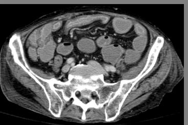
47 Jan Radiation Dose 11.6mSv Dec Radiation Dose 5.1 msv 23 yrs old with Crohn s Disease. Baseline and FU CT exam Axial & Coronal CT images demonstrating wall thickening and enhancement (red arrow) and increased perienteric vascularity (white arrow) 40% ASIR 55% Dose Reduction
 Axial CT Image demonstrating wall")
 19 yrs old with Crohn s")

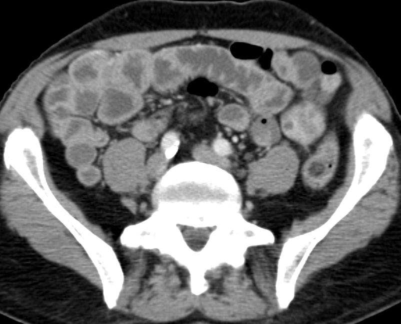
48 Coronal CT Image demonstrating wall thickening and mucosal Enhancement (red arrow) Axial CT Image demonstrating wall thickening and mucosal enhancement (red arrow) and increased perienteric vascularity (white arrow) 19 yrs old with Crohn s Disease. First CT exam 40% ASIR Coronal CT Image demonstrating a lymph node (yellow arrow) and increased perienteric vascularity (white arrow) RADIATION DOSE msv
49 Potential of MRI for evaluating IBD No ionizing radiation Superior soft tissue resolution compared with CT In routine clinical use for detecting perianal fistulae
50 IBD protocol MRI Technique Oral prep same as with CT given 45 mins prior Coronal T2WI screen Targeted axial T2W-FS 0.1mmol/kg wt. Coronal T1 FS pre- and 1, 3, 5 mins post Quantitative T1 maps to assess enhancement Hi-res axial T1 FS post-gado images


51 18F with known Crohn s SBFT shows narrowing of TI T2 shows segmental narrowing, wall thickening, fatty proliferation at same location


52 Progressive mucosal enhancement suggestive of active inflammation PRE 1 MIN POST 5 MIN POST
53 Summary Advances in MDCT and oral contrast has renewed interest in small bowel imaging with CT CTE using neutral oral contrast most promising non-invasive test Sensitive for the evaluation of luminal and extra luminal findings in IBD Disease activity Role evolving for other indication in small bowel
54 Summary Oral contrast protocol optimization is essential for the best results MPR s/3d are complimentary to axial data set and its routine use is encouraged Patient education for AE s Bowel motion Anticipate a short learning curve Use of MRVR s can minimize pitfalls
55 MRI Summary Evaluation of patients with IBD disease in young patients Monitoring disease activity Complications/Fistula


56 GIT Imaging: New Paradigm
57 Acknowledgment Mark Baker MD Cleveland Clinic Michael Gee MD MGH
children Crohn s disease in MR enterography for GI Complications Microscopy Characterization Primary sclerosing cholangitis Anorectal fistulae
 MR enterography for Crohn s disease in children BOAZ KARMAZYN, MD PEDIATRIC RADIOLOGY ASSOCIATE PROFESSOR Characterization Crohn disease Idiopathic chronic transmural IBD Increasing incidence Age 7/100,000
CT Angiography g of Lower Intestinal Bleeding
 CT Angiography g of Lower Intestinal Bleeding Jorge A. Soto, MD General concepts: Learning Objectives Clinical Importance Presentation, Location Etiologies CT Ttchniques: CT Angiography CT Enterography
Oral Contrast for Abdominal CT: Nay (Or Let s Make CT Great Again )
") Oral Contrast for Abdominal CT: Nay (Or Let s Make CT Great Again ) Mark E. Baker, MD, FACR, FSAR, FSCBT/MR Professor of Radiology Cleveland Clinic Lerner College of Medicine of CWRU Staff Radiologist,
Oral Contrast for Abdominal CT: Nay (Or Let s Make CT Great Again ) Mark E. Baker, MD, FACR, FSAR, FSCBT/MR Professor of Radiology Cleveland Clinic Lerner College of Medicine of CWRU Staff Radiologist,
IBD 101. Ronen Stein, MD Assistant Professor of Clinical Pediatrics Division of Gastroenterology, Hepatology, and Nutrition
 IBD 101 Ronen Stein, MD Assistant Professor of Clinical Pediatrics Division of Gastroenterology, Hepatology, and Nutrition Objectives Identify factors involved in the development of inflammatory bowel
Guided by Dr. Michal Amitai Head of Abdominal Imaging Department of Diagnostic Imaging Sheba Medical Center Sackler School of Medicine, Tel Aviv
 Guided by Dr. Michal Amitai Head of Abdominal Imaging Department of Diagnostic Imaging Sheba Medical Center Sackler School of Medicine, Tel Aviv University 1 SHARE study My year Collaboration between gastroenterology
Multidetector-CT (MDCT) with polyethylene glycol (PEG) solution versus multidetector-ct enteroclysis in small bowel disease
 with polyethylene glycol (PEG) solution versus multidetector-ct enteroclysis in small bowel disease") Multidetector-CT (MDCT) with polyethylene glycol (PEG) solution versus multidetector-ct enteroclysis in small bowel disease Poster No.: C-445 Congress: ECR 2009 Type: Scientific Exhibit Topic: Abdominal
Multidetector-CT (MDCT) with polyethylene glycol (PEG) solution versus multidetector-ct enteroclysis in small bowel disease Poster No.: C-445 Congress: ECR 2009 Type: Scientific Exhibit Topic: Abdominal
MDCT enterography for evaluation of Crohn's disease
 MDCT enterography for evaluation of Crohn's disease Poster No.: C-1588 Congress: ECR 2010 Type: Educational Exhibit Topic: GI Tract Authors: G. Ballester, Y. M. López-Álvarez, A. A. Gómez, E. A. Torres,
MDCT enterography for evaluation of Crohn's disease
 MDCT enterography for evaluation of Crohn's disease Poster No.: C-1588 Congress: ECR 2010 Type: Educational Exhibit Topic: GI Tract - Small Bowel Authors: G. Ballester, Y. M. López-Álvarez, A. A. Gómez,
Crohn s Disease. bowel. Two picks of diagnosis - 2nd and 6th decades of life. Multifactorial - Genetic and Environmental.
 Crohn s Disease Chronic and Inflammatory disease of the small and or large bowel. Two picks of diagnosis - 2nd and 6th decades of life. Multifactorial - Genetic and Environmental. In Israel: 40,000 patients.
CT Enteroclysis in the Diagnosis of Crohn's Disease (CD)
") CT Enteroclysis in the Diagnosis of Crohn's Disease (CD) Poster No.: C-2291 Congress: ECR 2012 Type: Scientific Exhibit Authors: I. Kiss, A. Rosztóczy, F. Nagy, T. Wittmann, A. Palko; Szeged/HU Keywords:
CT Enteroclysis in the Diagnosis of Crohn's Disease (CD) Poster No.: C-2291 Congress: ECR 2012 Type: Scientific Exhibit Authors: I. Kiss, A. Rosztóczy, F. Nagy, T. Wittmann, A. Palko; Szeged/HU Keywords:
MR ENTEROGRAPHY IN EVALUATING CROHN S DISEASE
 MR ENTEROGRAPHY IN EVALUATING CROHN S DISEASE LAMYA ATWEH MD NOVEMBER 20, 2015 Department of Diagnostic Radiolgy American University of Beirut Medical Center INTRODUCTION MRE established role in Crohn
The Role of Ultrasound in the Assessment of Inflammatory Bowel Disease
 The Role of Ultrasound in the Assessment of Inflammatory Bowel Disease Dr. Richard A. Beable Consultant Gastrointestinal Radiologist Queen Alexandra Hospital Portsmouth Hospitals NHS Trust Topics for Discussion
Review Article Small Bowel Imaging: Clinical Applications of the Different Imaging Modalities A Comprehensive Review
 ISRN Pathology Volume 2013, Article ID 419542, 13 pages http://dx.doi.org/10.1155/2013/419542 Review Article Small Bowel Imaging: Clinical Applications of the Different Imaging Modalities A Comprehensive
European evidence-based consensus on the use of imaging techniques in inflammatory bowel disease diagnosis and management
 European evidence-based consensus on the use of imaging techniques in inflammatory bowel disease diagnosis and management J. Martin-Comin Hospital U. Bellvitge Hospitalet de Llobregat Spain THE EUROPEAN
Utility of CT enterography in the evaluation of small bowel pathologies
 International Journal of Advances in Medicine Varma RU et al. Int J Adv Med. 2017 Oct;4(5):1328-1332 http://www.ijmedicine.com pissn 2349-3925 eissn 2349-3933 Original Research Article DOI: http://dx.doi.org/10.18203/2349-3933.ijam20174190
Imaging in gastric cancer
 Imaging in gastric cancer Gastric cancer remains a deadly disease because of late diagnosis. Adenocarcinoma represents 90% of malignant tumors. Diagnosis is based on endoscopic examination with biopsies.
The Egyptian Journal of Hospital Medicine (July 2018) Vol. 72 (3), Page
 Vol. 72 (3), Page") The Egyptian Journal of Hospital Medicine (July 2018) Vol. 72 (3), Page 4126-4133 Role of CT Enterography in the Diagnosis of Crohn's Disease Sherine K. Amine (1), Merhan A. Nasr (1), Ahmed M. Moustafa
The Egyptian Journal of Hospital Medicine (July 2018) Vol. 72 (3), Page 4126-4133 Role of CT Enterography in the Diagnosis of Crohn's Disease Sherine K. Amine (1), Merhan A. Nasr (1), Ahmed M. Moustafa
Staging Colorectal Cancer
 Staging Colorectal Cancer CT is recommended as the initial staging scan for colorectal cancer to assess local extent of the disease and to look for metastases to the liver and/or lung Further imaging for
Diagnostic imaging and radiation exposure in inflammatory bowel disease
 Submit a Manuscript: http://www.wjgnet.com/esps/ Help Desk: http://www.wjgnet.com/esps/helpdesk.aspx DOI: 10.3748/wjg.v22.i7.2165 World J Gastroenterol 2016 February 21; 22(7): 2165-2178 ISSN 1007-9327
Deep Enteroscopy Methods to Diagnose Small Bowel IBD
 Deep Enteroscopy Methods to Diagnose Small Bowel IBD Name: Institution: Peter Draganov University of Florida, Gainesville, FL Overview Types of enteroscopy Enteroscopy equipment Enetoscopy do and don'ts
Imaging techniques for assessment of inflammatory bowel disease: joint ECCO and ESGAR evidence-based consensus guidelines
 Zurich Open Repository and Archive University of Zurich Main Library Strickhofstrasse 39 CH-8057 Zurich www.zora.uzh.ch Year: 2013 Imaging techniques for assessment of inflammatory bowel disease: joint
Radiology of the abdomen Lecture -1-
 Radiology of the abdomen Lecture -1- Objectives To know radiology modalities used in abdomen imaging mainly GI tract. To know advantages and disadvantages of each modality. To know indications and contraindications
How do the Parameters affect Image Quality and Dose for Abdominal CT? Image Review
 How do the Parameters affect Image Quality and Dose for Abdominal CT? Image Review Mannudeep K. Kalra, MD, DNB Massachusetts General Hospital Harvard Medical School Financial Disclosure This presentation
INVESTIGATIONS OF GASTROINTESTINAL DISEAS
 INVESTIGATIONS OF GASTROINTESTINAL DISEAS Lecture 1 and 2 دز اسماعيل داود فرع الطب كلية طب الموصل Radiological tests of structure (imaging) Plain X-ray: May shows soft tissue outlines like liver, spleen,
The Use of Ultrasound in the Diagnosis of Crohn's Disease
 American Academy of Pediatrics CA2 Ashley Wachsman, MD Namita Singh, MD Newsletter June 2016 Cindy E. Kallman, MD The Use of Ultrasound in the Diagnosis of Crohn's Disease A few years ago, a prominent
Pediatric Imaging Pictorial Essay
 Pediatric Imaging Pictorial Essay Chalian et al. MR Enterography of IBD in Pediatric Patients Pediatric Imaging Pictorial Essay Majid Chalian 1 Arzu Ozturk 1 Maria Oliva-Hemker 2 Scott Pryde 1 Thierry
Current trends in virtual colonoscopy
 Current trends in virtual colonoscopy Zarina I Lockhat, FFRad(D)SA Department of Radiology, Pretoria Academic Hospital and Irma van de Werke, FRCR Department of Radiology, Kalafong Hospital and André du
Multiplanar and Multiparametric MR Enterography in Crohn's disease
 Multiplanar and Multiparametric MR Enterography in Crohn's disease Poster No.: C-2387 Congress: ECR 2015 Type: Educational Exhibit Authors: F. Barbiera, E. Murmura, L. S. Maltese, B. Murmura, M. 1 2 1
Abdomen and Pelvis CT (1) By the end of the lecture students should be able to:
 By the end of the lecture students should be able to:") RAD 451 Abdomen and Pelvis CT (1) By the end of the lecture students should be able to: State the common indications for Abdomen and pelvis CT exams Identify possible contra indications for Abdomen and
RAD 451 Abdomen and Pelvis CT (1) By the end of the lecture students should be able to: State the common indications for Abdomen and pelvis CT exams Identify possible contra indications for Abdomen and
Diffusion Weighted Imaging in IBD: An Update Ethan A. Smith, MD
 Diffusion Weighted Imaging in IBD: An Update Ethan A. Smith, MD Section of Pediatric Radiology C.S. Mott Children s Hospital University of Michigan ethans@med.umich.edu Disclosures Royalties from Elsevier
Crohn s disease: Activity, complications and treatment. Evaluation using MDCT enterography and endoscopy
 The Egyptian Journal of Radiology and Nuclear Medicine (2012) 43, 507 517 Egyptian Society of Radiology and Nuclear Medicine The Egyptian Journal of Radiology and Nuclear Medicine www.elsevier.com/locate/ejrnm
Advances in Emergency Imaging
 Hampton Symposium,, October 16 th, 2010 Advances in Emergency Imaging Robert A. Novelline, MD Professor of Radiology, Harvard Medical School Director of Emergency Radiology, Massachusetts General Hospital
INFLAMMATORY BOWEL DISEASE. Jean-Paul Achkar, MD Center for Inflammatory Bowel Disease Cleveland Clinic
 INFLAMMATORY BOWEL DISEASE Jean-Paul Achkar, MD Center for Inflammatory Bowel Disease Cleveland Clinic WHAT IS INFLAMMATORY BOWEL DISEASE (IBD)? Chronic inflammation of the intestinal tract Two related
Abdominal MRI in the Emergency Setting
 Abdominal MRI in the Emergency Setting Ivan Pedrosa, MD, FSCBTMR UT Southwestern Medical Center. Dallas, TX MRI Use in Tertiary Care ED (01-05) 391% increase in ED MRI (1,900 exams) 38.9% total MRI increase
Case Presentation of amyloidosis of the gastrointestinal tract with CT enterography
 Case Presentation of amyloidosis of the gastrointestinal tract with CT enterography Poster No.: C-1406 Congress: ECR 2015 Type: Educational Exhibit Authors: Y. C. Ho, O. S. H. Lo, W. L. Law, W. W. M. Lam;
Pattern based approach for differential diagnosis of small bowel neoplasms using MDCT
 Pattern based approach for differential diagnosis of small bowel neoplasms using MDCT Poster No.: C-1400 Congress: ECR 2014 Type: Educational Exhibit Authors: P. Bhari Thippeswamy, C. Anuradha, A. Polimood,
Surgical Management of IBD. Val Jefford Grand Rounds October 14, 2003
 Surgical Management of IBD Val Jefford Grand Rounds October 14, 2003 Introduction Important Features Clinical Presentation Evaluation Medical Treatment Surgical Treatment Cases Overview Introduction Two
Radiography CPD activity for Article: A4 (15)
") Radiography CPD activity for 2015 Article: A4 (15) TOPIC: CT AND MR ENTEROGRAPHY IN CHILDREN AND ADOLESCENTS WITH INFLAMMATORY BOWEL DISEASE Approved for two (2) Clinical Continuing Educational Units Note:
Radiography CPD activity for 2015 Article: A4 (15) TOPIC: CT AND MR ENTEROGRAPHY IN CHILDREN AND ADOLESCENTS WITH INFLAMMATORY BOWEL DISEASE Approved for two (2) Clinical Continuing Educational Units Note:
Inflammatory Bowel Diseases (IBD) Clinical aspects Nitsan Maharshak M.D., IBD Center, Department of Gastroenterology and Liver Diseases Tel Aviv Soura
 Clinical aspects Nitsan Maharshak M.D., IBD Center, Department of Gastroenterology and Liver Diseases Tel Aviv Soura") Inflammatory Bowel Diseases (IBD) Clinical aspects Nitsan Maharshak M.D., IBD Center, Department of Gastroenterology and Liver Diseases Tel Aviv Sourasky Medical Center Tel Aviv, Israel IBD- clinical features
Inflammatory Bowel Diseases (IBD) Clinical aspects Nitsan Maharshak M.D., IBD Center, Department of Gastroenterology and Liver Diseases Tel Aviv Sourasky Medical Center Tel Aviv, Israel IBD- clinical features
All along the colon: multimodality imaging and staging
 Satellite Symposium ESGAR 2011 All along the colon: multimodality imaging and staging Chairman: Prof. T. Lauenstein (Essen Germany) Invitation Sunday, May 22 nd, 2011 13:00-14:00 Venice Convention Centre,
Diagnostic Ionizing Radiation Exposure in a Population-Based Cohort of Children with Inflammatory Bowel Disease
 Diagnostic Ionizing Radiation Exposure in a Population-Based Cohort of Children with Inflammatory Bowel Disease Lena B. Palmer MD, Carol Q. Porter, Michael D. Kappelman MD MPH No relevant financial disclosures
CT findings of tumors and tumor-like conditions of small intestine.
 CT findings of tumors and tumor-like conditions of small intestine. Poster No.: C-1100 Congress: ECR 2012 Type: Educational Exhibit Authors: T. Tsuda, M. Takechi, H. Tanaka, T. Mochizuki; Ehime/ Keywords:
What is Crohn's disease?
 What is Crohn's disease? Crohn's disease is a chronic inflammatory disorder that causes inflammation of the digestive tract. It can affect any area of the GI tract, from the mouth to the anus, but it most
CT findings of tumors and tumor-like conditions of small intestine.
 CT findings of tumors and tumor-like conditions of small intestine. Poster No.: C-1100 Congress: ECR 2012 Type: Educational Exhibit Authors: T. Tsuda, M. Takechi, H. Tanaka, T. Mochizuki; Ehime/JP Keywords:
Mohamed EL-hemaly Gastro- intestinal surgical center, Mansoura University.
 Mohamed EL-hemaly Gastro- intestinal surgical center, Mansoura University. Chronic transmural inflammatory process of the bowel & affects any part of the gastro -intestinal tract from the mouth to the
Imaging of Neuroendocrine Metastases
 Imaging of Neuroendocrine Metastases Aoife Kilcoyne, Shaunagh McDermott, Colin McCarthy,Manuel Patino, Dushyant Sahani, Michael Blake Abdominal Imaging Division Massachusetts General Hospital Disclosure
CT Enterography in Crohn disease
 CT Enterography in Crohn disease Poster No.: C-0586 Congress: ECR 2014 Type: Scientific Exhibit Authors: D. C. Machado, C. E. Oliveira, G. B. Camilo, L. A. Chagas, A. S. PECANHA, Ú. D. Alves, L. S. Lacerda,
Autoimmune Pancreatitis: A Great Imitator
 Massachusetts General Hospital Harvard Medical School Autoimmune Pancreatitis: A Great Imitator Dushyant V Sahani MD dsahani@partners.org Autoimmune Pancreatitis: Learning Objectives Clinical manifestations
Entrustable Professional Activity
 Entrustable Professional Activity 1. EPA Title: Care of infants, children and adolescents with inflammatory bowel disease 2. Description of Activity Practicing subspecialists must be trained to care for
SMALL BOWEL ADENOCARCINOMA. Dr. C. Jeske
 SMALL BOWEL ADENOCARCINOMA Dr. C. Jeske Case presentation 54 year old female. Presents with OJ and weight loss. Abdominal examination only reveals a palpable gallbladder. ERCP reveals a circumferential
Gemstone Spectral Imaging quantifies lesion characteristics for a confident diagnosis
 GE Healthcare Gemstone Spectral Imaging quantifies lesion characteristics for a confident diagnosis CT clinical case study lesion characterization Desiree Morgan, MD Vice Chair of Clinical Research Professor
Imaging iconography of gallbladder cancer. Assessment by CT.
 1 REVISTA DE IMAGENOLOGIA- EII / Vol. XVI / Num. 2 Imaging iconography of gallbladder cancer. Assessment by CT. Doctors Crisci, Alejandro (1); Landó, Fernando.(2). CASMU CT Department Hospital of Tacuarembó
JBR BTR, 2012, 95: M.R. Oliva, S.M. Erturk, T. Ichikawa, T. Rocha, P.R. Ros, S.G. Silverman, K.J. Mortele
 JBR BTR, 2012, 95: 237-242. GASTROINTESTINAL TRACT WALL VISUALIZATION AND DISTENTION DURING ABDOMINAL AND PELVIC MULTIDETECTOR CT WITH A NEUTRAL BARIUM SULPHATE SUSPENSION: COMPARISON WITH POSITIVE BARIUM
Jonathan R. Dillman, MD, MSc. Associate Professor Department of Radiology Cincinnati Children s Hospital Medical Center
 MR Enterography in Children: Interpretation & Value-Added Jonathan R. Dillman, MD, MSc Associate Professor Department of Radiology Cincinnati Children s Hospital Medical Center Disclosures Crohn s disease
Role of DE-CT in Oncology
 Role of DE-CT in Oncology Dushyant Sahani, M.D Director of CT Associate Professor of Radiology Massachusetts General Hospital Harvard Medical School Email-dsahani@partners.org Disclosure Research Grant
Crohn s Disease: Should We Treat Based on Symptoms or Based on Objective Markers of Inflammation?
 Crohn s Disease: Should We Treat Based on Symptoms or Based on Objective Markers of Inflammation? Edward V. Loftus, Jr., M.D. Professor of Medicine Division of Gastroenterology and Hepatology Mayo Clinic
Magnetic Resonance Imaging
 5th SESSION HOW TO INVESTIGATE THE SMALL BOWEL Chairpeople: S. Cucchiara, F. Maccioni Magnetic Resonance Imaging Emanuele Casciani emanuelecasciani@gmail.com Responsabile U.O.S. Diagnostica del trauma
Imaging techniques in the diagnosis, staging and follow up of GI cancers. Moderators: Banke Agarwal, MD and Paul Schultz, MD
 Imaging techniques in the diagnosis, staging and follow up of GI cancers Moderators: Banke Agarwal, MD and Paul Schultz, MD Panelists Axel Grothey, MD Professor of Oncology Division of Medical Oncology
Corporate Medical Policy
 Corporate Medical Policy File Name: Origination: Last CAP Review: Next CAP Review: Last Review: capsule_endoscopy_wireless 5/2002 5/2016 5/2017 11/2016 Description of Procedure or Service Wireless capsule
Interactive Exhibit On Imaging Updates For Staging And Response Assessment In Pancreatic Cancer
 Interactive Exhibit On Imaging Updates For Staging And Response Assessment In Pancreatic Cancer 1 Vinit Baliyan, MD; 1 Hamed Kordbacheh, MD; 2 Eric P Tamm, MD; 3 Theodore S Hong, MD; 4 Carlos Fernandez-Del
Emergency MDCT in case of right lower quadrant pain
 Emergency MDCT in case of right lower quadrant pain Poster No.: C-0563 Congress: ECR 2015 Type: Educational Exhibit Authors: M. Lisitskaya, V. Sinitsyn; Moscow/RU Keywords: Abdomen, Emergency, Gastrointestinal
MDCT Features of Angiotensin- Converting Enzyme Inhibitor Induced Visceral Angioedema
 Gastrointestinal Imaging Pictorial Essay Vallurupalli and Coakley MDCT of Visceral ngioedema Gastrointestinal Imaging Pictorial Essay Kalyani Vallurupalli 1 Kevin J. Coakley 2 Vallurupalli K, Coakley KJ
Customizing Contrast Injection for Body MDCT: Algorithmic Approach
 Customizing Contrast Injection for Body MDCT: Algorithmic Approach Lincoln L. Berland, M.D., F.A.C.R. University of Alabama at Birmingham Before Contrast Prep and Hydration Hydration single most important
Back to Basics: What Imaging Test should I order? Jeanne G. Hill, M.D. Pediatric Radiology Medical University of South Carolina
 Back to Basics: What Imaging Test should I order? Jeanne G. Hill, M.D. Pediatric Radiology Medical University of South Carolina Disclosure Neither I nor any member of my immediate family has a relevant
General Imaging. Imaging modalities. Incremental CT. Multislice CT Multislice CT [ MDCT ]
![General Imaging. Imaging modalities. Incremental CT. Multislice CT Multislice CT [ MDCT ]](/thumbs/76/74079340.jpg "General Imaging. Imaging modalities. Incremental CT. Multislice CT Multislice CT [ MDCT ]") General Imaging Imaging modalities Conventional X-rays Ultrasonography [ US ] Computed tomography [ CT ] Radionuclide imaging Magnetic resonance imaging [ MRI ] Angiography conventional, CT,MRI Interventional
General Imaging Imaging modalities Conventional X-rays Ultrasonography [ US ] Computed tomography [ CT ] Radionuclide imaging Magnetic resonance imaging [ MRI ] Angiography conventional, CT,MRI Interventional
Effect of Adjusted Positioning on Gastric Distention and Fluid Distribution During CT Gastrography
 CT Gastrograph y Gastrointestinal Imaging Technical Innovation Se Hyung Kim 1 Jeong Min Lee 1,2 Joon Koo Han 1,2 Jae Young Lee 1,2 Han Kwang Yang 3 Hyuk-Joon Lee 3 Kyung-Sook Shin 4 Byung Ihn Choi 1,2
Hands-On MRI 2017 Abdomino-pelvic MRI. February 1-3, 2017 Paris/FR ESMRMB
 Hands-On MRI 2017 Abdomino-pelvic MRI February 1-3, 2017 Paris/FR MR Enterography Pr C.Hoeffel CHU Reims Disposition Technique and Protocol Crohn s disease Other diseases MR Enteroclysis or Enterography?
CT Colonography. What is CT Colonography?
 Scan for mobile link. CT Colonography Computed tomography (CT) colonography or virtual colonoscopy uses special x-ray equipment to examine the large intestine for cancer and growths called polyps. During
MR Enterography of Crohn Disease : the Keys to success for Junior Radiologists.
 MR Enterography of Crohn Disease : the Keys to success for Junior Radiologists. Poster No.: C-2048 Congress: ECR 2014 Type: Educational Exhibit Authors: K. El Karzazi, C. Santos Montón, P. Sanchez de Medina
Images In Gastroenterology
 Images In Gastroenterology Thong-Ngam D, et al. THAI J GASTROENTEROL 2005 Vol. 6 No. 2 May - Aug. 2005 105 Imaging of Gastrointestinal Stromal Tumors Pornpim Fuangtharnthip, M.D. Narumol Hargroove, M.D.
Upper GI Malignancies Imaging Guidelines for the Management of Gastric, Oesophageal & Pancreatic Cancers 2012
 Upper GI Malignancies Imaging Guidelines for the Management of Gastric, Oesophageal & Pancreatic Cancers 2012 Version Control This is a controlled document please destroy all previous versions on receipt
Cross-sectional Imaging of Neuroendocrine Tumors of the Gastrointestinal Tract
 Cross-sectional Imaging of Neuroendocrine Tumors of the Gastrointestinal Tract Eric J. May 1, Shannon P. Sheedy 1, Joel G. Fletcher 1, Mark J. Truty 2, Thomas C. Smyrk 3, Jeff L. Fidler 1 1. Radiology,
CLINICAL PRESENTATION AND RADIOLOGY QUIZ QUESTION
 Donald L. Renfrew, MD Radiology Associates of the Fox Valley, 333 N. Commercial Street, Suite 100, Neenah, WI 54956 09/17/2011 Radiology Quiz of the Week # 38 Page 1 CLINICAL PRESENTATION AND RADIOLOGY
CT imaging of chronic radiation enteritis in surgical and non surgical patients
 CT imaging of chronic radiation enteritis in surgical and non surgical patients Poster No.: C-0334 Congress: ECR 2017 Type: Educational Exhibit Authors: M. Zappa, S. Kemel, C. Bertin, M. Ronot, D. Cazals-Hatem,
Abdominal Imaging: Luminal organs. Rowland Illing MA BMBCh DM FLS MRCS(Eng) FRCR
 FRCR") Abdominal Imaging: Luminal organs Rowland Illing MA BMBCh DM FLS MRCS(Eng) FRCR Aims Reference text & resources Management of a patient Imaging what and when to use What to ask and how to describe Segments
Abdominal Imaging: Luminal organs Rowland Illing MA BMBCh DM FLS MRCS(Eng) FRCR Aims Reference text & resources Management of a patient Imaging what and when to use What to ask and how to describe Segments
Occult GI Bleed. July 2015
 Occult GI Bleed July 2015 Occult GI Bleed Occult vs Obscure Occult positive FOB and/or IDA, but no evidence of visible blood loss to pt or physician Obscure GI bleed that persist/ recurs without obvious
SPECIFIC PRINCIPLES FOR DOSE REDUCTION IN HEAD CT IMAGING. Rajiv Gupta, MD, PhD Neuroradiology, Massachusetts General Hospital Harvard Medical School
 SPECIFIC PRINCIPLES FOR DOSE REDUCTION IN HEAD CT IMAGING Rajiv Gupta, MD, PhD Neuroradiology, Massachusetts General Hospital Harvard Medical School OUTLINE 1 st Presentation: Dose optimization strategies
Low-Dose CT: Clinical Studies & the Radiologist Perspective
 Low-Dose CT: Clinical Studies & the Radiologist Perspective RD-ASiR RD-MBIR SD-FBP RD=0.35 msv (80% dose reduction) Perry J. Pickhardt, MD UW School of Medicine & Public Health Low-Dose CT: Clinical Overview
Pitfalls in the CT diagnosis of appendicitis
 The British Journal of Radiology, 77 (2004), 792 799 DOI: 10.1259/bjr/95663370 E 2004 The British Institute of Radiology Pictorial review Pitfalls in the CT diagnosis of appendicitis 1 C D LEVINE, 2 O
Inflammatory Bowel Disease When is diarrhea not just diarrhea?
 Inflammatory Bowel Disease When is diarrhea not just diarrhea? Jackie Kazik, MA, PA C CME Resources CAPA Annual Conference, 2011 Inflammatory Bowel Disease Objectives Discuss what is known about the pathophysiology
CT Colonography: Clinical case review. Beth G. McFarland, MD, FACR SSM St. Joseph, St. Charles, MO
 CT Colonography: Clinical case review Beth G. McFarland, MD, FACR SSM St. Joseph, St. Charles, MO Disclosures Consultant, Vital Images Part I CTC Overview of different morphologic types:» Focal polyp vs
RadioGraphics. Invited Commentary. From: Dean D. T. Maglinte, MD Department of Radiology, Indiana University Hospital Indianapolis, Indiana
 RG f Volume 25 Number 3 Hara et al 711 Invited Commentary From: Dean D. T. Maglinte, MD Department of Radiology, Indiana University Hospital Indianapolis, Indiana The concept of combining miniaturization
X-ray (Radiography) - Lower GI Tract
 - Lower GI Tract") Scan for mobile link. X-ray (Radiography) - Lower GI Tract Lower gastrointestinal tract radiography or lower GI uses a form of real-time x-ray called fluoroscopy and a barium-based contrast material to
Scan for mobile link. X-ray (Radiography) - Lower GI Tract Lower gastrointestinal tract radiography or lower GI uses a form of real-time x-ray called fluoroscopy and a barium-based contrast material to
Ultralow Dose Chest CT with MBIR
 Ultralow Dose Chest CT with MBIR Ella A. Kazerooni, M.D. Professor & Director Cardiothoracic Radiology Associate Chair for Clinical Affairs University of Michigan Disclosures Consultant: GE Healthcare
Radiation related cancer risk & benefit/risk assessment for screening procedures
 WHO Workshop on Justification of CT for IHA 15-17 Oct 2014 Radiation related cancer risk & benefit/risk assessment for screening procedures Elke A. Nekolla BfS Federal Office for Radiation Protection Radiation
ACG Clinical Guideline: Diagnosis and Management of Small Bowel Bleeding
 ACG Clinical Guideline: Diagnosis and Management of Small Bowel Bleeding Lauren B. Gerson, MD, MSc, FACG 1, Jeff L. Fidler 2, MD, David R. Cave, MD, PhD, FACG 3, Jonathan A. Leighton, MD, FACG 4 1 Division
Radiation Exposure in Pregnancy. John R. Mayo UNIVERSITY OF BRITISH COLUMBIA
 Radiation Exposure in Pregnancy John R. Mayo UNIVERSITY OF BRITISH COLUMBIA Illustrative Clinical Scenario 32 year old female 34 weeks pregnant with recent onset shortness of breath and central chest pain
New Horizons in the Imaging of the Lung
 New Horizons in the Imaging of the Lung Postprocessing. How to do it and when do we need it? Peter M.A. van Ooijen, MSc, PhD Principal Investigator, Radiology, UMCG Discipline Leader Medical Imaging Informatics
Plain abdomen The standard films are supine & erect AP views (alternative to erect, lateral decubitus film is used in ill patients).
.") Plain abdomen The standard films are supine & erect AP views (alternative to erect, lateral decubitus film is used in ill patients). The stomach can be readily identified by its location, gastric rugae
Plain abdomen The standard films are supine & erect AP views (alternative to erect, lateral decubitus film is used in ill patients). The stomach can be readily identified by its location, gastric rugae
Occult and Overt GI Bleeding: Small Bowel Imaging. Outline of Talk
 Occult and Overt GI Bleeding: Small Bowel Imaging Lauren B. Gerson MD, MSc Director of Clinical Research, GI Fellowship Program California Pacific Medical Center San Francisco, CA Outline of Talk Definition
Abdominal Ultrasound
 Abdominal Ultrasound What is Ultrasound Imaging of the Abdomen? What are some common uses of the procedure? How should I prepare? What does the equipment look like? How does the procedure work? How is
ENTEROCOLITIDES CAN YOU TELL THEM APART ON MDCT? Richard M. Gore, MD North Shore University Medical Center University of Chicago Evanston, Illinois
 ENTEROCOLITIDES CAN YOU TELL THEM APART ON MDCT? Richard M. Gore, MD North Shore University Medical Center University of Chicago Evanston, Illinois SCBT/MR 2010 San Diego, California March 8, 2010 13:40-14:00
B. CT protocols for the spine
 B. CT protocols for the spine Poster No.: A-003 Congress: ECR 2010 Type: Invited Speaker Topic: Neuro Authors: B. Tins; Oswestry/UK Keywords: CT, spine, diagnostic imaging protocol DOI: 10.1594/ecr2010/A-003
Value of Contrast Enhanced MDCT in Distinguishing Complicated from Non-Complicated Acute Appendicitis
 Med. J. Cairo Univ., Vol. 84, No. 2, September: 91-98, 2016 www.medicaljournalofcairouniversity.net Value of Contrast Enhanced MDCT in Distinguishing Complicated from Non-Complicated Acute Appendicitis
Dual-Energy CT: The Technological Approaches
 Dual-Energy CT: The Technological Approaches Dushyant Sahani, M.D Director of CT Associate Professor of Radiology Massachusetts General Hospital Harvard Medical School Email-dsahani@partners.org Disclosure
Upper Gastrointestinal (GI) Tract X-ray (Radiography)
 Tract X-ray (Radiography)") Upper Gastrointestinal (GI) Tract X-ray (Radiography) What is Upper Gastrointestinal (GI) Tract Radiography? What are some common uses of the procedure? How should I prepare? What does the equipment look
Upper Gastrointestinal (GI) Tract X-ray (Radiography) What is Upper Gastrointestinal (GI) Tract Radiography? What are some common uses of the procedure? How should I prepare? What does the equipment look
CT EVALUATION OF GASTRIC LESIONS:
 CT EVALUATION OF GASTRIC LESIONS: Pictural essay Hasni Bouraoui I, Kahloun A, Jemni H, Elouni F, Moulahi H, Daadoucha A, Ben Ali A, Sriha B, Tlili Graies K Departments of Radiology, Gastro enterology,
GIT RADIOLOGY. Water-soluble contrast media (e.g. gastrograffin) are the other available agents.which doesn t cause inflammatory peritonitis..
 are the other available agents.which doesn t cause inflammatory peritonitis..") GIT RADIOLOGY Imaging techniques-general principles: Contrast examinations: Barium sulphate is the best contrast for GIT (with good mucosal coating & excellent opacification & being inert); but is contraindicated
GIT RADIOLOGY Imaging techniques-general principles: Contrast examinations: Barium sulphate is the best contrast for GIT (with good mucosal coating & excellent opacification & being inert); but is contraindicated
Role of CT enterography in evaluation of small bowel disorders
 International Journal of Research in Medical Sciences Misra RN et al. Int J Res Med Sci. 2019 Feb;7(2):xxx-xxx www.msjonline.org pissn 2320-6071 eissn 2320-6012 Original Research Article DOI: http://dx.doi.org/10.18203/2320-6012.ijrms20185504