Preoperativna i intraoperativna dijagnostika u hirurškoj patologiji dojke
|
|
|
- Roland Armstrong
- 5 years ago
- Views:
Transcription
1 DANI SRPSKE MEDICINSKE DIJASPORE 2012, Srbija, Oktobar 4-6, 2012 Preoperativna i intraoperativna dijagnostika u hirurškoj patologiji dojke Tibor Tot, Švedska
2 Practical approach: preop The need for neoadjuvant therapy Type of surgical intervention: the breast Type of surgical intervention: the axilla
3 Practical approach: patient history Woman or man? Young or elderly? Previous breast pathology Recurrence? Ipsilateral or contralateral? Detection mode? Localization within the breast? Palpable? Discharge? Skin lesions? Axilla?
4 Practical approach: patient history Detection mode: screening outside screening interval cancer follow-up
5 The aim of preoperative diagnosis The get a minimum of sufficient material To motivate proper therapeutic interventions To not harm the patient
6 Primum non nocere!
7 Seeding of tumour cells following breast biopsy Loughran CF and Keeling CP, BJR 2011, 84:869-74, review 10 papers, 3643 patients There is histological evidence of seeding of tumor cells from primary neoplastic sites to adjacent breast tissue following biopsy. The incidence is declining with prolonged time between needling and surgery.
8 Seeding of tumour cells following breast biopsy Loughran CF and Keeling CP, BJR 2011, 84:869-74, review 10 papers, 3643 patients Needling procedure may cause hematogeneous dissemination of breast cells
9 Seeding of tumour cells following breast biopsy Loughran CF and Keeling CP, BJR 2011, 84:869-74, review 10 papers, 3643 patients Clinical recurrence at the site of needle biopsy is uncommon and the relationship between biopsy and later recurrence is difficult to confirm.
10 Seeding of tumour cells following breast biopsy Loughran CF and Keeling CP, BJR 2011, 84:869-74, review 10 papers, 3643 patients There is some evidence to suggest that cell seeding may be reduced when vacuum biopsy devices are deployed.


11 Needling artifacts in >80% of cases Angulated empty spaces in >60% Needle track in >20% Pseudotumor Epithelial displacement in <10%


12 The preoperative needle biopsies are necessary but not totally free of risk. Use the thinest effective needle!!! Limit the number of biopsier/case!!
13 Thinest effective! FNAB Needle biopsy Core Vacuum 26, 21 g 18 g 14 g 11 g


14
15 Individual planning
16 Preoperative diagnosis: planning Mammografic pathologic correlation The experience with different biopsy modalities (advantages and disadvantages) The clinical situation and the radiologic image
17 Preoperative diagnosis: planning Mammografic pathologic correlation The experience with different biopsy modalities (advantages and disadvantages) The clinical situation and the radiologic image
18 Mammographic appearance of histologically malignant lesions Mass only Calcifications only Combined. 64% (552/866) 19% (166/866) 17% (148/866) Tabár L, Tot T, Dean PB: The Art and Science of Mammography, Thieme 2005
19 Mammographic appearance of all mass lesions on open surgery 35% (245/694) 65% (449/694) Malignant: 60% Malignant: 93% Tabár L, Tot T, Dean PB: The Art and Science of Mammography, Thieme 2005


20
21 Proportion of circular lesions by histologic tumor type and grade in 1301 invasive carcinomas, Falun Lobular 9% (24/263) Tubular 13% (8/62) Ductal grade I 21% (52/245) Ductal grade II 33% (128/391) Ductal grade III 55% (147/267) Mucinous 96% (45/47) Medullary 100% 26/26) Tot T. The Subgross Morphology of normal and Pathologically Altered Breast Tissue. In Suri, Rangayyan eds. Recent Advances in Breast Imaging, Mammography and Computer Aided diagosis of Breast Cancer. Spie Press, Washington, USA, 2006, 1-49


22
23 Cumulative survival of women years with invasive breast cancer by tumor size 1,0,8,6,4,2 DCIS (6 / 169) 1-9 mm (30 / 354) mm (67 / 498) mm (98 / 461) mm (182 / 534) mm (152 / 295) 50+ mm (116 / 152), Courtesy of Professor László Tabár
24 Cumulative survival Cumulative survival in early (in situ and <15 mm invasive) breast carcinomas, Falun, ,9 0,8 0,7 0,6 0,5 0,4 0,3 94.3% 0,2 In situ and <15 mm invasive carcinoma (13/252) 0, Time since diagnosis (year) Up to 12 years follow-up, 8.34 at average, SD+/ years Kahán Z, Tot T. Breast Cancer, a Heterogeneous Disease Entity. The Very Early Stage Springer 2011.
25 Molecular characteristics of early vs more advanced invasive breast carcinomas Early BC < 15 mm Basal-like 5.9% (12/203) ER negative* Tripple negative Her-2 positive 12.3% (42/342) 6.4% (22/341) 8.9% (31/347) Grade % (46/355) Total 41.5% (362/873) Advanced BC >= 15 mm 15.1% (48/317) 18.2% (93/510) 10.5% (53/507) 13.3% (68/511) 29,5% (151/511) 58,5% (511/873) Total 11.5% (60/520) 15.8% (135/852) 8.8% (75/848) 11.5% (99/858) 22.0% (197/866) 100% (873/873) P-value = = = = < Kahán Zs., Tot T., eds. Breast Cancer, a Heterogeneous Disease Entity: The Very Early Stage. Springer 2011
26 Tumor phenotype by radiological tumor shape and histological size, Falun Size mm G3 ER - Triple - Her2 + Basal % (10/185) 5% (10/185) 2% (3/185) 4% (7/185) 1% (2/185) % (44/219) 9% (19/219) 5% (12/219) 7% (16/219) 5% (10/219) % (22/101) 16% (16/101) 13% (13/101) 7% (7/101) 7% (7/101) % (83/217) 26% (57/217) 19% (42/217) 8% (18/217) 13% (28/2127) Total 22% (159/722) 14% (102/722) 10% (70/722) 7% (48/722) 7% (47/722)
27 Tumor phenotype by radiological tumor shape and histological size, Falun Size mm G3 ER - Triple - Her2 + Basal % (10/185) 5% (10/185) 2% (3/185) 4% (7/185) 1% (2/185) % (44/219) 9% (19/219) 5% (12/219) 7% (16/219) 5% (10/219) % (22/101) 16% (16/101) 13% (13/101) 7% (7/101) 7% (7/101) % (83/217) 26% (57/217) 19% (42/217) 8% (18/217) 13% (28/2127) Total 22% (159/722) 14% (102/722) 10% (70/722) 7% (48/722) 7% (47/722)
28 Tumor phenotype by radiological tumor shape and histological size, Falun Size mm G3 ER - Triple - Her2 + Basal % (10/185) 5% (10/185) 2% (3/185) 4% (7/185) 1% (2/185) % (44/219) 9% (19/219) 5% (12/219) 7% (16/219) 5% (10/219) % (22/101) 16% (16/101) 13% (13/101) 7% (7/101) 7% (7/101) % (83/217) 26% (57/217) 19% (42/217) 8% (18/217) 13% (28/2127) Total 22% (159/722) 14% (102/722) 10% (70/722) 7% (48/722) 7% (47/722)
29 Mammographic appearance of all calcification cases sent to open surgery Casting type Cushed stones type Powdery. 19% (51/264) 45% (119/264) 36% (94/264) Malignancy rate 94% Malignancy rate 63% Malignancy rate 50%


30 DCIS III

31 Malignancy rate 94%


32 DCIS II

33 Malignancy rate 63%


34 DCIS I
35 Malignancy rate 50%
36
37

38


39 I n v a s i v e c o m p o n e n t 52% 33% 5% 10% Unifocal Multifocal Diffuse 33% 33% 24% 10% I n s i t u c o m p o n e n t
40 500 breast carcinomas by combined lesion distribution, Falun U U/Us U/U U/MF U/D MF MF/U MF/MF MF/D D D/U D/MF D/D Isu Ismf Isd mix Distribution of the lesions in 500 consecutive breast cancer cases documented on large-format histological sections Tot T. Cancer 110: , 2007
41 Tot T, Int J Breast Cancer, in press

42 Tot T, Int J Breast Cancer, in press

43 Combined lesion distribution T u m o r c o m p o n e n t In situ I n v a s i v e None Unifocal Multifocal Diffuse None - Unifocal Multifocal Diffuse Unifocal Unifocal Unifocal Multifocal Diffuse Multifocal Multifocal Multifocal Multifocal Diffuse Diffuse Diffuse Diffuse Diffuse Diffuse
44 Unifocal (170/500) Multifocal (180/500) Diffuse (138/500) Mixed (12/500) 2% 34% 28% 36% Distribution of the lesions in 500 consecutive breast cancer cases documented on large-format histological sections Tot T. Cancer 110: , 2007
45 33%, unifocal 33%, Multifocal in situ 33%, Multifocal invasive Diffuse invasive 5%

46 Combined (in situ + invasive) distribution of the lesions in early vs advanced invasive carcinomas, 1007 consecutive cases, Dalarna, mm 15+ mm Tot T. Large-format histology, a prerequisite for adequate assessment of early breast carcinomas. In Kahán, Tot, eds. Breast Cancer, a Heterogeneous Disease Entity: The Very Early Stage. Springer 2011.

47


48
49 Cumulative survival in 499 invasive breast carcinoma cases by distribution of the invasive component, Falun, ,9 0,8 0,7 0,6 0,5 0,4 0,3 0,2 0,1 0 Unifocal (30/311) Multifocal (28/122) Diffuse (8/26) % 76.0% 63.6% P < Time since diagnosis (years) Distribution unknown in 40 cases Tot et al. Breast cancer multifocality, disease extent, and survival.hum Path 2011
50 Survival rate A Luminal A 1 0,9 0,8 0,7 0,6 0,5 0,4 0,3 0,2 0,1 Unifocal (13/212), HR=1.00 Multifocal (14/79), HR=2.98(1.40, 6.34) Diffuse (8/25), HR=6.63(2.75, 16.04) p< Time since diagnosis (years) Cumulative disease-specific survival in 444 consecutive invasive breast carcinoma cases by subgross lesion distribution and molecular phenotypes in A) 316 luminal A cancers Pekár G et al Cancer, in press
51 55% Unifocal eller multifocal, limited extent 45% M u l t i f o c a l, d i f f u s e extensive
52 Extensive tumors >= 4 cm Non-extensive tumors <4cm Total Relative risk Significan ce level Mastectomy 7.3% (9/124) 9.3% (8/86) 8.1% (17/210) RR= (CI: ) P= Breast Conserving Surgery 20.5% (9/44) 7.4% (20/269) 8.9% (29/313) RR= (CI: ) P= Sum 10.7% (18/168) 7.9% (28/355) 8.6% (46/523) RR= (CI: ) P= Relative risk RR= (CI: ) RR= (CI: ) RR== (CI: ) Significance level P= P= P= Ipsilateral local recurrence rates by disease extent and type of surgery: extensive tumor defined as those occupying an area 4 cm or larger in greatest dimension. Falun, , 10-year follow-up results. Tot T. Int J Breast Cancer 2011
53 Cumulative survival Cumulative survival in a consecutive series of 574 newly diagnosed breast cancer cases by the extent of the disease, Falun ,9 0,8 0,7 0,6 0,5 0,4 91.6% 75.5% 0,3 0,2 0,1 Limited (28/347) Extensive (36/164) p < Time since diagnosis (year) Extensive = extent >= 40mm, limited extent < 40 mm, unknown in 63 cases Tot T et al: Breast cancer multifocality, disease extent, and survival. Human Pathology 2011:42,1761-9


54 E-cadherin E-cadherin ER ER Ki67 Ki67
55 When describing malignant lesions in the breast, the following morphologic parameters should be listed (independent of the used imaging method): the distribution of the lesions (as unifocal, multifocal or diffuse) separately for invasive and in situ lesions, the extent of the disease (representing the whole area including all the invasive, in situ and intravascular malignant structures), the size of the tumor corresponding to the largest diameter of the lagest individual invasive tumor focus, evidence for intratumoral or intertumoral heterogeneity.
56 It is far not sufficient to preoperatively verify malignancy; lesion distribution, disease extent, localization, tumor size, tumor stage should also be assessed for adequate therapeutic decision
57 There are no indications for frozen section on breast tissue in modern breast pathology!
58 Preoperative conference

59 Radiological pathological correlation
60 Preoperative interdisciplinary consensus diagnosis Benign or malignant (suspicious) In situ or invasive Tumor size Unifocal, multifocal or diffuse Extensive or of limited extent Position within the breast Lobular invasive, high-grade invasive, associated high - grade in situ component


61 Unifocal Multifocal Diffuse

62 Limited extent < 4 cm Extensive >= 4cm

63
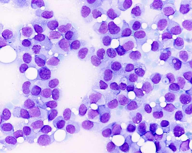
64 Preoperative diagnosis: planning Mammografic pathologic correlation The experience with different biopsy modalities (advantages and disadvantages) The clinical situation and the radiologic image
65 FNAB - categories 1. unsatisfactory 2. benign 3. atypia. probably benign 4. suspicious for malignancy 5. malignant European guidelines... I not performed II insufficient material III benign IV suspicious V malignant unsatisfactory benign suspicious.

66 II insufficient material

67 III benign


68 IV suspicious


69 V malignant The interpreter should feel at ease in making such a diagnosis J Sloane
70
71 Cytological criteria Benign Malignant cellularity - + myoepithelium + - cochesiveness + - atypia - +
72 False positive rate = 0
73 Diagnostic accuracy of FNAB in 240 breast cancers % GIII Med Muc GII GI Lob DCIS Tub all cases Tot, Tabár, Gere: The role of core needle biopsy of breast lesions when fine needle biopsy is inconclusive Acta Cytol 1999;43(4)
74 FNAB : CORE accuracy/tumor type % ductal med muc lob tub FNAB CORE Tot, Tabár, Gere: The role of core needle biopsy of breast lesions when fine needle biopsy is inconclusive Acta Cytol 1999;43(4)

75


76


77
78 False positive rate = 0 Sapino A et al. Ultrasonographically-guided fineneedle aspiration of axillary lymph nodes: role in breast cancer management. Br J Cancer 88(5):702-6, 2003
79 CNB FNAB P value Inadequacy rate 6.9% 17.7% <0.001 Inadequacy rate for malignant lesion 0.9% 4.5% <0.001 Absolute sensitivity 93.1% 74.1% <0.001 Complete sensitivity 97.4% 93.8% =0.001 Absolute diagnostic accuracy 84.5% 71.9% <0.001 False negativity 1.7% 1.7% Specificity 88.3% 95.4% <0.001 Complete diagnostic accuracy 93.2% 95.4% <0.008 Brancato B, Croetti E, Bianchi S et al. Accuracy of needle biopsy of breast lesions visible on ultrasound: Audit of fine needle versus core needlöe biopsy in 3233 consecutive samplings with ascertained outcomes. Breast 21: , 2012


80


81
82 Preoperative diagnosis in 141 cases of fibroadenoma uns ben FA susp mal cyt. core Tot, unpublished results


83
84 False positivity = 0
85 Malignant FNAB = malignant
86 Non - malignant FNAB = anything
87 Diagnostic accuracy of FNAB and Core biopsy, Falun % 90% 80% 70% 60% 50% 40% 30% 20% 10% 0% 65% II III IV V 91% FNAB Core
88 FNAB and CORE biopsy in 2500 breast carcinoma cases (Falun, ) 100% 90% 80% 70% 60% 50% 40% 30% 20% 10% 0% FNAB CORE
89 Vacuum biopsy 3-4 mm thick cores May include in toto an enlarged TDLU May include a transsection of a distended duct May represent an alternative to open surgical intervention in some cases


90
91 INTACT biopsy

92

93

94

95
96 Preoperative diagnosis: planning Mammografic pathologic correlation The experience with different biopsy modalities (advantages and disadvantages) The clinical situation and the radiologic image
97 What I am supposed to do?? To prove malignancy To prove invasion To prove benign character - cystic - solid To role out malignancy
98 What I am supposed to do?? To prove malignancy FNAB/CORE To prove invasion CORE To prove benign character - cystic FNAB - solid CORE To role out malignancy
99 What is the purpose? Prove malignancy! Thinest effective!
100 Diagnostic accuracy of FNAB in 240 breast cancers % GIII Med Muc GII GI Lob DCIS Tub all cases


101 FNAB

102 ductal med muc lob tub


103
104
105 Preoperative diagnosis The aim is to categorize the case as Operable vs non-operable Early vs advanced Extensive vs non-extensive
106 Intra-operative diagnosis Margin assessment Sentinel lymph node procedure
107


108

109
110 Sentinel node Turner et al : 103 patient with metastasis - free sentinel node immunhistology detected metastasis in 14,3 % non-sentinel node of the same series 1/1087 contained micrometastasis Guliano, Kelemen Cancer aug. 1.

111


112

113

114

115


116


117
118 Rapportera: Metastas > 2,0 mm Mikrometastas > 0,2 mm eller fler än 200 tumörceller i ett histologiskt snitt Isolerade tumörceller, ITC (Submikrometastas): <= 0,2 mm eller färr än 200 tumörceller I ett histologiskt snitt Reaktiv lymfkörtel: utan påvisade tumörceller
119 Tumor size Mastectomy LVI LNM ILC n U* 16.3 mm (9,4941) 33% (49+3/158) 18% (28/158) 28% (44/158) 4% (6/158) 158 U invasive MF in situ 14.0 mm (7,3696) 35% (15+7/62) 19% (12/62) 11% (7/62) 10% (6/62) 62 U invasive D in situ 14.0 mm (9,5018) 43% (8+9/40) 28% (11/40) 30% (12/40) 3% (1/40) 40 MF invasive 20.2 mm (11,3253) 66% (51+16/101) 42% (42/101) 48% (48/101) 25% (25/101) 101 MF invasive D in situ 16.5 mm (9,5371) 76% (29+13/55) 53% (29/55) 42% (23/55) 5% (3/55) 55 D invasive Unclassified All invasives n.a. n.a mm (9,8998) 85% (14+3/20) 17% (2/12) 49% (219/448) 10% (2/20) n.a. 28% (124/448) 60% (12/20) 33% (4/12) 33% (150/448) 65% (13/20) 8% (1/12) 12% (55/448) P-value < < < < Tot T. Cancer 110: , 2007
120 Proportion of cases with SN 80.4% (442/550) Unifocal Multifocal Diffuse Comparison of proportions MF vs U 51.1% (182/356) 42.9% (18/42) P< ( ) Comparison of proportions D vs U P< ( ) Negative SN 79.9% (353/442) 60.4% (110/182) 55.6% (10/18) P< ( ) P= ( ) ITC 5.0% (22/442) 6.6% (12/182) 5.6% (1/18) P= ( ) P= ( ) Micrometastasis 5.4% (24/442) 9.9% (18/182) 5.6% (1/18) P= ( ) P= ( ) Macrometastasis 9.7% (43/442) 23.1% (42/182) 33.3% (6/18) P< ( ) P= ( ) False negative SN 1.1% (5/442) 3.3% (6/182) 11.1% (2/18) P= ( ) P= ( ) Radiologically abnormal nodes 11.6% (64/550) 27.2% (97/356) 35.7% (15/42) P< ( ) P< ( ) Complete axillary intervention 26.4% (145/550) 67.7% (241/356) 73.8% (31/42) P< ( ) P< ( ) Tot T.Axillary lymph node status in unifocal, multifocal, and diffuse breast carcinomas: Differences are related to macrometastatic disease. Ann Surg Oncol, 2012
121 Tu size (mm) Unifocal tumors Multifocal tumors Relative risk of macrometastatic Isoletad cells Macrometastasis Micrometastasis Macrometastasis Micrometastasis Isoletad cells diasese (P-value, 95% CI) % (9/115) 1.7% (2/115) 4.7% (7/115) 16.0% (8/50) 8.0% (4/50) 2% (1/50) (P=0.1164, ) % (22/150) 4.0% (6/150) 2.7% (4/150) 36.0% (27/75) 4.0% (3/75) 1.3% (1/75) (P=0.0003, ) % (33/138) 3.6% (5/138) 6.5% (9/138) 46.6% (34/73) 5.5% (4/73) 6.8% (5/73) (P=0.0007, ) % (28/105) 9.5% (10/105) 3.8% (4/105) 59.8% (64/107) 5.6% (6/107) 3.7% (4/107) (P<0.0001, ) % (16/33) 15.2% (5/33) 0.0% (0/33) 78.1% (25/32) 3.1% (1/32) 6.3% (2/32) (P=0.0184, ) % (4/9) 0.0% (0/9) 11.1% (1/9) 73.7% (14/19) 0.0% (0/19) 0.0% (0/19) (P=0.2030, ) Total 20.4% (112/550) 5.1% (28/550) 4.5% (25/550) 48.3% (172/356) 5.1% (18/356) 3.7% (13/356) (P<0.0001, ) Tot T.Axillary lymph node status in unifocal, multifocal, and diffuse breast carcinomas: Differences are related to macrometastatic disease. Ann Surg Oncol, 2012
122 Unifocal Multifocal Diffuse Total RR multifocal vs unifocal RR diffuse vs unifocal Grade 1 9.7%/18.2% (15/28/154) 19.2%/32.8% (11/19/58) 50.0%/50.0% (1/1/2) 12.6%/22.4% (27/48/214) (P=0.0685, ) (P=0.0288, ) Grade %/31.7% (57/85/268) 48.8%/55.5% (102/116/209) 59.5%/69.0% (22/24/37) 35.2%/43.4% (181/223/514) (P<0.0001, ) (P<0.0001, ) Grade %/40.5% (39/51/126) 66.3%/76.4% (59/68/89) 100.0%/100.0% (3/3/3) 46.3%/56.0% (101/122/218) (P<0.0001, ) (P<0.0001, ) Total 20.3%/29.9% (111/164/548) 48.3%/57.0% (172/203/356) 62.0%/66.7% (26/28/42) 32.7%/41.8% (309/395/946) (P<0.0001, ) (P<0.0001, ) Tot T.Axillary lymph node status in unifocal, multifocal, and diffuse breast carcinomas: Differences are related to macrometastatic disease. Ann Surg Oncol, 2012
123 It is far not sufficient to preoperatively verify malignancy; lesion distribution, disease extent, localization, tumor size, tumor stage should also be assessed for adequate therapeutic decision
124 There are no indications for frozen section on breast tissue in modern breast pathology!

125 Tack!
Multiparameter characterization of breast carcinoma: subgross, microscopy, proteins, and genes
 World Congress on Breast Cancer August 1-3, 2015, Birmingham, UK Multiparameter characterization of breast carcinoma: subgross, microscopy, proteins, and genes Tibor Tot Falun, Sweden Lake Varpan, Falun,
Case study 1. Rie Horii, M.D., Ph.D. Division of Pathology Cancer Institute Hospital, Japanese Foundation for Cancer Research
 NCCN/JCCNB Seminar in Japan April 15, 2012 Case study 1 Rie Horii, M.D., Ph.D. Division of Pathology Cancer Institute Hospital, Japanese Foundation for Cancer Research Present illness: A 50y.o.premenopausal
Breast Cancer. Most common cancer among women in the US. 2nd leading cause of death in women. Mortality rates though have declined
 Breast Cancer Most common cancer among women in the US 2nd leading cause of death in women Mortality rates though have declined 1 in 8 women will develop breast cancer Breast Cancer Breast cancer increases
Breast Cancer. Saima Saeed MD
 Breast Cancer Saima Saeed MD Breast Cancer Most common cancer among women in the US 2nd leading cause of death in women 1 in 8 women will develop breast cancer Incidence/mortality rates have declined Breast
Advanced Course on Multimodality Detection and Diagnosis of Breast Diseases
 BREAST SEMINAR Advanced Course on Multimodality Detection and 3D image of the breast tissue Invited speaker LÁSZLÓ TABÁR, MD,FACR (Hon) Falun, Sweden Nov 22-23, 2014 ATHENS, Greece Royal Olympic Hotel
ACRIN 6666 Therapeutic Surgery Form
 S1 ACRIN 6666 Therapeutic Surgery Form 6666 Instructions: Complete a separate S1 form for each separate area of each breast excised with the intent to treat a cancer (e.g. each lumpectomy or mastectomy).
IBCM 2, April 2009, Sarajevo, Bosnia and Herzegovina
 Preoperative diagnosis and treatment planning in breast cancer The pathologist s perspective L. Mazzucchelli Istituto Cantonale di Patologia Locarno, Switzerland IBCM 2, 23-25 April 2009, Sarajevo, Bosnia
Interpretation of Breast Pathology in the Era of Minimally Invasive Procedures
 Shahla Masood, M.D. Professor and Chair Department of Pathology and Laboratory Medicine University of Florida College of Medicine Jacksonville Medical Director, UF Health Breast Center Chief of Pathology
The role of the cytologist in breast cancer screening
 The role of the cytologist in breast cancer screening I.Seili-Bekafigo, MD, PhD Clinical cytologist KBC Rijeka Croatian Society for Clinical Cytology Fine needle aspiration (FNA, FNAB, FNAC) Fine needle
Mammography Education, Inc.
 Hands-on Breast Screening and Diagnosis Course * Screening of 315 full field digital mammography cases. * Reading a mixture of normals and proven abnormals at high resolution viewing stations. * Immediate
How to Use MRI Following Neoadjuvant Chemotherapy (NAC) in Locally Advanced Breast Cancer
 in Locally Advanced Breast Cancer") Global Breast Cancer Conference 2016 & 5 th International Breast Cancer Symposium April 29 th 2016, 09:40-10:50 How to Use MRI Following Neoadjuvant Chemotherapy (NAC) in Locally Advanced Breast Cancer
Global Breast Cancer Conference 2016 & 5 th International Breast Cancer Symposium April 29 th 2016, 09:40-10:50 How to Use MRI Following Neoadjuvant Chemotherapy (NAC) in Locally Advanced Breast Cancer
Lesion Imaging Characteristics Mass, Favoring Benign Circumscribed Margins Intramammary Lymph Node
 Lesion Imaging Characteristics Mass, Favoring Benign Circumscribed Margins Intramammary Lymph Node Oil Cyst Mass, Intermediate Concern Microlobulated Margins Obscured Margins Mass, Favoring Malignant Indistinct
Quality ID #263: Preoperative Diagnosis of Breast Cancer National Quality Strategy Domain: Effective Clinical Care
 Quality ID #263: Preoperative Diagnosis of Breast Cancer National Quality Strategy Domain: Effective Clinical Care 2018 OPTIONS FOR INDIVIDUAL MEASURES: REGISTRY ONLY MEASURE TYPE: Process DESCRIPTION:
Surgical Pathology Issues of Practical Importance
 Surgical Pathology Issues of Practical Importance Anne Moore, MD Medical Oncology Syed Hoda, MD Surgical Pathology The pathologist is central to the team approach needed to manage the patient with breast
FIBROEPITHELIAL LESIONS
 DEFINITIONS FIBROEPITHELIAL LESIONS Suzanne Moore FIBROADENOMA- A discrete benign tumour showing evidence of connective tissue and epithelial proliferation- WHO Fibrous stromal element of these tumours
PAAF vs Core Biopsy en Lesiones Mamarias Case #1
 5/19/2014 PAAF vs Core Biopsy en Lesiones Mamarias Case #1 Fine Needle Aspiration Cytology of Breast: Correlation with Needle Core Biopsy 64-year-old woman Mass in breast Syed Hoda, MD CD31 Post-Radiation
Ultrasound or FNA for Predicting Node Positive in Breast Cancer
 Ultrasound or FNA for Predicting Node Positive in Breast Cancer Chiun Sheng Huang, MD, PhD, MPH Professor and Chairman Department of Surgery Director of Breast Care Center National Taiwan University Hospital
Tibor Tot. 1. Introduction
 International Breast Cancer Volume 2012, Article ID 395415, 8 pages doi:10.1155/2012/395415 Review Article The Role of Large-Format Histopathology in Assessing Subgross Morphological Prognostic Parameters:
ROLE OF MRI IN SCREENING, DIAGNOSIS AND MANAGEMENT OF BREAST CANCER. B.Zandi Professor of Radiology
 ROLE OF MRI IN SCREENING, DIAGNOSIS AND MANAGEMENT OF BREAST CANCER B.Zandi Professor of Radiology Introduction In the USA, Breast Cancer is : The Most Common Non-Skin Cancer The Second Leading cause of
Mammographic imaging of nonpalpable breast lesions. Malai Muttarak, MD Department of Radiology Chiang Mai University Chiang Mai, Thailand
 Mammographic imaging of nonpalpable breast lesions Malai Muttarak, MD Department of Radiology Chiang Mai University Chiang Mai, Thailand Introduction Contents Mammographic signs of nonpalpable breast cancer
Basement membrane in lobule.
 Bahram Memar, MD Basement membrane in lobule. Normal lobule-luteal phase Normal lobule-follicular phase Lactating breast Greater than 95% are adenocarcinomas in situ carcinomas and invasive carcinomas.
Breast Cancer: Basic and Clinical Research 2014:8
 Open Access: Full open access to this and thousands of other papers at http://www.la-press.com. Breast Cancer: Basic and Clinical Research A Proposal to Unify the Classification of Breast and Prostate
Imaging in breast cancer. Mammography and Ultrasound Donya Farrokh.MD Radiologist Mashhad University of Medical Since
 Imaging in breast cancer Mammography and Ultrasound Donya Farrokh.MD Radiologist Mashhad University of Medical Since A mammogram report is a key component of the breast cancer diagnostic process. A mammogram
Treatment options for the precancerous Atypical Breast lesions. Prof. YOUNG-JIN SUH The Catholic University of Korea
 Treatment options for the precancerous Atypical Breast lesions Prof. YOUNG-JIN SUH The Catholic University of Korea Not so benign lesions? Imaging abnormalities(10% recall) lead to diagnostic evaluation,
Atypical proliferative lesions diagnosed on core biopsy - 6 year review
 Atypical proliferative lesions diagnosed on core biopsy - 6 year review Dr Angela Harris, Dr Julie Weigner & Dr Ricardo Vilain NSW Health Pathology Pathology North, Hunter Anatomical Pathology & Cytology
Mammography Education, Inc.
 Hands-on Breast Screening and Diagnosis Course * Screening of 315 full field digital mammography cases. * Reading a mixture of normals and proven abnormals at high resolution viewing stations. * Immediate
ANNEX 1 OBJECTIVES. At the completion of the training period, the fellow should be able to:
 1 ANNEX 1 OBJECTIVES At the completion of the training period, the fellow should be able to: 1. Breast Surgery Evaluate and manage common benign and malignant breast conditions. Assess the indications
Mammography Education, Inc.
 Mammography Education, Inc. 2018 3D image of the breast tissue BREAST SEMINAR SERIES Faculty LÁSZLÓ TABÁR, MD, FACR (Hon) Professor emeritus of Radiology The Critical Role of the Breast Imaging Technologists
Pitfalls and Limitations of Breast MRI. Susan Orel Roth, MD Professor of Radiology University of Pennsylvania
 Pitfalls and Limitations of Breast MRI Susan Orel Roth, MD Professor of Radiology University of Pennsylvania Objectives Review the etiologies of false negative breast MRI examinations Discuss the limitations
Ana Sofia Preto 19/06/2013
 Ana Sofia Preto 19/06/2013 Understanding the underlying pathophysiologic processes leading to the various types of calcifications Description and illustration of the several types of calcifications, according
Consensus Guideline on Image-Guided Percutaneous Biopsy of Palpable and Nonpalpable Breast Lesions
 Consensus Guideline on Image-Guided Percutaneous Biopsy of Palpable and Nonpalpable Breast Lesions Purpose: To outline the use of minimally invasive biopsy techniques (MIBT) for palpable and nonpalpable
Surgical Therapy: Sentinel Node Biopsy and Breast Conservation
 Surgical Therapy: Sentinel Node Biopsy and Breast Conservation Stephen B. Edge, MD Professor of Surgery and Oncology Roswell Park Cancer Institute University at Buffalo Dr. Roswell Park: Tradition in Cancer
Mammography Education, Inc.
 Mammography Education, Inc. 2018 3D image of the breast tissue BREAST SEMINAR SERIES Faculty LÁSZLÓ TABÁR, MD, FACR (Hon) Professor emeritus of Radiology Using the Multimodality Approach. A FULLY INTERACTIVE,
Pathology of Lobular & Ductal Preneoplasia. Syed A Hoda, MD Weill-Cornell, New York, NY
 Pathology of Lobular & Ductal Preneoplasia Syed A Hoda, MD Weill-Cornell, New York, NY Proliferative Epithelial Changes in Breast A wide range of proliferative epithelial changes occur in the breast There
Problems in staging breast carcinoma
 Problems in staging breast carcinoma Primary systemic therapy (PST) of breast carcinoma pathologists tasks Dr. Janina Kulka, 2nd Department of Pathology, Semmelweis University Budapest Austro-Hungarian
Papillary Lesions of the breast
 Papillary Lesions of the breast Emad Rakha Professor of Breast Pathology The University of Nottingham Papillary lesions of the breast are a heterogeneous group of disease, which are characterised by neoplastic
The relationship of multifocality and tumor burden with various tumor characteristics and survival in early breast cancer
 566 Neoplasma 59, 5, 2012 doi:10.4149/neo_2012_073 The relationship of multifocality and tumor burden with various tumor characteristics and survival in early breast cancer G. KELEMEN 1, V. FARKAS 1, J.
04/10/2018 HIGH RISK BREAST LESIONS. Pathology Perspectives of High Risk Breast Lesions ELEVATED RISK OF BREAST CANCER HISTORICAL PERSPECTIVES
 Pathology Perspectives of High Risk Breast Lesions Savitri Krishnamurthy MD Professor of Pathology Deputy Division Head Director of Clinical Trials, Research and Development The University of Texas MD
16/09/2015. ACOSOG Z011 changing practice. Presentation outline. Nodal mets #1 prognostic tool. Less surgery no change in oncologic outcomes
 ACOSOG Z011 changing practice The end of axillary US/FNA? Preoperative staging of the axilla in the era of Z011 Adena S Scheer MD MSc FRCSC Surgical Oncologist, St. Michael s Hospital Assistant Professor,
Successful Breast MRI Program : The ingredients
 Successful Breast MRI Program : The ingredients Dr. Smriti Hari Associate Professor Deptt. Of Radiology All India Institute of Medical Sciences New Delhi How to perform Breast MRI Breast MRI descriptors
Breast Cancer Diagnosis, Treatment and Follow-up
 Breast Cancer Diagnosis, Treatment and Follow-up What is breast cancer? Each of the body s organs, including the breast, is made up of many types of cells. Normally, healthy cells grow and divide to produce
Proliferative Breast Disease: implications of core biopsy diagnosis. Proliferative Breast Disease
 Proliferative Breast Disease: implications of core biopsy diagnosis Jean F. Simpson, M.D. Breast Pathology Consultants, Inc. Nashville, TN Proliferative Breast Disease Must be interpreted in clinical and
STAGE CATEGORY DEFINITIONS
 CLINICAL Extent of disease before any treatment y clinical staging completed after neoadjuvant therapy but before subsequent surgery TX Tis Tis (DCIS) Tis (LCIS) Tis (Paget s) T1 T1mi T1a T1b T1c a b c
Ductal Carcinoma in Situ (DCIS)
") Diagnosis and Treatment of Patients with Primary and Metastatic Breast Cancer Ductal Carcinoma in Situ (DCIS) Ductal Carcinoma in Situ DCIS Versions 2002 2017: Audretsch / Blohmer / Brunnert / Budach /
Diagnosis and Treatment of Patients with Primary and Metastatic Breast Cancer Ductal Carcinoma in Situ (DCIS) Ductal Carcinoma in Situ DCIS Versions 2002 2017: Audretsch / Blohmer / Brunnert / Budach /
Case Report Synchronous Bilateral Solid Papillary Carcinomas of the Breast
 Case Reports in Surgery Volume 2013, Article ID 812129, 4 pages http://dx.doi.org/10.1155/2013/812129 Case Report Synchronous Bilateral Solid Papillary Carcinomas of the Breast Noriko Yoshimura, 1 Shigeru
Carcinoma mammario: le istologie non frequenti. Valentina Guarneri Università di Padova IOV-IRCCS
 Carcinoma mammario: le istologie non frequenti Valentina Guarneri Università di Padova IOV-IRCCS Histological diversity of breast adenocarcinomas Different histological types are defined according to specific
Breast pathology. 2nd Department of Pathology Semmelweis University
 Breast pathology 2nd Department of Pathology Semmelweis University Breast pathology - Summary - Benign lesions - Acute mastitis - Plasma cell mastitis / duct ectasia - Fat necrosis - Fibrocystic change/
CURRENT METHODS IN IMAGE GUIDED BREAST BIOPSY
 CURRENT METHODS IN IMAGE GUIDED BREAST BIOPSY Stuart Silver April 24, 2004 OBJECTIVES Review development of current techniques Discuss stereotactic breast biopsy Discuss US guided breast biopsy 1 OBJECTIVES
Diagnosis and Treatment of Patients with Primary and Metastatic Breast Cancer. Pathology. AGO e. V. in der DGGG e.v. sowie in der DKG e.v.
 Diagnosis and Treatment of Patients with Primary and Metastatic Breast Cancer Pathology Pathology Versions 2004 2017: Blohmer / Costa / Fehm / Friedrichs / Huober / Kreipe / Lück / Schneeweis / Sinn /
Newly Diagnosed Breast Cancer: Preoperative Imaging and Localization
 Newly Diagnosed Breast Cancer: Preoperative Imaging and Localization Debra Monticciolo, MD Professor of Radiology Texas A&M University no disclosures Debra Monticciolo, MD Professor of Radiology Texas
BreastScreen Victoria Annual Statistical Report
 BreastScreen Victoria Annual Statistical Report 29 BREASTSCREEN VICTORIA: ANNUAL STATISTICAL REPORT, 29 Produced by: BreastScreen Victoria Coordination Unit Level, 3 Pelham Street, Carlton South Victoria
Post Neoadjuvant therapy: issues in interpretation
 Post Neoadjuvant therapy: issues in interpretation Disclosure: Overview D Prognostic features in assessment of post treatment specimens: Tumor size Cellularity Grade Receptors LN Neoadjuvant chemotherapy:
Atypical Ductal Hyperplasia and Papillomas: A Comparison of Ultrasound Guided Breast Biopsy and Stereotactic Guided Breast Biopsy
 Atypical Ductal Hyperplasia and Papillomas: A Comparison of Ultrasound Guided Breast Biopsy and Stereotactic Guided Breast Biopsy Breast Cancer is the most common cancer diagnosed in women in the United
Descriptor Definition Author s notes TNM descriptors Required only if applicable; select all that apply multiple foci of invasive carcinoma
 S5.01 The tumour stage and stage grouping must be recorded to the extent possible, based on the AJCC Cancer Staging Manual (7 th Edition). 11 (See Tables S5.01a and S5.01b below.) Table S5.01a AJCC breast
Mammography Education, Inc.
 Mammography Education, Inc. 2018 3D image of the breast tissue Sept 18-21 Hs-on Screening Course combined with Multimodality Diagnosis of Breast Diseases with Emphasis on Breast MRI SIGTUNA Hotel Kristina
Histological Type. Morphological and Molecular Typing of breast Cancer. Nottingham Tenovus Primary Breast Cancer Study. Survival (%) Ian Ellis
 Ian Ellis") Morphological and Molecular Typing of breast Cancer Ian Ellis Molecular Medical Sciences, University of Nottingham Department of Histopathology, Nottingham University Hospitals NHS Trust Histological Type
Morphological and Molecular Typing of breast Cancer Ian Ellis Molecular Medical Sciences, University of Nottingham Department of Histopathology, Nottingham University Hospitals NHS Trust Histological Type
DIAGNOSIS. Biopsy, Pathology and Subtypes. Knowledge Summary
 DIAGNOSIS Biopsy, Pathology and Subtypes Knowledge Summary DIAGNOSIS Biopsy, Pathology and Subtypes INTRODUCTION The success of an effective breast health care program is directly related to the availability
Recent Update in Surgery for the Management of Breast Cancer
 Recent Update in Surgery for the Management of Breast Cancer Wonshik Han, MD, PhD Professor, Department of Surgery, Seoul National University College of Medicine Chief of Breast Care Center, Seoul National
Breast Pathology. Breast Development
 Breast Pathology Lecturer: Hanina Hibshoosh, M.D. Reading: Kumar, Cotran, Robbins, Basic Pathology, 6th Edition, pages 623-635 Breast Development 5th week - thickening of the epidermis - milk line 5th
Breast MRI: Friend or Foe?
 Breast MRI: Friend or Foe? UCSF Postgraduate Course May 18, 2013 Cheryl Ewing, MD Clinical Professor of Surgery UCSF Department of Surgery APPLEGATE HAS DOUBLE MASTECTOMY IN CANCER SCARE DIAGNOSED WITH
Radioactive Seed Localization of Nonpalpable Breast Lesions
 Subject: Radioactive Seed Localization of Page: 1 of 7 Last Review Status/Date: March 2017 Radioactive Seed Localization of Description Radioactive seed localization is used to detect nonpalpable breast
BREAST PATHOLOGY. Fibrocystic Changes
 BREAST PATHOLOGY Lesions of the breast are very common, and they present as palpable, sometimes painful, nodules or masses. Most of these lesions are benign. Breast cancer is the 2 nd most common cause
Fine-Needle Aspiration and Cytologic Findings of Surgical Scar Lesions in Women With Breast Cancer
 148 Fine-Needle Aspiration and Cytologic Findings of Surgical Scar Lesions in Women With Breast Cancer Ehud Malberger, DMD, FIAC,* Yeouda Edoute, MD, PhD,t Osnaf Toledano, MD,* and Dov Sapir, MDS Benign
BREAST MRI. Elizabeth A. Rafferty, M.D. Avon Comprehensive Breast Center Massachusetts General Hospital Harvard Medical School
 BREAST MRI Elizabeth A. Rafferty, M.D. Avon Comprehensive Breast Center Massachusetts General Hospital Harvard Medical School BREAST MRI Any assessment of the breast parenchyma requires the administration
Study of Fine Needle Aspiration Cytology of Breast Lump: Correlation of Cytologically Malignant Cases with Their Histological Findings
 Study of Fine Needle Aspiration Cytology of Breast Lump: Correlation of Cytologically Malignant Cases with Their Histological Findings Touhid Uddin Rupom 1, Tamanna Choudhury 2, Sultana Gulshana Banu 3
BreastScreen Victoria Annual Statistical Report
 BreastScreen Victoria Annual Statistical Report 005 Produced by: BreastScreen Victoria Coordination Unit Level, Pelham Street, Carlton South Victoria 05 PH 0 9660 6888 FX 0 966 88 EM info@breastscreen.org.au
Case Scenario 1: This case has been slightly modified from the case presented during the live session to add clarity.
 Case Scenario 1: This case has been slightly modified from the case presented during the live session to add clarity. Background: 46 year old married premenopausal female with dense breasts has noticed
National Diagnostic Imaging Symposium 2013 SAM - Breast MRI 1
 National Diagnostic Imaging Symposium 2013 December 8-12, 2013 Disney s Yacht Club Resort Lake Buena Vista, Florida Self Assessment Module Questions, Answers and References Day SAM Title - Each SAM title
Papillary Lesions of the Breast: WHO Update
 Papillary Lesions of the Breast: WHO Update Stuart J. Schnitt, M.D. Department of Pathology Beth Israel Deaconess Medical Center and Harvard Medical School Boston, MA, USA Papillary Lesions of the Breast
2017BREAST SEMINAR SERIES
 Hands-on Breast Screening and Diagnosis Course * Screening of 510 full field digital mammography cases. * Reading a mixture of normals and proven abnormals at high resolution viewing stations. * Immediate
BREAST MRI. Elizabeth A. Rafferty, M.D. Avon Comprehensive Breast Center Massachusetts General Hospital Harvard Medical School
 BREAST MRI Elizabeth A. Rafferty, M.D. Avon Comprehensive Breast Center Massachusetts General Hospital Harvard Medical School BREAST MRI Any assessment of the breast parenchyma requires the administration
Evaluation of Pathologic Response in Breast Cancer Treated with Primary Systemic Therapy
 Evaluation of Pathologic Response in Breast Cancer Treated with Primary Systemic Therapy Eun Yoon Cho, MD, PhD Department of Pathology and Translational Genomics Samsung Medical Center Sungkyunkwan University
Diagnosis and staging of breast cancer and multidisciplinary team working
 1 Diagnosis and staging of breast cancer and multidisciplinary team working Common symptoms and signs Over 90% of breast cancers (BCs) are local or regional when first detected. At least 60% of patients
Research Article Ductal Breast Carcinoma In Situ: Mammographic Features and Its Relation to Prognosis and Tumour Biology in a Population Based Cohort
 Hindawi International Journal of Breast Cancer Volume 2017, Article ID 4351319, 9 pages https://doi.org/10.1155/2017/4351319 Research Article Ductal Breast Carcinoma In Situ: Mammographic Features and
The EBC Council Manifesto on optimal breast pathology
 ECP 2017 Amsterdam Joint Symposium Breast Pathology / Pathology in favour of developing countries: Diagnostic management of breast cancer in low resource settings The EBC Council Manifesto on optimal breast
Case Scenario 1: This case has been slightly modified from the case presented during the live session to add clarity.
 Case Scenario 1: This case has been slightly modified from the case presented during the live session to add clarity. Background: 46 year old married premenopausal female with dense breasts has noticed
Imaging the Symptomatic Patient. Avice M.O Connell MD,FACR,FSBI Professor of Imaging Sciences Director, Women s Imaging University of Rochester
 Imaging the Symptomatic Patient Avice M.O Connell MD,FACR,FSBI Professor of Imaging Sciences Director, Women s Imaging University of Rochester The four most common symptoms Mass Pain Discharge Infection
What is Cancer? Petra Ketterl, MD Medical Oncology and Functional Medicine
 What is Cancer? Petra Ketterl, MD Medical Oncology and Functional Medicine What is Cancer? Layman s terms: cancer starts when cells grow out of control (in any place in the body) and crowd out normal cells
04/10/2018. Intraductal Papillary Neoplasms Of Breast INTRADUCTAL PAPILLOMA
 Intraductal Papillary Neoplasms Of Breast Savitri Krishnamurthy MD Professor of Pathology Deputy Division Head The University of Texas MD Anderson Cancer Center 25 th Annual Seminar in Pathology Pittsburgh,
Utility of Fine Needle Aspiration Cytology in Evaluation of Breast Lesions.
 Utility of Fine Needle Aspiration Cytology in Evaluation of Breast Lesions. Komal Joshi 1*, Nandita Mehta 2, Hansa Goswami 3 1 2 nd Year Resident, Ahmedabad. 2 Professor, 3 Professor & Head, Pathology
Scottsdale, AZ Plaza Hotel, 7200 N. Scottsdale Rd
 Mammography Education, Inc. 2014 3D image of the breast tissue LÁSZLÓ TABÁR, M.D.,F.A.C.R (Hon) and STAMATIA DESTOUNIS, M.D., F.A.C.R. The normal TDLUs have bud-like acini Multimodality Approach to Detection
Contrast-enhanced Breast MRI RSSA 2013
 Contrast-enhanced Breast MRI RSSA 2013 Prof. dr. Maurice van den Bosch University Medical Center Utrecht, the Netherlands Index 1) Breast cancer 2) Why MRI of the breast 3) Technique 4) Interpretation
Atypical And Suspicious Categories In Fine Needle Aspiration Cytology Of The Breast
 IOSR Journal of Dental and Medical Sciences (IOSR-JDMS) e-issn: 2279-853, p-issn: 2279-861.Volume 15, Issue 1 Ver. III (October. 216), PP 57-61 www.iosrjournals.org Atypical And Suspicious Categories in
Mammography Education, Inc.
 Mammography Education, Inc. 2018 3D image of the breast tissue BREAST SEMINAR SERIES Faculty LÁSZLÓ TABÁR, MD, FACR (Hon) Professor emeritus of Radiology and GILLIAN NEWSTEAD, MD, FACR Professor emeritus
CPC 4 Breast Cancer. Rochelle Harwood, a 35 year old sales assistant, presents to her GP because she has noticed a painless lump in her left breast.
 CPC 4 Breast Cancer Rochelle Harwood, a 35 year old sales assistant, presents to her GP because she has noticed a painless lump in her left breast. 1. What are the most likely diagnoses of this lump? Fibroadenoma
Image guided core biopsies:
 Recommendations on the Surgical, Radiologic and Pathologic Approaches to Breast Disease: Using best practices based on multidisciplinary methodologies developed through the Allina Breast Committee. Image
Mammographic evaluation of palpable breast masses with pathological correlation: a tertiary care centre study in Nepal
 Original article 21 Mammographic evaluation of palpable breast masses with pathological correlation: a tertiary care centre study in Nepal G. Gurung, R. K. Ghimire, B. Lohani Department of Radiology and
Table of contents. Page 2 of 40
 Page 1 of 40 Table of contents Introduction... 4 1. Background Information... 6 1a: Referral source for the New Zealand episodes... 6 1b. Invasive and DCIS episodes by referral source... 7 1d. Age of the
Cytological study of palpable breast lumps with their histological correlation in a tertiary care hospital
 Original Research Article Cytological study of palpable breast lumps with their histological correlation in a tertiary care hospital Sunita Mistry 1*, Jignasha Patel 2, Kamlesh Shah 3, Ajit Patel 4 1 Assistant
3/27/2017. Disclosure of Relevant Financial Relationships. Papilloma???
 Management of Papillary Lesions Diagnosed at Rad Path Concordant Core Biopsy (CNB) Disclosure of Relevant Financial Relationships USCAP requires that all planners (Education Committee) in a position to
Resection Margins in Breast Conserving Surgery. Alberto Costa, MD Canton Ticino Breast Unit Lugano, Switzerland
 Resection Margins in Breast Conserving Surgery Alberto Costa, MD Canton Ticino Breast Unit Lugano, Switzerland Breast Conserving Surgery 1 Probably one of the most important innovation in cancer surgery
Angela Gilliam, MD University of Colorado Surgical Grand Rounds November 3, 2008
 Angela Gilliam, MD University of Colorado Surgical Grand Rounds November 3, 2008 Breast Cancer Most common cancer in American women 180,000 new cases per year Second most common cause of cancer death 44,000
Case 1. BREAST CANCER From Diagnosis to Treatment: The Role of Primary Care
 BREAST CANCER From Diagnosis to Treatment: The Role of Primary Care Leah Karliner, MD MAS University of California San Francisco Primary Care Medicine Update 2009 April 2009 Case 1 AR, a 60 year old African
Papillary Lesions of the Breast
 Papillary Lesions of the Breast Laura C. Collins, M.D. Associate Professor of Pathology Associate Director, Division of Anatomic Pathology Beth Israel Deaconess Medical Center and Harvard Medical School
Papillary Lesions of the Breast A Practical Approach to Diagnosis. (Arch Pathol Lab Med. 2016;140: ; doi: /arpa.
 Papillary Lesions of the Breast A Practical Approach to Diagnosis (Arch Pathol Lab Med. 2016;140:1052 1059; doi: 10.5858/arpa.2016-0219-RA) Papillary lesions of the breast Span the spectrum of benign,
Benign Breast Disease and Breast Cancer Risk
 Benign Breast Disease and Breast Cancer Risk Jean F. Simpson, M.D. Vanderbilt University Nashville, Tennessee December 1, 2011 Nashville Nashville Lebanon 1 Cedars of Lebanon State Park The American University
Use of a Protease Activated System for Real-time Breast Cancer Lumpectomy Margin Assessment
 Use of a Protease Activated System for Real-time Breast Cancer Lumpectomy Margin Assessment Barbara L. Smith, MD, PhD Professor of Surgery, Harvard Medical School Division of Surgical Oncology Massachusetts
Current Status of Supplementary Screening With Breast Ultrasound
 Current Status of Supplementary Screening With Breast Ultrasound Stephen A. Feig, M.D., FACR Fong and Jean Tsai Professor of Women s Imaging Department of Radiologic Sciences University of California,
Case Scenario 1. 2/15/2011 The patient received IMRT 45 Gy at 1.8 Gy per fraction for 25 fractions.
 Case Scenario 1 1/3/11 A 57 year old white female presents for her annual mammogram and is found to have a suspicious area of calcification, spread out over at least 4 centimeters. She is scheduled to
Diagnostic Dilemmas of Breast Imaging
 Diagnostic Dilemmas of Breast Imaging Common Causes of Error in Breast Cancer Detection By: Jason Cord, M.D. Mammography: Initial Imaging The standard for detection of breast cancer Screening mammography