Mammography Education, Inc.
|
|
|
- Edith Baker
- 5 years ago
- Views:
Transcription


1 Mammography Education, Inc D image of the breast tissue BREAST SEMINAR SERIES Faculty LÁSZLÓ TABÁR, MD, FACR (Hon) Professor emeritus of Radiology Using the Multimodality Approach. A FULLY INTERACTIVE, UNIQUE LEARNING EXPERIENCE <10 mm invasive breast cancer NEW course design October 19-21, 2018 OTTAWA, Canada Shaw Centre in downtown Ottawa 55 Colonel By Drive, Ottawa, Canada Sea urchin 21 hours of Category I CME credits Designed for: Radiologists Surgeons Pathologists Gynecologists Radiology Technologists This course provides extensive knowledge about diagnostic breast imaging, differential diagnosis of breast diseases, implications for management and newest diagnostic technologies


2 FACULTY. Professor emeritus of Radiology, Department of Mammography Falun, Sweden Images from the non-profit Tabar Foundation for Research and Education for Breast Cancer II

3 Mammography Education, Inc. is accredited by the Accreditation Council for Continuing Medical Education to sponsor continuing medical education for physicians. Mammography Education, Inc. designed these medical education activities for a maximum of 21 credit hours incategory I of the Physicians' Recognition Award of the American Medical Association. Each physician should claim only those hours of credit that he / she actually spent in the educational activity. NEW COURSE DESIGN * The lectures on each major subject will be followed by interactive screening sessions consisting of a mixture of normal and early cancer cases presented on the large screen exactly as they appear on a viewing station at screening. Using a specially provided polling program downloaded to each participant's smartphone or tablet, the attendees will be asked to vote anonymously on each case. The aggreate results will appear instantly for discussion and evaluation. This new course design gives immediate feedback demonstrating the effectiveness of various screening methods. * During the course the attendees will progressively improve their interpretive expertise, as they learn the full spectrum of normal breast images, with all important findings explained with the help of 3-dimensional histology images. * These skills will lead to fewer call-backs and greater confidence in reading a large number of mammograms. * Immediate feedback and discussion of every case throughout every reading session. * Special emphasis will be placed on finding early phase breast cancers. * All abnormal cases are fully worked up and the complete imaging workup will be presented in detail, including ultrasound, MRI and large section histopathology. CREDITS We would like to thank the sponsors for their support of the teaching seminars of Mammography Education, Inc (list of vendors will be presented at the beginning of the course) III


 histopathologic correlation enables the radiologist to account for")
4 Day 1 Morning lectures between 8:30 AM - 12:00 PM. Breaks: 10:00 AM, 11:00 AM 8:30 INTRODUCTION FOLLOWED BY DIDACTIC LECTURES COVERING: A NEW ERA in the DIAGNOSIS and TREATMENT of BREAST CANCER. A SHORT HISTORY. HOW TO READ A MAMMOGRAM. THE BASIS FOR SKILLFUL AND EFFICIENT INTERPRETATION OF THE MAMMOGRAPHIC IMAGE Correlating 3-dimensional, subgross anatomy with mammography of the normal breast results in increased confidence in reading a mammogram and finding small abnormalities. Special training in large format thin and thick section (3D) histopathologic correlation enables the radiologist to account for every linear and nodular density on the mammogram. The breast, unlike any other organ, has five structurally different mammographic parenchymal patterns. 12:00 PM - 1:00 PM L u n c h IV


.")
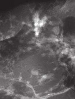
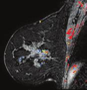
5 Day 1 Afternoon lectures: 1:00 PM - 4:30 PM. Breaks at 2:30 and 3:30 PM ALGORITHM FOR CLASSIFYING BREAST DISEASES ACCORDING TO THEIR SITE OF ORIGIN HOW TO FIND THE INVASIVE BREAST CANCER WHEN IT IS STILL SMALL. Malignant stellate and circular/oval-shaped lesions originating from the TDLUs (AAB): clinical presentation, histology, mammographic - MRI - ultrasound appearance and outcome. A systematic method for viewing mammograms. Areas on the mammogram where most breast cancers will be found. Viewing dense breasts. Viewing relatively easyto-read breasts. The role of hand-held ultrasound / 3D automated ultrasound / MRI in the detection and workup of the findings. The multimodality approach Interactive screening session (see description next page). Example: Multifocal invasive and in situ carcinoma, where the extensive micropapillary cancer originating from the major ducts was well demonstrated on breast MRI. 4:30 PM. End of Day 1. V

 Continuation A systematic method for viewing mammograms.")


6 Day 2 Morning lectures between 8:30 AM - 12:00 PM. Breaks:10:00 AM, 11:00 AM 8:30 HOW TO FIND THE INVASIVE BREAST CANCER WHEN IT IS STILL SMALL. SCREENING COMBINED WITH AN ANALYTICAL APPROACH FOR THE DIFFERENTIAL DIAGNOSIS OF STELLATE / SPICULATED LESIONS (AAB) Continuation A systematic method for viewing mammograms. Areas on the mammogram where most breast cancers will be found. Viewing dense breasts. Partenchymal contour changes, non-calcified architectural distortion, unifocal / multifocal / diffuse breast cancers. Interactive screening session: Using what has just been taught, each participant will assess a mixture of normal and early cancer cases, and vote anonymously using a smartphone or tablet. The combined results will appear instantly for discussion. and evaluation. Multifocal invasive and in situ carcinoma on an area measuring 180X60 mm pn 4/9. 12:00 PM - 1:00 PM L u n c h VI


7 Day 2 Afternoon lectures: 1:00 PM - 4:30 PM. Breaks at 2:30 and 3:30 PM 1:00 PM ASYMMETRIC DENSITIES ON THE MAMMOGRAM Didactic workup of non-specific asymmetric densities without architectural distortion Didactic workup of non-specific asymmetric densities with architectural distortion ANALYSIS of BENIGN RADIATING STRUCTURES on the mammogram, originating in the ducts - Radial scar. A suggested algorithm for the workup of stellate lesions Radial scar Neoductgenesis ANALYSIS of MALIGNANT LESIONS PRESENTED as non-calcified RADIATING STRUCTURES on the mammogram. Clinical presentation, mammographic appearance and outcome: - Diffuse invasive breast cancer: the most deceptive and frequently missed cancer of the breast. The value of ultrasound and MRI in finding and diagnosing diffusely invasive breast cancer subtypes. Case demonstrations - Neoductgenesis cases presenting on the mammogram as architectural distortion - A suggested algorithm for the workup of lesions with architectural distortion Interactive session for detecting architectural distortion on the mammogram. 4:30 PM. End of Day 2 Multimodality workup of a huge diffuse invasive carcinoma. VII


 Fragmented casting type calcifications b) Dotted")
")
 pearl necklace-like.")


8 Day Morning lectures: 8:00 AM - 12:00 PM. Breaks at 9:30 and 11:00 AM 8:00 AM INTERACTIVE LECTURE SERIES WILL COVER THE FOLLOWING TOPICS. ALGORITHM FOR CLASSIFYING BREAST DISEASES ACCORDING TO THEIR SITE OF ORIGIN Breast diseases originating in the major ducts Benign type calcifications originating in the major ducts a) Secretory disease type calcifications Malignant type calcifications originating in the major ducts Interactive calcification analysis. a) Fragmented casting type calcifications b) Dotted casting type calcifications * Four different malignant type calcifications developing in the major ducts: a) fragmented casting type b) dotted casting type c) skipping stone-like d) pearl necklace-like. * The concept of neoductgenesis. Long-term follow-up results. New aspects, correct terminology. * The role of breast MRI examination in demonstrating the extent of Gr 3 in situ carcinoma. * Mammographic/3D histologic correlation helping to explain the underlying pathophysiology and outcome. 5:30 End of Day 2 VIII c) Skipping stone-like calcifications 12:00 PM - 1:00 PM L u n c h d) Pearl necklace-like calcifica-


, powdery calcifications (B), pleomorphic A")
 and associated with calcifications on")

9 Day 3 Afternoon lectures: 1:00 PM - 4:30 PM. Breaks: 2:30 PM and 3:30 PM 1:00 PM. ALGORITHM FOR CLASSIFYING BREAST DISEASES ACCORDING TO THEIR SITE OF ORIGIN Benign breast diseases originating in the TDLU and associated with calcifications on the mammogram - - Fibrocystic change. Fibroadenoma. Different types of adenosis. Understanding pathophysiology leading to calcified and non-calcified hyperplastic breast changes. Conventional and 3D histology images of small breast cysts containing sediment of psammoma body-like calcifications, seen as "teacup-like calcifications on the mammogram. - Detailed analysis of calcifications associated with hyperplastic breast changes Weddellites (A), powdery calcifications (B), pleomorphic calcifications on the mammogram. A B Malignant breast diseases originating in the TDLU(s) and associated with calcifications on the mammogram (Grade 1 and 2). Grade 2 cancer in situ: Mammographic / 3-D histologic / MRI correlation of cases with crushed stone-like/pleomorphic calcifications on the mammogram. 4:30 PM End of the course IX

 419 0219 e-mail: info@mammographyed.com Internet: www.")


10 For more information and registration please contact: Mammography Education, Inc E. Spur Drive CAVE CREEK, AZ 85331, USA Phone: (480) Fax: (480) Internet: The schedule is subject to change without notice and does not represent a commitment on the part of M.E.I. All rights reserved including the right of reproduction in whole or in part of any form. Visit us on the Internet: Copyright Computer simulation images of the development of Grade 2 in situ carcinoma within the TDLU. The lobule becomes gradually distended and deformed. Calcifications are formed within the necrotic debris and are seen on the mammogram as crushed stone-like calcifications. X


11


12


13


14


, Part 5")


15 2018 László Tabár, MD Tibor Tot, MD, Peter B. Dean, MD Ductal Adenocarcinoma of the Breast (DAB), Part 5 Fluid producing DAB subtypes associated with calcifications In 3D László Tabár, MD Tibor Tot, MD, Peter B. Dean, MD Bloody and serous nipple discharge Ductal Adenocarcinoma of the Breast (DAB), Part 6 In 3D


16 László Tabár, MD Tibor Tot, MD, Peter B. Dean, MD In 3D Breast cancer originating from the major ducts Ductal Adenocarcinoma of the Breast (DAB), Part 7 Architectural distorion on the mammogram without calcifications or nipple discharge Mammographic-MRI-subgross (3D) histologic correlation of this extensive micropapillary cancer originating from the major ducts presenting as architectural distortion.
17
Mammography Education, Inc.
 Mammography Education, Inc. 2018 3D image of the breast tissue BREAST SEMINAR SERIES Faculty LÁSZLÓ TABÁR, MD, FACR (Hon) Professor emeritus of Radiology Using the Multimodality Approach A FULLY INTERACTIVE,
Mammography Education, Inc.
 Mammography Education, Inc. 2018 3D image of the breast tissue BREAST SEMINAR SERIES Faculty LÁSZLÓ TABÁR, MD, FACR (Hon) Professor emeritus of Radiology The Critical Role of the Breast Imaging Technologists
Mammography Education, Inc.
 Mammography Education, Inc. 2018 3D image of the breast tissue BREAST SEMINAR SERIES Faculty LÁSZLÓ TABÁR, MD, FACR (Hon) Professor emeritus of Radiology and GILLIAN NEWSTEAD, MD, FACR Professor emeritus
2017BREAST SEMINAR SERIES
 Hands-on Breast Screening and Diagnosis Course * Screening of 510 full field digital mammography cases. * Reading a mixture of normals and proven abnormals at high resolution viewing stations. * Immediate
Scottsdale, AZ Plaza Hotel, 7200 N. Scottsdale Rd
 Mammography Education, Inc. 2014 3D image of the breast tissue LÁSZLÓ TABÁR, M.D.,F.A.C.R (Hon) and STAMATIA DESTOUNIS, M.D., F.A.C.R. The normal TDLUs have bud-like acini Multimodality Approach to Detection
2017BREAST SEMINAR SERIES
 Hands-on Breast Screening and Diagnosis Course * Screening of 510 full field digital mammography cases. * Reading a mixture of normals and proven abnormals at high resolution viewing stations. * Immediate
Mammography Education, Inc.
 Hands-on Breast Screening and Diagnosis Course * Screening of 315 full field digital mammography cases. * Reading a mixture of normals and proven abnormals at high resolution viewing stations. * Immediate
Mammography Education, Inc.
 Hands-on Breast Screening and Diagnosis Course * Screening of 315 full field digital mammography cases. * Reading a mixture of normals and proven abnormals at high resolution viewing stations. * Immediate
Mammography Education, Inc.
 Mammography Education, Inc. 2018 3D image of the breast tissue Sept 18-21 Hs-on Screening Course combined with Multimodality Diagnosis of Breast Diseases with Emphasis on Breast MRI SIGTUNA Hotel Kristina
Advanced Course on Multimodality Detection and Diagnosis of Breast Diseases
 BREAST SEMINAR Advanced Course on Multimodality Detection and 3D image of the breast tissue Invited speaker LÁSZLÓ TABÁR, MD,FACR (Hon) Falun, Sweden Nov 22-23, 2014 ATHENS, Greece Royal Olympic Hotel
Mammography Education, Inc. Multimodality approach to screening, diagnosis and differential diagnosis of breast diseases
 Mammography Education, Inc. 2015 BREAST SEMINAR SERIES The Parliament building in Pest viewed from Buda LÁSZLÓ TABÁR, MD, FACR (Hon) Multimodality approach to screening, diagnosis and differential diagnosis
Breast Cancer: Basic and Clinical Research 2014:8
 Open Access: Full open access to this and thousands of other papers at http://www.la-press.com. Breast Cancer: Basic and Clinical Research A Proposal to Unify the Classification of Breast and Prostate
Ana Sofia Preto 19/06/2013
 Ana Sofia Preto 19/06/2013 Understanding the underlying pathophysiologic processes leading to the various types of calcifications Description and illustration of the several types of calcifications, according
MEDICAL IMAGING AND BREAST DISEASE HOW CAN WE HELP YOU?
 MEDICAL IMAGING AND BREAST DISEASE HOW CAN WE HELP YOU? Barbara M. Preston, M.D. SCREENING MAMMOGRAPHY AVERAGE RISK PATIENTS KAISER RECOMMENDATION: ALL WOMEN (INCLUDING TRANSGENDER FEMALES) Every 1-21
Imaging in breast cancer. Mammography and Ultrasound Donya Farrokh.MD Radiologist Mashhad University of Medical Since
 Imaging in breast cancer Mammography and Ultrasound Donya Farrokh.MD Radiologist Mashhad University of Medical Since A mammogram report is a key component of the breast cancer diagnostic process. A mammogram
Diagnostic Dilemmas of Breast Imaging
 Diagnostic Dilemmas of Breast Imaging Common Causes of Error in Breast Cancer Detection By: Jason Cord, M.D. Mammography: Initial Imaging The standard for detection of breast cancer Screening mammography
One or Two Clusters of Crushed Stone like Calcifications on the Mammogram Produced by Malignancy
 66 One or Two Clusters of Crushed Stone like Calcifications on the Mammogram Produced by Malignancy Example 2.13 A 36-year-old woman who recentlyfelt a small hard lump in the upper-outer quadrant of her
EARLY DETECTION: MAMMOGRAPHY AND SONOGRAPHY
 EARLY DETECTION: MAMMOGRAPHY AND SONOGRAPHY Elizabeth A. Rafferty, M.D. Avon Comprehensive Breast Center Massachusetts General Hospital Harvard Medical School Breast Cancer Screening Early detection of
Multiparameter characterization of breast carcinoma: subgross, microscopy, proteins, and genes
 World Congress on Breast Cancer August 1-3, 2015, Birmingham, UK Multiparameter characterization of breast carcinoma: subgross, microscopy, proteins, and genes Tibor Tot Falun, Sweden Lake Varpan, Falun,
EARLY DETECTION: MAMMOGRAPHY AND SONOGRAPHY
 EARLY DETECTION: MAMMOGRAPHY AND SONOGRAPHY Elizabeth A. Rafferty, M.D. Avon Comprehensive Breast Center Massachusetts General Hospital Harvard Medical School Breast Cancer Screening Early detection of
Non-mass Enhancement on Breast MRI. Aditi A. Desai, MD Margaret Ann Mays, MD
 Non-mass Enhancement on Breast MRI Aditi A. Desai, MD Margaret Ann Mays, MD Breast MRI Important screening and diagnostic tool, given its high sensitivity for breast cancer detection Breast MRI - Indications
RADIOLOGIC EVALUATION OF BREAST CANCER
 RADIOLOGIC EVALUATION OF BREAST CANCER Orsolya Farkas, Gabriella Bodrogi and Gábor Szalai Department of Radiology, Pécs University Orsifarkas@yahoo.com Complex evaluation of the breast Patient history
Microcalcifications detected on mammography classified as BIRADS 4 and 5 and their correlations with histopatologic findigns
 Microcalcifications detected on mammography classified as BIRADS 4 and 5 and their correlations with histopatologic findigns Poster No.: C-0401 Congress: ECR 2010 Type: Educational Exhibit Topic: Breast
Leonard M. Glassman MD Analysis of Breast Calcifications
 Importance of Calcification Leonard M. Glassman MD FACR American Institute for Radiologic Pathology Washington Radiology Associates, PC Washington DC 45% of all breast cancers present as calcification
Breast Imaging Lexicon
 9//201 200 BI RADS th Edition 201 BI RADS th Edition Breast Imaging Lexicon Mammographic Pathology and Assessment Categories Deborah Thames, R.T.(R)(M)(QM) The Advanced Health Education Center Nonmember:
In multiple published papers digital breast
 2015 September 26-27, 2015 San Diego, CA In multiple published papers digital breast tomosynthesis (DBT) has demonstrated the ability to improve the detection of breast cancer while, at the same time,
Armed Forces Institute of Pathology.
 Armed Forces Institute of Pathology www.radpath.com Armed Forces Institute of Pathology Breast Disease www.radpath.org Armed Forces Institute of Pathology Evaluation of Breast Calcifications Leonard M.
ORIGINAL ARTICLE EVALUATION OF BREAST LESIONS USING X-RAY MAMMOGRAM WITH HISTOPATHOLOGICAL CORRELATION
 Available online at www.journalijmrr.com INTERNATIONAL JOURNAL OF MODERN RESEARCH AND REVIEWS IJMRR ISSN: 2347-8314 Int. J. Modn. Res. Revs. Volume 3, Issue 10, pp 807-814, October, 2015 ORIGINAL ARTICLE
Detailed Program of the second BREAST IMAGING AND INTERVENTIONS PROGRAM am am : Clinician s requirements from breast imaging
 Detailed Program of the second BREAST IMAGING AND INTERVENTIONS PROGRAM 2012 Day one, 2 nd November BREAST IMAGING AND INTERVENTIONS PROGRAM 2012 9.00 AM 9.10 am Introduction 9.10 am - 9.30 am : Clinician
Lesion Imaging Characteristics Mass, Favoring Benign Circumscribed Margins Intramammary Lymph Node
 Lesion Imaging Characteristics Mass, Favoring Benign Circumscribed Margins Intramammary Lymph Node Oil Cyst Mass, Intermediate Concern Microlobulated Margins Obscured Margins Mass, Favoring Malignant Indistinct
DCIS of the Breast--MRI findings with mammographic correlation.
 DCIS of the Breast--MRI findings with mammographic correlation. Poster No.: C-1560 Congress: ECR 2013 Type: Educational Exhibit Authors: N. B. Ibrahim, P. Morris, S. ANANDAN; Burlington, MA/US Keywords:
Mammographic evaluation of palpable breast masses with pathological correlation: a tertiary care centre study in Nepal
 Original article 21 Mammographic evaluation of palpable breast masses with pathological correlation: a tertiary care centre study in Nepal G. Gurung, R. K. Ghimire, B. Lohani Department of Radiology and
2016 Clinical Breast Imaging:
 ENTIRE PROGRAM: 22.25 AMA PRA Category 1 Credit(s) TM 11.75 AMA PRA Category 1 Credit(s) TM 8.5 AMA PRA Category 1 Credit(s) TM 5.5 AMA PRA Category 1 Credit(s) TM 10.5 AMA PRA Category 1 Credit(s) TM
BI-RADS and Breast MRI. Kathy Borovicka, M.D. Thursday February 15, 2018
 BI-RADS and Breast MRI Kathy Borovicka, M.D. Thursday February 15, 2018 Learning Objectives Be familiar with the Breast Imaging Reporting and Data System (BI-RADS) Understand the components of a breast
Armed Forces Institute of Pathology.
 Armed Forces Institute of Pathology www.radpath.com Armed Forces Institute of Pathology Breast Disease www.radpath.org Armed Forces Institute of Pathology Interpretation of Breast MRI Leonard M. Glassman
Leonard M. Glassman MD
 BI-RADS The New BI-RADS Leonard M. Glassman MD FACR Former Chief of Breast Imaging American Institute for Radiologic Pathology Washington Radiology Associates, PC Breast Imaging Reporting and Data System
Imaging the Symptomatic Patient. Avice M.O Connell MD,FACR,FSBI Professor of Imaging Sciences Director, Women s Imaging University of Rochester
 Imaging the Symptomatic Patient Avice M.O Connell MD,FACR,FSBI Professor of Imaging Sciences Director, Women s Imaging University of Rochester The four most common symptoms Mass Pain Discharge Infection
Amammography report is a key component of the breast
 Review Article Writing a Mammography Report Amammography report is a key component of the breast cancer diagnostic process. Although mammographic findings were not clearly differentiated between benign
04/10/2018 HIGH RISK BREAST LESIONS. Pathology Perspectives of High Risk Breast Lesions ELEVATED RISK OF BREAST CANCER HISTORICAL PERSPECTIVES
 Pathology Perspectives of High Risk Breast Lesions Savitri Krishnamurthy MD Professor of Pathology Deputy Division Head Director of Clinical Trials, Research and Development The University of Texas MD
IBCM 2, April 2009, Sarajevo, Bosnia and Herzegovina
 Preoperative diagnosis and treatment planning in breast cancer The pathologist s perspective L. Mazzucchelli Istituto Cantonale di Patologia Locarno, Switzerland IBCM 2, 23-25 April 2009, Sarajevo, Bosnia
Research Article Ductal Breast Carcinoma In Situ: Mammographic Features and Its Relation to Prognosis and Tumour Biology in a Population Based Cohort
 Hindawi International Journal of Breast Cancer Volume 2017, Article ID 4351319, 9 pages https://doi.org/10.1155/2017/4351319 Research Article Ductal Breast Carcinoma In Situ: Mammographic Features and
Breast Health and Imaging Glossary
 Contact: Lorna Vaughan HerSpace Breast Imaging & Biopsy Associates 300 State Route 35 South W. Long Branch, NJ 07764 732-571-9100, ext. 104 lorna@breast-imaging.com Breast Health and Imaging Glossary Women
In multiple published papers digital breast
 2016 Case-Based Review & Advanced Breast Imaging Course: DIGITAL BREAST TOMOSYNTHESIS NEW FOR 2016! Dedicated case review time on individual DBT workstations* September 17-18, 2016 San Diego, CA Register
Stanford Breast in the West: Multi-Modality Breast Imaging Symposium for Radiologists
 Course Overview Stanford Breast in the West: Multi-Modality Breast Imaging Symposium for Radiologists October 18-20, 2018 Monterey Plaza Hotel Monterey, CA A Continuing Medical Education Conference presented
Mammographic imaging of nonpalpable breast lesions. Malai Muttarak, MD Department of Radiology Chiang Mai University Chiang Mai, Thailand
 Mammographic imaging of nonpalpable breast lesions Malai Muttarak, MD Department of Radiology Chiang Mai University Chiang Mai, Thailand Introduction Contents Mammographic signs of nonpalpable breast cancer
AMSER Case of the Month: November 2018
 AMSER Case of the Month: November 2018 52 year old female with an abnormal screening mammogram Areeg Rehman, MS 4 Nova Southeastern University Rebecca T. Sivarajah, MD Penn State University College of
Current Status of Supplementary Screening With Breast Ultrasound
 Current Status of Supplementary Screening With Breast Ultrasound Stephen A. Feig, M.D., FACR Fong and Jean Tsai Professor of Women s Imaging Department of Radiologic Sciences University of California,
Benign, Reactive and Inflammatory Lesions of the Breast
 Benign, Reactive and Inflammatory Lesions of the Breast Marilin Rosa, MD Associate Member Section Head of Breast Pathology Department of Anatomic Pathology Program Director, Breast Pathology Fellowship
Atypical proliferative lesions diagnosed on core biopsy - 6 year review
 Atypical proliferative lesions diagnosed on core biopsy - 6 year review Dr Angela Harris, Dr Julie Weigner & Dr Ricardo Vilain NSW Health Pathology Pathology North, Hunter Anatomical Pathology & Cytology
3D Automated Breast Ultrasound (ABUS): The dense breast screening tool and its potential role for preoperative staging
: The dense breast screening tool and its potential role for preoperative staging") 3D Automated Breast Ultrasound (ABUS): The dense breast screening tool and its potential role for preoperative staging Introduction Breast cancer is by far the most common cancer amongst women across Europe,
3D Automated Breast Ultrasound (ABUS): The dense breast screening tool and its potential role for preoperative staging Introduction Breast cancer is by far the most common cancer amongst women across Europe,
2016 INTENSIVE BREAST ULTRASOUND
 New lectures based on new classification of breast disease 2016 INTENSIVE BREAST ULTRASOUND A Histopathologically based Approach to Diagnostic and Screening Breast Ultrasound SEPTEMBER 15-18, 2016 THE
BI-RADS Update. Martha B. Mainiero, MD, FACR, FSBI Brown University Rhode Island Hospital
 BI-RADS Update Martha B. Mainiero, MD, FACR, FSBI Brown University Rhode Island Hospital No Disclosures BI-RADS History 1980s Quality Issues ACR Accreditation BI-RADS 1994 2003 4 th Edition MRI, US January
Diseases of the breast (1 of 2)
") Diseases of the breast (1 of 2) Introduction A histology introduction Normal ducts and lobules of the breast are lined by two layers of cells a layer of luminal cells overlying a second layer of myoepithelial
Diseases of the breast (1 of 2) Introduction A histology introduction Normal ducts and lobules of the breast are lined by two layers of cells a layer of luminal cells overlying a second layer of myoepithelial
Mousa. Israa Ayed. Abdullah AlZibdeh. 0 P a g e
 1 Mousa Israa Ayed Abdullah AlZibdeh 0 P a g e Breast pathology The basic histological units of the breast are called lobules, which are composed of glandular epithelial cells (luminal cells) resting on
Breast imaging in general practice
 Breast series CLINICAL PRACTICE Breast imaging in general practice Nehmat Houssami, MBBS, FAFPHM, FASBP, PhD, is Associate Clinical Director, NSW Breast Cancer Institute, Westmead Hospital, Honorary Senior
Breast calcification: Management and Pictorial Review
 Breast calcification: Management and Pictorial Review Poster No.: C-0692 Congress: ECR 2014 Type: Educational Exhibit Authors: V. de Lara Bendahan, M. F. Ramos Solis, A. Amador Gil, C. 1 2 3 2 4 4 Gómez
Breast Disease: What PCPs Need to Know. Eunice Cho MD FACS
 Breast Disease: What PCPs Need to Know Eunice Cho MD FACS New Breast Cancer Screening Guideline for women with average risk Every other year AGE 40 AGE 45 AGE 55 AGE 55 + Talk with your doctor about when
Mammography. What is Mammography?
 Scan for mobile link. Mammography Mammography is a specific type of breast imaging that uses low-dose x-rays to detect cancer early before women experience symptoms when it is most treatable. Tell your
Breast pathology. 2nd Department of Pathology Semmelweis University
 Breast pathology 2nd Department of Pathology Semmelweis University Breast pathology - Summary - Benign lesions - Acute mastitis - Plasma cell mastitis / duct ectasia - Fat necrosis - Fibrocystic change/
Intracystic papillary carcinoma of the breast
 Intracystic papillary carcinoma of the breast Poster No.: C-1932 Congress: ECR 2011 Type: Educational Exhibit Authors: V. Dimarelos, F. TZIKOS, N. Kotziamani, G. Rodokalakis, 1 2 3 1 1 1 2 T. MALKOTSI
CLINICAL UPDATE IN THE Management of Breast Diseases
 Friday October 29, 2004 8 am to 5:30 pm Phillips Ambulatory Care Center 10 Union Square East CLINICAL UPDATE IN THE Course Directors: Manjeet Chadha, MD Alison Estabrook, MD Sponsored by: Continuum Cancer
Radiologic-pathologic correlation of the mammographic findings retrospectively detected in inflammatory breast cancer: usefulness in clinical practice
 Radiologic-pathologic correlation of the mammographic findings retrospectively detected in inflammatory breast cancer: usefulness in clinical practice Francesca Caumo, Erminia Manfrin, Franco Bonetti,
8/31/2016 HIDING IN PLAIN SITE, ARCHITECTURAL DISTORTIONS AND BREAST ASYMMETRIES ARCHITECTURAL DISTORTIONS ARCHITECTURAL DISTORTIONS
 HIDING IN PLAIN SITE, ARCHITECTURAL DISTORTIONS AND BREAST ASYMMETRIES DEBORAH THAMES R.T. (R)(M)(QM) ARCHITECTURAL DISTORTIONS Definition is disruption of the natural flow of breast pattern towards the
Pictorial Essay Singapore Med J 2009; 50(9) :
 :") 907 Pictorial Essay CME Article Breast calcifications: which are malignant? Muttarak M, Kongmebhol P, Sukhamwang N ABSTRACT Most calcifications depicted on mammograms are benign. However, calcifications
907 Pictorial Essay CME Article Breast calcifications: which are malignant? Muttarak M, Kongmebhol P, Sukhamwang N ABSTRACT Most calcifications depicted on mammograms are benign. However, calcifications
Papillary lesions of the breast - Imaging findings and diagnostic challenges
 Papillary lesions of the breast - Imaging findings and diagnostic challenges Poster No.: R-0146 Congress: RANZCR-AOCR 2012 Type: Educational Exhibit Authors: P. Jagmohan, F. J. Pool Keywords: Breast, Mammography,
Breast Cancer Management 2014
 Breast Cancer Management 2014 Program Co-Directors: Sheldon Marc Feldman, MD, FACS & Dawn L. Hershman, MD, MS Friday, January 31, 2014 8:45am 5:50pm NewYork-Presbyterian/Columbia University Medical Center
Here are examples of bilateral analog mammograms from the same patient including CC and MLO projections.
 Good afternoon. It s my pleasure to be discussing Diagnostic Breast Imaging over the next half hour. I m Wei Yang, Professor of Diagnostic Radiology and Chief, the Section of Breast Imaging as well as
Mammography is a most effective imaging modality in early breast cancer detection. The radiographs are searched for signs of abnormality by expert
 Abstract Methodologies for early detection of breast cancer still remain an open problem in the Research community. Breast cancer continues to be a significant problem in the contemporary world. Nearly
BI-RADS CATEGORIZATION AND BREAST BIOPSY categorization in the selection of appropriate breast biopsy technique is also discussed. Patients and method
 Original Article Positive Predictive Value of BI-RADS Categorization in an Asian Population Yah-Yuen Tan, Siew-Bock Wee, Mona P.C. Tan and Bee-Kiang Chong, 1 Departments of General Surgery and 1Diagnostic
MBP AP 3 Core Curriculum
 MBP AP 3 Core Curriculum The MBP AP3 core curriculum focuses on providing pathologists with the knowledge and skills needed to be a vital member of the patient care team. Further, the curriculum fulfills
2018 Clinical Breast Imaging and Interventions Update
 Educational Symposia A CME Teaching Activity 2018 Clinical Breast Imaging and Interventions Update Designated for SA-CME APPLICABLE FOR MQSA REQUIREMENTS & ACR ACCREDITATION Breast MRI 10.0 Hours Breast
Ultrasound of the Breast BASICS FOR THE ORDERING CLINICIAN
 Ultrasound of the Breast BASICS FOR THE ORDERING CLINICIAN Breast Ultrasound Anatomy Skin Breast Parenchyma Pectoralis Fascia Pectoralis Breast Ultrasound Anatomy Indications for Breast Ultrasound Palpable
CPC 4 Breast Cancer. Rochelle Harwood, a 35 year old sales assistant, presents to her GP because she has noticed a painless lump in her left breast.
 CPC 4 Breast Cancer Rochelle Harwood, a 35 year old sales assistant, presents to her GP because she has noticed a painless lump in her left breast. 1. What are the most likely diagnoses of this lump? Fibroadenoma
Malignant transformation of fibroadenomas
 Malignant transformation of fibroadenomas Poster No.: C-2503 Congress: ECR 2013 Type: Educational Exhibit Authors: L. N. Elias, M. A. Rudner, L. M. Yano, P. C. Moraes, Y. 1 1 1 1 1 1 2 1 2 Chang, M. B.
Mammography. What is Mammography? What are some common uses of the procedure?
 Mammography What is Mammography? Mammography is a specific type of imaging that uses a low-dose x-ray system to examine breasts. A mammography exam, called a mammogram, is used to aid in the early detection
Breast Imaging & You
 Breast Imaging & You What s Inside: Breast Imaging... 2 Digital Breast Tomosynthesis (DBT) mammograms... 4 Breast cancer screening... 6 Dense breast tissue... 8 Automated breast ultrasound (ABUS)... 9
1 NORMAL HISTOLOGY AND METAPLASIAS
 1 NORMAL HISTOLOGY AND METAPLASIAS, MD Anatomy and Histology 1 Metaplasias 2 ANATOMY AND HISTOLOGY The female breast is composed of a branching duct system, which begins at the nipple with the major lactiferous
Imaging & Care of the Breast Cancer Patient
 Imaging & Care of the Breast Cancer Patient Presented by Department of Radiology Sponsored by Saturday September 13, 2008 One Baylor Plaza Alkek Building, N315 Houston, TX Saturday, September 13, 2008
Breast Imaging & You
 Breast Imaging & You What s Inside: Breast Imaging... 2 Digital Breast Tomosynthesis (DBT) mammograms... 4 Breast cancer screening... 6 Dense breast tissue... 8 Automated Breast Ultrasound (ABUS)... 9
Triple-negative breast cancer: which typical features can we identify on conventional and MRI imaging?
 Triple-negative breast cancer: which typical features can we identify on conventional and MRI imaging? Poster No.: C-1862 Congress: ECR 2013 Type: Educational Exhibit Authors: V. Bertani 1, A. Gualano
CURRICULUM FOR THE BREAST PATHOLOGY ROTATION UNIVERSITY OF FLORIDA DEPARTMENT OF PATHOLOGY
 CURRICULUM FOR THE BREAST PATHOLOGY ROTATION UNIVERSITY OF FLORIDA DEPARTMENT OF PATHOLOGY JULY, 2003 The following is a conceptual curriculum and set of guidelines for Pathology Residents on the Breast
Screening Mammograms: Questions and Answers
 CANCER FACTS N a t i o n a l C a n c e r I n s t i t u t e N a t i o n a l I n s t i t u t e s o f H e a l t h D e p a r t m e n t o f H e a l t h a n d H u m a n S e r v i c e s Screening Mammograms:
PELVIC MRI COURSE. November 12-14, 2016
 Memorial Sloan Kettering Cancer Center is pleased to announce: PELVIC MRI COURSE November 12-14, 2016 Conference Location: Memorial Sloan Kettering Cancer Center Rockefeller Research Laboratories 430 East
Alena Levit MD Avice O Connell MD University of Rochester, Rochester, NY
 Alena Levit MD Avice O Connell MD University of Rochester, Rochester, NY Purpose Review imaging spectrum of both common benign and malignant breast lesions Describe and demonstrate CT features with mammogram,
Breast Health. Learning Objectives. Breast Anatomy. Poll Question. Breast Anatomy
 Learning Objectives Describe breast anatomy to a patient Breast Health Answer questions about causes of breast pain and masses Explain breast cancer screening/diagnostic modalities Appropriately triage
FRIDAY. Breast Cancer Symposium. The Eleventh Annual. Controversies and Advances in the Management of Breast Cancer. 8:00am 1:00pm.
 The Eleventh Annual A CME Conference designated for 4 AMA PRA Category I Credits TM FRIDAY October 30, 2015 8:00am 1:00pm Chappaqua Crossing Auditorium 480 Bedford Road A member of the North Shore LIJ
S. Murgo, MD. Chr St-Joseph, Mons Erasme Hospital, Brussels
 S. Murgo, MD Chr St-Joseph, Mons Erasme Hospital, Brussels? Introduction Mammography reports are sometimes ambiguous and indecisive. ACR has developped the BIRADS. BIRADS consists of a lexicon in order
Prebiopsy Localization of Nonpalpable Breast Lesions
 ORIGINAL ARTICLE Prebiopsy Localization of Nonpalpable Breast Lesions A. Zulfiqar, MMed* v. Param, DMRD** F.A. Meah, FRACS* s. Nair, FRCS** M.A. Siti-Aishah, DCP* A. N orizan, DCP** * Departments of Radiology,
Treatment options for the precancerous Atypical Breast lesions. Prof. YOUNG-JIN SUH The Catholic University of Korea
 Treatment options for the precancerous Atypical Breast lesions Prof. YOUNG-JIN SUH The Catholic University of Korea Not so benign lesions? Imaging abnormalities(10% recall) lead to diagnostic evaluation,
WHICH INDICATION FOR BREAST MRI?
 WHICH INDICATION FOR BREAST MRI? Dr. P. De Visschere, Prof. Dr. G. Villeirs Genitourinary Radiology and Mammography University Hospital Gent Symposium Belgian Menopause Society 13/03/2010 Which Indication
Case study 1. Rie Horii, M.D., Ph.D. Division of Pathology Cancer Institute Hospital, Japanese Foundation for Cancer Research
 NCCN/JCCNB Seminar in Japan April 15, 2012 Case study 1 Rie Horii, M.D., Ph.D. Division of Pathology Cancer Institute Hospital, Japanese Foundation for Cancer Research Present illness: A 50y.o.premenopausal
Presented by: Lillian Erdahl, MD
 Presented by: Lillian Erdahl, MD Learning Objectives What is Breast Cancer Types of Breast Cancer Risk Factors Warning Signs Diagnosis Treatment Options Prognosis What is Breast Cancer? A disease that
THE BREAST COURSE 2010
 THE BREAST COURSE 2010 SUNDAY AM 21 FEBRUARY 2010 SESSION 1 BREAST INTERVENTIONS (LIVE CASES) Moderator: R. Kuske 8:00 AM Introduction to The Breast Course R. Kuske Interdisciplinary Breast Care Management
DOCTORAL THESIS SUMMARY
 UNIVERSITY OF MEDICINE AND PHARMACY CRAIOVA FACULTY OF MEDICINE DOCTORAL THESIS SUMMARY CLINICO-IMAGING STUDY OF INVASIVE DUCTAL BREAST CARCINOMAS CORRELATED TO HORMONAL RECEPTORS AND HER2/NEU ONCOPROTEIN
Current Imaging Diagnosis of the Breast Tumors
 Breast Cancer Current Imaging Diagnosis of the Breast Tumors JMAJ 45(6): 258 264, 2002 Tokiko ENDO Director of the Department of Radiology, National Nagoya Hospital Abstract: Breast masses include the
BREAST ULTRASOUND WITH TOM STAVROS
 2017 BREAST ULTRASOUND WITH TOM STAVROS With Emphasis on a New Classification System for Breast Cancer SEPTEMBER 14-17, 2017 THE RITZ-CARLTON PENTAGON CITY ARLINGTON, VA WorldClassCME.com (888) 207-9105
Guidance on the management of B3 lesions
 Guidance on the management of B3 lesions Lesion diagnosed on 14g or vacuumassisted biopsy (VAB) Risk of upgrade Recommended investigation Suggested approach for follow-up if no malignancy on VAE awaiting
High risk lesions of the breast : Review of the current diagnostic and management strategies
 High risk lesions of the breast : Review of the current diagnostic and management strategies Poster No.: C-1204 Congress: ECR 2016 Type: Educational Exhibit Authors: P. Jagmohan, F. J. Pool, P. G. Pillay,
Triple Negative Breast Cancer: Clinical Presentation and Multimodality Imaging Characteristics
 Triple Negative Breast Cancer: Clinical Presentation and Multimodality Imaging Characteristics Poster No.: R-0141 Congress: RANZCR-AOCR 2012 Type: Scientific Exhibit Authors: O. H. Woo, S. Jang, K. R.