Chemoprevention, the use of drugs or natural substances to inhibit. carcinogenesis, is an important and rapidly evolving subject of cancer research.
|
|
|
- Griselda Sutton
- 5 years ago
- Views:
Transcription
1 Discussion
2 Chemoprevention, the use of drugs or natural substances to inhibit carcinogenesis, is an important and rapidly evolving subject of cancer research. There has recently been a surge of interest in marine bioresources, particularly seaweeds, as sources of bioactive substances. Several preparations of seaweeds such as polysaccaharide, peptide and phycobiliproteins were shown to affect the multiplication of tumor cells (Schwartz et a/. 1988; Noda ef al. 1989; Riou ef al., 1996). Aqueous extracts of green, brown and red algae were shown to possess bioactivity against murine immunocytes (Sadnori ef al., 1993). C- Phycocyanin (C-PC) from Spirulina platensis was shown to reduce the viability of mouse myeloma cells after irradiation by 300 J cm" 2 at 514 nm for 3 days (Morcos ef al., 1988). C-PC is one of the major water-soluble biliprotein present in Spirulina platensis. This water-soluble protein pigment is gaining a lot of importance these days because of its various biological and pharmacological properties. C-PC is extensively used as a food colorant and in cosmetics because of its blue colour and its strong fluorescence in the visible region. It is also non-carcinogenic. However, most of its pharmacological properties are not known except a few. Morcos ef al. (1988 & 1992) have shown its photodynamic properties and its use in cancer treatment. They have shown that, C-PC specifically binds to cancer cells, and thus can be used for anatomical imaging of tumors in vivo (Morcos ef al., 1988). Recently its hepatoprotective (Vadiraja ef al., 1998), anti-oxidant (Romay ef al., 1998 a), radical scavenging (Vadiraja and Madyastha, 2000) and anti- inflammatory properties have been demonstrated (Romay ef al., 1998 b). Earlier studies from the laboratory revealed that C-PC is a selective inhibitor of 60
3 COX-2 (Reddy ef a/., 2000) and induces apoptosis in mouse macrophage cell line, RAW induced with LPS (Reddy ef a/., 2003) and in rat histiocytoma cell line, AK5 (Pardhasaradhi ef a/., 2003). Epidemiological studies showed that use of non-steroidal antiinflammatory drugs (NSAIDs) was associated with a reduced risk of developing several malignant diseases including colorectal cancer (Giardiello ef a/., 1993; Giovannucci ef a/., 1994; 1995; Hanif ef a/., 1996; Barnes and Lee, 1998; Kawamori ef a/., 1998; Reddy ef a/., 1993; 2000). Selective COX-2 inhibitors cause fewer serious adverse effects than traditional NSAIDs. The improved safety profile of selective COX 2 inhibitors makes it realistic to consider their long-term use in individuals at low to moderate risk of cancer. Earlier studies from the laboratory revealed that C-PC is a selective inhibitor of COX-2 (Reddy ef a/., 2000). Similar to other NSAIDs, C-PC is known to exhibit anti-cancer properties (Liu ef a/., 2000). When compared to the toxicities associated with the currently available COX-2 selective anti-inflammatory drugs, C-PC would likely provide safer therapeutic alternative since it is as efficacious as currently used NSAIDs, if not more. But most importantly, this water-soluble biliprotein is from a natural source with least toxic effects. In an effort to gain insight into effects of C-PC on other cancer cell lines and tounderstand the biochemical mechanism underlying COX-2 inhibitor induced apoptosis, in the present study we have evaluated the effects of C-PC (a natural COX-2 inhibitor) and Celecoxib (a synthetic COX-2 inhibitor) on the proliferation and apoptosis of chronic myeloid leukemia cells (K562 cells). The effects of C-PC and Celecoxib on the viability of K562 cells were evaluated after 61
4 24, 48, 72, 96 h in culture. These studies have clearly shown the inhibition in the growth of K562 cells in a dose and time dependent manner. A dose dependent decrease in K562 cell proliferation was observed until 48 hours after C-PC (Fig. 1) and Celecoxib (Fig.2) treatment with maximum decrease in cell proliferation being at 50 \M where the percent inhibition was 49% and 53% respectively. This reduction in the growth of K562 cells in the presence of C-PC / Celecoxib could be due to either apoptosis or necrosis. In order to test the factors responsible for reduced growth of K562 cells, further studies were undertaken on the characteristic markers of apoptosis. Although a clinical significance of COX-2 inhibitors is well established, the mechanism of the chemopreventive action of these compounds is largely unknown. As COX-2 inhibitors such as Celecoxib and Nimesulide are already in use in several clinical trials, it will be tremendously important to decipher and understand the pathways that underlie the apoptosis promoting activities of these compounds. Indeed, there is accumulating evidence suggesting that the antineoplastic effect of NSAIDs may not be solely mediated by the inhibition of COX-2 activity and a subsequent decrease of prostaglandin E 2 (PGE 2 ) synthesis, but by other cellular targets besides COX-2. This assumption is largely based on the observation that significantly higher concentrations of NSAIDs are necessary to inhibit cell growth and to induce apoptosis than those required for the inhibition of prostaglandin production (Tegeder et a/., 2001). Furthermore, NSAIDs reduced cell survival not only in COX-2 expressing cells, but also in COX-2 deficient cell lines (Elder et a/., 1997; Zhang et a/., 1999; Grosch ef a/., 2001). In the present study the effect of C-PC and Celecoxib was monitored on COX-1 and 62
5 COX-2 expression in K562 cell line. Both C-PC and Celecoxib showed significant reduction in growth only at very high concentrations i.e 50 um, while their COX-2 IC50 values are in 0.18 and 0.26 um range. It is to be noted at this juncture, that both C-PC and Celecoxib inhibit COX-1 also at high concentrations employed in the present study. COX-1 IC 50 for C-PC and Celecoxib are 4.5 and 16.3 um respectively (Reddy et a/., 2000). The concentrations of C-PC and Celecoxib required for inducing apoptosis in K562 cells is much higher compared to the IC50 values obtained for in vitro inhibition of enzyme. The present data indicate that the effects of C-PC and Celecoxib on K562 cells may not necessarily be related to inhibition of prostaglandin synthesis. However, the effective concentrations of C-PC and Celecoxib available in the cells for inhibition of COX- 1 and COX-2 is to be evaluated in the present study. Recent accumulating evidence suggests that defects in the process of apoptosis may be closely associated with carcinogenesis and that many cancer cells have defective machinery for self-destruction (Yano ef a/., 1994). It is suggested that the susceptibility to apoptosis-inducing effects of chemotherapeutic drugs may depend on the intrinsic ability of tumor cells to respond to apoptosis (Yano et a/., 1994; Tseng ef a/., 2002). It has been reported that sulindac sulfide can induce apoptosis in promyelocytic leukemia cell line HL- 60, which suggests that nonsteroidal anti-inflammatory drugs (NSAIDs) have anti-leukemic effect (Shiff ef a/., 1995). Recently, it has been shown that aspirin and salicylate induce apoptosis of B-CLL cells (Bellosillo ef a/., 1998). The present study demonstrates the induction of apoptosis in K562 cells treated with C-PC and Celecoxib, the selective COX-2 inhibitors. Apoptosis is a specific mode 63
6 of cell death recognized by a characteristic pattern of morphological, biochemical, and molecular changes. C-PC and Celecoxib treated cells showed pronounced morphological changes like cell shrinkage, formation of membrane blebs, and micronuclei characteristic of apoptosis as evidenced by fluorescence and electron microscopic studies. A ladder-like DNA fragmentation pattern, a biochemical marker of apoptosis (cleavage of DNA into nucleosomal size fragments of bp) was observed in K562 cells treated with C-PC and Celecoxib. The flow cytometer has recently become the instrument of choice for analysis of cell kinetics and offers a mean for the rapid and accurate analysis of a large population of individual cells. Flow cytometric analysis of treated cells showed the increase of hypodiploid apoptotic cells in a concentration-dependent manner and the decrease of the cells at S and G2 phase of cell cycle. This result suggested a possibility that C-PC and Celecoxib induced apoptosis occurs at S and G2 phase of the cell cycle. This result is similar to that found by Hanif et al. (1996) in the study of colon cancer cells for NSAIDs. In mitochondria, cytochrome c is required as an electron carrier in oxidative phosphorylation, a process which generates the majority of intracellular ATP (Hatefi, 1985). Cytochrome c resides in the space between the outer and inner membranes of mitochondria, where it snuggles up to the cytochrome c oxidase complex located in the inner membrane. Several apoptosis inducing agents are known to trigger mitochondrial uncoupling leading to the rupture of outer membrane. This inturn causes the release of pro-apoptotic factors such as apoptosis inducing factor (AIF), cytochrome c and the apoptosis proteaseactivating factor (Apaf-1) into the cytosol. In cytoplasm, cytochrome c is known to 64
7 get associated with caspase-9, Apaf-1 and datp to form the apoptosome complex (Li ef a/., 1997), which inturn activates caspase-9, 3 and 7. Caspase activation leads to the cleavage of cellular substrates and apoptosis. The molecular mechanism responsible for the translocation of cytochrome c from mitochondria to cytosol is at present unknown. A principal hypothesis for how cytochrome c exits mitochondria during apoptosis is that the permeability transition pore in the mitochondrial IM (inner membrane) opens, causing mitochondrial swelling and rupture of the mitochondrial OM (outer membrane) (Szabo and Zoratti, 1991; 1992; Von Ashen et al., 2000). The anti-apoptotic protein Bcl-2 acts on mitochondria to stabilize membrane integrity and prevent the opening of the megachannel (Yang et al., 1997; Susin et al, 1998; Tsujimoto and Shimizu, 2000). In the present study, it is examined whether cytochrome c is released or not into the cytosol in response to C-PC and Celecoxib treatment by employing Western blot analysis. These studies have shown the release of cytochrome c after treatment of C-PC and Celecoxib treatment. Cytochrome c release was observed as early as 6 h after treatment with C-PC, with later increase upto 24 h. In this report, we show that the release of cytochrome c from mitochondria to cytosol is an early event in the apoptotic process, preceding morphological signs of apoptosis. This is in support with the earlier finding that C- PC indeed apoptosis in RAW 26.7cells through cytochrome c release into the cytosol (Reddy ef al., 2003). NS-398, the selective inhibitor of COX-2, was also shown to induce apoptosis in colon cancer cells by cytochrome c dependent pathway (Lief al.,2001). 65
8 In the present model a biochemical evidence is provided for cellular damage in the from of activation of potential substrates for an ICE/CED 3 like proteases during apoptosis called, Poly (ADP) ribose polymearse (PARP). Activation of caspases leads to cell demise (Nicholson and Thornberry, 1997) via cleavage of cellular substrates, such as actin (Mashima et a/., 1997), fodrin (Martin et a/., 1995), PARP (Lazebnik et a/., 1994) and gelsolin (Kothakota et a/., 1997). By processes that are not altogether clear, poly (ADP) ribosylation of variety of proteins facilitates DNA repair. Activation of PARP by DNA damage depletes energy stores and thus may prevent apoptosis. PARP cleavage, on the other hand, seems to be important to preserve the energetic substrates for apoptotic events. In the present study, C-PC and Celecoxib accelerated the cleavage of PARP leading to the formation of a 23 kda product. This cleavage of PARP might then preclude the catalytic domains of PARP being recruited to the sites of DNA damage, and presumably disable PARP from coordinating subsequent repair and maintain genome integrity. Also PARP is known to negatively regulate the Ca +2 and Mg* 2 dependent endonucleases (Yoshihara et al., 1975; Yoshihara ef a/., 1974;Tanaka et a/, 1984). Since C-PC and Celecoxib are promoting the PARP cleavage in K562 cells, it may result in activation of Ca +2 and Mg* 2 dependent endonucleases, which would eventually cleave DNA into oligonucleosomal fragments. Caspase-3 cleavage product was not detected in the present study, although PARP cleavage was observed. It is possible that the amount of caspase-3 cleavage was too low to be detected by Western blot analysis or, alternatively, that other caspases may be participating in C-PC induced apoptosis. 66
9 Bcl-2 belongs to a growing family of proteins, which can either inhibit (Bcl- 2, Bcl-X L. Bcl-2w, Mcl-1, Bfl-1, A1 etc.) or favor (Bax, Bcl-X s, Bad, Bak, Bik etc.) apoptosis, which in cells reside predominantly in the outer mitochondrial membrane, endoplasmic reticulum, and the outer nuclear envelope (Adams and Cory 1998; Zamzami ef a/., 1996). Bcl-2 is known to protect cells against apoptosis triggered by a wide range of factors. Activated bcl-2 gene could prevent apoptosis induced by c-myc (Bissonnette et a/., 1992), and Bax, another bcl-2 family gene was observed to be increased when c-myc was overexpressed (Sakamuro et a/., 1995). Enhanced expression of Bcl-2 or of its apoptosisinhibitory homologs is involved in the pathogenesis of numerous human cancers. Overexpression of Bcl-2 is correlated with the progression of prostate carcinoma (McDonnell et a/., 1992). Ectopic expression of Bcl-2 was shown to impair apoptotic signaling by inactivating c-jun NH2-terminal kinase, leading to apoptosis (Herrmann et a/., 1997). Bcl-2 regulates apoptosis atleast inpart, due to their capacity to act on mitochondria, perhaps as an endogenous inhibitor of the pore forming protein Bax (Antonsson et a/., 1997). Bcl-2 proteins regulate the translocation of mitochondrial ions or proteins (cytochrome c) into the cytoplasm (Kluck ef a/., 1997). Bax might function as a death effector molecule that is neutralized by Bcl-2. However, the inhibitory effect of Bcl-2 on apoptosis is determined by the interaction with Bax, a 21 kda protein with a degree of homology to Bcl-2. Bcl-2 can form heterodimers with Bax and lose its protective effect. When Bcl-2 is present in excess, cells are protected from apoptosis. However, when Bax is in excess and the homodimers of Bax dominate, cells are susceptible to programmed cell death. So, it appears to be the relative ratios of 67
10 Bcl-2 and Bax that determine the fate of a cell, rather than the absolute concentrations of either (Oltvai ef al., 1993). C-PC induced apoptosis in AK5 cells through the down regulation of Bcl-2 (Pardhasaradhy ef al., 2003). To determine whether the cell death induced by C-PC and Celecoxib in K562 cells has any relation to the expression of Bcl-2, C-PC and Celecoxib treated cells were analyzed for changes in the levels of Bcl-2 protein. Bcl-2 and Bax are expressed in cultured K562 cells. However, the changes in these two proteins induced by C- PC and Celecoxib were different. The level of Bcl-2 in K562 cells decreased with increasing C-PC and Celecoxib concentrations. In contrast to the decreased Bcl- 2 levels, the expression of Bax showed no apparent changes after treatment with C-PC and Celecoxib at various concentrations. The net effect resulted in a lowered ratio of Bcl-2/Bax, which might be responsible for C-PC and Celecoxib induced apoptosis in K562 cells. Similar operation was reported in Indomethacin induced apoptosis in chronic myeloid leukemia cells (Zhang et al., 2000). Apoptosis is modulated by the expression of a number of regulatory genes, especially some oncogenes and tumor suppressor genes, such as p53, bcl-2, bax and c-myc (Staunton and Gaffney, 1998). The c-myc proto-oncogene has two coupled functions: proliferation and apoptosis. These opposing roles of c-myc require the interaction with other gene products to determine the final outcome of cells. A candidate for such a modifying gene is bcl-2 (Reed, 1994). The interaction between the proto-oncogene c-myc and members of the Bcl-2 family may play an important role in the control of cell apoptosis (Bissonnette ef al., 1992). Myc proteins are known to be critical regulators of apoptotic mechanisms (Furhmann ef al., 1999; Thompson, 1998). Among various 68
11 proapoptotic factors, Myc oncoprotein has been reported to promote apoptotic responses as well as proliferative activity, although the precise mechanism by which Myc regulates both unlinked pathways remains obscure (Shi ef al., 1992; Sakamuro ef al., 1995; Evan and Littlewood, 1998). Constitutive c-myc expression in an II-3 dependent myeloid cell line suppresses cell cycle arrest and accelerates apoptosis (Askew ef al., 1991). The present results have demonstrated an increase in the expression of c-myc in response to C-PC and Celecoxib treatment. Recent experiments have demonstrated that c-myc-induced sensitization to apoptotic stimuli is mediated by changes in the mitochondrial membrane resulting in the release of cytochrome c into cytoplasm (Juin ef al., 1999). The translocation of mitochondrial cytochrome c into the cytoplasm observed in the present study supports such a possibility. The expression of other regulatory proteins, with which c-myc complexes, in response to C-PC/ Celecoxib treatment might be responsible for driving the cells towards apoptosis (Morris ef al., 2003). Further studies, however, are required to elucidate the mechanism(s) of c-myc- induced apoptosis in the present study, and explain how c-myc can promote both cell growth and cell death and what are the factors influencing that decision. Overexpression of PKC is associated with carcinogenesis, whereas inactivation of PKC is associated with tumor suppression, cell cycle arrest, decreased proliferation, and apoptosis. Inhibitors of PKC have been studied as potential anticancer agents precisely because they are effective in inducing apoptosis (Caponigro et al, 1997). C-PC and Celecoxib inhibited PKC activity in K562 cells, suggesting that the antiproliferative signal from C-PC and Celecoxib 69
12 may be partially transmitted through PKC. However, the target of PKC-mediated suppression of proliferation in K562 cells is not known. Also the present data demonstrating the induction of apoptosis is preceded by arrest of cells in the S and G2 phase of the cell cycle suggesting that C-PC and Celecoxib probably interact with components of the cell cycle engine such as cyclin-dependent kinases and phosphorylation events regulated by other kinases. Mode of action of C-PC C-PC has been shown to induce apoptosis in mouse macrophage cell line (RAW264.7) stimulated with LPS (Reddy ef a/., 2003). In addition to inducing apoptosis, this biliprotein is known to have anti-inflammatory (Romay et a/., 1998 b), anti-oxidant (Romay ef a/., 1998 a) properties with potent inhibition of COX-2 activity (Reddy ef a/., 2000). It is not clear whether any of these activities contribute to the ability of C-PC to induce apoptosis. To understand the biochemical properties and to raise polydonal antibodies to C-PC it was necessary to have pure C-PC. C-PC from Spirulina platensis was isolated and purified to homogeneity and the procedure involved precipitation with ammonium sulfate (30-50 % saturation), and ion exchange column chromatography on a DEAE-cellulose column. As reported earlier, the A620/280 ratio of C-PC can be used to check the purity of C-PC preparation and if the A620/280 ratio is >4, it is considered as pure. So peak fractions having A620/A280 ratio above > 4.0 were pooled. The UV-Visible spectrum exactly matches the reported spectra of C-PC from Spirulina platensis (Boussiba, s 1979). The native PAGE and SDS-PAGE data indicate that, the C-PC is pure and contains two subunits, a (17 kda) and (3 (20 kda). In order to study the mode 70
13 of action of C-PC, polyclonal antibodies were raised against purified C-PC in rabbits. Western blot analysis with increasing concentrations of C-PC confirmed that the antibody is specific to protein of interest. Heat shock proteins are a family of highly conserved molecules involved in protein folding in both prokaryotic and eukaryotic cells. Hsp60 strictly interacts in a two-step folding mechanism in normal prokaryotic and eukaryotic cells. The role of Hsps during carcinogenesis was postulated and investigated in vivo in many sites, as lung (Koomagi et al., 1996), breast (Yano ef al., 1996), esophageal (Nakajima et a/., 2002) and ovarian (Yamamoto et al., 2001) melanoma (Protti et al., 1994), lymphoblastic leukemia and nephroblastoma (Stammler and Volm, 1996). Mitochondria, that can release pro-apoptotic factors such as cytochrome-c and pro-caspase, are central regulators of the apoptosis. It has been suggested that the activation of caspase-3 and -6, key components of the apoptotic machinery forms a part of a multiprotein complex which contains the mitochondrial Hsp60, and pro-caspase-3 was activated and dissociated from the Hsp60 complex following induction of apoptosis, suggesting that Hsp60 may serve as positive regulator of caspases during apoptosis (Samali ef al., 1999; Xanthoudakis ef al., 1999). Heat shock proteins are known to modulate apoptotic cell death induced by various stimuli and would modulate the balance between cell death and survival (Ciocca et al., 1992; Jaattela, 1999). Recent results from several laboratories have made it clear that cytoplasmic Hsp70 and Hsp27 prevent apoptosis induced by several anticancer drugs as well as other apoptotic stimuli, suggesting that these heat shock proteins have anti-apoptotic action and thus limit the efficacy of cancer therapy (Dix ef al., 1996; Creagh and 71
14 Cotter 1999; Mosser et a/, 1999). In contrast, mitochondrial heat shock proteins such as Hsp60 and mthsp70 were upregulated by the treatment of anticancer drug in HeLa cells (Kim ef a/., 1999). Also, it has been revealed that Hsp60 accelerates the maturation of pro-caspase-3 by upstream activator proteases during apoptosis in Jurkat cells (Samali ef a/., 1999; Xanthoudakis et a/., 1999). The present study also demonstrated an increase in mitochondrial Hsp60 levels in a dose and time dependent manner during C-PC induced apoptosis. Telomerase activation and telomerase catalytic subunit gene (htert) are correlated with the deregulation of apoptosis. Telomerase is active in stabilizing telomeres of certain self-renewing cell populations and most malignant cells and, in humans, telomeres in these cells are maintained at about 15 kilobase pairs (kbp). In contrast, telomerase is not expressed in most normal human somatic cells, and telomere length is significantly shorter (Greider, 1998). The telomere hypothesis postulates stabilization of telomere length and telomerase activation as key events in cellular immortalization and carcinogenesis (Hayakawa et a/., 1999). Accordingly, telomerase could be a novel and highly selective target for antitumor drug design, a number of reports have examined factors believed to inhibit telomerase (Boklan ef a/., 2002). The present study indicates a decrease in telomerase activity in K562 cells with C-PC treatment for up to 5 days suggesting that this biliproetin from Spirulina platensis could be a candidate for cancer therapy. The positive correlation was also detected between htert or telomerase activation and Bcl-2 expression (Wang ef a/., 2000). Recently, it is shown that down-regulation of Bcl-2 and telomerase is considered to play an important role in drug-induced apoptosis (Lyu ef a/., 2002; Ji ef a/., 2002). It is 72
15 reported that telomerase activity in the colorectal carcinomas with expression of Bcl-2 is higher than in the carcinomas without expression of Bcl-2, suggesting that expression of Bcl-2 is importantly associated with telomerase activity. In this study, C-PC decreased the expression of anti-apoptotic gene, Bcl-2 suggesting that C-PC exerts an inhibitory effect on the cell growth of K562 cells through down regulation of Bcl-2, but not the Bax gene mediated pathway. Immunological studies employing C-PC polyclonal antibodies and fluorescent in situ hybridisation on confocal microscope suggest that intact C-PC enters into the K562 cells and is concentrated in the cytosol. However, it is not clear whether the entry of C-PC into the cells is mediated by any cell surface receptors or by other mechanisms. It was demonstrated earlier that C-PC is specifically taken up by actively proliferating cancer cells (Morcos et at., 1988), suggesting such a possibility in K562 cells also. In summary, this work presents evidence that COX-2 inhibitors C-PC and Ceiecoxib, induce apoptosis in human chronic myeloid leukemia cells. Both C-PC and Ceiecoxib induced ultrastructural changes such as cell shrinkage, formation of membrane blebs, and micronuclei characteristic of cells undergoing apoptosis. This induction of apoptosis in K562 cells by C-PC and Ceiecoxib appears to be mediated by cytochrome c release, PARP cleavage, Bcl-2 down regulation, c- Myc upregulation, overexpression of Hsp60 and inhibition of PKC activity., and by inhibiting PKC activity. Since very high concentrations of C-PC and Ceiecoxib (much higher than IC 50 values for COX-2) are required to induce apoptosis in these cells, it is suggested that COX-2 independent pathways also may be operating in inducing apoptosis. Howevr, it is not clear at this juncture that how 73
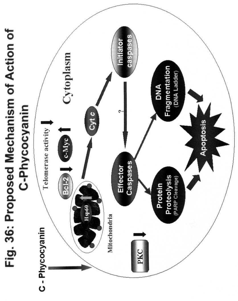
16 much of C-PC/ Celecoxib supplied to K562 cells is available for action. The present study also demonstrated that C-PC, being a protein of 39 kda enters into the K562 cells. However, the mechanism of its entry is quite unclear. The overall signal transduction mechanism involved in C-PC induced apoptosis in K562 cells is presented in Fig. 36. Since C-PC being a natural pigment and a component of edible Spirulina extracts, it forms a good alternative to highly toxic chemotherapeutic products in the market. Also it can form a good candidate for enhancing the sensitivity of cancer cells to conventional anticancer drugs and thus ultimately reducing their toxic side effects. However, further studies are required to test the efficacy of C- PC and Celecoxib on CML and other leukemias in animal models. 74
17
C-Phycocyanin (C-PC) is a n«sjfc&c- waefc-jduble phycobiliprotein. pigment isolated from Spirulina platensis. This water- soluble protein pigment is
 is a n«sjfc&c- waefc-jduble phycobiliprotein. pigment isolated from Spirulina platensis. This water- soluble protein pigment is") ' ^Summary C-Phycocyanin (C-PC) is a n«sjfc&c- waefc-jduble phycobiliprotein pigment isolated from Spirulina platensis. This water- soluble protein pigment is of greater importance because of its various
' ^Summary C-Phycocyanin (C-PC) is a n«sjfc&c- waefc-jduble phycobiliprotein pigment isolated from Spirulina platensis. This water- soluble protein pigment is of greater importance because of its various
Apoptosis Oncogenes. Srbová Martina
 Apoptosis Oncogenes Srbová Martina Cell Cycle Control point Cyclin B Cdk1 Cyclin D Cdk4 Cdk6 Cyclin A Cdk2 Cyclin E Cdk2 Cyclin-dependent kinase (Cdk) have to bind a cyclin to become active Regulation
Molecular biology :- Cancer genetics lecture 11
 Molecular biology :- Cancer genetics lecture 11 -We have talked about 2 group of genes that is involved in cellular transformation : proto-oncogenes and tumour suppressor genes, and it isn t enough to
p53 and Apoptosis: Master Guardian and Executioner Part 2
 p53 and Apoptosis: Master Guardian and Executioner Part 2 p14arf in human cells is a antagonist of Mdm2. The expression of ARF causes a rapid increase in p53 levels, so what would you suggest?.. The enemy
Introduction to pathology lecture 5/ Cell injury apoptosis. Dr H Awad 2017/18
 Introduction to pathology lecture 5/ Cell injury apoptosis Dr H Awad 2017/18 Apoptosis = programmed cell death = cell suicide= individual cell death Apoptosis cell death induced by a tightly regulated
Cancer and Gene Alterations - 1
 Cancer and Gene Alterations - 1 Cancer and Gene Alteration As we know, cancer is a disease of unregulated cell growth. Although we looked at some of the features of cancer when we discussed mitosis checkpoints,
Cell cycle and apoptosis
 Cell cycle and apoptosis Cell cycle Definition Stages and steps Cell cycle Interphase (G1/G0, S, and G2) Mitosis (prophase, metaphase, anaphase, telophase, karyokinesis, cytokinesis) Control checkpoints
The Biochemistry of apoptosis
 The Biochemistry of apoptosis 1 1 The apoptosis is composed of multiple biochemical events 2 2 Biochemical, cellular, and molecular events in Apoptosis 1. Membrane blebbing; phosphatidyl serine exposure
Introduction. Cancer Biology. Tumor-suppressor genes. Proto-oncogenes. DNA stability genes. Mechanisms of carcinogenesis.
 Cancer Biology Chapter 18 Eric J. Hall., Amato Giaccia, Radiobiology for the Radiologist Introduction Tissue homeostasis depends on the regulated cell division and self-elimination (programmed cell death)
Apoptosome dysfunction in human cancer
 Apoptosis 2004; 9: 691 704 C 2004 Kluwer Academic Publishers Apoptosome dysfunction in human cancer K. M. Hajra and J. R. Liu Department of Obstetrics and Gynecology, University of Michigan Medical School,
Programmed Cell Death (apoptosis)
") Programmed Cell Death (apoptosis) Stereotypic death process includes: membrane blebbing nuclear fragmentation chromatin condensation and DNA framentation loss of mitochondrial integrity and release of
Programmed Cell Death (apoptosis) Stereotypic death process includes: membrane blebbing nuclear fragmentation chromatin condensation and DNA framentation loss of mitochondrial integrity and release of
Problem Set 8 Key 1 of 8
 7.06 2003 Problem Set 8 Key 1 of 8 7.06 2003 Problem Set 8 Key 1. As a bright MD/PhD, you are interested in questions about the control of cell number in the body. Recently, you've seen three patients
Cancer. The fundamental defect is. unregulated cell division. Properties of Cancerous Cells. Causes of Cancer. Altered growth and proliferation
 Cancer The fundamental defect is unregulated cell division. Properties of Cancerous Cells Altered growth and proliferation Loss of growth factor dependence Loss of contact inhibition Immortalization Alterated
Apoptosis Chapter 9. Neelu Yadav PhD
 Apoptosis Chapter 9 Neelu Yadav PhD Neelu.Yadav@Roswellpark.org 1 Apoptosis: Lecture outline Apoptosis a programmed cell death pathway in normal homeostasis Core Apoptosis cascade is conserved Compare
Determination Differentiation. determinated precursor specialized cell
 Biology of Cancer -Developmental Biology: Determination and Differentiation -Cell Cycle Regulation -Tumor genes: Proto-Oncogenes, Tumor supressor genes -Tumor-Progression -Example for Tumor-Progression:
Introduction: 年 Fas signal-mediated apoptosis. PI3K/Akt
 Fas-ligand (CD95-L; Fas-L) Fas (CD95) Fas (apoptosis) 年 了 不 度 Fas Fas-L 力 不 Fas/Fas-L T IL-10Fas/Fas-L 不 年 Fas signal-mediated apoptosis 度降 不 不 力 U-118, HeLa, A549, Huh-7 MCF-7, HepG2. PI3K/Akt FasPI3K/Akt
Cell Injury MECHANISMS OF CELL INJURY
 Cell Injury MECHANISMS OF CELL INJURY The cellular response to injurious stimuli depends on the following factors: Type of injury, Its duration, and Its severity. Thus, low doses of toxins or a brief duration
Under the Radar Screen: How Bugs Trick Our Immune Defenses
 Under the Radar Screen: How Bugs Trick Our Immune Defenses Session 8: Apoptosis Marie-Eve Paquet and Gijsbert Grotenbreg Whitehead Institute for Biomedical Research Myxoma virus Poxvirus Infects rabbits
GMS 6644: Apoptosis. Introduction
 GMS 6644: Apoptosis Introduction (Feb. 15, 2006) Lei Xiao, Ph.D. Department of Anatomy & Cell Biology UF Shands Cancer Center ARB Rm R4-250, 846-1199, lxiao@ufl.edu Outline of the Lecture Different types
Mechanisms of Cell Death
 Mechanisms of Cell Death CELL DEATH AND FORMATION OF THE SEMICIRCULAR CANALS Carol M. Troy August 25, 2008 FROM: Fekete et al., Development 124: 2451 (1997) PHENOMENOLOGY OF CELL DEATH I. DEVELOPMENT A.
609G: Concepts of Cancer Genetics and Treatments (3 credits)
") Master of Chemical and Life Sciences Program College of Computer, Mathematical, and Natural Sciences 609G: Concepts of Cancer Genetics and Treatments (3 credits) Text books: Principles of Cancer Genetics,
Master of Chemical and Life Sciences Program College of Computer, Mathematical, and Natural Sciences 609G: Concepts of Cancer Genetics and Treatments (3 credits) Text books: Principles of Cancer Genetics,
#19 Apoptosis Chapter 9. Neelu Yadav PhD
 #19 Apoptosis Chapter 9 Neelu Yadav PhD Neelu.Yadav@Roswellpark.org Why cells decide to die? - Stress, harmful, not needed - Completed its life span Death stimulation or Stress Cell Survival Death Functions
Apoptotic Pathways in Mammals Dr. Douglas R. Green
 Apoptotic Pathways in Mammals Douglas R. Green 1 Apoptosis A form of cell death that is defined morphologically, and features a number of biochemical events Programmed cell death Cell death that occurs
RAS Genes. The ras superfamily of genes encodes small GTP binding proteins that are responsible for the regulation of many cellular processes.
 ۱ RAS Genes The ras superfamily of genes encodes small GTP binding proteins that are responsible for the regulation of many cellular processes. Oncogenic ras genes in human cells include H ras, N ras,
Cancer. Throughout the life of an individual, but particularly during development, every cell constantly faces decisions.
 Cancer Throughout the life of an individual, but particularly during development, every cell constantly faces decisions. Should it divide? Yes No--> Should it differentiate? Yes No-->Should it die? Yes-->Apoptosis
Deregulation of signal transduction and cell cycle in Cancer
 Deregulation of signal transduction and cell cycle in Cancer Tuangporn Suthiphongchai, Ph.D. Department of Biochemistry Faculty of Science, Mahidol University Email: tuangporn.sut@mahidol.ac.th Room Pr324
Part-4. Cell cycle regulatory protein 5 (Cdk5) A novel target of ERK in Carb induced cell death
 A novel target of ERK in Carb induced cell death") Part-4 Cell cycle regulatory protein 5 (Cdk5) A novel target of ERK in Carb induced cell death 95 1. Introduction The process of replicating DNA and dividing cells can be described as a series of coordinated
Part-4 Cell cycle regulatory protein 5 (Cdk5) A novel target of ERK in Carb induced cell death 95 1. Introduction The process of replicating DNA and dividing cells can be described as a series of coordinated
Chapt 15: Molecular Genetics of Cell Cycle and Cancer
 Chapt 15: Molecular Genetics of Cell Cycle and Cancer Student Learning Outcomes: Describe the cell cycle: steps taken by a cell to duplicate itself = cell division; Interphase (G1, S and G2), Mitosis.
Cancer genetics
 Cancer genetics General information about tumorogenesis. Cancer induced by viruses. The role of somatic mutations in cancer production. Oncogenes and Tumor Suppressor Genes (TSG). Hereditary cancer. 1
The Hallmarks of Cancer
 The Hallmarks of Cancer Theresa L. Hodin, Ph.D. Clinical Research Services Theresa.Hodin@RoswellPark.org Hippocrates Cancer surgery, circa 1689 Cancer Surgery Today 1971: Nixon declares War on Cancer
Cancer Genetics. What is Cancer? Cancer Classification. Medical Genetics. Uncontrolled growth of cells. Not all tumors are cancerous
 Session8 Medical Genetics Cancer Genetics J avad Jamshidi F a s a U n i v e r s i t y o f M e d i c a l S c i e n c e s, N o v e m b e r 2 0 1 7 What is Cancer? Uncontrolled growth of cells Not all tumors
NFκB What is it and What s the deal with radicals?
 The Virtual Free Radical School NFκB What is it and What s the deal with radicals? Emily Ho, Ph.D Linus Pauling Institute Scientist Department of Nutrition and Food Management Oregon State University 117
INTERACTION DRUG BODY
 INTERACTION DRUG BODY What the drug does to the body What the body does to the drug Receptors - intracellular receptors - membrane receptors - Channel receptors - G protein-coupled receptors - Tyrosine-kinase
In vivo prediction of anti-tumor effect of 3- bromopyruvate in Hepatocellular Carcinoma using Tc-99m labeled annexin-v imaging
 In vivo prediction of anti-tumor effect of 3- bromopyruvate in Hepatocellular Carcinoma using Tc-99m labeled annexin-v imaging Won Kim, Jung-Hwan Yoon*, Gi Jeong Cheon, Tae Sup Lee, Chung Yong Kim Department
Transformation of Normal HMECs (Human Mammary Epithelial Cells) into Metastatic Breast Cancer Cells: Introduction - The Broad Picture:
 into Metastatic Breast Cancer Cells: Introduction - The Broad Picture:") Transformation of Normal HMECs (Human Mammary Epithelial Cells) into Metastatic Breast Cancer Cells: Introduction - The Broad Picture: Spandana Baruah December, 2016 Cancer is defined as: «A disease caused
Transformation of Normal HMECs (Human Mammary Epithelial Cells) into Metastatic Breast Cancer Cells: Introduction - The Broad Picture: Spandana Baruah December, 2016 Cancer is defined as: «A disease caused
Biology is the only subject in which multiplication is the same thing as division
 The Cell Cycle Biology is the only subject in which multiplication is the same thing as division Why do cells divide? For reproduction asexual reproduction For growth one-celled organisms from fertilized
Cell cycle, signaling to cell cycle, and molecular basis of oncogenesis
 Cell cycle, signaling to cell cycle, and molecular basis of oncogenesis MUDr. Jiří Vachtenheim, CSc. CELL CYCLE - SUMMARY Basic terminology: Cyclins conserved proteins with homologous regions; their cellular
The discovery of Bcl-2
 The discovery of Bcl-2 Bcl-2: B-cell lymphoma 2 The pro-survival subfamily of Bcl-2 protein family Cloning of Bcl-2 as the oncogene which is deregulated at t(14;18) lymphomas Pioneer works by Tsujimoto
shehab Moh Tarek ... ManarHajeer
 3 shehab Moh Tarek... ManarHajeer In the previous lecture we discussed the accumulation of oxygen- derived free radicals as a mechanism of cell injury, we covered their production and their pathologic
Signaling Apoptosis. Scott André Oakes, M.D. Dept. of Pathology Univ. of Calif-San Francisco. Cyt c Release BAX/BAK. Apoptosome Formation
 Signaling Apoptosis Cyt c Release BAX/BAK Apoptosome Formation Caspase Activation Scott André Oakes, M.D. Dept. of Pathology Univ. of Calif-San Francisco Why do we need cell death? Sculpt Organs Control
Cancer Biology How a cell responds to DNA Damage
 1 Cancer Biology How a cell responds to DNA Damage Jann Sarkaria Department of Oncology Mayo Clinic 2 EDUCATIONAL GOALS How proteins can transmit signals to each other. The definition of a tumor suppressor
CELL CYCLE MOLECULAR BASIS OF ONCOGENESIS
 CELL CYCLE MOLECULAR BASIS OF ONCOGENESIS Summary of the regulation of cyclin/cdk complexes during celll cycle Cell cycle phase Cyclin-cdk complex inhibitor activation Substrate(s) G1 Cyclin D/cdk 4,6
BIOL 4374/BCHS 4313 Cell Biology Exam #1 February 13, 2001
 BIOL 4374/BCHS 4313 Cell Biology Exam #1 February 13, 2001 SS# Name This exam is worth a total of 100 points. The number of points each question is worth is shown in parentheses. Good luck! 1. (2) The
Introduction to Cancer Biology
 Introduction to Cancer Biology Robin Hesketh Multiple choice questions (choose the one correct answer from the five choices) Which ONE of the following is a tumour suppressor? a. AKT b. APC c. BCL2 d.
Phospho-AKT Sampler Kit
 Phospho-AKT Sampler Kit E 0 5 1 0 0 3 Kits Includes Cat. Quantity Application Reactivity Source Akt (Ab-473) Antibody E021054-1 50μg/50μl IHC, WB Human, Mouse, Rat Rabbit Akt (Phospho-Ser473) Antibody
Multistep nature of cancer development. Cancer genes
 Multistep nature of cancer development Phenotypic progression loss of control over cell growth/death (neoplasm) invasiveness (carcinoma) distal spread (metastatic tumor) Genetic progression multiple genetic
number Done by Corrected by Doctor Maha Shomaf
 number 19 Done by Waseem Abo-Obeida Corrected by Abdullah Zreiqat Doctor Maha Shomaf Carcinogenesis: the molecular basis of cancer. Non-lethal genetic damage lies at the heart of carcinogenesis and leads
REGULATION OF ENZYME ACTIVITY. Medical Biochemistry, Lecture 25
 REGULATION OF ENZYME ACTIVITY Medical Biochemistry, Lecture 25 Lecture 25, Outline General properties of enzyme regulation Regulation of enzyme concentrations Allosteric enzymes and feedback inhibition
Epigonal Conditioned Media from Bonnethead Shark, Sphyrna tiburo, Induces Apoptosis in a T-Cell Leukemia Cell Line, Jurkat E6-1
 Mar. Drugs 2013, 11, 3224-3257; doi:10.3390/md11093224 Article OPEN ACCESS marine drugs ISSN 1660-3397 www.mdpi.com/journal/marinedrugs Epigonal Conditioned Media from Bonnethead Shark, Sphyrna tiburo,
Mitosis and the Cell Cycle
 Mitosis and the Cell Cycle Chapter 12 The Cell Cycle: Cell Growth & Cell Division Where it all began You started as a cell smaller than a period at the end of a sentence Getting from there to here Cell
PREPARED BY P.DHARANI PRASAD II YEAR B.PHARM II SEM SUB:PATHOPHYSIOLOGY
 CELL INJURY UNIT I PREPARED BY P.DHARANI PRASAD II YEAR B.PHARM II SEM SUB:PATHOPHYSIOLOGY DETECTION OF CELLULAR CHANGES AFTER INJURY BY: LIGHT MICROSCOPY OR GROSS EXAMINATION DETECT CHANGES HOURS TO DAYS
Page 39 of 44. 8h LTA & AT h PepG & AT h LTA
 Page 39 of 44 Fig. S1 A: B: C: D: 8h LTA 8h LTA & AT7519 E: F: 8h PepG G: 8h PepG & AT7519 Fig. S1. AT7519 overrides the survival effects of lipoteichoic acid (LTA) and peptidoglycan (PepG). (A) Human
Regulators of Cell Cycle Progression
 Regulators of Cell Cycle Progression Studies of Cdk s and cyclins in genetically modified mice reveal a high level of plasticity, allowing different cyclins and Cdk s to compensate for the loss of one
A class of genes that normally suppress cell proliferation. p53 and Rb..ect. suppressor gene products can release cells. hyperproliferation.
 Tumor Suppressor Genes A class of genes that normally suppress cell proliferation. p53 and Rb..ect Mutations that inactivate the tumor suppressor gene products can release cells from growth suppression
Chapter 12. Regulation of Cell Division. AP Biology
 Chapter 12. Regulation of Cell Division Coordination of cell division! Multicellular organism " need to coordinate across different parts of organism! timing of cell division! rates of cell division "
Supplementary Figures
 Supplementary Figures Figure S1. Validation of kinase regulators of ONC201 sensitivity. Validation and screen results for changes in cell viability associated with the combination of ONC201 treatment (1
Regulation of Cell Division (Ch. 12)
") Regulation of Cell Division (Ch. 12) Coordination of cell division A multicellular organism needs to coordinate cell division across different tissues & organs critical for normal growth, development &
Regulation of Cell Division (Ch. 12) Coordination of cell division A multicellular organism needs to coordinate cell division across different tissues & organs critical for normal growth, development &
Regulation of Cell Division
 Regulation of Cell Division Two HeLa cancer cells are just completing cytokinesis. Explain how the cell division of cancer cells like these is misregulated. Identify genetic and other changes that might
#19 Apoptosis Chapter 9. Neelu Yadav PhD
 #19 Apoptosis Chapter 9 Neelu Yadav PhD Neelu.Yadav@Roswellpark.org Why cells decide to die? - Stress, harmful, not needed - Completed its life span Death stimulation or Stress Cell Survival Death Functions
mirna Dr. S Hosseini-Asl
 mirna Dr. S Hosseini-Asl 1 2 MicroRNAs (mirnas) are small noncoding RNAs which enhance the cleavage or translational repression of specific mrna with recognition site(s) in the 3 - untranslated region
Cell Communication. Chapter 11. Biology Eighth Edition Neil Campbell and Jane Reece. PowerPoint Lecture Presentations for
 Chapter 11 Cell Communication PowerPoint Lecture Presentations for Biology Eighth Edition Neil Campbell and Jane Reece Lectures by Chris Romero, updated by Erin Barley with contributions from Joan Sharp
Lecture 6 9/17 Dr. Hirsh Organization of Cells, continued
 Cell structure of Eukaryotic cells Lecture 6 9/17 Dr. Hirsh Organization of Cells, continued Lots of double-membraned organelles Existence of an Endo-membrane system separation of areas of cell, transport
Chapter 9. Cellular Signaling
 Chapter 9 Cellular Signaling Cellular Messaging Page 215 Cells can signal to each other and interpret the signals they receive from other cells and the environment Signals are most often chemicals The
Cell Cycle and Cancer
 142 8. Cell Cycle and Cancer NOTES CELL CYCLE G 0 state o Resting cells may re-enter the cell cycle Nondividing cells (skeletal and cardiac muscle, neurons) o Have left the cell cycle and cannot undergo
Activation of cellular proto-oncogenes to oncogenes. How was active Ras identified?
 Dominant Acting Oncogenes Eugene E. Marcantonio, M.D. Ph.D. Oncogenes are altered forms of normal cellular genes called proto-oncogenes that are involved in pathways regulating cell growth, differentiation,
Regulation of Cell Division. AP Biology
 Regulation of Cell Division 2006-2007 Coordination of cell division A multicellular organism needs to coordinate cell division across different tissues & organs critical for normal growth, development
Lecture 14 - The cell cycle and cell death
 02.17.10 Lecture 14 - The cell cycle and cell death The cell cycle: cells duplicate their contents and divide The cell cycle may be divided into 4 phases The cell cycle triggers essential processes (DNA
Chemical and Biochemical Mechanism Of Cell Injury.
 Chemical and Biochemical Mechanism Of Cell Injury. Professor Dr. M. Tariq Javed Dept. of Pathology Faculty of Vet. Science The University Of Agriculture Faisalabad Cell Injury When the cell is exposed
APPLICATION NOTE 1850 Millrace Drive, Suite 3A Eugene, Oregon
 APPLICATION NOTE 185 Millrace Drive, Suite 3A Eugene, Oregon 973 In-Cell ELISA (ICE) Assay Platform Monitoring apoptosis in cells: a high-throughput, quantitative cellbased assay. Rev. Introduction: Apoptosis:
Biochemistry 673 Lecture 2 Jason Kahn, UMCP Introduction to steroid hormone receptor (nuclear receptor) signalling
 signalling") Biochemistry 673 Lecture 2 Jason Kahn, UMCP Introduction to steroid hormone receptor (nuclear receptor) signalling Resources: Latchman Lodish chapter 10, 20 Helmreich, chapter 11 http://www.nursa.org,
Biochemistry 673 Lecture 2 Jason Kahn, UMCP Introduction to steroid hormone receptor (nuclear receptor) signalling Resources: Latchman Lodish chapter 10, 20 Helmreich, chapter 11 http://www.nursa.org,
Cell Communication. Chapter 11. Biology Eighth Edition Neil Campbell and Jane Reece. PowerPoint Lecture Presentations for
 Chapter 11 Cell Communication PowerPoint Lecture Presentations for Biology Eighth Edition Neil Campbell and Jane Reece Lectures by Chris Romero, updated by Erin Barley with contributions from Joan Sharp
Silibinin i activates p53-caspase-2 pathway and causes caspase-mediated cleavage of Cip1/p21 in apoptosis
 Silibinin i activates p53-caspase-2 pathway and causes caspase-mediated cleavage of Cip1/p21 in apoptosis induction in bladder transitional-cell papilloma RT4 cells: Evidence for a regulatory loop between
Determination of the temporal pattern and importance of BALF1 expression in Epstein-Barr viral infection
 Determination of the temporal pattern and importance of BALF1 expression in Epstein-Barr viral infection Melissa Mihelidakis May 6, 2004 7.340 Research Proposal Introduction Apoptosis, or programmed cell
Division Ave. High School AP Biology
 Regulation of Cell Division 2008-2009 Coordination of cell division A multicellular organism needs to coordinate cell division across different tissues & organs u critical for normal growth, development
Regulation of Gene Expression in Eukaryotes
 Ch. 19 Regulation of Gene Expression in Eukaryotes BIOL 222 Differential Gene Expression in Eukaryotes Signal Cells in a multicellular eukaryotic organism genetically identical differential gene expression
Cell Signaling (part 1)
") 15 Cell Signaling (part 1) Introduction Bacteria and unicellular eukaryotes respond to environmental signals and to signaling molecules secreted by other cells for mating and other communication. In multicellular
15 Cell Signaling (part 1) Introduction Bacteria and unicellular eukaryotes respond to environmental signals and to signaling molecules secreted by other cells for mating and other communication. In multicellular
Neoplasia 18 lecture 6. Dr Heyam Awad MD, FRCPath
 Neoplasia 18 lecture 6 Dr Heyam Awad MD, FRCPath ILOS 1. understand the role of TGF beta, contact inhibition and APC in tumorigenesis. 2. implement the above knowledge in understanding histopathology reports.
Mitochondria in apoptosis. Jean-Claude Martinou, MD, Ph.D Department of cell biology University of Geneva Geneva, Switzerland
 Mitochondria in apoptosis Jean-Claude Martinou, MD, Ph.D Department of cell biology University of Geneva Geneva, Switzerland The dual role of mitochondria in life and death QuickTime and a TIFF (Uncompressed)
Apoptosis-based Therapies: Mechanisms and Applications
 Apoptosis-based Therapies: Mechanisms and Applications Perspective Bcl-2 family members, potential usage of BH3 domains as drug targets Bcl-2/xL inhibitors - Antisense, inhibitors of protein-protein interactions,
Cell Communication. Chapter 11. Biology. Eighth Edition Neil Campbell and Jane Reece. PowerPoint Lecture Presentations for
 Chapter 11 Cell Communication PowerPoint Lecture Presentations for Biology Eighth Edition Neil Campbell and Jane Reece Lectures by Chris Romero, updated by Erin Barley with contributions from Joan Sharp
Mechanisms of Cell Injury: Loss of Calcium Homeostasis
 Mechanisms of Cell Injury: Loss of Calcium Homeostasis SCPA610: Cellular Pathology Amornrat N. Jensen, Ph.D. amornrat.nar@mahidol.ac.th Leading questions Why is intracellular calcium important for the
Molecular Cell Biology Problem Drill 16: Intracellular Compartment and Protein Sorting
 Molecular Cell Biology Problem Drill 16: Intracellular Compartment and Protein Sorting Question No. 1 of 10 Question 1. Which of the following statements about the nucleus is correct? Question #01 A. The
BCHM3972 Human Molecular Cell Biology (Advanced) 2013 Course University of Sydney
 2013 Course University of Sydney") BCHM3972 Human Molecular Cell Biology (Advanced) 2013 Course University of Sydney Page 2: Immune Mechanisms & Molecular Biology of Host Defence (Prof Campbell) Page 45: Infection and Implications for Cell
BCHM3972 Human Molecular Cell Biology (Advanced) 2013 Course University of Sydney Page 2: Immune Mechanisms & Molecular Biology of Host Defence (Prof Campbell) Page 45: Infection and Implications for Cell
Objectives. Abbas Chapter 11: Immunological Tolerance. Question 1. Question 2. Question 3. Definitions
 Objectives Abbas Chapter 11: Immunological Tolerance Christina Ciaccio, MD Children s Mercy Hospitals and Clinics February 1, 2010 To introduce the concept of immunologic tolerance To understand what factors
Convergent and Divergent Mechanisms in Aging and Cancer
 Convergent and Divergent Mechanisms in Aging and Cancer Mariana S. De Lorenzo, PhD Department of Cell Biology & Molecular Medicine delorems@umdnj.edu LEARNING OBJECTIVES 1. To identify convergent and divergent
Screening of novel thalidomide analogues with label-free assays and appling in the in vivo model of hepatocellular carcinoma.
 Screening of novel thalidomide analogues with label-free assays and appling in the in vivo model of hepatocellular carcinoma Lajos István Nagy Ph.D thesis summary Supervisor: László Puskás, PhD, DSc Scientific
Genome of Hepatitis B Virus. VIRAL ONCOGENE Dr. Yahwardiah Siregar, PhD Dr. Sry Suryani Widjaja, Mkes Biochemistry Department
 Genome of Hepatitis B Virus VIRAL ONCOGENE Dr. Yahwardiah Siregar, PhD Dr. Sry Suryani Widjaja, Mkes Biochemistry Department Proto Oncogen and Oncogen Oncogen Proteins that possess the ability to cause
Section D: The Molecular Biology of Cancer
 CHAPTER 19 THE ORGANIZATION AND CONTROL OF EUKARYOTIC GENOMES Section D: The Molecular Biology of Cancer 1. Cancer results from genetic changes that affect the cell cycle 2. Oncogene proteins and faulty
Cell cycle and Apoptosis. Chalermchai Mitrpant
 Cell cycle and Apoptosis 2556 Chalermchai Mitrpant Overview of the cell cycle Outline Regulatory mechanisms controlling cell cycle Progression of the cell cycle Checkpoint of the cell cycle Phases of the
Cell Cycle. Trends in Cell Biology
 Cell Cycle Trends in Cell Biology Cell Cycle The orderly sequence of events by which a cell duplicates its contents and divides into two Daughter Cells Activities of a cell from one cell division to the
LECTURE-15. itraq Clinical Applications HANDOUT. Isobaric Tagging for Relative and Absolute quantitation (itraq) is a quantitative MS
 is a quantitative MS") LECTURE-15 itraq Clinical Applications HANDOUT PREAMBLE Isobaric Tagging for Relative and Absolute quantitation (itraq) is a quantitative MS based method for quantifying proteins subject to various different
LECTURE-15 itraq Clinical Applications HANDOUT PREAMBLE Isobaric Tagging for Relative and Absolute quantitation (itraq) is a quantitative MS based method for quantifying proteins subject to various different
2015 AP Biology Unit #4 Quiz 1 Cell Communication, Cancer and The Cell Cycle Week of November
 Name: Class: Date: 2015 AP Biology Unit #4 Quiz 1 Cell Communication, Cancer and The Cell Cycle Week of 16-20 November Multiple Choice Identify the choice that best completes the statement or answers the
Name Class Date. 1. Cellular respiration is the process by which the of "food"
 Name Class Date Cell Respiration Introduction Cellular respiration is the process by which the chemical energy of "food" molecules is released and partially captured in the form of ATP. Carbohydrates,
Cell Cycle Notes chromatin, somatic cells gametes mitosis sister chromatids, centromere cytokinesis binary fission,
 Cell Cycle Notes 1. Importance of Cell Division a. For single celled organisms, cell division increases the number of individuals. b. In a multicellular organism, cell division functions to repair and
Breaking Up is Hard to Do (At Least in Eukaryotes) Mitosis
 Mitosis") Breaking Up is Hard to Do (At Least in Eukaryotes) Mitosis Prokaryotes Have a Simpler Cell Cycle Cell division in prokaryotes takes place in two stages, which together make up a simple cell cycle 1. Copy
Breaking Up is Hard to Do (At Least in Eukaryotes) Mitosis Prokaryotes Have a Simpler Cell Cycle Cell division in prokaryotes takes place in two stages, which together make up a simple cell cycle 1. Copy
Cell Communication. Local and Long Distance Signaling
 Cell Communication Cell to cell communication is essential for multicellular organisms Some universal mechanisms of cellular regulation providing more evidence for the evolutionary relatedness of all life
How do mutated oncogenes and tumor suppressor genes cause cancer?
 Medical Oncology (1998) 15, 20-26 9 1998 Stockton Press All rights reserved 0736-0118/98 $12.00 REVIEW How do mutated oncogenes and tumor suppressor genes cause cancer? Dan Grand6r Research Laboratory
MOLECULAR BASIS OF ONCOGENESIS
 MOLECULAR BASIS OF ONCOGENESIS MUDr. Jiří Vachtenheim, CSc. 1 Cell processes which result also in cell cycle effects. Differentiation. Differentiated cells are usually in the G0 phase of the cell cycle.
BIO 5099: Molecular Biology for Computer Scientists (et al)
") BIO 5099: Molecular Biology for Computer Scientists (et al) Lecture 15: Being a Eukaryote: From DNA to Protein, A Tour of the Eukaryotic Cell. Christiaan van Woudenberg Being A Eukaryote Basic eukaryotes
BIO 5099: Molecular Biology for Computer Scientists (et al) Lecture 15: Being a Eukaryote: From DNA to Protein, A Tour of the Eukaryotic Cell. Christiaan van Woudenberg Being A Eukaryote Basic eukaryotes