PREPARED BY P.DHARANI PRASAD II YEAR B.PHARM II SEM SUB:PATHOPHYSIOLOGY
|
|
|
- Ashlee Lyons
- 5 years ago
- Views:
Transcription

1 CELL INJURY UNIT I PREPARED BY P.DHARANI PRASAD II YEAR B.PHARM II SEM SUB:PATHOPHYSIOLOGY


2 DETECTION OF CELLULAR CHANGES AFTER INJURY BY: LIGHT MICROSCOPY OR GROSS EXAMINATION DETECT CHANGES HOURS TO DAYS AFTER INJURY HISTOCHEMICAL OR ULTRASTRUCTURAL TECHNIQUES DETECT CHANGES MINUTES TO HOURS AFTER INJURY
3

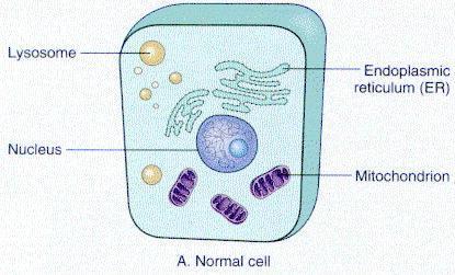
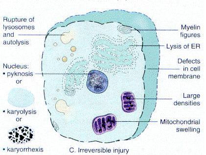
4 Patterns of Acute Cell Injury Reversible Injury Cellular swelling: Ultrastructural changes plasma membrane blebbing, blunting and distortion of microvilli mitochondrial swelling, phospholipid-rich amorphous densities dilation of endoplasmic reticulum with detachment of ribosomes and dissociation of polysomes disaggregation of granular and fibrillar elements on nucleus
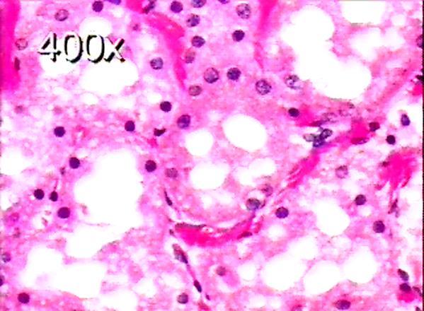
5 Patterns of Acute Cell Injury Reversible Injury 2. Fatty change: Vacuolation of cells due to accumulation of lipid droplets Results due to disturbance of ribosomal function The liver is commonly affected Occurs in hypoxic injury, toxic (alcohol), metabolic (diabetes mellitus) Moderate fatty changes are reversible, but sever changes may not be

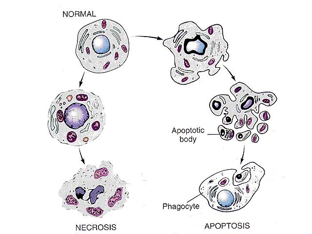
6 Patterns of Acute Cell Injury Irreversible injury: Cell death It is suggested that cell membrane is the central factor in the pathogenesis of irreversible cell injury Also due to: sever mitochondrial dysfunction lysosomal rupture Two patterns of cell death: Necrosis Apoptosis
7 Patterns of Acute Cell Injury Irreversible injury: Cell death 1. Necrosis: Definition: sequence of morphologic changes that follow cell death in living tissue The morphologic appearance of necrosis is due to: Enzymatic digestion of cell: Autolysis: hydrolytic enzymes are derived from the dead cells themselves Heterolysis: hydrolytic enzymes are derived from invading inflammatory cells Denaturation of proteins
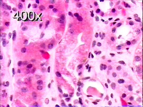
8 Patterns of Acute Cell Injury Microscopic appearance of Necrotic dead cells: Cytoplasmic changes eosinophilia (pink) increased due to eosin binding to denatured proteins Decreased basophilia (blue) mainly imparted by RNA Glassy homogenous cytoplasm due to loss of glycogen Clacification may occur late Nuclear changes due to break down of DNA Karyolysis: decrease basophilia of chromatin Pyknosis: nuclear shrinkage and increased basophilia Karyorrhexis: fragmentation of pyknotic nucleus
9

10 Kidney, necrosis of tubular cells
11 Patterns of Acute Cell Injury Specific Morphologic Patterns of Necrosis Coagulative necrosis Liquefactive necrosis Gangrenous necrosis Caseous necrosis Fat necrosis Others (fibrinoid necrosis)
12 Specific Morphologic Patterns of Necrosis 1. Coagulative Necrosis: Preservation of the structural outline of the dead (coagulated) cell for days The most common form of necrosis (particularly in myocardium, liver, kidney) characteristic of hypoxic cell death in all tissues except in the brain Myocardial infarction is a very good example Mechanism: denaturation of proteins and enzymes blocking cellular proteolysis preserve cell outline
13 Specific Morphologic Patterns of Necrosis Morphology of Coagulative Necrosis: Gross: pale color, normal firm texture at the beginning become soft later due to digestion by macrophages (may lead to rupture of infarcted myocardium) Microscopic: first few hours no abnormalities later progressive loss of nuclear staining, with preservation of cell boundaries finally damaged cells are removed by macrophages (the presence of necrotic tissue usually evokes inflammatory response followed by repair)
14 Fate of Necrosis Most of necrotic tissue is removed by leukocyte (Phagocytosis) combined with extracellular enzyme digestion If necrotic tissue is not eliminated it attracts Ca ++ salts dystrophic calcification
15 Patterns of Acute Cell Injury Apoptosis (a falling away from) Definition: Programmed cell death It is an active (energy-dependant) programmed single cell death to delete the unwanted or defective cells It has an important role in physiological processes and pathological conditions
16 Apoptosis Physiological processes: during embryogenesis (implantation, organogenesis, developmental involution, separation of digits in limb development) hormone -dependent involution (endometrium during menstruation, lactating breast after weaning) cell deletion in proliferating populations intestinal crypt epithelium deletion of autoreactive T cells in thymus (failure might result in autoimmunity) Pathological conditions: pathologic atrophy-prostate after castration (hormone -dependent involution) Cell death in tumors Cell death induced by cytotoxic drugs and ionizing radiation Councilman s bodies due to viral hepatitis

17 Apoptosis Morphology: Involves single cells or small clusters Cells shrink rapidly, retain intact plasma membrane Formation of cytoplasmic buds Fragmentation into apoptotic bodies Apoptotic bodies phagocytosed or rapidly degraded No inflammatory response Entire process from 5 to 30 minutes

18 Apoptosis
19 Necrosis Vs Apoptosis Necrosis Grp of cells or part of tissue passive process Always pathologic Mechanism is ATP depletion, mb damage Histology: coagulation. liquefaction inflammation Apoptosis: Single cell death in living tissue Active process Physiologic or pathologic Endonucleases Apoptotic bodies No inflammation
number Done by Corrected by Doctor Heyam Awad
 number 4 Done by Waseem Abu Obeida Corrected by Saad Al-Hayek Doctor Heyam Awad Cell injury -in the previous lectures we talked about the causes (etiology) and the mechanism (pathogenesis) of cell injury.
Cellular responses to stress
 Cellular responses to stress (Adaptations, injury and death) (2 of 5) Most injurious stimuli are grouped into: Oxygen deprivation Chemical agents Infectious agents Immunologic reactions Genetic factors
Cellular Injury, Necrosis, Apoptosis
 Cellular Injury, Necrosis, Apoptosis Cell injury results when cells are stressed and can no longer adapt Injury may progress through a reversible stage Reversible Cell Injury Reduced oxidative phosphorylation
Mechanisms of Cell Injury
 Causes of Cell Injury 1- oxygen deprivation (anoxia) 2- physical agents 3- chemical agents 4- infections agents 5- immunologic reactions 6- genetic defects 7- nutritional imbalances Mechanisms of Cell
Introduction to pathology lecture 5/ Cell injury apoptosis. Dr H Awad 2017/18
 Introduction to pathology lecture 5/ Cell injury apoptosis Dr H Awad 2017/18 Apoptosis = programmed cell death = cell suicide= individual cell death Apoptosis cell death induced by a tightly regulated
SECTION 2 CELL INJURY
 Adapted myocyte Normal myocyte Reversibly-injured myocyte SECTION 2 CELL INJURY Cell death 5/4/2014 1 5/4/2014 2 Reversible Degeneration Irreversible Cellular Swelling Fatty Change Hyaline Change Amyloid
Cell Adaptation, Cell Injury and Cell Death
 Cell Adaptation, Cell Injury and Cell Death Pathology:- is the study of structural and functional abnormalities that are expressed as diseases of organs and systems. Modern pathology, proposed that injury
INTRODUCTION TO HEALTH AND DISEASE BLOCK
 MBBS 1 st Yr. Lecture Dr. Annie Cheung September 25, 2002, 8:30AM 9:30 AM LT1, G/F, Academic and Administration Block Faculty of Medicine Building INTRODUCTION TO HEALTH AND DISEASE BLOCK CELL INJURY AND
Histopathology: Cell necrosis and cytoplasmic accumulations
 Histopathology: Cell necrosis and cytoplasmic accumulations These presentations are to help you identify basic histopathological features. They do not contain the additional factual information that you
Chapter 1 CELL INJURY CELL DEATH CELL ADAPTATIONS. M.G.Rajanandh, Dept. of Pharmacy Practice, SRM College of Pharmacy, SRM University.
 Chapter 1 CELL INJURY CELL DEATH CELL ADAPTATIONS M.G.Rajanandh, Dept. of Pharmacy Practice, SRM College of Pharmacy, SRM University. CONCEPTS IN CELL INJURY The clinical signs and symptoms are several
APOPTOSIS, NECROSIS AND CANCER. Dr. S. P. Pattanayak
 APOPTOSIS, NECROSIS AND CANCER Dr. S. P. Pattanayak LEARNING OBJECTIVES At the end of the lecture, students should be able to: Know the importance of cell death. Define various modes of cell death. Identify
Quiz 1 Review. More Cowbell
 Quiz 1 Review More Cowbell Quiz 1 review Inflamma7on Repair Cell Injury and Adapta7on Quiz 1 review Inflamma7on Injury Acute inflammation Chronic inflammation Abscess Resolution Repair Time course Inflammation
Lecture-2 / Dr Hussain Abady Aljebori Over view of cell injury and cell death; Cell injury results when: a. cells are stressed so severely that they
 Lecture-2 / Dr Hussain Abady Aljebori Over view of cell injury and cell death; Cell injury results when: a. cells are stressed so severely that they are no longer able to adapt or b. when cells are exposed
Necrosis is death of cells and tissues in the living animal. Focal/ Multifocal necrosis- terms used for one
 Necrosis Necrosis Necrosis is death of cells and tissues in the living animal. Focal/ Multifocal necrosis- terms used for one or more, small, clearly defined areas of necrosis. Diffuse necrosis- term used
NECROSIS, GANGRENE. I. practical training 2 rd year Dentistry
 NECROSIS, GANGRENE. I. practical training 2 rd year Dentistry Signs of death Cardiac arrest (no pulse) Pallor mortis, paleness which happens in the 15 120 minutes after death Livor mortis, a settling of
The basis of Disease
 General Curriculum The basis of Disease ZHOU REN 周韧 Prof., M.D., Ph.D. Institute of Pathology & Forensic Medicine Department of Pathology & Patho-physiology Zhenjiang University Judicial Evidence & Evaluation
Cellular Pathology (VPM 152) Lecture 4 (Web) Paul Hanna Jan 2018
 Lecture 4 (Web) Paul Hanna Jan 2018") Cellular Pathology (VPM 152) Lecture 4 (Web) Paul Hanna Jan 2018 IRREVERSIBLE CELL INJURY 1) Necrosis describes the range of morphologic changes that follow cell death in living tissue the morphologic
Cellular Pathology (VPM 152) Lecture 4 (Web) Paul Hanna Jan 2018 IRREVERSIBLE CELL INJURY 1) Necrosis describes the range of morphologic changes that follow cell death in living tissue the morphologic
CELL INJURY, DEATH, AND ADAPTATION
 CELL INJURY, DEATH, AND ADAPTATION Definitons Pathology is a dicipline bridging clinical practice and basic sience To render diagnosis and guide therapy - Identity changes in gross - Morphology ( microscopy
Cellular response to stress
 Cellular pathology - cell injury, death and adaptations Pathology Göran Andersson Cellular response to stress Cells differ in their capacity to tolerate changes in their microenvironment Acute, severe
Stages in the cellular response to stress & injurious stimuli
 Blok BBS 2 Departemen Patologi Anatomi Fakultas Kedokteran Universitas Sumatera Utara Medan -2011 Stages in the cellular response to stress & injurious stimuli 3/28/2011 2 1 Table 1-1.Cellular Responses
[General Pathology] Introduction to Pathology
![[General Pathology] Introduction to Pathology](/thumbs/73/69585662.jpg "[General Pathology] Introduction to Pathology") Introduction to Pathology Pathology: Literally translated, pathology is the study (logos) of disease (pathos, suffering). It involves the investigation of the causes of disease and the associated changes
Introduction to Pathology Pathology: Literally translated, pathology is the study (logos) of disease (pathos, suffering). It involves the investigation of the causes of disease and the associated changes
Hashem Al-Dujaily. Tamer Barakat. Manar Hajeer
 1 Hashem Al-Dujaily Tamer Barakat... Manar Hajeer Introduction Pathology comes from Patho: disease/suffering and Logy: study. Therefore, Pathology is the study of disease. Pathology is the bridge between
Cell Injury MECHANISMS OF CELL INJURY
 Cell Injury MECHANISMS OF CELL INJURY The cellular response to injurious stimuli depends on the following factors: Type of injury, Its duration, and Its severity. Thus, low doses of toxins or a brief duration
I. ADAPTATION TO ENVIRONMENTAL STRESS. A. Hypertrophy:
 د.جواهر محي الدين Lec:2&3 Cellular Reaction to Injury I. ADAPTATION TO ENVIRONMENTAL STRESS II. HYPOXIC CELL INJURY III. FREE RADICAL INJURY IV. CHEMICAL CELL INJURY V. NECROSIS VI. APOPTOSIS VII. REVERSIBLE
Coagulative Necrosis of Myocardium. Dr Rodney Itaki Division of Pathology
 Coagulative Necrosis of Myocardium Dr Rodney Itaki Division of Pathology Coagulative Necrosis Gross pathology: 3 day old infarct: Yellow necrosis surrounded by hyperemic borders. Arrow points to a transmural
Pathology MCQs. lipid. protein. glycogen. lipofuscin. water. Karyolysis. Cellular swelling. Involvement of a large number of cells
 Pathology MCQs 1. In hypoxic cell injury, cell swelling occurs because of increased intracellular: lipid protein glycogen lipofuscin water 2. Which of the following is a feature of apoptosis? Karyolysis
DEGENERATION NECROSIS AND INFILTRATION
 DEGENERATION NECROSIS AND INFILTRATION Cellular Degenerations and Infiltrations 1. Cloudy swelling and hydropic degeneration Cloudy swelling and hydropic degeneration occur when the regulatory mechanisms
CELL INJURY AND CELL DEATH
 CELL INJURY AND CELL DEATH INTRODUCTION Cell Injury is a result of the sequence of events that occur if the limits of the adaptive capability of cells are exceeded or there is no adaptive response is possible,
Pathophysiology lab 2. Cellular injury and adaptation
 Pathophysiology lab 2 Cellular injury and adaptation Adaptation Cellular changes that aim to preserve cell viability and prevent cell injury. The adaptive responses include: 1. Atrophy 2. Hypertrophy 3.
shehab Moh Tarek ... ManarHajeer
 3 shehab Moh Tarek... ManarHajeer In the previous lecture we discussed the accumulation of oxygen- derived free radicals as a mechanism of cell injury, we covered their production and their pathologic
Cellular Injury. Intracellular degeneration. By Dr. Hemn Hassan Othman PhD, Pathology Fall /20/2018 1
 Cellular Injury Intracellular degeneration By Dr. Hemn Hassan Othman PhD, Pathology Fall 2018 10/20/2018 1 Types of cell injury Cell injury is divided into: 1. Reversible cell injury 2. Irreversible cell
PATHOLOGY Intracellular Degeneration LAB 1
 PATHOLOGY Intracellular Degeneration LAB 1 Cellular swelling Liver Organ :- Liver Lesion :- 1. Narrowing of hepatic sinusoids due to the swelling of hepatocyte. 2. The cytoplasm of affected hepatocyte
Extracellular degeneration
 Extracellular degeneration By Dr. Hemn Hassan Othman PhD, Pathology Fall 2016 1/17/2017 1 Extracellular Degenerations I / Hyaline Degeneration (Hyalinization): The ward hyaline is derived from the Latin
Chemical and Biochemical Mechanism Of Cell Injury.
 Chemical and Biochemical Mechanism Of Cell Injury. Professor Dr. M. Tariq Javed Dept. of Pathology Faculty of Vet. Science The University Of Agriculture Faisalabad Cell Injury When the cell is exposed
Cell injury, adaptation and death. Unite one Second Lab.
 Cell injury, adaptation and death Unite one Second Lab. The two lung abscesses seen here are examples of liquefactive necrosis in which there is a liquid center in an area of tissue injury. One abscess
The basis of Disease
 General Curriculum The basis of Disease ZHOU REN 周韧 Prof., M.D., Ph.D. Institute of Pathology & Forensic Medicine Department of Pathology & Patho-physiology Zhenjiang University Judicial Evidence & Evaluation
Cellular Injury and Adaptation
 General pathology Introduction to pathology Literal translation of the word pathology is the study (logos) of suffering (Pathos). It is a discipline that bridges clinical practice and basic sciences. Pathology
Types of insult - hypoxia
 Introduction This presentation will be a guide to cell injury and cell death outline causes and pathogenesis of cell injury/death describe the morphological changes of cell injury/death Describe the process
Cellular Pathology. Histopathology Lab #2 (web) Paul Hanna Jan 2018
 Paul Hanna Jan 2018") Cellular Pathology Histopathology Lab #2 (web) Paul Hanna Jan 2018 Slide #91 Clinical History: a necropsy was performed on an aged cat the gross pathological changes included: widespread subcutaneous edema
Cellular Pathology Histopathology Lab #2 (web) Paul Hanna Jan 2018 Slide #91 Clinical History: a necropsy was performed on an aged cat the gross pathological changes included: widespread subcutaneous edema
2 nd Practice. Cell injury, adaptation, storage disorders. Semmelweis University 2nd Department of Pathology
 2 nd Practice Cell injury, adaptation, storage disorders Semmelweis University 2nd Department of Pathology Cell and tissue injury Cellular response to injury depends on the type, the duration and the severity
SBI3U7 Cell Structure & Organelles. 2.2 Prokaryotic Cells 2.3 Eukaryotic Cells
 SBI3U7 Cell Structure & Organelles 2.2 Prokaryotic Cells 2.3 Eukaryotic Cells No nucleus Prokaryotic Cells No membrane bound organelles Has a nucleus Eukaryotic Cells Membrane bound organelles Unicellular
First discovered in 1665 since then every organism observed with microscopes shows cells
 The Cell Cell theory (1838): 1. All organisms are composed of one or more cells, and the life processes of metabolism and heredity occur within these cells. 2. Cells are the smallest living things, the
Cell Overview. Hanan Jafar BDS.MSc.PhD
 Cell Overview Hanan Jafar BDS.MSc.PhD THE CELL is made of: 1- Nucleus 2- Cell Membrane 3- Cytoplasm THE CELL Formed of: 1. Nuclear envelope 2. Chromatin 3. Nucleolus 4. Nucleoplasm (nuclear matrix) NUCLEUS
Biology 12 Cell Structure and Function. Typical Animal Cell
 Biology 12 Cell Structure and Function Typical Animal Cell Vacuoles: storage of materials and water Golgi body: a series of stacked disk shaped sacs. Repackaging centre stores, modifies, and packages proteins
Chapters 2 and 3. Pages and Pages Prayer Attendance Homework
 Chapters 2 and 3 Pages 44-45 and Pages 59-62 Prayer Attendance Homework The Cell The cell is the basic unit of life on Earth, separated from its environment by a membrane and sometimes an outer wall. Prokaryotic
WSC , Conference 9, Case 1. Tissue from a nyala.
 WSC 2009-2010, Conference 9, Case 1. Tissue from a nyala. MICROSCOPIC DESCRIPTION: Heart, atrium (1 pt.): Approximately 40% of the atrial myocardium is replaced by areas of fibrous connective tissue (1
lysosomes Ingested materials Defective cell components Degrades macromolecules of all types:
 lysosomes Digests Ingested materials Defective cell components Degrades macromolecules of all types: Proteins Nucleic acids Carbohydrates Lipids Single membrane bound vesicle, contains up to 50 digestive
QUESTIONSHEET 1. The diagram shows some of the cell structures involved in the secretion of an extracellular enzyme. C D
 QUESTIONSHEET 1 The diagram shows some of the cell structures involved in the secretion of an extracellular enzyme. C D A (a) Identify A,, C, and D. A:... :... C:... D:... [4] (b) Outline the role of each
Plants, Animals, Fungi and Protists have Eukaryotic Cell(s)
") Cell Structure Plants, Animals, Fungi and Protists have Eukaryotic Cell(s) Plant Cell Animal Cell straight edges curved edges Cell Organization cytoplasm cell membrane Eukaryotic cells have 3 major parts:
Cell Structure Plants, Animals, Fungi and Protists have Eukaryotic Cell(s) Plant Cell Animal Cell straight edges curved edges Cell Organization cytoplasm cell membrane Eukaryotic cells have 3 major parts:
(A) Cell membrane (B) Ribosome (C) DNA (D) Nucleus (E) Plasmids. A. Incorrect! Both prokaryotic and eukaryotic cells have cell membranes.
 Cell membrane (B) Ribosome (C) DNA (D) Nucleus (E) Plasmids. A. Incorrect! Both prokaryotic and eukaryotic cells have cell membranes.") High School Biology - Problem Drill 03: The Cell No. 1 of 10 1. Which of the following is NOT found in prokaryotic cells? #01 (A) Cell membrane (B) Ribosome (C) DNA (D) Nucleus (E) Plasmids Both prokaryotic
High School Biology - Problem Drill 03: The Cell No. 1 of 10 1. Which of the following is NOT found in prokaryotic cells? #01 (A) Cell membrane (B) Ribosome (C) DNA (D) Nucleus (E) Plasmids Both prokaryotic
CELL INJURY. Severity of Cell Injury
 GENERAL PATHOLOGY LECTURE - 3 DR. M. TARIQ JAVED Professor Department of Pathology, Faculty of Veterinary Science, University of Agriculture, Faisalabad, Pakistan. 9/11/2009 1 CELL INJURY No adaptive response
Histopathology: Glomerulonephritis and other renal pathology
 Histopathology: Glomerulonephritis and other renal pathology These presentations are to help you identify basic histopathological features. They do not contain the additional factual information that you
Looking Inside Cells
 Looking Inside Cells Inner Life of a Cell http://www.bing.com/videos/search?q=inside +cell+animation&form=hdrsc3#view=detail &mid=4ba834420ea307a061374ba834420ea 307A06137 Cell Defined Cells-Basic unit
By: Brooke Sheppard
 By: Brooke Sheppard What is a Cell? Cells are the basic structure of life for all organisms. Cells are microscopic, which means we can only view cells under a microscope. There are animal cells and plant
Basophilic. Basophilic structures are stained by basic dyes: Mnemonic: Basophilic = Blue
 Cell Overview Basophilic Basophilic structures are stained by basic dyes: Basic dyes are positive Basophilic structures are negative (ex. DNA, RNA, ribosomes, RER) Mnemonic: Basophilic = Blue Acidophilic
A Tour of the Cell. reference: Chapter 6. Reference: Chapter 2
 A Tour of the Cell reference: Chapter 6 Reference: Chapter 2 Monkey Fibroblast Cells stained with fluorescent dyes to show the nucleus (blue) and cytoskeleton (yellow and red fibers), image courtesy of
7-2 : Plasma Membrane and Cell Structures
 7-2 : Plasma Membrane and Cell Structures Plasma Membrane of aveolar sac But first... Let s Review What is cell theory? Light microscopes vs. electron microscopes Prokaryotic vs. eukaryotic Basic Cell
CHAPTER ONE INTRODUCTION TO PATHOLOGY
 Dr. Nabeel Abdulwadood Rasheed 1 CHAPTER ONE INTRODUCTION TO PATHOLOGY The literal translation of the word pathology is the study (logos) of suffering (pathos). It is a discipline that bridges clinical
The Study of Cells The diversity of the cells of the body The following figure shows the proportion of cell size of the variety of cells in the body
 Adapted from Martini Human Anatomy 7th ed. Chapter 2 Foundations: The Cell Introduction There are trillions of cells in the body Cells are the structural building blocks of all plants and animals Cells
A Tour of the Cell. reference: Chapter 6. Reference: Chapter 2
 A Tour of the Cell reference: Chapter 6 Reference: Chapter 2 Monkey Fibroblast Cells stained with fluorescent dyes to show the nucleus (blue) and cytoskeleton (yellow and red fibers), image courtesy of
7-2 : Plasma Membrane and Cell Structures
 7-2 : Plasma Membrane and Cell Structures Plasma Membrane of aveolar sac But first... Let s Review What is cell theory? Light microscopes vs. electron microscopes Prokaryotic vs. eukaryotic Basic Cell
Cells and Tissues. Lesson 2.1: Molecules of Life Lesson 2.2: Cells Lesson 2.3: Tissues
 2 Cells and Tissues Lesson 2.1: Molecules of Life Lesson 2.2: Cells Lesson 2.3: Tissues Chapter 2: Cells and Tissues Lesson 2.1 Molecules of Life Molecules of Life carbohydrates proteins lipids nucleic
Ischaemia It means local anemia, it is characterized by a decrease amount of blood in an organ or region. Causes of Ischemia: *1.
 المرحلة الثالثة م. هالة عباس ناجي Ischaemia It means local anemia, it is characterized by a decrease amount of blood in an organ or region. Causes of Ischemia: *1.External pressure upon an artery e.g:
1. or is the study of cellular structure and function. 2. What is the purpose and characteristics of the plasma membrane?
 Chapter 3 Reading Guide The Cellular Level of Organization Name 1. or is the study of cellular structure and function. Section 3.1 Parts of a Cell 2. What is the purpose and characteristics of the plasma
Cells & Cell Organelles
 Cells & Cell Organelles The Building Blocks of Life AP Biology 2008-2009 Types of cells bacteria cells Prokaryote - no organelles Eukaryotes - organelles animal cells plant cells Cell size comparison Animal
Special Staining (I)
") Special Staining (I) Carbohydrates 1- PERIODIC ACID SCHIFF'S (PAS ) Purpose: Glycogen is present in liver, kidney, skeletal and cardiac muscle. The PAS stain is used to demonstrate neutral polysaccharides
Special Staining (I) Carbohydrates 1- PERIODIC ACID SCHIFF'S (PAS ) Purpose: Glycogen is present in liver, kidney, skeletal and cardiac muscle. The PAS stain is used to demonstrate neutral polysaccharides
Cell Anatomy Anatomy = the study of the structures and components of an organism
 Cell Anatomy Anatomy = the study of the structures and components of an organism -Types of Cells: 1) Prokaryotic = simple, primitive = no membrane bound nucleus, only a dense, nuclear area = single-celled
Consultant Medical Laboratory Scientist Assistant Professor of Histopathology & Cytopathology
 بسم اهلل الرحمن الرحيم By: PhD (Histopathology & Cytopathology), M.BA (Total Quality Management) Consultant Medical Laboratory Scientist Assistant Professor of Histopathology & Cytopathology Introduction
Identification and characterization of genes responsive to apoptosis: Application of DNA chip technology and mrna differential display
 Histol Histopathol (2000) 15: 1271-1 284 http://www.ehu.es/histol-histopathol Histology and H istopat hology Cellular and Molecular Biology Invited Revie W Identification and characterization of genes
Chapter 2 Cell. Zhou Li Prof. Dept. of Histology and Embryology
 Chapter 2 Cell Zhou Li Prof. Dept. of Histology and Embryology The inner life of the cell Ⅰ. Plasma membrane (Plasmalemma) 1.1 The structure Unit membrane: inner layer 3-layered structure outer layer mediat
Cell Theory Vocabulary Flashcards
 Mr. Powner Biology Cell Theory Vocabulary Flashcards Instructions: Cut out the flashcards from the following pages. The following word list is the vocabulary for studying cell theory. Write each word on
Cell Theory. Cells are the basic unit of life.
 3.1 7.1 Cell Theory Cells are the basic unit of life. 3.1 7.1 Cell Theory The cell theory grew out of the work of many scientists Galileo (1610) made the first microscope Hooke (1665) made up the term
A Tour of the Cell Lecture 2, Part 1 Fall 2008
 Cell Theory 1 A Tour of the Cell Lecture 2, Part 1 Fall 2008 Cells are the basic unit of structure and function The lowest level of structure that can perform all activities required for life Reproduction
Cell Structure & Function. Source:
 Cell Structure & Function Source: http://koning.ecsu.ctstateu.edu/cell/cell.html Definition of Cell A cell is the smallest unit that is capable of performing life functions. http://web.jjay.cuny.edu/~acarpi/nsc/images/cell.gif
Cell Theory Vocabulary Flashcards
 Mr. Powner Biology Cell Theory Vocabulary Flashcards Instructions: Cut out the flashcards from the following pages. Use the following words to label the backside of the flashcards. The words are not listed
AP Biology Cells: Chapters 4 & 5
 AP Biology Cells: Chapters 4 & 5 Multiple Choice Identify the choice that best completes the statement or answers the question. 1. The was the first unifying principle of biology. a. spontaneous generation
Plasma Membrane. comprised of a phospholipid bilayer and embedded proteins separates the cells s contents from its surroundings
 Cell Organelles Plasma Membrane comprised of a phospholipid bilayer and embedded proteins separates the cells s contents from its surroundings Cytosol the fluid Cytoplasm cell interior, everything outside
Overview of the Cellular Basis of Life. Copyright 2009 Pearson Education, Inc., publishing as Benjamin Cummings
 Overview of the Cellular Basis of Life Cells and Tissues Cells: Carry out all chemical activities needed to sustain life Cells are the building blocks of all living things Tissues Cells vary in length,
6 DISTURBANCES IN CELL METABOLISM
 6 DISTURBANCES IN CELL METABOLISM Cloudy Swelling Hydropic Degeneration Mucinous Degeneration Mucoid Degeneration Psuedomucin Amyloid Infilteration Hyaline Degeneration Fatty Changes Glycogen Infilteration
Blood Cells. Dr. Sami Zaqout. Dr. Sami Zaqout Faculty of Medicine IUG
 Blood Cells Dr. Sami Zaqout Blood Blood Blood cells (45%) Erythrocytes Platelets Leukocytes Plasma (55%) Hematocrit tubes with blood Composition of Plasma Plasma Aqueous solution (90%) Substances (10%)
Cell Theory All living matter is composed of one or more The cell is the structural and functional unit of life All cells come from pre-existing cell
 Cell Theory All living matter is composed of one or more The cell is the structural and functional unit of life All cells come from pre-existing cell Prokeryotic Bacteria or archaea Cell wall, small circular
This is Learning Component 6 in Learning Module 1. We will show examples of features ( things ) including mineral deposits, urates, pigments, dust,
 including mineral deposits, urates, pigments, dust,") This is Learning Component 6 in Learning Module 1. We will show examples of features ( things ) including mineral deposits, urates, pigments, dust, plant material, and amyloid. 1 Calcium salts are the
This is Learning Component 6 in Learning Module 1. We will show examples of features ( things ) including mineral deposits, urates, pigments, dust, plant material, and amyloid. 1 Calcium salts are the
Canadian Scientific Journal. Histochemical polymorphism of keratin pearls in squamous cell carcinoma of the lung
 Canadian Scientific Journal 2 (2014) Contents lists available at Canadian Scientific Journal Canadian Scientific Journal journal homepage: Histochemical polymorphism of keratin pearls in squamous cell
The Cell Organelles. Eukaryotic cell. The plasma membrane separates the cell from the environment. Plasma membrane: a cell s boundary
 Eukaryotic cell The Cell Organelles Enclosed by plasma membrane Subdivided into membrane bound compartments - organelles One of the organelles is membrane bound nucleus Cytoplasm contains supporting matrix
BIOSC 041. v Today s lecture. v Today s lab. v Note- Monday is a holiday good time to do some reading!
 BIOSC 041 v Today s lecture Review questions Chapter 6, Cells More review questions v Today s lab Quick review of lab safety The Scientific Method start thinking about which environments you might want
Name: Date: Block: Biology 12
 Name: Date: Block: Biology 12 Provincial Exam Review: Cell Processes and Applications January 2003 Use the following diagram to answer questions 1 and 2. 1. Which labelled organelle produces most of the
Chapter 7: Cells Review Packet Name: 1. endoplasmic reticulum The organelle made up of internal membranes where lipids and proteins are synthesized
 Chapter 7: Cells Review Packet Name: Organelles 1. endoplasmic reticulum The organelle made up of internal membranes where lipids and proteins are synthesized 2. Golgi apparatus Enzymes in this apparatus
Journal Club Semmler Lorenz
 Beer et al. 2015 - Analysis of the Secretome of Apoptotic Peripheral Blood Mononuclear Cells: Impact of Released Proteins and Exosomes for Tissue Regeneration Journal Club 13.11.2017 1 Introduction to
Cells & Cell Organelles. Doing Life s Work
 Cells & Cell Organelles Doing Life s Work AP Biology 2009-2010 Types of cells bacteria cells Prokaryote - no organelles Eukaryotes - organelles animal cells plant cells Cell size comparison Animal cell
A Tour of the Cell. Ch. 7
 A Tour of the Cell Ch. 7 Cell Theory O All organisms are composed of one or more cells. O The cell is the basic unit of structure and organization of organisms. O All cells come from preexisting cells.
The Microscopic World of Cells. The Microscopic World of Cells. The Microscopic World of Cells 9/21/2012
 Organisms are either: Single-celled, such as most prokaryotes and protists or Multicelled, such as plants, animals, and most fungi How do we study cells? Light microscopes can be used to explore the structures
Yara shwabkeh. Osama Alkhader. Heba Kalbouneh
 2 Yara shwabkeh Osama Alkhader Heba Kalbouneh CELL OVERVIEW -Note ; the important thing is to know how the organelles appear under the microscope - the stains we usually use in Histology are composed of
Chapter 3 Review Assignment
 Class: Date: Chapter 3 Review Assignment Multiple Choice 40 MC = 40 Marks Identify the choice that best completes the statement or answers the question. 1. Which of the following organelles produces transport
Notes Chapter 7 Cell Structure and Function Hooke looked at cork under a simple microscope and found tiny chambers he named cells.
 Notes Chapter 7 Cell Structure and Function 7.1 Cell discovery and Theory 1665 Hooke looked at cork under a simple microscope and found tiny chambers he named cells. Cells are the basic structural and
Starch grains - excess sugars
 (a) Membrane system - site of light reactions (photosynthesis) - chlorpophyll pigments - enzymes - electron carriers - flattened, fluid-filled sacs (called thylakoids which are stacked to form grana) -
five lineages of stem cells producing all of the various formed elements.
 Chapter 6 Blood Tissue 6.1. Basic Composition of Blood Blood is a connective tissue composed of free cells in a fluid matrix. Unlike other types of connective tissues, blood lacks fibers except during
Title: Sep 10 7:59 PM (1 of 36) Ch 3 Cell Organelles and Transport
 Ch 3 Cell Organelles and Transport") Title: Sep 10 7:59 PM (1 of 36) Ch 3 Cell Organelles and Transport Title: Sep 10 8:02 PM (2 of 36) Cell organelles Nucleus: contains DNA Title: Sep 10 8:03 PM (3 of 36) Nuclear envelope double membrane
Title: Sep 10 7:59 PM (1 of 36) Ch 3 Cell Organelles and Transport Title: Sep 10 8:02 PM (2 of 36) Cell organelles Nucleus: contains DNA Title: Sep 10 8:03 PM (3 of 36) Nuclear envelope double membrane
Keystone Biology Remediation A4: Homeostasis and Transport
 Keystone Biology Remediation A4: Homeostasis and Transport Assessment Anchors: to describe how the structure of the plasma allows it to function as a regulatory structure and/or protective barrier for
Basic Structure of a Cell
 Basic Structure of a Cell 1 Introduction to Cells Cells are the basic units of organisms Cells can only be observed under microscope Basic types of cells: Animal Cell Plant Cell Bacterial Cell 2 Number
/searchlist/6850.html Tour of the Cell 1
 http://www.studiodaily.com/main /searchlist/6850.html Tour of the Cell 1 2011-2012 Cytology: science/study of cells To view cells: Light microscopy resolving power: measure of clarity Electron microscopy