Eosinophilia: A Diagnostic Approach and Test Utilization Strategies for Bone Marrow Evaluation
|
|
|
- Clement Spencer
- 5 years ago
- Views:
Transcription
1 Eosinophilia: A Diagnostic Approach and Test Utilization Strategies for Bone Marrow Evaluation American Society for Clinical Pathology 2014 Annual Meeting Presented by: Matthew T. Howard, MD Assistant Professor Division of Hematopathology Mayo Clinic, Rochester, MN howard.matthew@mayo.edu 1
2 Table of Contents I. Introduction: (page 3) a. Normal absolute count of eosinophils in peripheral blood and bone marrow b. Normal cytologic aspects of eosinophils c. Normal function of eosinophils d. Harmful consequences of excessive eosinophil tissue infiltration II. Definitions of Eosinophilia: (page 3) a. Criteria for mild, moderate and marked eosinophilia in peripheral blood III. Causes of Eosinophilia (pages 4-7) a. Reactive/non-clonal (a.k.a. secondary) b. Clonal (a.k.a. primary) IV. Genetics Underlying Clonal Eosinophilia: (pages 8-10) V. Therapeutic Implications of Key Genetic Alterations in Neoplastic Eosinophilia (pages 11-12) a. Imatinib sensitive genetic alterations b. Imatinib insensitive genetic alterations VI. Algorithmic Approach to the Diagnostic Workup of Eosinophilia in Bone Marrow (page 13) VII. References (page 17) 2
3 I. Introduction A. Normal numbers: Eosinophils are a normal constituent of the peripheral blood and bone marrow, accounting for <0.5 x 10 9 /L of cells in the blood and 1-5% of cells in the bone marrow. B. Cytologic features: Eosinophils are morphologically distinctive on a Wrightstained smear given their prominent, large, bright orange cytoplasmic granules. Hence, they are rarely a morphologic diagnostic dilemma. Mature eosinophils have 2-3 lobed nuclei. C. Function: Eosinophils derive from the granulocyte-monocyte precursor cell in the bone marrow and subsequently migrate to extramedullary tissues to perform important functions in normal immune hemostasis. They are recruited to ingest and remove foreign material such as parasites and allergens, for example. D. Consequences: In some instances, the release of excess eosinophilic granule proteins including major basic protein and peroxidase may incite exuberant tissue inflammation resulting in unwanted and harmful tissue destruction or fibrosis. This latter phenomenon may occur in association with both reactive and clonal causes of eosinophilia and requires aggressive treatment to avert the particularly devastating consequences in the myocardium and central nervous system. II. Definitions of Eosinophilia (absolute counts in the peripheral blood) A. Criteria for degree of eosinophilia: 1. Mild eosinophilia: <1.5 x 10 9 /L 2. Moderate eosinophilia: x 10 9 /L 3. Marked eosinophilia: > 5.0 x 10 9 /L 3
4 III. Causes of Eosinophilia A. Secondary/Non-clonal/reactive eosinophilia (Table 1) 1. General comments a. The eosinophilia is often the result of released eosinophilic cytokines (e.g. IL-3, IL-5) which promote eosinophil proliferation. b. Secondary eosinophilia is by far the most common. c. Hypereosinophilia in and of itself is not a diagnosis and refers merely to an excess of eosinophils. The etiology is not specified and may be primary or secondary in nature. d. Idiopathic hypereosinophilia is the term used when no etiology has been recognized and there is no associated direct end-organ damage. e. Idiopathic hypereosinophilic syndrome (IHES) is the term used when no etiology has been recognized and there is associated direct end-organ damage. 2. Secondary to a non-neoplastic disorder (Table 1A) a. There are a significant number of non-neoplastic disorders that may be associated with a non-clonal eosinophilia; these greatly outnumber other etiologies for eosinophilia. b. These are quite diverse and may have been clinically investigated to varying degrees prior to coming to pathologic examination. c. The most common etiologies include allergic reaction, helminthic infection, drug/medication reaction and atopic disorder. 4
5 d. Lymphocytic variant of hypereosinophilic syndrome a. Definition: Lymphocytic disorder characterized by a nonmalignant, clonal, expansion of a T-cell population producing IL-5 in patients fulfilling HES diagnostic criteria b. Diagnostic criteria: hypereosinophilia, aberrant T-cell population and clonal T-cell receptor, absence of myeloid or lymphoid malignancy c. Phenotype: Most commonly surface CD3 negative, CD4 positive, CD5 positive and CD7 negative d. Clinical features: Affects males and females equally, cutaneous manifestations, malignant transformation to lymphoma may occur rarely e. Treatment: Corticosteroids first-line, interferon alpha, cytotoxic drugs, anti-il5 monoclonal antibody e. See Table 1A for a list of underlying non-neoplastic disorders that may be associated with a non-clonal eosinophilia 3. Secondary to an underlying neoplastic disorder (Table 1B) a. There are a number of underlying neoplastic disorders that may be associated with a non-clonal eosinophilia. a. The most common disorders include hematopoietic neoplasms such as T-cell lymphoma and classical Hodgkin lymphoma. 5
6 b. Rarely, carcinomas may be associated with reactive eosinophilia. b. Reactive eosinophilia secondary to underlying neoplasia may be quite exuberant and mask the underlying tumor (e.g. acute lymphoblastic leukemia, T-cell lymphoma). c. See Table 1B for a list of underlying neoplastic disorders that may be associated with a non-clonal eosinophilia B. Primary/Clonal eosinophilia (modified from WHO 2008, reference 2) (Table 2) 1. General comments a. Clonal eosinophilic syndromes derive largely from myeloid neoplasms although a subset of cases are clearly lymphoid (e.g. lymphoblastic leukemia/lymphoma) b. Clonal eosinophilic syndromes run the gamut from chronic myeloid disorders to acute leukemia c. A variety of diagnostic ancillary tests are necessary to sort through the vast array of possible underlying diagnoses d. The utilization of ancillary tests is absolutely predicated on the clinical and morphologic findings and should not be performed in every case. a. See our proposed algorithm for more details (Figure) b. These ancillary methods include, but are not limited to: i. Conventional karyotyping, 6
7 ii. Flow cytometry for aberrant T-cell population and T-cell receptor gene rearrangement iii. Immunohistochemistry for tryptase and CD25, iv. FISH for PDGFRA rearrangement detection and, v. KIT D816V mutation e. Recurring genetic abnormalities characterize a subset of these disorders (See Table 2 and handout section IV, page 8). f. Targeted drug therapies are available for several of these disorders, adding to our growing knowledge and classification of hematologic disorders by their molecular underpinnings with an emphasis on potential specific treatment strategies. 2. See Table 2 for a list of clonal eosinophilic disorders 7
8 IV. Genetics Underlying Clonal Eosinophilia A. General comments 1. The diagnostic and therapeutic importance of detecting specific genetic alterations in clonal disorders including eosinophils is becomingly recognized. a. The introduction of tyrosine kinase inhibitor therapy (TKI), targeting various tyrosine kinases, has resulted in therapeutic benefits for patients with a variety of diseases. b. TKI sensitive mutations: PDGFRA, PDGFRB and BCR-ABL1 c. TKI insensitive mutations: FGFR1, KIT D816V B. Given the exquisite sensitivity of several disorders to Imatinib (first generation TKI), identification of specific genetic alterations is essential. C. Bone marrow morphology in these various disorders may range from: 1. Unremarkable aside from the proliferation of eosinophils, 2. To remarkable for the presence of a distinct myeloid or lymphoid neoplasm and eosinophilia D. PDGFRA rearrangement 1. Abnormalities involving 4q12 2. Cytogenetically cryptic; requires FISH testing 3. Most often submicroscopic deletion results in PDGFRA-FIP1L1 fusion 4. Bone marrow is generally hypercellular with scattered, individuallydistributed interstitial spindled mast cells; KIT D816V mutation negative. 8
9 5. Associated clonal eosinophilia 6. Rare cases may be morphologically indistinguishable from classic systemic mastocytosis with multifocal compact dense aggregates of spindled mast cells E. PDGFRB rearrangement 1. Translocations involving 5q31-q33; often t(5;12)(q31-33;p12); chromosomes necessary with FISH to subsequently confirm PDGFRB rearrangement 2. Associated clonal eosinophilia 3. Most cases morphologically resemble chronic myelomonocytic leukemia; less commonly atypical chronic myeloid leukemia, chronic eosinophilic leukemia and myeloproliferative neoplasm, NOS. F. FGFR1 rearrangement 1. Abnormalities involving 8p11; chromosomes necessary with FISH to subsequently confirm FGFR1 rearrangement 2. Associated clonal eosinophilia 3. Cases have a heterogeneous presentation ranging from myeloproliferative neoplasm, acute myeloid leukemia to acute lymphoblastic leukemia/lymphoma. G. BCR-ABL1 genetic fusion [associated with t(9;22)(q34;q11.2)] 9
10 1. Genetic fusion underlies all cases of suspected chronic myelogenous leukemia (CML) 2. Morphology shows mild to moderate clonal eosinophilia in some cases in association with the typical hypercellular bone marrow with granulocytic hyperplasia and left shift, basophilia, lack of dysplasia and small monolobated megakaryocytes. H. CBFB-MHY11 (associated with inv16) 1. Subtype of Acute Myeloid Leukemia 2. Associated clonal eosinophilia 3. Favorable prognosis I. RUNX1-RUNX1T1 fusion [associated with t(8;21)(q22;q22)] 1. Subtype of Acute Myeloid Leukemia 2. Associated clonal eosinophilia 3. Favorable prognosis J. Other 1. PCM1-JAK2 fusion and ETV6-FLT3 fusions have also been described. a. Small molecule inhibitors of JAK2 and FLT3 could potentially be considered in those cases. 10
11 V. Therapeutic Implications of Key Genetic Alterations in Neoplastic Eosinophilia A. General comments 1. The discovery of molecular alterations underlying various hematopoietic disorders has led to the elucidation of targeted drug therapies that aid in disease management. 2. Goal: The aim of therapy in the group of CEL, IHES and PDGFRA, PDGFRB and FGFR1-associated eosinophilias is to abrogate eosinophilmediated damage. 3. Tyrosine kinase inhibitors (TKIs) are the primary drug group. This includes Imatinib and second and third-generation TKIs such as dasatinib, nilotinib, etc. 4. For the lymphocytic variant of HES, corticosteroids are first-line therapy. 5. In eosinophilias with a more aggressive disease course (e.g. AML), then cytotoxic treatments and transplantation come into consideration. 6. Imatinib is overall a fairly safe drug. B. Imatinib sensitive molecular alterations 1. BCR-ABL1 fusion, PDGFRA rearrangements and PDGFRB rearrangements a. Given the exquisite sensitivity of these molecular events to Imatinib, their identification is of vital therapeutic importance. b. Imatinib is considered the drug of choice. c. Imatinib is sufficient to induce long-term remission. 11
12 d. While acquisition of Imatinib resistance is known for CML (BCR- ABL1 fusion), very few instances have been identified in PDGFRA-rearranged disease. e. PDGFRA alterations are typically cytogenetically cryptic and therefore require FISH for detection. f. PDGFRB alterations are typically cytogenetically evident and therefore FISH is only required for confirmation of a cytogenetic abnormality. g. BCR-ABL1 genetic fusion can be suspected from the karyotype t(9;22)(q34;q11.2). Rarely BCR-ABL1 fusion is cryptic therefore utilization of an additional detection technique such as FISH is suggested. C. Imatinib insensitive molecular alterations 1. KIT D816V mutation (seen in mastocytosis), FGFR1 rearrangements 2. In FGFR1-rearranged disease, the clinical course is typically quite aggressive and requires the use of intensive chemotherapy and transplantation. 12
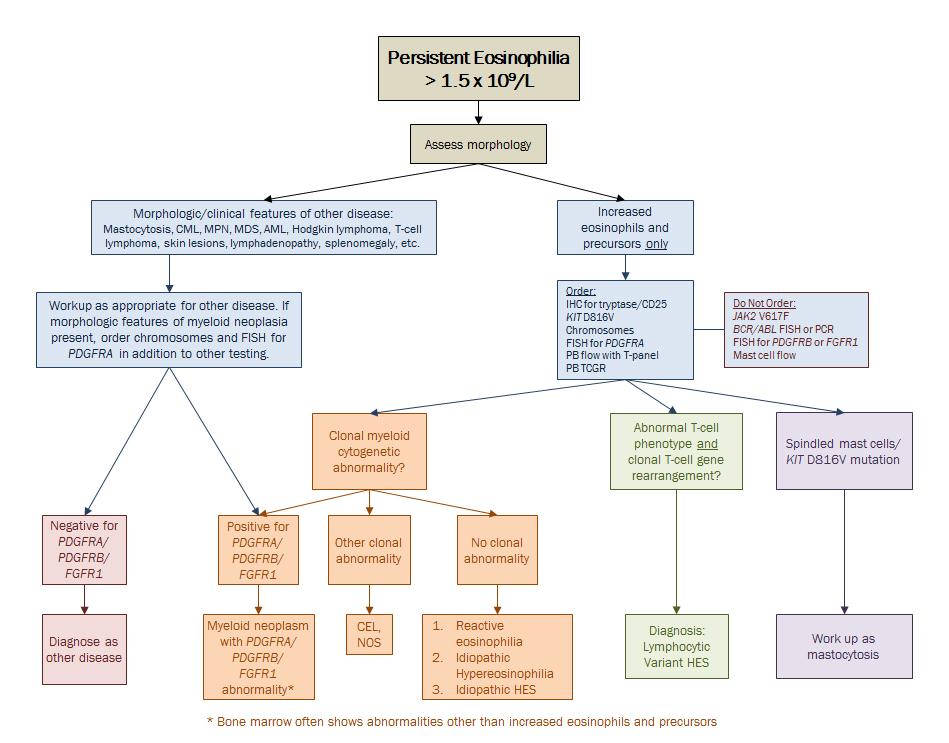
13 VI. Algorithmic approach to the Bone Marrow Workup of Eosinophilia (see flow chart on following page) A. Goal: Diagnostic testing strategy to exclude clonal disorders of eosinophilia B. Initial testing is predicated on peripheral blood /bone marrow morphologic findings and clinical history/presentation/symptomatology. C. Diagnoses are driven by ancillary testing findings, in conjunction with morphology and clinical history. 13
14 14
15 Table 1: Secondary (a.k.a. reactive, non-clonal) Causes of Eosinophilia A. Secondary to a non-neoplastic disorder Infection: Helminths, protozoa, fungi, others Allergic Disorders: Allergies, atopia, asthma, hypersensitivity Drug/Medication Reaction Rheumatologic disorders/connective tissue disorders/toxin-associated disorders: Eosinophilic granulomatosis with polyangiitis (a.k.a. Churg- Strauss), Dermatomyositis, Severe rheumatoid arthritis, Systemic lupus erythematosus, Behçet's disease Progressive systemic sclerosis, Sjögren's syndrome, Wegener's syndrome, Eosinophilic fasciitis Other rare disorders: o Primary immunodeficiency: hyper-ige syndrome, Omenn syndrome, autoimmune lymphoproliferative syndrome (ALPS) o Secondary immunodeficiency/chronic irritation/inflammation: status post transplantation, bullous pemphigoid, sarcoidosis, radiation B. Secondary to an underlying neoplastic disorder Hematopoietic neoplasms: T-cell lymphomas, Classical Hodgkin lymphoma, rarely, acute lymphoblastic leukemia/lymphoma (B and T); typically associated with t(5;14)(q35;q32); rarely, some B-lineage non-hodgkin lymphomas Non-hematopoietic neoplasms: Carcinoma (e.g. lung, gastrointestinal tract), sarcoma Other: Eosinophilia secondary to clonal proliferation of T-cells (not lymphoma or leukemia) currently termed the lymphocytic variant of hypereosinophilia 15
16 Table 2: Primary (a.k.a. clonal) Causes of Eosinophilia Chronic Eosinophilic Leukemia, Not Otherwise Specified Myeloid and lymphoid neoplasms with eosinophilia and rearrangements of PDGFRA, PDGFRB and FGFR1 Acute myeloid leukemia (uncommon) o AML with inv(16) Myeloproliferative Neoplasm o Chronic myelogenous leukemia, BCR-ABL1 positive o Systemic mastocytosis Myelodysplastic syndrome (less than 5% of cases have eosinophilia) Myelodysplastic/myeloproliferative neoplasm lacking rearrangements of PDGFRA, PGFRB and FGFR1 16
17 References: 1. Eosinophilia diagnostic workup algorithm, Mayo Clinic Rochester, Division of Hematopathology, WHO Gotlib J. World Health Organization-defined eosinophilic disorders: 2014 update on diagnosis, risk stratification, and management. Am J Hematol Mar;89(3): Foucar MK, Reichard KK and Czuchlewski DR. Bone Marrow Pathology. ASCP Press Tefferi A, Gotlib J, Pardanani A. Hypereosinophilic syndrome and clonal eosinophilia: point-of-care diagnostic algorithm and treatment update. Mayo Clin Proc Feb;85(2): Pardanani A, Tefferi A. Primary eosinophilic disorders: a concise review. Curr Hematol Malig Rep Jan;3(1): Havelange V, Demoulin JB. Review of current classification, molecular alterations, and tyrosine kinase inhibitor therapies in myeloproliferative disorders with hypereosinophilia. J Blood Med Aug 9;4: The author of this document has no financial disclosures to report. 17
Diagnostic Approach for Eosinophilia and Mastocytosis. Curtis A. Hanson, M.D.
 Diagnostic Approach for Eosinophilia and Mastocytosis Curtis A. Hanson, M.D. 2014 MFMER slide-1 DISCLOSURES: Relevant Financial Relationship(s) None Off Label Usage None 2014 MFMER slide-2 Molecular Classification
Supervisor: Prof. Dr. P Vandenberghe Dr. C Brusselmans
 Contribution of molecular diagnosis in eosinophilia/hypereosinophilia Eosinophilia Hypereosinophilia Hypereosinophilic syndrome Immune mediated hypereosinophilia Chronic eosinophilic leukemia (NOS)/ Idiopathic
Hypereosinophili c syndrome
 Hypereosinophili c syndrome Eosinophilia Eosinophilia is commonly defined as an elevated percentage of eosinophils, with an absolute eosinophil count > 500 cells per cubic millimeter Secondary Primary
2007 Workshop of Society for Hematopathology & European Association for Hematopathology Indianapolis, IN, USA Case # 228
 2007 Workshop of Society for Hematopathology & European Association for Hematopathology Indianapolis, IN, USA Case # 228 Vishnu V. B Reddy, MD University of Alabama at Birmingham Birmingham, AL USA 11/03/07
Case #16: Diagnosis. T-Lymphoblastic lymphoma. But wait, there s more... A few weeks later the cytogenetics came back...
 Case #16: Diagnosis T-Lymphoblastic lymphoma But wait, there s more... A few weeks later the cytogenetics came back... 46,XY t(8;13)(p12;q12)[12] Image courtesy of Dr. Xinyan Lu Further Studies RT-PCR
WHO Classification of Myeloid Neoplasms with Defined Molecular Abnormalities
 WHO Classification of Myeloid Neoplasms with Defined Molecular Abnormalities Robert W. McKenna, M.D. 1/2009 WHO Classification of Myeloid Neoplasms (4th Edition)--2008 Incorporates new information that
Integrated Diagnostic Approach to the Classification of Myeloid Neoplasms. Daniel A. Arber, MD Stanford University
 Integrated Diagnostic Approach to the Classification of Myeloid Neoplasms Daniel A. Arber, MD Stanford University What is an integrated approach? What is an integrated approach? Incorporating all diagnostic
Conditions that mimic neoplasia in the bone marrow. Kaaren K. Reichard Mayo Clinic Rochester
 Conditions that mimic neoplasia in the bone marrow Kaaren K. Reichard Mayo Clinic Rochester reichard.kaaren@mayo.edu Nothing to disclose Conflict of Interest Learning Objectives Multiple conditions in
Case Report. Introduction. Mastocytosis associated with CML Hematopathology - March K. David Li 1,*, Xinjie Xu 1, and Anna P.
 Mastocytosis associated with CML Hematopathology - March 2016 Case Report Systemic mastocytosis with associated clonal hematologic non-mast cell lineage disease (SM-AHNMD) involving chronic myelogenous
Welcome to Master Class for Oncologists. Session 3: 9:15 AM - 10:00 AM
 Welcome to Master Class for Oncologists Session 3: 9:15 AM - 10:00 AM Miami, FL December 18, 2009 Myeloproliferative Neoplasms: Bringing Order to Complexity and Achieving Optimal Outcomes Speaker: Andrew
Classification of Hematologic Malignancies. Patricia Aoun MD MPH
 Classification of Hematologic Malignancies Patricia Aoun MD MPH Objectives Know the basic principles of the current classification system for hematopoietic and lymphoid malignancies Understand the differences
Beyond the CBC Report: Extended Laboratory Testing in the Evaluation for Hematologic Neoplasia Disclosure
 Beyond the CBC Report: Extended Laboratory Testing in the Evaluation for Hematologic Neoplasia Disclosure I am receiving an honorarium from Sysmex for today s presentation. 1 Determining the Etiology for
Bone Marrow. Procedures Blood Film Aspirate, Cell Block Trephine Biopsy, Touch Imprint
 Bone Marrow Protocol applies to acute leukemias, myelodysplastic syndromes, myeloproliferative disorders, chronic lymphoproliferative disorders, malignant lymphomas, plasma cell dyscrasias, histiocytic
SH A CASE OF PERSISTANT NEUTROPHILIA: BCR-ABL
 SH2017-0124 A CASE OF PERSISTANT NEUTROPHILIA: BCR-ABL NEGATIVE John R Goodlad 1, Pedro Martin-Cabrera 2, Catherine Cargo 2 1. Department of Pathology, NHS Greater Glasgow & Clyde, QEUH, Glasgow 2. Haematological
MPL W515L K mutation
 MPL W515L K mutation BCR-ABL genotyping The exact chromosomal defect in Philadelphia chromosome is a translocation. Parts of two chromosomes, 9 and 22, switch places. The result is a fusion gene, created
Case Workshop of Society for Hematopathology and European Association for Haematopathology
 Case 148 2007 Workshop of Society for Hematopathology and European Association for Haematopathology Robert P Hasserjian Department of Pathology Massachusetts General Hospital Boston, MA Clinical history
Practical Diagnosis of Hematologic Disorders. Vol 2 Malignant Disorders
 5 th ed Practical Diagnosis of Hematologic Disorders Vol 2 Malignant Disorders Vol2_FrontMatter_v03_0804 final.indd i 8/6/2009 10:00:11 PM Authors Carl R Kjeldsberg, MD Professor of Pathology, University
MDS/MPN MPN MDS. Discolosures. Advances in the Diagnosis of Myeloproliferative Neoplasms. Myeloproliferative neoplasms
 Discolosures Advances in the Diagnosis of Myeloproliferative Neoplasms Consulting income from Promedior, Inc. Robert P Hasserjian, MD Associate Professor Massachusetts General Hospital and Harvard Medical
Template for Reporting Results of Biomarker Testing for Myeloproliferative Neoplasms
 Template for Reporting Results of Biomarker Testing for Myeloproliferative Neoplasms Version: MPNBiomarkers 1.0.0.2 Protocol Posting Date: June 2017 This biomarker template is NOT required for accreditation
WBCs Disorders 1. Dr. Nabila Hamdi MD, PhD
 WBCs Disorders 1 Dr. Nabila Hamdi MD, PhD ILOs Compare and contrast ALL, AML, CLL, CML in terms of age distribution, cytogenetics, morphology, immunophenotyping, laboratory diagnosis clinical features
Update on the WHO Classification of Acute Myeloid Leukemia. Kaaren K. Reichard, MD Mayo Clinic Rochester
 Update on the WHO Classification of Acute Myeloid Leukemia Kaaren K. Reichard, MD Mayo Clinic Rochester reichard.kaaren@mayo.edu Nothing to disclose Conflict of Interest Objectives Present a practical
Extramedullary precursor T-lymphoblastic transformation of CML at presentation
 Extramedullary precursor T-lymphoblastic transformation of CML at presentation Neerja Vajpayee, Constance Stein, Bernard Poeisz & Robert E. Hutchison Clinical History 30 year old man presented to the emergency
2010 Hematopoietic and Lymphoid ICD-O Codes - Alphabetical List THIS TABLE REPLACES ALL ICD-O-3 Codes
 Acute basophilic leukemia 9870/3 Acute biphenotypic leukemia [OBS] 9805/3 Acute erythroid leukemia 9840/3 Acute megakaryoblastic leukemia 9910/3 Acute monoblastic and monocytic leukemia 9891/3 Acute myeloid
2012 Hematopoietic and Lymphoid ICD-O Codes - Numerical List THIS TABLE REPLACES ALL ICD-O-3 Codes
 Malignant lymphoma, NOS 9590/3 Non-Hodgkin lymphoma, NOS 9591/3 B-cell lymphoma, unclassifiable, with features intermediate between diffuse large B-cell lymphoma and classical Hodgkin lymphoma 9596/3 Primary
Katrina L. Lancaster-Shorts, Joanna Chaffin, Natasha M. Savage. Department of Pathology, Augusta University, Augusta, GA, USA;
 Atlas of Genetics and Cytogenetics in Oncology and Haematology OPEN ACCESS JOURNAL AT INIST-CNRS Leukaemia Section Review Katrina L. Lancaster-Shorts, Joanna Chaffin, Natasha M. Savage Department of Pathology,
1. Challenging cases of eosinophilia (peripheral, bone marrow, or lymph node/tissue-based) falling into any of the following categories
 falling into any of the following categories") Society for Hematopathology/European Association for Haematopathology 2019 Workshop Addressing the Challenges of Eosinophilia and Mastocytosis September 12-14, 2019, Phoenix, AZ https://www.sh-eahp.org/index.php/meetings
Society for Hematopathology/European Association for Haematopathology 2019 Workshop Addressing the Challenges of Eosinophilia and Mastocytosis September 12-14, 2019, Phoenix, AZ https://www.sh-eahp.org/index.php/meetings
Myelodysplastic syndrome (MDS) & Myeloproliferative neoplasms
 & Myeloproliferative neoplasms") Myelodysplastic syndrome (MDS) & Myeloproliferative neoplasms Myelodysplastic syndrome (MDS) A multipotent stem cell that can differentiate into any of the myeloid lineage cells (RBCs, granulocytes, megakaryocytes)
Myelodysplastic syndrome (MDS) & Myeloproliferative neoplasms Myelodysplastic syndrome (MDS) A multipotent stem cell that can differentiate into any of the myeloid lineage cells (RBCs, granulocytes, megakaryocytes)
Combinations of morphology codes of haematological malignancies (HM) referring to the same tumour or to a potential transformation
 referring to the same tumour or to a potential transformation") Major subgroups according to the World Health Organisation (WHO) Classification Myeloproliferative neoplasms (MPN) Myeloid and lymphoid neoplasms with eosinophilia and abnormalities of PDGFRA, PDGFRB or
Major subgroups according to the World Health Organisation (WHO) Classification Myeloproliferative neoplasms (MPN) Myeloid and lymphoid neoplasms with eosinophilia and abnormalities of PDGFRA, PDGFRB or
Leukocytosis - Some Learning Points
 Leukocytosis - Some Learning Points Koh Liang Piu Department of Hematology-Oncology National University Cancer Institute National University Health System Objectives of this talk: 1. To provide some useful
Test Name Results Units Bio. Ref. Interval. Positive
 LL - LL-ROHINI (NATIONAL REFERENCE 135091534 Age 36 Years Gender Female 1/9/2017 120000AM 1/9/2017 105316AM 2/9/2017 104147AM Ref By Final LEUKEMIA GENETIC ROFILE ANY SIX MARKERS, CR QUALITATIVE AML ETO
Differential diagnosis of hematolymphoid tumors composed of medium-sized cells. Brian Skinnider B.C. Cancer Agency, Vancouver General Hospital
 Differential diagnosis of hematolymphoid tumors composed of medium-sized cells Brian Skinnider B.C. Cancer Agency, Vancouver General Hospital Lymphoma classification Lymphoma diagnosis starts with morphologic
MYELOPROLIFERATIVE NEOPLASMS
 9 : 2 MYELOPROLIFERATIVE NEOPLASMS Introduction William Dameshek in 1951 introduced the term Myeloproliferative disorders (MPD). This included polycythemia vera (PV), essential thrombocythemia (ET), primary
Opportunities for Optimal Testing in the Myeloproliferative Neoplasms. Curtis A. Hanson, MD
 Opportunities for Optimal Testing in the Myeloproliferative Neoplasms Curtis A. Hanson, MD 2013 MFMER slide-1 DISCLOSURES: Relevant Financial Relationship(s) None Off Label Usage None 2013 MFMER slide-2
HEMATOLOGIC MALIGNANCIES BIOLOGY
 HEMATOLOGIC MALIGNANCIES BIOLOGY Failure of terminal differentiation Failure of differentiated cells to undergo apoptosis Failure to control growth Neoplastic stem cell FAILURE OF TERMINAL DIFFERENTIATION
Heme 9 Myeloid neoplasms
 Heme 9 Myeloid neoplasms The minimum number of blasts to diagnose acute myeloid leukemia is 5% 10% 20% 50% 80% AML with the best prognosis is AML with recurrent cytogenetic abnormality AML with myelodysplasia
Case Report Chronic Eosinophilic Leukemia Not Otherwise Specified (NOS) in the Background of a Large Cell Lymphoma
 in the Background of a Large Cell Lymphoma") Case Reports in Hematology Volume 2013, Article ID 458303, 4 pages http://dx.doi.org/10.1155/2013/458303 Case Report Chronic Eosinophilic Leukemia Not Otherwise Specified (NOS) in the Background of a Large
Case Reports in Hematology Volume 2013, Article ID 458303, 4 pages http://dx.doi.org/10.1155/2013/458303 Case Report Chronic Eosinophilic Leukemia Not Otherwise Specified (NOS) in the Background of a Large
John L Frater, MD Jeffery M Klco, MD, PhD Department of Pathology and Immunology Washington University School of Medicine St Louis, Missouri
 Myeloproliferative Neoplasms: New Approaches to Diagnosis and Disease Monitoring John L Frater, MD Jeffery M Klco, MD, PhD Department of Pathology and Immunology Washington University School of Medicine
Pathology. #11 Acute Leukemias. Farah Banyhany. Dr. Sohaib Al- Khatib 23/2/16
 35 Pathology #11 Acute Leukemias Farah Banyhany Dr. Sohaib Al- Khatib 23/2/16 1 Salam First of all, this tafreegh is NOT as long as you may think. If you just focus while studying this, everything will
JAK2 V617F analysis. Indication: monitoring of therapy
 JAK2 V617F analysis BCR-ABL genotyping The exact chromosomal defect in Philadelphia chromosome is a translocation. Parts of two chromosomes, 9 and 22, switch places. The result is a fusion gene, created
Mixed Phenotype Acute Leukemias
 Mixed Phenotype Acute Leukemias CHEN GAO; AMY M. SANDS; JIANLAN SUN NORTH AMERICAN JOURNAL OF MEDICINE AND SCIENCE APR 2012 VOL 5 NO.2 INTRODUCTION Most cases of acute leukemia can be classified based
Recommended Timing for Transplant Consultation
 REFERRAL GUIDELINES Recommended Timing for Transplant Consultation Published jointly by the National Marrow Donor Program /Be The Match and the American Society for Blood and Marrow Transplantation BeTheMatchClinical.org
9/25/2017. Disclosure. I have nothing to disclose. Young S. Kim MD Dept. of Pathology
 Disclosure MAST CELLNEOPLASM I have nothing to disclose. Young S. Kim MD Dept. of Pathology 1 Objectives What is mast cell lineage? Changes in updated WHO 2016 mastocytosis Issues of Mastocytosis CD30
Hematopathology Case Study
 www.medfusionservices.com Hematopathology Case Study CV3515-14 JUNE Clinical Presentation: Clinical Information: A 42 year old male with history of chronic myelogenous leukemia (CML) presents with an elevated
Test Name Results Units Bio. Ref. Interval. Positive
 LL - LL-ROHINI (NATIONAL REFERENCE 135091533 Age 28 Years Gender Male 1/9/2017 120000AM 1/9/2017 105415AM 4/9/2017 23858M Ref By Final LEUKEMIA DIAGNOSTIC COMREHENSIVE ROFILE, ANY 6 MARKERS t (1;19) (q23
HEMATOPATHOLOGY (SHANDS HOSPITAL AT THE UNIVERSITY OF FLORIDA): Rotation Director: Ying Li, M.D., Ph.D., Assistant Professor
: Rotation Director: Ying Li, M.D., Ph.D., Assistant Professor") HEMATOPATHOLOGY (SHANDS HOSPITAL AT THE UNIVERSITY OF FLORIDA): Rotation Director: Ying Li, M.D., Ph.D., Assistant Professor I. Description of the rotation: During this rotation, the resident will gain
HEMATOPATHOLOGY (SHANDS HOSPITAL AT THE UNIVERSITY OF FLORIDA): Rotation Director: Ying Li, M.D., Ph.D., Assistant Professor I. Description of the rotation: During this rotation, the resident will gain
Molecular techniques in a case of concurrent BCR-ABL1 positive CML and CMML
 reprinted from november 2014 pathology laboratory medicine laboratory management Molecular techniques in a case of concurrent BCR-ABL1 positive CML and CMML CAP TODAY and the Association for Molecular
74y old Female with chronic elevation of Platelet count. August 18, 2005 Faizi Ali, MD Hematopathology Fellow
 74y old Female with chronic elevation of Platelet count August 18, 2005 Faizi Ali, MD Hematopathology Fellow Clinical History Patient is a 74y old otherwise healthy Caucasian female with no major complaint
Acute myeloid leukemia. M. Kaźmierczak 2016
 Acute myeloid leukemia M. Kaźmierczak 2016 Acute myeloid leukemia Malignant clonal disorder of immature hematopoietic cells characterized by clonal proliferation of abnormal blast cells and impaired production
Myeloid neoplasms. Early arrest in the blast cell or immature cell "we call it acute leukemia" Myoid neoplasm divided in to 3 major categories:
 Myeloid neoplasms Note: Early arrest in the blast cell or immature cell "we call it acute leukemia" Myoid neoplasm divided in to 3 major categories: 1. AML : Acute myeloid leukemia(stem cell with myeloid
Hypereosinophilic Syndrome. Fellow: Eunpi Cho Faculty Discussant: Michael Linenberger
 Hypereosinophilic Syndrome Fellow: Eunpi Cho Faculty Discussant: Michael Linenberger Case 19 yo M admitted to Evergreen Hospital after cardiac arrest. Troponin 8.41, BNP 12,087 Utox pos for amphetamines
Presenter Disclosure Information
 Welcome to Master Class for Oncologists Session 3: 2: PM 3:3 PM Pasadena, CA May 1, 21 Myeloproliferative Neoplasms 21 Speaker: Ayalew Tefferi Mayo Clinic, Rochester, MN Presenter Disclosure Information
Reactive and Neoplastic Lymphocytosis
 Reactive and Neoplastic Lymphocytosis Koranda A. Walsh, VMD, BS Assistant Professor, Clinical Pathobiology University of Pennsylvania School of Veterinary Medicine PLEASE NOTE: These notes are meant as
Corrigenda. WHO Classification of Tumours of Haematopoietic and Lymphoid Tissues (revised 4th edition): corrections made in second print run
: corrections made in second print run") Corrigenda WHO Classification of Tumours of Haematopoietic and Lymphoid Tissues (revised 4th edition): corrections made in second print run In addition to corrections of minor typographical errors, corrections
Corrigenda WHO Classification of Tumours of Haematopoietic and Lymphoid Tissues (revised 4th edition): corrections made in second print run In addition to corrections of minor typographical errors, corrections
Gleevec. Gleevec (imatinib) Description
 Description") Federal Employee Program 1310 G Street, N.W. Washington, D.C. 20005 202.942.1000 Fax 202.942.1125 5.21.74 Subject: Gleevec Page: 1 of 6 Last Review Date: June 24, 2016 Gleevec Description Gleevec (imatinib)
Federal Employee Program 1310 G Street, N.W. Washington, D.C. 20005 202.942.1000 Fax 202.942.1125 5.21.74 Subject: Gleevec Page: 1 of 6 Last Review Date: June 24, 2016 Gleevec Description Gleevec (imatinib)
Hematology Unit Lab 2 Review Material
 Objectives Hematology Unit Lab 2 Review Material - 2018 Laboratory Instructors: 1. Assist students during lab session Students: 1. Review the introductory material 2. Study the case histories provided
WHO Update to Myeloproliferative Neoplasms
 WHO Update to Myeloproliferative Neoplasms Archana M Agarwal, MD, Associate Professor of Pathology University of Utah Department of Pathology/ARUP Laboratories Myeloproliferative Neoplasms The categories
ADx Bone Marrow Report. Patient Information Referring Physician Specimen Information
 ADx Bone Marrow Report Patient Information Referring Physician Specimen Information Patient Name: Specimen: Bone Marrow Site: Left iliac Physician: Accession #: ID#: Reported: 08/19/2014 - CHRONIC MYELOGENOUS
HENATOLYMPHOID SYSTEM THIRD YEAR MEDICAL STUDENTS- UNIVERSITY OF JORDAN AHMAD T. MANSOUR, MD. Part 4 MYELOID NEOPLASMS
 HENATOLYMPHOID SYSTEM THIRD YEAR MEDICAL STUDENTS- UNIVERSITY OF JORDAN AHMAD T. MANSOUR, MD Part 4 MYELOID NEOPLASMS Introduction: o Myeloid neoplasms are divided into three major categories: o Acute
Lymphoblastic Leukemia / Lymphoma
 1 5014 - Topics in Pediatric Hematopathology: Acute Lymphoblastic Leukemia, Including Changes in the Revised WHO Classification, and Unusual Pediatric Myeloid Neoplasms Robert W. McKenna, MD MASCP * Elizabeth
Diagnostic challenge: Acute leukemia with biphenotypic blasts and BCR-ABL1 translocation
 Case Study Diagnostic challenge: Acute leukemia with biphenotypic blasts and BCR-ABL1 translocation Ling Wang 1 and Xiangdong Xu 1,2,* 1 Department of Pathology, University of California, San Diego; 2
Recent Advances in the Diagnosis and Treatment of Hypereosinophilic Syndromes
 Recent Advances in the Diagnosis and Treatment of Hypereosinophilic Syndromes Amy D. Klion Hypereosinophilic syndromes (HES) are a heterogeneous group of disorders characterized by marked peripheral blood
Diagnostic Challenges during Pretreatment Long-term Follow-up in a Patient with FIP1L1-PDGFRA-positive Eosinophilia
 CASE REPORT Diagnostic Challenges during Pretreatment Long-term Follow-up in a Patient with FIP1L1-PDGFRA-positive Eosinophilia Danijela Lekovic 1, Andrija Bogdanovic 1,2, Maja Perunicic-Jovanovic 1, Gradimir
Myeloproliferative Disorders - D Savage - 9 Jan 2002
 Disease Usual phenotype acute leukemia precursor chronic leukemia low grade lymphoma myeloma differentiated Total WBC > 60 leukemoid reaction acute leukemia Blast Pro Myel Meta Band Seg Lymph 0 0 0 2
GENETICS OF HEMATOLOGICAL MALIGNANCIES
 de DUVE INSTITUTE GENETICS OF HEMATOLOGICAL MALIGNANCIES INTERUNIVERSITY CERTIFICATE IN HUMAN GENETICS Université catholique de Louvain Brussels,19/02/2016 Professor Hélène Antoine-Poirel, MD, PhD Center
Hypereosinophilic syndromes recent advances in definition, classification and therapeutic approach
 M K pag 146 Mædica - a Journal of Clinical Medicine STATE TE-OF OF-THE THE-AR ART Hypereosinophilic syndromes recent advances in definition, classification and therapeutic approach Irina VOICAN, MD; Ana
5/21/2018. Disclosures. Objectives. Normal blood cells production. Bone marrow failure syndromes. Story of DNA
 AML: Understanding your diagnosis and current and emerging treatments Nothing to disclose. Disclosures Mohammad Abu Zaid, MD Assistant Professor of Medicine Indiana University School of Medicine Indiana
Template for Reporting Results of Biomarker Testing for Myeloproliferative Neoplasms
 Template for Reporting Results of Biomarker Testing for Myeloproliferative Neoplasms Template web posting date: December 2014 Authors Todd W. Kelley, MD, FCAP University of Utah and ARUP Laboratories,
NUP214-ABL1 Fusion: A Novel Discovery in Acute Myelomonocytic Leukemia
 Case 0094 NUP214-ABL1 Fusion: A Novel Discovery in Acute Myelomonocytic Leukemia Jessica Snider, MD Medical University of South Carolina Case Report - 64 year old Caucasian Male Past Medical History Osteoarthritis
Chronic Myelogenous Leukemia (Hematology) By DEISSEROTH READ ONLINE
 By DEISSEROTH READ ONLINE") Chronic Myelogenous Leukemia (Hematology) By DEISSEROTH READ ONLINE If searched for the ebook by DEISSEROTH Chronic Myelogenous Leukemia (Hematology) in pdf format, in that case you come on to correct
Chronic Myelogenous Leukemia (Hematology) By DEISSEROTH READ ONLINE If searched for the ebook by DEISSEROTH Chronic Myelogenous Leukemia (Hematology) in pdf format, in that case you come on to correct
CHAPTER:4 LEUKEMIA. BY Mrs. K.SHAILAJA., M. PHARM., LECTURER DEPT OF PHARMACY PRACTICE, SRM COLLEGE OF PHARMACY 8/12/2009
 LEUKEMIA CHAPTER:4 1 BY Mrs. K.SHAILAJA., M. PHARM., LECTURER DEPT OF PHARMACY PRACTICE, SRM COLLEGE OF PHARMACY Leukemia A group of malignant disorders affecting the blood and blood-forming tissues of
Case Presentation. Attilio Orazi, MD
 Case Presentation Attilio Orazi, MD Weill Cornell Medical College/ NYP Hospital Department of Pathology and Laboratory Medicine New York, NY United States History 60 year old man presented with anemia
Objectives. Morphology and IHC. Flow and Cyto FISH. Testing for Heme Malignancies 3/20/2013
 Molecular Markers in Hematologic Malignancy: Ways to locate the needle in the haystack. Objectives Review the types of testing for hematologic malignancies Understand rationale for molecular testing Marcie
Allogeneic Hematopoietic Stem-Cell Transplantation for Myelodysplastic Syndromes and Myeloproliferative Neoplasms. Policy Specific Section:
 Medical Policy Allogeneic Hematopoietic Stem-Cell Transplantation for Myelodysplastic Syndromes and Myeloproliferative Type: Medical Necessity and Investigational / Experimental Policy Specific Section:
UnitedHealthcare Pharmacy Clinical Pharmacy Programs
 UnitedHealthcare Pharmacy Clinical Pharmacy Programs Program Number 2017 P 1037-6 Program Prior Authorization/Notification Medication Gleevec (imatinib mesylate) P&T Approval Date 8/2008, 6/2009, 6/2010,
Disclosures. Myeloproliferative Neoplasms: A Case-Based Approach. Objectives. Myeloproliferative Neoplasms. Myeloproliferative Neoplasms
 Myeloproliferative Neoplasms: A Case-Based Approach Disclosures No conflicts of interests regarding the topic being presented Adam M. Miller, MD PGY-4 Resident Physician Department of Pathology and Laboratory
Jordi Esteve Hospital Clínic (Barcelona) Acute Leukemia Working Party. The European Group for Blood and Marrow Transplantation
 Acute Leukemia Working Party. The European Group for Blood and Marrow Transplantation") 36th EBMT & 9th Data Management Group Annual Meeting Vienna, 23 March 2010 Jordi Esteve Hospital Clínic (Barcelona) Acute Leukemia Working Party The European Group for Blood and Marrow Transplantation
36th EBMT & 9th Data Management Group Annual Meeting Vienna, 23 March 2010 Jordi Esteve Hospital Clínic (Barcelona) Acute Leukemia Working Party The European Group for Blood and Marrow Transplantation
Hematology Page 1 of 8
 Hematology Page 1 of 8 Hematology Major Category Code Headings Revised 12/17 1 Basic methodology and test armamentarium 20000 2 Normal hematopoiesis & hemostasis 20100 3 RBC disorders, non-neoplastic 20340
From Morphology to Molecular Pathology: A Practical Approach for Cytopathologists Part 1-Cytomorphology. Songlin Zhang, MD, PhD LSUHSC-Shreveport
 From Morphology to Molecular Pathology: A Practical Approach for Cytopathologists Part 1-Cytomorphology Songlin Zhang, MD, PhD LSUHSC-Shreveport I have no Conflict of Interest. FNA on Lymphoproliferative
Changes to the Hematopoietic and Lymphoid Neoplasm Coding Manual
 Changes to the Hematopoietic and Lymphoid Neoplasm Coding Manual KCR 2018 SPRING TRAINING 2018 Hematopoietic Database Updates Updates were done to the Hematopoietic Database based on the WHO Hematopoietic
Update on Myelodysplastic Syndromes and Myeloproliferative Neoplasms. Kaaren Reichard Mayo Clinic Rochester
 Update on Myelodysplastic Syndromes and Myeloproliferative Neoplasms Kaaren Reichard Mayo Clinic Rochester Reichard.kaaren@mayo.edu Nothing to disclose Conflict of Interest Learning Objectives Present
89 Beaumont Avenue, Given E-214-UVM363, Burlington, VT 05405, USA
 Case Reports in Medicine Volume 2016, Article ID 8324791, 4 pages http://dx.doi.org/10.1155/2016/8324791 Case Report Myeloid Neoplasms with t(5;12) and ETV6-ACSL6 Gene Fusion, Potential Mimickers of Myeloid
Group of malignant disorders of the hematopoietic tissues characteristically associated with increased numbers of white cells in the bone marrow and
 Group of malignant disorders of the hematopoietic tissues characteristically associated with increased numbers of white cells in the bone marrow and / or peripheral blood Classified based on cell type
Case #1. 65 yo man with no prior history presented with leukocytosis and circulating blasts: Bone marrow biopsy was performed
 Case #1 65 yo man with no prior history presented with leukocytosis and circulating blasts: WBC 187.4K/uL ; Hgb 10.0gm/dL; Platelet 68K/uL Neutrophil % 25.0% Lymphocyte % 38.0% Monocyte % 12.0% Metamyelocyte
Myelodysplastic Syndromes: Everyday Challenges and Pitfalls
 Myelodysplastic Syndromes: Everyday Challenges and Pitfalls Kathryn Foucar, MD kfoucar@salud.unm.edu Henry Moon lecture May 2007 Outline Definition Conceptual overview; pathophysiologic mechanisms Incidence,
Molecular Markers. Marcie Riches, MD, MS Associate Professor University of North Carolina Scientific Director, Infection and Immune Reconstitution WC
 Molecular Markers Marcie Riches, MD, MS Associate Professor University of North Carolina Scientific Director, Infection and Immune Reconstitution WC Overview Testing methods Rationale for molecular testing
Done By : WESSEN ADNAN BUTHAINAH AL-MASAEED
 Done By : WESSEN ADNAN BUTHAINAH AL-MASAEED Acute Myeloid Leukemia Firstly we ll start with this introduction then enter the title of the lecture, so be ready and let s begin by the name of Allah : We
Lymphoma: What You Need to Know. Richard van der Jagt MD, FRCPC
 Lymphoma: What You Need to Know Richard van der Jagt MD, FRCPC Overview Concepts, classification, biology Epidemiology Clinical presentation Diagnosis Staging Three important types of lymphoma Conceptualizing
Mast Cell Disease. Daniel A. Arber, MD Stanford University, Stanford CA
 Mast Cell Disease Daniel A. Arber, MD Stanford University, Stanford CA Mast cell disease, or mastocytosis, includes a variety of disorders that are characterized by the presence of mast cell aggregates
Systemic Mastocytosis: Seldomly Seen, Multiple Manifestations
 Systemic Mastocytosis: Seldomly Seen, Multiple Manifestations Ryan Cassaday, MD HematologyFellows Conference June 3, 2011 Outline Why this topic, and case discussion Brief background Classification i of
Αιχμές στην Παθολογία
 2 η ΕΠΙΣΤΗΜΟΝΙΚΗ ΗΜΕΡΙΔΑ Αιχμές στην Παθολογία ΔΙΑΓΝΩΣΤΙΚΗ ΠΡΟΣΕΓΓΙΣΗ ΗΩΣΙΝΟΦΙΛΙΑΣ Σταυρούλα Γιαννούλη Λέκτορας Παθολογίας-Αιματολογίας Β Πανεπιστημιακή Παθολογική Κλινική ΓΝΑ Ιπποκράτειο Εosinophil: biology
Bone marrow morphology in reactive conditions. Kaaren K. Reichard, MD Mayo Clinic Rochester
 Bone marrow morphology in reactive conditions Kaaren K. Reichard, MD Mayo Clinic Rochester reichard.kaaren@mayo.edu Nothing to disclose Conflict of Interest Outline of Presentation Brief introduction General
Bone Marrow Morphology after Therapy and Stem Cell Transplantation. Arash Mohtashamian, MD Naval Medical Center, San Diego
 Bone Marrow Morphology after Therapy and Stem Cell Transplantation Arash Mohtashamian, MD Naval Medical Center, San Diego Objectives Bone marrow findings after myeloablative therapy. Effects of recombinant
Molecular Markers in Acute Leukemia. Dr Muhd Zanapiah Zakaria Hospital Ampang
 Molecular Markers in Acute Leukemia Dr Muhd Zanapiah Zakaria Hospital Ampang Molecular Markers Useful at diagnosis Classify groups and prognosis Development of more specific therapies Application of risk-adjusted
AML: WHO classification, biology and prognosis. Dimitri Breems, MD, PhD Internist-Hematoloog Ziekenhuis Netwerk Antwerpen
 AML: WHO classification, biology and prognosis Dimitri Breems, MD, PhD Internist-Hematoloog Ziekenhuis Netwerk Antwerpen Acute myeloid leukemia Clonal expansion of undifferentiated myeloid precursors Impaired
Case 1. Sa A.Wang, MD UT MD Anderson Cancer Center Houston, TX
 Case 1 Sa A.Wang, MD UT MD Anderson Cancer Center Houston, TX Disclosure of Relevant Financial Relationships The USCAP requires that anyone in a position to influence or control the content of all CME
Molecular Advances in Hematopathology
 Molecular Advances in Hematopathology HOW MOLECULAR METHODS HAVE CHANGED MY PRACTICE Objectives Understand the importance of cytogenetic/molecular studies in hematolymphoid diseases Know some of the important