Conditions that mimic neoplasia in the bone marrow. Kaaren K. Reichard Mayo Clinic Rochester
|
|
|
- Jesse Nichols
- 5 years ago
- Views:
Transcription
1 Conditions that mimic neoplasia in the bone marrow Kaaren K. Reichard Mayo Clinic Rochester
2 Nothing to disclose Conflict of Interest
3 Learning Objectives Multiple conditions in bone marrow/peripheral blood that may mimic neoplastic disorders Select examples Present key tips for discriminating normal from abnormal
4 Hematogones A.k.a. normal B-lymphocyte precursors Typically found in small numbers in most adult bone marrow specimens Occur in larger numbers In some healthy infants and young children In a variety of disease states in both children and adults particularly prominent in regeneration following chemotherapy or bone marrow transplantation autoimmune disorders, congenital cytopenias, neoplasms, viral infections and immunodeficiency states In some cases, they constitute 5% to 50% of bone marrow cells.
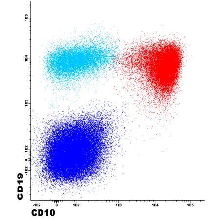
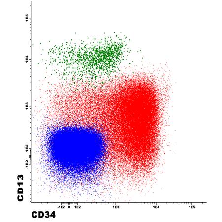
5 Hematogones


6 Hematogone maturation Hematogone maturation Cyan- Early stage 1 Dark Blue-Middle stage Magenta -Naïve B-cells Consistent and predicable spectrum of sequential antigen expression

7 Hematogones Microarchitectural pattern Reside in the bone marrow interstitium in an individually distributed, non-clustered fashion Background trilineage hematopoiesis should be intact and normal With increasing numbers of hematogones, one may encounter cases where they are more closely situated to one another Lack of distinctive clustering of more than 5-10 cells, particularly if they all express TdT TdT
 Lymphomas Metastatic neoplasm Early bone marrow involvement by")
8 Hematogones may mimic neoplasms Lymphoblastic leukemia/ lymphoma (LL) Lymphomas Metastatic neoplasm Early bone marrow involvement by B-LL
9 Lymphoblasts Share morphologic, and some immunophenotypic features with hematogones In particular, distinguishing residual/recurrent B- lymphoblasts from hematogones after treatment for B-lymphoblastic leukemia/lymphoma can be challenging Recognition of the typical maturational profile for hematogones and lack of aberrant antigen expression are two useful tools to help with this distinction
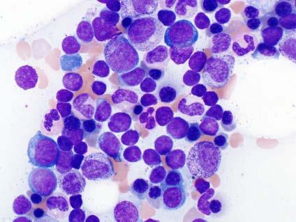
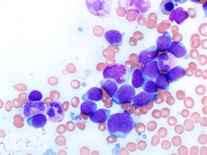
10 B-lymphoblasts
11 B-lymphoblasts Aberrant antigen expression


12 Hematogones may mimic neoplasms T-cell prolymphocytic leukemia High grade B-cell lymphoma with MYC and BCL2 rearrangement Metastatic small cell carcinoma
13 Quiz B-lymphoblastic leukemia Hematogones
14 Eosinophilia Numerous causes, both neoplastic and nonneoplastic Broad workup can be daunting Methodical algorithmic approach Manage ancillary testing Ensure thorough, appropriate workup Diagnose/exclude specific disease entities
15 Eosinophilia Etiology Primary (clonal) Chronic eosinophilic leukemia (CEL, NOS) Myeloid and lymphoid neoplasms with eosinophilia and rearrangements of PDGFRA, PDGFRB, FGFR1, PCM1-JAk2 AML [particularly with inv(16)] CML MDS MPN Systemic mastocytosis Secondary to underlying neoplasm Lymphoma T-cell lymphoma classical Hodgkin lymphoma Lymphoblastic leukemia Carcinoma Lymphocytic variant of hypereosinophilic syndrome (LV-HES) Secondary to nonneoplastic disorder Infection Allergic disorders Drug reaction Rheumatologic disorders Immunodeficiency
16 Eosinophilia Assess Morphology Increased eosinophils and precursors only Normal bone marrow with increased eosinophils Take care in assessing eosinophil cytology Abnormal forms may be found in secondary causes Eosinophil cytology alone should not be used to indicate clonal eosinophilic disease
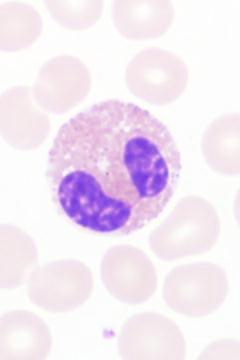
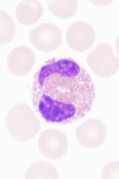
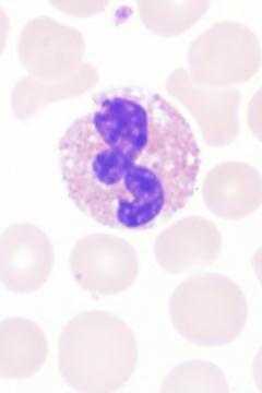
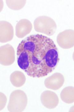
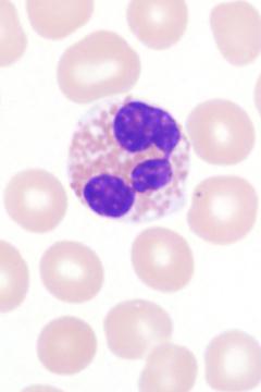
17 Idiopathic hypereosinophilia Peripheral blood Images courtesy of M. Howard, MD
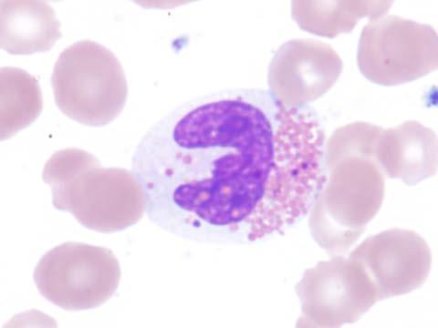
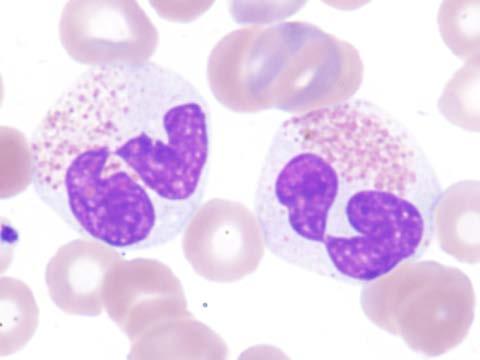
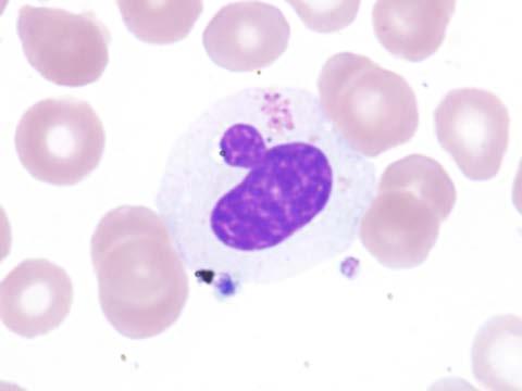
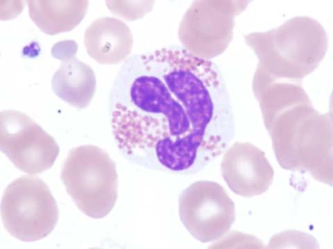
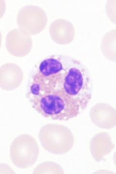
18 Peripheral T-cell lymphoma Peripheral blood Images courtesy of M. Howard, MD
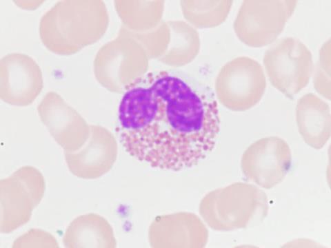
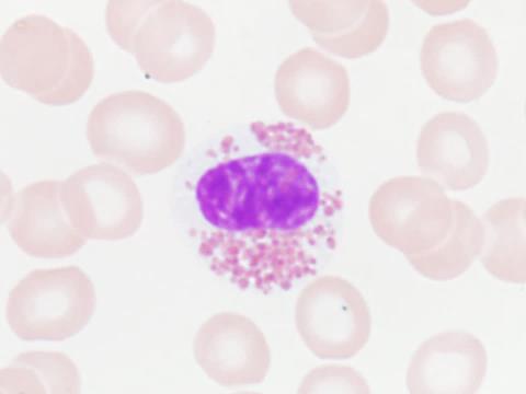
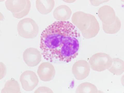
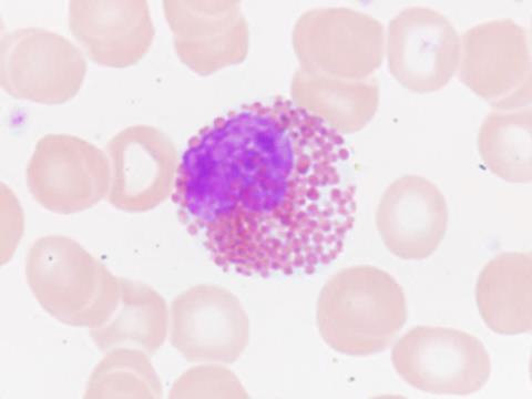
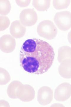
19 Chronic eosinophilic leukemia, NOS Peripheral blood Images courtesy of M. Howard, MD
20 Eosinophilia Assess other pathology Monocytosis, dysplasia, increased or abnormal blasts, basophilia Fibrosis Dysplasia Mast cell clusters Lymphoid infiltrates/hodgkin lymphoma Skin lesions, splenomegaly, known diagnosis, etc. Granulomas


21 Neoplastic eosinophilia Myeloid neoplasm with eosinophilia and PDGFRA-FIP1L1


22 Neoplastic eosinophilia Chronic myeloid leukemia, BCR-ABL1 positive


23 Non-neoplastic eosinophilia masking a clonal neoplasm Peripheral T-cell lymphoma, NOS


")

24 Non-neoplastic eosinophilia masking a clonal neoplasm Abnormal T-cells (in red) Lymphocytic variant hypereosinophilic syndrome (LV-HES)


25 Sustained Eosinophilia Assess/identify secondary causes No Assess peripheral blood/ bone marrow morphology Yes Workup as appropriate Workup as appropriate for other diseases Yes Morphologic features of other diseases, e.g. mastocytosis, myeloid neoplasm, lymphoma etc. No Increased eosinophils and precursors only Order: Immunohistochemistry for tryptase and/or CD117 and CD25 KIT D816V Chromosomes FISH for PDGFRA-rearrangement T-cell flow cytometry Consider T cell receptor gene rearrangement Do Not Routinely Order: JAK2 V617F BCR/ABL1 FISH or PCR FISH for PDGFRB or FGFR1 Mast cell flow cytometry Chromosome and/or FISH result Abnormal T-cell phenotype and clonal T-cell receptor gene rearrangement Spindled CD25 positive mast cells and KIT D816V mutation Positive for: PDGFRA rearrangement PDGFRB rearrangement FGFR1 rearrangement PCM1-JAK2 fusion Other clonal myeloid abnormality** No clonal abnormality Myeloid neoplasm with*: PDGFRA rearrangement PDGFRB rearrangement FGFR1 rearrangement PCM1-JAK2 fusion Consider chronic eosinophilic leukemia, not otherwise specified Consider: Reactive eosinophilia Idiopathic hypereosinophilia Idiopathic hypereosinophilic syndrome Consider lymphocytic variant hypereosinophilic syndrome Work up as mastocytosis * Bone marrow typically shows additional morphologic abnormalities ** Trisomy 8, deletion 20q, and Y as sole abnormalities do not necessary implicate the presence of a clonal myeloid disease Key: FISH, fluorescence in situ hybridization


26 Quiz Drug-induced hypereosinophilia B-lymphoblastic leukemia with eosinophilia
27 Post-therapy effect Recombinant therapy Therapeutic agents Selected medications Post myeloablative chemotherapy/stem cell transplantation


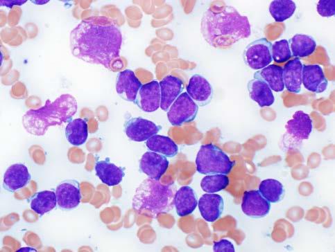
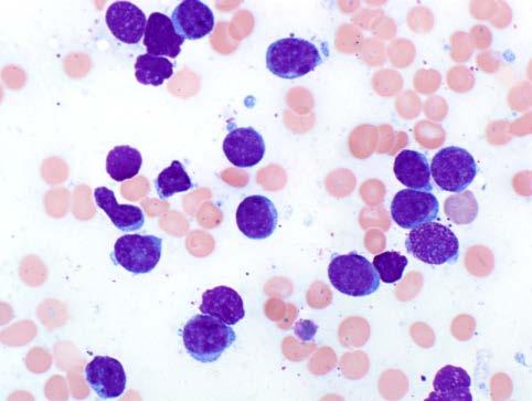
28 Granulocyte colony stimulating factor Peripheral Blood Bone marrow core biopsy Leukocytosis with granulocytic left shift, toxic changes, occasional monocytosis and circulating blasts Hypercellular with granulocytic hyperplasia, prominent left shift and occasional transient increase in blasts and monocytes


29 G-CSF/GM-CSF may mimic neoplasia Peripheral blood G-CSF administration after delayed hematopoietic recovery for treatment of lymphoma mimics acute leukemia 72 y.o. treated for high grade lymphoma mimics chronic myelomonocytic leukemia
30 Thrombopoietin receptor agonists (TPO-RAs) A number of thrombopoietin-like molecules have been developed to treat thrombocytopenia TPO-RAs are being used with increasing frequency to treat thrombocytopenia associated with a variety of conditions such as hepatitis C associated thrombocytopenia, myelodysplastic syndrome, and aplastic anemia.
31 TPO Receptor Agonists Mechanism of Action Stimulate megakaryocyte and platelet production by binding to the TPO receptor Generally reserved for third-line therapy Recombinant TPO products are not used in the USA due to reports of anti-tpo antibody development


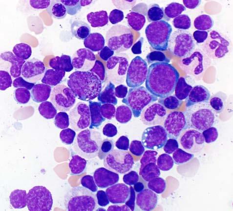
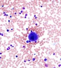
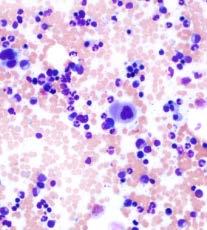
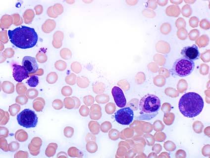
32 Exemplary case Peripheral blood Slight leukocytosis Occasional basophils and nucleated RBC


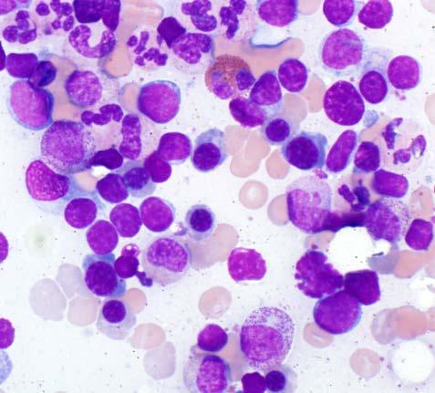
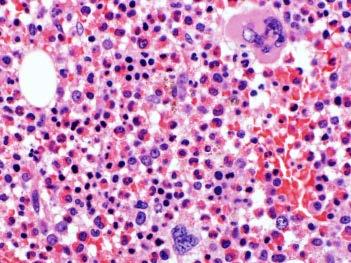
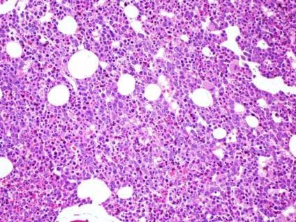
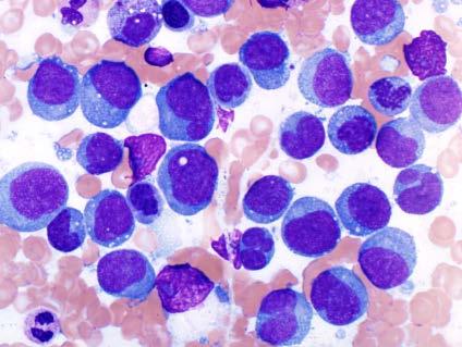
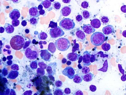
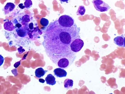
33 Bone Marrow Aspirate Numerous spicules with megakaryocytic hyperplasia and morpholgoic spectrum


34 Bone Marrow Core Biopsy Reticulin Megakaryocytic hyperplasia, loose clustering and large, hyperlobulated forms Mild reticulin fibrosis, grade 1 of 3 (MF-1)
35 Bone Marrow Pathology with Thrombopoietin Receptor Agonists Leukoerythroblastic reaction seen in small number Hypercellular with panmyelosis ~10% showed erythroid/granulocytic hyperplasia (Brynes) Increased megakaryocytes, pleomorphism, cluster formation: MPN-like features (ET/PVlike) Boiocchi L, et al. Mod Pathol. 2012;25:65-74; Ghanima W et al. Haematologica. 2014;99:937-44; Brynes RK, et al. Am J Hematol. 2015;90:
36 Bone Marrow Pathology with Thrombopoietin Receptor Agonists Increase in reticulin fibrosis; mainly mild (MF- 1), some with MF-2/3; no collagen fibrosis; not clinically relevant No significant bone marrow dysfunction Karyotype normal Nonclonal Boiocchi L, et al. Mod Pathol. 2012;25:65-74; Ghanima W et al. Haematologica. 2014;99:937-44; Brynes RK, et al. Am J Hematol. 2015;90:
37 TPO-RA neoplastic mimics The main diagnostic trap is with myeloproliferative neoplasms Similarities include splenomegaly (particularly if history is ITP), leukoerythroblastosis, transient thrombocytosis, occasional thrombosis, and megakaryocyte hyperplasia with large, hyperlobulated and occasionally bizarre/hyperchromatic forms which may loosely cluster Features to help distinguish include history of TPO-RA use, normal karyotype, non-clonal, no mutations in JAK2, CALR, MPL


38 Quiz TPO-RA Early MPN (JAK2 V167F 7%)


39 Drug effect mimicking myelodysplasia Pseudo Pelger-Huët abnormalities Etiology: number of medications Neutrophils demonstrate hyposegmentation with abnormally clumped chromatin; Dohle bodies also Cytologic changes typically abate after removal of the offending agent Be careful to not over interpret as myelodysplasia! Sirolimus Pseudo-Pelger Huet change Tacrolimus


40 Drug effect mimicking myelodysplasia Arsenic induced RBC changes in APL Dyserythropoiesis due to colchicine


41 Post myeloblative chemotherapy changes and neoplastic mimics Post-therapy Myelodysplastic syndrome


42 Post myeloblative chemotherapy changes and neoplastic mimics Post-therapy Myelodysplastic syndrome
43 Megaloblastic anemia due to vitamin B12 deficiency May be due to inadequate dietary intake At risk populations include the elderly, alcoholics, those with poor diet and pregnant women May also be due to absorption defects This includes deficiencies of and antibodies against intrinsic factor including the autoimmune disorder (pernicious anemia) Some medications that may impair absorption (e.g. aminosalicylates)


44 Peripheral Blood findings Macrocytic anemia Pancytopenia in severe cases there may be pancytopenia Increasing anisopoikilocytosis with increasing severity of the deficiency often includes schistocytes Hypersegmented neutrophils Oval macrocytes are typical Rarely, Howell-Jolly bodies or Cabot rings are seen

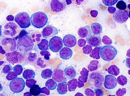
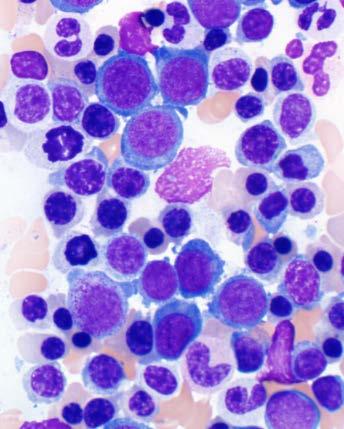
45 Bone Marrow Findings


46 Megaloblastic anemia may mimic Pure erythroid leukemia Myeloid neoplasm with abundant erythroid precursors Other neoplasms: lymphoma, carcinoma Key tips in megaloblastic anemia: Be careful to not overcall as neoplastic Systematic approach PEL, Case 1 PEL, Case 2
47 Neoplastic mimics DLBCL MDS-RS-MLD
48 Quiz Myelodysplastic syndrome Megaloblastic anemia
49 Copper Deficiency Definition Decreased serum copper or ceruloplasmin levels General features/etiology Insufficient copper absorption from gastrointestinal tract; long-term TPN without copper supplementation, malabsorption after gastric bypass surgery or celiac disease, excess serum zinc
50 Copper Deficiency Clinical presentation Sensory and motor difficulties, neurocognitive defects, cytopenias (anemia and neutropenia) Prognosis Hematologic abnormalities are fully reversible with restoration of serum copper levels Neurologic disorders may not resolve but are less likely to progress

")
51 Copper Deficiency Peripheral blood findings Anemia is typical may be normocytic, macrocytic, or microcytic anemia minimal polychromasia Neutropenia (which can be marked) is generally also seen

52 Bone marrow findings Variable cellularity May be hypocellular, normocellular, or hypercellular Possible erythroid predominance Cytologic Distinctive cytoplasmic vacuoles in granulocytic and erythroid precursors Megakaryocytes may show some slight atypia May be some subtle nuclearcytoplasmic dyssynchrony in the erythroid or granulocytic precursors No overt dysplastic features Blasts not increased Ring sideroblasts are common Copper Deficiency
53 Copper Deficiency

54 Copper Deficiency Main neoplastic differential diagnosis is with low-grade MDS Should not see vacuolated granulocytic precursors in MDS Clear cut dysplasia in MDS fulfilling WHO criteria
55 Germline predisposition syndromes New category in the WHO 2016 Classification of Haematopoietic neoplasms Important to recognize and diagnose for future management Be careful not to overinterpret abnormal megakaryocytes in a young patient with thrombocytopenia as MDS Recognize different propensities to develop frank hematopoietic malignancies
, although occasional small, atypical")
56 Bone Marrow Aspirate Smear Cellular aspirate; intact trilineage hematopoiesis; morphologically unremarkable megakaryocytes (circle), although occasional small, atypical forms (arrow) noted


57 Bone Marrow Aspirate Smear Cellular aspirate; intact trilineage hematopoiesis; occasional small, atypical megakaryocytes (arrow) noted


58 Bone Marrow Core Biopsy Occasional small, atypical megakaryocytes (arrow) noted amidst otherwise intact granulopoiesis and erythropoiesis
59 Myeloid neoplasms with germ line predisposition and preexisting platelet disorders Disease Inheritance Gene involved Bleeding tendency FPDMM AD RUNX1 Mild due to an aspirin-like functional platelet defect Degree of Thrombocytopenia Thrombocytopenia 2 ETV6-related thrombocytopenia Moderate Bone marrow morphology Normal number of megakaryocytes with abnormal (small, hypolobated) forms AD ANKRD26 None to mild Moderate Normal number of megakaryocytes with abnormal (small, hypolobated) forms AD ETV6 None to mild Mild-moderate Normal number of megakaryocytes with abnormal (small, hypolobated) forms Comments >40% develop a myeloid neoplasm. Occasional T-LL ~10% develop a myeloid neoplasm. Some patients have hemoglobin. Increased risk of lymphoblastic leukemia Overlapping features
60 Summary Numerous bone marrow reactive conditions Important to recognize morphologic mimics so as to neither overdiagnose or underdiagnose a neoplastic condition A systematic morphologic approach with judicious use of specialized testing technologies along with clinical history is optimal
61 Questions?
Bone marrow morphology in reactive conditions. Kaaren K. Reichard, MD Mayo Clinic Rochester
 Bone marrow morphology in reactive conditions Kaaren K. Reichard, MD Mayo Clinic Rochester reichard.kaaren@mayo.edu Nothing to disclose Conflict of Interest Outline of Presentation Brief introduction General
Eosinophilia: A Diagnostic Approach and Test Utilization Strategies for Bone Marrow Evaluation
 Eosinophilia: A Diagnostic Approach and Test Utilization Strategies for Bone Marrow Evaluation American Society for Clinical Pathology 2014 Annual Meeting Presented by: Matthew T. Howard, MD Assistant
Diagnostic Approach for Eosinophilia and Mastocytosis. Curtis A. Hanson, M.D.
 Diagnostic Approach for Eosinophilia and Mastocytosis Curtis A. Hanson, M.D. 2014 MFMER slide-1 DISCLOSURES: Relevant Financial Relationship(s) None Off Label Usage None 2014 MFMER slide-2 Molecular Classification
Myelodysplastic Syndromes: Everyday Challenges and Pitfalls
 Myelodysplastic Syndromes: Everyday Challenges and Pitfalls Kathryn Foucar, MD kfoucar@salud.unm.edu Henry Moon lecture May 2007 Outline Definition Conceptual overview; pathophysiologic mechanisms Incidence,
Disclosures. Myeloproliferative Neoplasms: A Case-Based Approach. Objectives. Myeloproliferative Neoplasms. Myeloproliferative Neoplasms
 Myeloproliferative Neoplasms: A Case-Based Approach Disclosures No conflicts of interests regarding the topic being presented Adam M. Miller, MD PGY-4 Resident Physician Department of Pathology and Laboratory
Juvenile Myelomonocytic Leukemia (JMML)
") Juvenile Myelomonocytic Leukemia (JMML) JMML: Definition Monoclonal hematopoietic disorder of childhood characterized by proliferation of the granulocytic and monocytic lineages Erythroid and megakaryocytic
Juvenile Myelomonocytic Leukemia (JMML) JMML: Definition Monoclonal hematopoietic disorder of childhood characterized by proliferation of the granulocytic and monocytic lineages Erythroid and megakaryocytic
ADx Bone Marrow Report. Patient Information Referring Physician Specimen Information
 ADx Bone Marrow Report Patient Information Referring Physician Specimen Information Patient Name: Specimen: Bone Marrow Site: Left iliac Physician: Accession #: ID#: Reported: 08/19/2014 - CHRONIC MYELOGENOUS
2007 Workshop of Society for Hematopathology & European Association for Hematopathology Indianapolis, IN, USA Case # 228
 2007 Workshop of Society for Hematopathology & European Association for Hematopathology Indianapolis, IN, USA Case # 228 Vishnu V. B Reddy, MD University of Alabama at Birmingham Birmingham, AL USA 11/03/07
Hematology Unit Lab 2 Review Material
 Objectives Hematology Unit Lab 2 Review Material - 2018 Laboratory Instructors: 1. Assist students during lab session Students: 1. Review the introductory material 2. Study the case histories provided
WHO Update to Myeloproliferative Neoplasms
 WHO Update to Myeloproliferative Neoplasms Archana M Agarwal, MD, Associate Professor of Pathology University of Utah Department of Pathology/ARUP Laboratories Myeloproliferative Neoplasms The categories
Beyond the CBC Report: Extended Laboratory Testing in the Evaluation for Hematologic Neoplasia Disclosure
 Beyond the CBC Report: Extended Laboratory Testing in the Evaluation for Hematologic Neoplasia Disclosure I am receiving an honorarium from Sysmex for today s presentation. 1 Determining the Etiology for
Heme 9 Myeloid neoplasms
 Heme 9 Myeloid neoplasms The minimum number of blasts to diagnose acute myeloid leukemia is 5% 10% 20% 50% 80% AML with the best prognosis is AML with recurrent cytogenetic abnormality AML with myelodysplasia
SH A CASE OF PERSISTANT NEUTROPHILIA: BCR-ABL
 SH2017-0124 A CASE OF PERSISTANT NEUTROPHILIA: BCR-ABL NEGATIVE John R Goodlad 1, Pedro Martin-Cabrera 2, Catherine Cargo 2 1. Department of Pathology, NHS Greater Glasgow & Clyde, QEUH, Glasgow 2. Haematological
Integrated Diagnostic Approach to the Classification of Myeloid Neoplasms. Daniel A. Arber, MD Stanford University
 Integrated Diagnostic Approach to the Classification of Myeloid Neoplasms Daniel A. Arber, MD Stanford University What is an integrated approach? What is an integrated approach? Incorporating all diagnostic
Myelodysplastic Syndromes Myeloproliferative Disorders
 Myelodysplastic Syndromes Myeloproliferative Disorders Myelodysplastic Syndromes characterized by maturation defects that are associated with ineffective hematopoiesis and a high risk of transformation
Hematopathology Case Study
 www.medfusionservices.com Hematopathology Case Study CV3515-14 JUNE Clinical Presentation: Clinical Information: A 42 year old male with history of chronic myelogenous leukemia (CML) presents with an elevated
HENATOLYMPHOID SYSTEM THIRD YEAR MEDICAL STUDENTS- UNIVERSITY OF JORDAN AHMAD T. MANSOUR, MD. Part 4 MYELOID NEOPLASMS
 HENATOLYMPHOID SYSTEM THIRD YEAR MEDICAL STUDENTS- UNIVERSITY OF JORDAN AHMAD T. MANSOUR, MD Part 4 MYELOID NEOPLASMS Introduction: o Myeloid neoplasms are divided into three major categories: o Acute
MDS/MPN MPN MDS. Discolosures. Advances in the Diagnosis of Myeloproliferative Neoplasms. Myeloproliferative neoplasms
 Discolosures Advances in the Diagnosis of Myeloproliferative Neoplasms Consulting income from Promedior, Inc. Robert P Hasserjian, MD Associate Professor Massachusetts General Hospital and Harvard Medical
Pathology. #11 Acute Leukemias. Farah Banyhany. Dr. Sohaib Al- Khatib 23/2/16
 35 Pathology #11 Acute Leukemias Farah Banyhany Dr. Sohaib Al- Khatib 23/2/16 1 Salam First of all, this tafreegh is NOT as long as you may think. If you just focus while studying this, everything will
Update on Myelodysplastic Syndromes and Myeloproliferative Neoplasms. Kaaren Reichard Mayo Clinic Rochester
 Update on Myelodysplastic Syndromes and Myeloproliferative Neoplasms Kaaren Reichard Mayo Clinic Rochester Reichard.kaaren@mayo.edu Nothing to disclose Conflict of Interest Learning Objectives Present
Supervisor: Prof. Dr. P Vandenberghe Dr. C Brusselmans
 Contribution of molecular diagnosis in eosinophilia/hypereosinophilia Eosinophilia Hypereosinophilia Hypereosinophilic syndrome Immune mediated hypereosinophilia Chronic eosinophilic leukemia (NOS)/ Idiopathic
Myeloid neoplasms. Early arrest in the blast cell or immature cell "we call it acute leukemia" Myoid neoplasm divided in to 3 major categories:
 Myeloid neoplasms Note: Early arrest in the blast cell or immature cell "we call it acute leukemia" Myoid neoplasm divided in to 3 major categories: 1. AML : Acute myeloid leukemia(stem cell with myeloid
Faculty of Medicine Dr. Tariq Aladily
 Iron deficiency anemia The most common anemia worldwide Only 10% of ingested iron is absorbed Most dietary iron occurs in meat products Absorbed in duodenum Hepcidin By inhibiting ferroportin, hepcidin
Myelodysplastic syndrome (MDS) & Myeloproliferative neoplasms
 & Myeloproliferative neoplasms") Myelodysplastic syndrome (MDS) & Myeloproliferative neoplasms Myelodysplastic syndrome (MDS) A multipotent stem cell that can differentiate into any of the myeloid lineage cells (RBCs, granulocytes, megakaryocytes)
Myelodysplastic syndrome (MDS) & Myeloproliferative neoplasms Myelodysplastic syndrome (MDS) A multipotent stem cell that can differentiate into any of the myeloid lineage cells (RBCs, granulocytes, megakaryocytes)
Opportunities for Optimal Testing in the Myeloproliferative Neoplasms. Curtis A. Hanson, MD
 Opportunities for Optimal Testing in the Myeloproliferative Neoplasms Curtis A. Hanson, MD 2013 MFMER slide-1 DISCLOSURES: Relevant Financial Relationship(s) None Off Label Usage None 2013 MFMER slide-2
Case Presentation No. 075
 Case Presentation No. 075 Session 4. Myelodysplastic Syndrome Cristina Montalvo, MD Baylor College of Medicine Houston, Texas 2007 Workshop of Society for Hematopathology and European Association for Haematopathology
Case #16: Diagnosis. T-Lymphoblastic lymphoma. But wait, there s more... A few weeks later the cytogenetics came back...
 Case #16: Diagnosis T-Lymphoblastic lymphoma But wait, there s more... A few weeks later the cytogenetics came back... 46,XY t(8;13)(p12;q12)[12] Image courtesy of Dr. Xinyan Lu Further Studies RT-PCR
MYELOPROLIFERATIVE NEOPLASMS
 9 : 2 MYELOPROLIFERATIVE NEOPLASMS Introduction William Dameshek in 1951 introduced the term Myeloproliferative disorders (MPD). This included polycythemia vera (PV), essential thrombocythemia (ET), primary
Chronic Idiopathic Myelofibrosis (CIMF)
") Chronic Idiopathic Myelofibrosis (CIMF) CIMF Synonyms Agnogenic myeloid metaplasia Myelosclerosis with myeloid metaplasia Chronic granulocytic-megakaryocytic myelosis CIMF Megakaryocytic proliferation
Chronic Idiopathic Myelofibrosis (CIMF) CIMF Synonyms Agnogenic myeloid metaplasia Myelosclerosis with myeloid metaplasia Chronic granulocytic-megakaryocytic myelosis CIMF Megakaryocytic proliferation
Polycthemia Vera (Rubra)
") Polycthemia Vera (Rubra) Polycthemia Vera (Rubra) Increased red cells Clonal Myeloid lineages also increased 2-13 cases per million Mean age: 60 years Sites of Involvement Bone marrow Peripheral blood
Polycthemia Vera (Rubra) Polycthemia Vera (Rubra) Increased red cells Clonal Myeloid lineages also increased 2-13 cases per million Mean age: 60 years Sites of Involvement Bone marrow Peripheral blood
2013 AAIM Pathology Workshop
 2013 AAIM Pathology Workshop John Schmieg, M.D., Ph.D. None Disclosures 1 Pathology Workshop Objectives Define the general philosophy of reviewing pathology reports Review the various components of Bone
MYELODYSPLASTIC SYNDROMES
 MYELODYSPLASTIC SYNDROMES Babak Tamizi Far MD. Assistant professor of internal medicine Al-zahra university hospital, Isfahan university of medical sciences Key Features ESSENTIALS OF DIAGNOSIS Cytopenias
Leukocytosis - Some Learning Points
 Leukocytosis - Some Learning Points Koh Liang Piu Department of Hematology-Oncology National University Cancer Institute National University Health System Objectives of this talk: 1. To provide some useful
Classification of Hematologic Malignancies. Patricia Aoun MD MPH
 Classification of Hematologic Malignancies Patricia Aoun MD MPH Objectives Know the basic principles of the current classification system for hematopoietic and lymphoid malignancies Understand the differences
Disclosure BCR/ABL1-Negative Classical Myeloproliferative Neoplasms
 Disclosure BCR/ABL1-Negative Classical Myeloproliferative Neoplasms Sonam Prakash declares affiliation with Incyte Corporation: Advisor for Hematopathology Publications Steering Committee Sonam Prakash,
Approaching myeloid neoplasms: diagnostic algorithms
 Approaching myeloid neoplasms: diagnostic algorithms Alexandar Tzankov Histopathology Pathology Content Integration of clinical and laboratory data Bone marrow evaluation approaching Myeloproliferative
Welcome to Master Class for Oncologists. Session 3: 9:15 AM - 10:00 AM
 Welcome to Master Class for Oncologists Session 3: 9:15 AM - 10:00 AM Miami, FL December 18, 2009 Myeloproliferative Neoplasms: Bringing Order to Complexity and Achieving Optimal Outcomes Speaker: Andrew
20/20 PATHOLOGY REPORTS
 20/20 PATHOLOGY REPORTS Improving the Physician Experience. Enhancing the Patient Experience. We are LabTest Diagnostics. LabTest Diagnostics has a full-service laboratory with state-of-the-art equipment
Participants Identification No. % Evaluation. Mitotic figure Educational Erythrocyte precursor, abnormal/
 Cell Identification BMD-09 Participants Identification No. % Evaluation Mitotic figure 233 96.7 Educational Erythrocyte precursor, abnormal/ 4 1.7 Educational dysplastic nuclear features Erythrocyte precursor
Myeloproliferative Disorders - D Savage - 9 Jan 2002
 Disease Usual phenotype acute leukemia precursor chronic leukemia low grade lymphoma myeloma differentiated Total WBC > 60 leukemoid reaction acute leukemia Blast Pro Myel Meta Band Seg Lymph 0 0 0 2
Participants Identification No. % Evaluation. Mitotic figure Educational Erythrocyte precursor, abnormal 1 0.
 Cell Identification Mitotic figure 212 99.5 Educational Erythrocyte precursor, abnormal BMD-02 The arrowed cell is a mitotic figure. It was correctly identified by 99.5% of the participants. A cell containing
Hypereosinophili c syndrome
 Hypereosinophili c syndrome Eosinophilia Eosinophilia is commonly defined as an elevated percentage of eosinophils, with an absolute eosinophil count > 500 cells per cubic millimeter Secondary Primary
Evaluation of Bone Marrow Biopsies and Aspirates ANNA PORWIT DEPARTMENT OF PATHOLOGY, LUND UNIVERSITY
 Evaluation of Bone Marrow Biopsies and Aspirates ANNA PORWIT DEPARTMENT OF PATHOLOGY, LUND UNIVERSITY DISCLOSURES NONE Learning objectives To review the rules of BMA evaluation To review the main issues
Bone marrow histopathology in Ph - CMPDs. - the new WHO classification - Juergen Thiele Cologne, Germany
 Bone marrow histopathology in Ph - CMPDs - the new WHO classification - Juergen Thiele Cologne, Germany Current issues in MPNs concerning morphology 1.Prodromal stages of disease 2.Impact of histopathology
Ordering Physician CLIENT,CLIENT. Collected REVISED REPORT
 HPWET Hematopathology Consultation, MML Embed Client Hematopathology Consult REVISED INAL DIAGNOSIS Interpretation Peripheral blood, bone marrow aspirate and biopsies, bilateral iliac crests: 1. Normocellular
Case Workshop of Society for Hematopathology and European Association for Haematopathology
 Case 148 2007 Workshop of Society for Hematopathology and European Association for Haematopathology Robert P Hasserjian Department of Pathology Massachusetts General Hospital Boston, MA Clinical history
Differential diagnosis of hematolymphoid tumors composed of medium-sized cells. Brian Skinnider B.C. Cancer Agency, Vancouver General Hospital
 Differential diagnosis of hematolymphoid tumors composed of medium-sized cells Brian Skinnider B.C. Cancer Agency, Vancouver General Hospital Lymphoma classification Lymphoma diagnosis starts with morphologic
Extramedullary precursor T-lymphoblastic transformation of CML at presentation
 Extramedullary precursor T-lymphoblastic transformation of CML at presentation Neerja Vajpayee, Constance Stein, Bernard Poeisz & Robert E. Hutchison Clinical History 30 year old man presented to the emergency
WHO Classification 7/2/2009
 Least Malignant Myeloproliferative Disorders Myelodysplastic Syndromes Most Malignant Acute Leukemia Classifying Hematopoietic Disorders French-American-British (FAB) World Health Organization (WHO) Thanks
WHO Classification of Myeloid Neoplasms with Defined Molecular Abnormalities
 WHO Classification of Myeloid Neoplasms with Defined Molecular Abnormalities Robert W. McKenna, M.D. 1/2009 WHO Classification of Myeloid Neoplasms (4th Edition)--2008 Incorporates new information that
Update on the WHO Classification of Acute Myeloid Leukemia. Kaaren K. Reichard, MD Mayo Clinic Rochester
 Update on the WHO Classification of Acute Myeloid Leukemia Kaaren K. Reichard, MD Mayo Clinic Rochester reichard.kaaren@mayo.edu Nothing to disclose Conflict of Interest Objectives Present a practical
When Cancer Looks Like Something Else: How Does Mutational Profiling Inform the Diagnosis of Myelodysplasia?
 Transcript Details This is a transcript of a continuing medical education (CME) activity accessible on the ReachMD network. Additional media formats for the activity and full activity details (including
Myelodysplastic syndromes
 Myelodysplastic syndromes Robert P Hasserjian Massachusetts General Hospital, Boston, MA Disclosure of Relevant Financial Relationships Dr. Hasserjian declares he has no conflict(s) of interest to disclose.
Hematopathology Lab. Third year medical students
 Hematopathology Lab Third year medical students Objectives Identify the lesion Know the specific name of the lesion Know associated disease Know relevant pathologic background Spherocytes: appear small,
Template for Reporting Results of Biomarker Testing for Myeloproliferative Neoplasms
 Template for Reporting Results of Biomarker Testing for Myeloproliferative Neoplasms Version: MPNBiomarkers 1.0.0.2 Protocol Posting Date: June 2017 This biomarker template is NOT required for accreditation
Patterns of Lymphoid Neoplasia in Peripheral Blood. Leon F. Baltrucki, M.D. Leon F. Baltrucki, M.D. Disclosure
 Patterns of Lymphoid Neoplasia in Peripheral Blood Leon F. Baltrucki, M.D. Leon F. Baltrucki, M.D. Disclosure Dr Baltrucki has received an honorarium for his participation as a faculty presenter in this
Mild Megakaryocyte Atypia in a Patient with Presumed Germline GATA2 Mutation, and Active Mycobacterial Infection.
 CASE TYPE: GERMLINE MUTATIONS OR FAMILIAL SYNDROMES PREDISPOSING TO MYELOID OR LYMPHOID NEOPLASMS. Mild Megakaryocyte Atypia in a Patient with Presumed Germline GATA2 Mutation, and Active Mycobacterial
HEMATOPATHOLOGY (SHANDS HOSPITAL AT THE UNIVERSITY OF FLORIDA): Rotation Director: Ying Li, M.D., Ph.D., Assistant Professor
: Rotation Director: Ying Li, M.D., Ph.D., Assistant Professor") HEMATOPATHOLOGY (SHANDS HOSPITAL AT THE UNIVERSITY OF FLORIDA): Rotation Director: Ying Li, M.D., Ph.D., Assistant Professor I. Description of the rotation: During this rotation, the resident will gain
HEMATOPATHOLOGY (SHANDS HOSPITAL AT THE UNIVERSITY OF FLORIDA): Rotation Director: Ying Li, M.D., Ph.D., Assistant Professor I. Description of the rotation: During this rotation, the resident will gain
John L Frater, MD Jeffery M Klco, MD, PhD Department of Pathology and Immunology Washington University School of Medicine St Louis, Missouri
 Myeloproliferative Neoplasms: New Approaches to Diagnosis and Disease Monitoring John L Frater, MD Jeffery M Klco, MD, PhD Department of Pathology and Immunology Washington University School of Medicine
Case Report. Introduction. Mastocytosis associated with CML Hematopathology - March K. David Li 1,*, Xinjie Xu 1, and Anna P.
 Mastocytosis associated with CML Hematopathology - March 2016 Case Report Systemic mastocytosis with associated clonal hematologic non-mast cell lineage disease (SM-AHNMD) involving chronic myelogenous
Case Presentation. Attilio Orazi, MD
 Case Presentation Attilio Orazi, MD Weill Cornell Medical College/ NYP Hospital Department of Pathology and Laboratory Medicine New York, NY United States History 60 year old man presented with anemia
Done By : WESSEN ADNAN BUTHAINAH AL-MASAEED
 Done By : WESSEN ADNAN BUTHAINAH AL-MASAEED Acute Myeloid Leukemia Firstly we ll start with this introduction then enter the title of the lecture, so be ready and let s begin by the name of Allah : We
Myeloproliferative Neoplasms
 Myeloproliferative Neoplasms (MPN and MDS/MPN) Attilio Orazi, MD, FRCPath Weill Cornell Medical College/ NY Presbyterian Hospital, New York, NY USA EAHP EDUCATIONAL SESSION: Updated WHO classification
2010 Hematopoietic and Lymphoid ICD-O Codes - Alphabetical List THIS TABLE REPLACES ALL ICD-O-3 Codes
 Acute basophilic leukemia 9870/3 Acute biphenotypic leukemia [OBS] 9805/3 Acute erythroid leukemia 9840/3 Acute megakaryoblastic leukemia 9910/3 Acute monoblastic and monocytic leukemia 9891/3 Acute myeloid
2012 Hematopoietic and Lymphoid ICD-O Codes - Numerical List THIS TABLE REPLACES ALL ICD-O-3 Codes
 Malignant lymphoma, NOS 9590/3 Non-Hodgkin lymphoma, NOS 9591/3 B-cell lymphoma, unclassifiable, with features intermediate between diffuse large B-cell lymphoma and classical Hodgkin lymphoma 9596/3 Primary
Chronic Myelomonocytic Leukemia with molecular abnormalities SH
 Chronic Myelomonocytic Leukemia with molecular abnormalities SH2017-0351 Madhu P. Menon MD,PhD, Juan Gomez MD, Kedar V. Inamdar MD,PhD and Kristin Karner MD Madhu P Menon, MD, PhD Henry Ford Hospital Patient
HEMATOPATHOLOGY SUMMARY REPORT RL;MMR;
 HEMATOPATHOLOGY SUMMARY REPORT RL;MMR; Page 1 of 1 05/15/20XX HP000000-20XX 05/21/20XX (212) 123-457 (51) 32-3455 (51) 123-457 Age: 78 DOB: 0/05/19XX SS#: 45-45-45 Clinical Information: 78 y/o female with
HEMATOLOGIC MORPHOLOGY- AECOM HEMATOLOGY COURSE
 Log Out Help current login :lcytryn@montefiore.org HEMATOLOGIC MORPHOLOGY- AECOM HEMATOLOGY COURSE Lawrence Cytryn, M.D. - Course Director 1998 Edward Burns, M.D. Images used by permission within AECOM
74y old Female with chronic elevation of Platelet count. August 18, 2005 Faizi Ali, MD Hematopathology Fellow
 74y old Female with chronic elevation of Platelet count August 18, 2005 Faizi Ali, MD Hematopathology Fellow Clinical History Patient is a 74y old otherwise healthy Caucasian female with no major complaint
MYELOPROLIFERATIVE DISEASE. Dr Mere Kende MBBS (UPNG), MMED (Path),MAACB, MACTM, MACRRM (Aus) Lecturer-SMHS UPNG
, MMED (Path),MAACB, MACTM, MACRRM (Aus) Lecturer-SMHS UPNG") MYELOPROLIFERATIVE DISEASE Dr Mere Kende MBBS (UPNG), MMED (Path),MAACB, MACTM, MACRRM (Aus) Lecturer-SMHS UPNG Myeloproliferative Diseases Essential to diagnosis Acquired clonal abnormalities of the hematopoietic
MYELOPROLIFERATIVE DISEASE Dr Mere Kende MBBS (UPNG), MMED (Path),MAACB, MACTM, MACRRM (Aus) Lecturer-SMHS UPNG Myeloproliferative Diseases Essential to diagnosis Acquired clonal abnormalities of the hematopoietic
Chapter 3 Diseases of the Blood and Bloodforming Organs and Certain Disorders Involving the Immune Mechanism D50-D89
 Chapter 3 Diseases of the Blood and Bloodforming Organs and Certain Disorders Involving the Immune Mechanism D50-D89 Presented by Jennifer Kurkulonis 1 FOUR MAJOR TYPES OF BLOOD CELLS White blood cells
WBCs Disorders 1. Dr. Nabila Hamdi MD, PhD
 WBCs Disorders 1 Dr. Nabila Hamdi MD, PhD ILOs Compare and contrast ALL, AML, CLL, CML in terms of age distribution, cytogenetics, morphology, immunophenotyping, laboratory diagnosis clinical features
Combinations of morphology codes of haematological malignancies (HM) referring to the same tumour or to a potential transformation
 referring to the same tumour or to a potential transformation") Major subgroups according to the World Health Organisation (WHO) Classification Myeloproliferative neoplasms (MPN) Myeloid and lymphoid neoplasms with eosinophilia and abnormalities of PDGFRA, PDGFRB or
Major subgroups according to the World Health Organisation (WHO) Classification Myeloproliferative neoplasms (MPN) Myeloid and lymphoid neoplasms with eosinophilia and abnormalities of PDGFRA, PDGFRB or
MPL W515L K mutation
 MPL W515L K mutation BCR-ABL genotyping The exact chromosomal defect in Philadelphia chromosome is a translocation. Parts of two chromosomes, 9 and 22, switch places. The result is a fusion gene, created
Myeloproliferative Disorders: Diagnostic Enigmas, Therapeutic Dilemmas. James J. Stark, MD, FACP
 Myeloproliferative Disorders: Diagnostic Enigmas, Therapeutic Dilemmas James J. Stark, MD, FACP Medical Director, Cancer Program and Palliative Care Maryview Medical Center Professor of Medicine, EVMS
Bone Marrow Morphology after Therapy and Stem Cell Transplantation. Arash Mohtashamian, MD Naval Medical Center, San Diego
 Bone Marrow Morphology after Therapy and Stem Cell Transplantation Arash Mohtashamian, MD Naval Medical Center, San Diego Objectives Bone marrow findings after myeloablative therapy. Effects of recombinant
SESSION 1 Reactive cytopenia and dysplasia
 SESSION 1 Reactive cytopenia and dysplasia Falko Fend, Tübingen & Alexandar Tzankov, Basel 1 Disclosure of speaker s interests (Potential) conflict of interest none Potentially relevant company relationships
Lymphoma Tumor Board Quiz! Laboratory Hematology: Basic Cell Morphology
 Lymphoma Tumor Board Quiz! Laboratory Hematology: Basic Cell Morphology CABOT RINGS Cabot rings in a patient with hemolytic anemia. Cabot ring (red arrow) and Howell-Jolly body (blue arrow). Observed in
Hematology Page 1 of 8
 Hematology Page 1 of 8 Hematology Major Category Code Headings Revised 12/17 1 Basic methodology and test armamentarium 20000 2 Normal hematopoiesis & hemostasis 20100 3 RBC disorders, non-neoplastic 20340
Disclosures for Ayalew Tefferi
 Disclosures for Ayalew Tefferi Principal investigator role Employee Consultant Major Stockholder Speakers Bureau Scientific Advisory Board Janssen, Geron, Celgene, Sanofi-Aventis, Gilead Sciences, Incyte
Group of malignant disorders of the hematopoietic tissues characteristically associated with increased numbers of white cells in the bone marrow and
 Group of malignant disorders of the hematopoietic tissues characteristically associated with increased numbers of white cells in the bone marrow and / or peripheral blood Classified based on cell type
Cost-Effective Strategies in the Workup of Hematologic Neoplasm. Karl S. Theil, Claudiu V. Cotta Cleveland Clinic
 Cost-Effective Strategies in the Workup of Hematologic Neoplasm Karl S. Theil, Claudiu V. Cotta Cleveland Clinic In the past 12 months, we have not had a significant financial interest or other relationship
Myelodysplastic/Myeloproliferative Neoplasms (MDS/MPN) Updated
 Updated") Myelodysplastic/Myeloproliferative Neoplasms (MDS/MPN) Updated Attilio Orazi, MD, FRCPath. (Engl.) Professor of Pathology and Laboratory Medicine Weill Cornell Medical College/NYP Hospital New York, NY
Myelodysplastic/Myeloproliferative Neoplasms (MDS/MPN) Updated Attilio Orazi, MD, FRCPath. (Engl.) Professor of Pathology and Laboratory Medicine Weill Cornell Medical College/NYP Hospital New York, NY
ACCME/Disclosures. History. Hematopathology Specialty Conference Case #4 4/13/2016
 Hematopathology Specialty Conference Case #4 Sherrie L. Perkins MD, PhD University of Utah ACCME/Disclosures The USCAP requires that anyone in a position to influence or control the content of CME disclose
Allogeneic Hematopoietic Stem-Cell Transplantation for Myelodysplastic Syndromes and Myeloproliferative Neoplasms. Policy Specific Section:
 Medical Policy Allogeneic Hematopoietic Stem-Cell Transplantation for Myelodysplastic Syndromes and Myeloproliferative Type: Medical Necessity and Investigational / Experimental Policy Specific Section:
Myelodysplastic Syndrome: Let s build a definition
 1 MDS: Diagnosis and Treatment Update Gail J. Roboz, M.D. Director, Leukemia Program Associate Professor of Medicine Weill Medical College of Cornell University The New York Presbyterian Hospital Myelodysplastic
Refresher in Blood Cell Morphology. Tracy I. George, MD Professor of Pathology University of Utah
 Refresher in Blood Cell Morphology Tracy I. George, MD Professor of Pathology University of Utah No financial conflicts of interest Objectives Recognize non-neoplastic and neoplastic disorders in the peripheral
Disclosures. I do not have anything to disclose. Shared Features of MPNs. Overview. Diagnosis and Molecular Monitoring in the
 Myeloproliferative Neoplasms: Diagnosis and Molecular Monitoring in the Target Therapy Era C. Cameron Yin, M.D., Ph.D. Department of Hematopathology UT MD Anderson Cancer Center Disclosures I do not have
CHALLENGING CASES PRESENTATION
 CHALLENGING CASES PRESENTATION Michael C. Wiemann, MD, FACP Program Co-Chair and Vice President Indy Hematology Education President, Clinical St. John Providence Physician Network Detroit, Michigan 36
Pathology of Hematopoietic and Lymphoid tissue
 CONTENTS Pathology of Hematopoietic and Lymphoid tissue White blood cells and lymph nodes Quantitative disorder of white blood cells Reactive lymphadenopathies Infectious lymphadenitis Tumor metastasis
Bone Marrow. Procedures Blood Film Aspirate, Cell Block Trephine Biopsy, Touch Imprint
 Bone Marrow Protocol applies to acute leukemias, myelodysplastic syndromes, myeloproliferative disorders, chronic lymphoproliferative disorders, malignant lymphomas, plasma cell dyscrasias, histiocytic
Year 2003 Paper two: Questions supplied by Tricia
 QUESTION 65 A 36-year-old man presents in a post-ictal state after an observed generalised seizure. Full blood investigation shows: haemoglobin 0 g/l [128-175] mean corpuscular volume (MCV) 106 fl [80-7]
SWOG ONCOLOGY RESEARCH PROFESSIONAL (ORP) MANUAL LEUKEMIA FORMS CHAPTER 16A REVISED: DECEMBER 2017
 MANUAL LEUKEMIA FORMS CHAPTER 16A REVISED: DECEMBER 2017") LEUKEMIA FORMS The guidelines and figures below are specific to Leukemia studies. The information in this manual does NOT represent a complete set of required forms for any leukemia study. Please refer
LEUKEMIA FORMS The guidelines and figures below are specific to Leukemia studies. The information in this manual does NOT represent a complete set of required forms for any leukemia study. Please refer
Myelodysplasia/Myeloproliferative Neoplasms (MDS/MPN) Post-HCT Data
 Post-HCT Data") Instructions for Myelodysplasia/Myeloproliferative Neoplasms (MDS/MPN) Post-HCT Data (Form 2114) This section of the CIBMTR Forms Instruction Manual is intended to be a resource for completing the Myelodysplasia/Myeloproliferative
Instructions for Myelodysplasia/Myeloproliferative Neoplasms (MDS/MPN) Post-HCT Data (Form 2114) This section of the CIBMTR Forms Instruction Manual is intended to be a resource for completing the Myelodysplasia/Myeloproliferative
DETERMINATION OF A LYMPHOID PROCESS
 Chapter 2 Applications of Touch Preparation Cytology to Intraoperative Consultations: Lymph Nodes and Extranodal Tissues for Evaluation of Hematolymphoid Disorders INTRODUCTION As discussed in Chap. 1,
Myeloproliferative Neoplasms: Diagnosis and Molecular Monitoring in the Era of Target Therapy
 Myeloproliferative Neoplasms: Diagnosis and Molecular Monitoring in the Era of Target Therapy C. Cameron Yin, M.D., Ph.D. Department of Hematopathology UT MD Anderson Cancer Center Disclosures I do not
MDS 101. What is bone marrow? Myelodysplastic Syndrome: Let s build a definition. Dysplastic? Syndrome? 5/22/2014. What does bone marrow do?
 101 May 17, 2014 Myelodysplastic Syndrome: Let s build a definition Myelo bone marrow Gail J. Roboz, M.D. Director, Leukemia Program Associate Professor of Medicine What is bone marrow? What does bone