저작권법에따른이용자의권리는위의내용에의하여영향을받지않습니다.
|
|
|
- Stuart Bruce
- 5 years ago
- Views:
Transcription


1 저작자표시 - 비영리 - 동일조건변경허락 2.0 대한민국 이용자는아래의조건을따르는경우에한하여자유롭게 이저작물을복제, 배포, 전송, 전시, 공연및방송할수있습니다. 이차적저작물을작성할수있습니다. 다음과같은조건을따라야합니다 : 저작자표시. 귀하는원저작자를표시하여야합니다. 비영리. 귀하는이저작물을영리목적으로이용할수없습니다. 동일조건변경허락. 귀하가이저작물을개작, 변형또는가공했을경우에는, 이저작물과동일한이용허락조건하에서만배포할수있습니다. 귀하는, 이저작물의재이용이나배포의경우, 이저작물에적용된이용허락조건을명확하게나타내어야합니다. 저작권자로부터별도의허가를받으면이러한조건들은적용되지않습니다. 저작권법에따른이용자의권리는위의내용에의하여영향을받지않습니다. 이것은이용허락규약 (Legal Code) 을이해하기쉽게요약한것입니다. Disclaimer
2 의학박사학위논문 Assessment of HER2 status in invasive breast cancer with increased centromere 17 copy number by fluorescence in situ hybridization 형광제자리보합법상 17 번염색체동원체의수적증가를보이는침윤성유방암에서 HER2 의평가 2015 년 2 월 서울대학교대학원의학과병리학전공장민혜
3 ABSTRACT Background: A subset of breast cancers shows increased copy numbers of chromosome 17 centromere on in situ hybridization (ISH). However, recent studies have revealed that true polysomy 17 is a rare event in breast cancer, and that an increased copy number of centromere 17 represents amplification or copy number gain in and around the centromeric region. In such instances, the use of chromosome enumeration probe targeting centromere 17 (CEP17) in HER2 ISH is limited; thus, alternative methods for precise assessment of HER2 status are necessary. Performing ISH using probes for other genes on chromosome 17 as additional reference genes has been proposed by 2013 ASCO/CAP guidelines as well as several previous studies. In this study, we applied this method to breast cancers with increased CEP17 copy numbers ( 2.6) and compared it with conventional methods based on the 2007 and 2013 ASCO/CAP guidelines. Methods: After reviewing all HER2 fluorescence in situ hybridization (FISH) reports recorded from June 2004 to December 2011 at Seoul National University Bundang Hospital, we identified 300 cases (29.6%) with CEP17 copy number 2.6 from 1013 breast cancers. We performed FISH with i
4 probes for RARA, SMS, and TP53 genes on 253 breast cancers that had available tissue blocks, using tissue microarrays. If one or more gene had a mean copy number <2.6 the largest number for that gene(s) was chosen as an alternative to the CEP17 copy number, and we re-graded the HER2 status based on HER2: alternative gene ratio. After re-grading the HER2 status, we selected 8 cases which represent the various patterns of copy number alterations on chromosome 17, and performed high-resolution array-based comparative genomic hybridization (acgh) to confirm the genomic copy number variation. Results: Of the 243 cases in which re-grading were possible, only 2 had copy numbers 2.6 for RARA, SMS and TP53 gene. Of 151 breast cancers classified as HER2 non-amplified by the 2007 ASCO/CAP guidelines using the HER2:CEP17 ratio (<1.8), 42 (27.8%) were re-graded as amplified and 33 (21.8%) as equivocal after FISH using additional reference genes. Of the 101 HER2 non-amplified cases by the 2013 ASCO/CAP guidelines, 2 (2.0%) were reclassified as amplified and 24 (23.8%) as equivocal. Of 46 equivocal cases, 35 (76.1%) were re-graded as amplified. After re-grading, amplified cases were significantly increased, and the concordance between HER2 FISH and immunohistochemistry decreased. Of the 8 cases analyzed by acgh, six were upgraded from non-amplified to amplified by additional FISH studies. ii
5 However, only 3 cases were proven to have HER2 amplification on acgh. Two cases which were assumed to have true polysomy 17 by additional FISH studies were proven not to be polysomic. We also reviewed the pathologic features of the cases whose HER2 status were upgraded to be amplified by additional FISH, but some pathologic features were not matched with those of HER2-amplied tumors. Conclusion: Using additional reference genes in combination might be an option for accurate HER2 evaluation in breast cancer with increased CEP17 copy numbers. However, it has some limitations. It can cause over-grading of HER2 status, when the tumor has loss of new reference genes. Especially three genes that we used in current study (SMS, TP53 and RARA) were not suitable for alternative reference gene when used independently. Moreover, copy number alterations detected by additional FISH and those by acgh were not well-correlated. Thus, use of alternative genes on chromosome 17 such as SMS, RARA and TP53 instead of CEP17 is not still suitable to be applied in daily practice. Additional studies to search the most stable gene that rarely shows copy number alteration will be needed. Keywords: Breast cancer, HER2, Centromere 17, polysomy 17, FISH, reference gene Student number: iii
6 CONTENTS Abstract... i Contents... iv List of tables... v List of figures... vi List of abbreviations... vii Introduction... 1 Materials and Methods... 5 Results Discussion Conclusion Appendices References Abstract in Korean iv
7 LIST OF TABLES Table 1 Comparison of HER2 status and increased CEP17 copy number using three different criteria in 1013 invasive breast cancers Table 2 Copy number variation of additional reference genes on chromosome 17 in 243 tissue microarray samples by FISH analyses Table 3 Copy number variation of 934 breast cancers by acgh from TCGA dataset Table 4 HER2 status by FISH analyses using additional reference genes on chromosome Table 5 Correlation between HER2 FISH and HER2 immunohistochemistry Table 6 Comparison between FISH and acgh in the select 8 cases v
8 LIST OF FIGURES Figure 1 Distribution of mean HER2 and CEP17 copy numbers in 1013 invasive breast cancers Figure 2 A representative example with copy number loss in TP Figure 3 Schematic drawing of presumed extent of amplicons on chromosome 17 based on the results of FISH analyses using additional reference genes Figure 4 Two cases with suspected true polysomy 17 by FISH analyses Figure 5 Chromosome plots of 8 cases by acgh vi
9 LIST OF ABBREVIATIONS IHC: immunohistochemistry ISH: in situ hybridization ASCO/CAP: American Society of Clinical Oncology/College of American Pathologists FISH: Fluorescence in situ hybridization CEP17: chromosome enumeration probe 17 IBC: invasive breast cancer TMA: tissue microarray FFPE: formalin- fixed, paraffin- embedded acgh: array based comparative genomic hybridization ADM2: Aberration Detection Method 2 TCGA: The Cancer Genome Atlas CNG: copy number gain CNL: copy number loss IDC: invasive ductal carcinoma vii
10 NOS: not otherwise specified ILC: invasive lobular carcinoma TNBC: triple negative breast cancer MPLC: metaplastic carcinoma MPC: micropapillary carcinoma viii
11 INTRODUCTION HER2 is a proto-oncogene that encodes epidermal growth factor receptor with tyrosine kinase activity, located on chromosome 17 at q21 (1, 2). In breast cancers, HER2 protein overexpression is mostly caused by gene amplification, and HER2 amplification is recognized in 15% ~ 20% of invasive breast cancers (IBCs) (3, 4). HER2 amplification is associated with poor prognosis and is a predictive biomarker for response to anthracyclinebased chemotherapies (3, 5-8). Most importantly, it is a sole predictive marker for treatment benefits from HER2 targeting agents such as trastuzumab, a humanized monoclonal antibody of HER2 (9). HER2 targeted therapy is exclusively effective for HER2-amplified primary or metastatic breast cancers and thus, a standard of treatment as a single agent or in combination with other chemotherapeutic agents in such cancers (10-14). Therefore, precise assessment of HER2 status is an essential step for treatment of breast cancer. Immunohistochemistry (IHC) and in situ hybridization (ISH) of HER2 are standard methods for assessing HER2 status (9, 15). American Society of Clinical Oncology (ASCO)/College of American Pathologists (CAP) recommended performing IHC at the screening step and to carry out ISH to 1
12 confirm HER2 amplification if HER2 IHC is equivocal (2+) (9). Fluorescence in situ hybridization (FISH) is the most commonly used in situ hybridization technique. There are 3 HER2 FISH kits approved by FDA: PathVysion (Abbott Molecular, Downers Grove, IL, USA), PharmDx (DAKO, Glostrup, Denmark), and INFORM (Ventana Medical Systems, Tucson, AZ, USA). Among them, PathVysion and PharmDx kits use dual probes-, HER2 and chromosome enumeration probe targeting centromere 17 (CEP17). In the FISH scoring process, correction of specific gene copy numbers with the copy numbers of centromere enumeration probe as a surrogate of chromosome that the gene is located has long been considered crucial (16). By this method, we can compensate for the loss of signals by tissue sectioning and adjust for the natural increase in the number of chromosome during replication. It also helps to detect the chromosomal aneuploidy of tumors (17). Therefore, dual-colored FISH using a CEP is preferred over single-colored. Chromosome 17 is one of the smallest and most-condensed human chromosomes, and complex structural and numerical aberrations of chromosome 17 have been identified in many genomic based studies (18-20). Aneuploidy of chromosome 17 is frequently observed in breast cancer. We can easily encounter alteration of CEP17 copy numbers during ISH for diagnostic purposes. This phenomenon was thought to result from increasing 2
13 number of whole chromosome 17 which is usually referred to as polysomy 17. Until now, there have been no standardized criteria defining polysomy 17. Thus, the reported incidence varied upon the individual criteria used in the studies. ASCO/CAP guidelines in 2007 stated that if polysomy 17 were to be defined as more than 3.0 CEP17 copy numbers, its incidence was approximately 8% of all breast cancers (9). During recent decades, many researchers have investigated the effect of polysomy 17 on HER2 overexpression as well as response to trastuzumab. Even though there still exist some controversies on this issue, polysomy 17 itself does not seem to be associated with protein overexpression or increased gene dosage of HER2 (21-23) nor does it make any differences in response to trastuzumab (24, 25). However, it is obvious that copy number changes of CEP17 can influence the interpretation of HER2 status defined by ISH using HER2:CEP17 ratio (26-30). Recent studies have revealed that true polysomy 17 is a very rare event in breast cancers. Yeh et al. reported that there was no true polysomy 17 among 99 cases of breast cancers by comparative genomic hybridization using frozen tissues (31). Moelans et al. also found that true polysomy 17 is a rare event by multiplex ligation-dependent probe study (32). They identified that increased copy number of CEP17 represents amplification or copy number 3
14 gain in the centromeric or pericentromeric regions (19, 31-33). It brings into question whether CEP17 is a reliable surrogate for chromosome 17. If the copy numbers of HER and CEP17 are increased together, the HER2:CEP17 ratio can become less than 2 even though HER2 gene copy number itself can be considered amplified. Thus, alternative methods for precise assessment of HER2 status are needed to avoid this delicate problem ISH using probes of other genes on chromosome 17 as additional reference genes have been proposed by several studies. Toxwell et al. and Marchio et al. proposed the use of SMS and RARA as surrogates for chromosome 17 in breast cancers with altered CEP17, and applied this method to limited number of cases (33, 34). Varga et al. also used RARA, TOP2 and TP53 as surrogates (35). Recently, Tse et al. applied a new method using SMS, RARA, and TP53 on 171 breast cancers with polysomy 17 as additional surrogates and suggested a new HER2 test algorithm (17). In this study, we applied this method to IBCs with increased CEP17 copy numbers ( 2.6) and compared it with the conventional method based on 2007 and 2013 ASCO/CAP guidelines. 4
15 MATERIALS AND METHODS 1. Patient population and tissue collection We reviewed all HER2 FISH reports, which had been recorded from June 2004 to December 2011 at Seoul National University Bundang Hospital, to search for IBCs with increased CEP17 copy numbers. In total, 1435 HER2 FISH analyses had been performed. Among them, 1,230 cases were primary or metastatic invasive breast cancers. We excluded 87 cases from outside hospitals and 130 cases in which tissue were obtained by needle biopsy, mammotome excision, or fine needle aspiration. As a result, 1,013 cases of IBCs that were surgically resected from 1,006 patients were selected for this study. The data including mean HER2 copy number, mean CEP17 copy number, HER2/CEP17 ratio, and the number of counted nuclei were obtained from the FISH reports. Polysomy 17 was defined as mean copy number of CEP17 2.6, in accordance with the previous study by Tse et al (17). Although there are no standard criteria for polysomy 17, we defined the cutoff value for polysomy 17 as CEP1 2.6 considering the possible truncation effect of FISH. Using this criterion, 300 from 1013 (29.6%) IBCs were designated as having polysomy 17. 5
16 2. Tissue microarray construction Of the 300 cases with CEP17 2.6, forty-seven cases with little residual tumor tissue were excluded, and the remaining 253 cases were used for construction of tissue microarray (TMA) blocks. To overcome sampling errors caused by TMA evaluation, all hematoxylin and eosin-stained slides and immunohistochemical stained slides for HER2 were reviewed, and sections most representative of the tumor were chosen for TMA construction. Three tissue columns of invasive carcinomas (2.0 mm in diameter) were taken from different areas of the tumors and arranged in new tissue microarray blocks using a trephine apparatus (Superbiochips Laboratories, Seoul, Korea). In each TMA block, two columns of normal breast tissue were included as negative control. 3. Immunohistochemistry of HER2 Expression of HER2 was re-evaluated on TMA sections. Four ųm-thick tissue sections were cut, dried, deparaffinized, and rehydrated following standard procedures. All sections were subjected to heat-induced antigen retrieval. Immunohistochemical staining was carried out in a BenchMark XT autostainer (Ventana Medical Systems, Tucson, AZ, USA) using an i-view detection kit (Ventana Medical Systems) for HER2 (rabbit monoclonal; 4B5; 6
17 Ventana). According to 2007 ASCO/CAP guidelines, HER2 expression was scored as follows: 0, no staining; 1+, weak and incomplete membranous staining in 10% of the tumor cells; 2+, weak to moderate, complete membranous staining in 10% of the tumor cells; 3+, strong, complete membranous staining in 30% of the tumor cells. 4. Fluorescent in situ hybridization To evaluate each copy number of SMS, HER2, CEP17, RARA, and TP53, we performed FISH on TMA slides using commercially available locus-specific probes and chromosome enumeration probe (CEP); SMS (17p11.2):RARA (17q21.2) and TP53 (17p13.1):CEP17 (Abbott Molecular, Downers Grove, IL, USA). HER2 FISH was performed using the PathVysion assay (Abbott Molecular). Briefly, 4µm deparaffinized TMA sections were incubated in pretreatment solution (Abbott Molecular) at 80 C for 30 minutes, then in protease solution (Abbott Molecular) for 20 minutes at 37 C. Probes were diluted in tden-hyb-2 hybridization buffer (InSitus Biotechnologies, Albuquerque, NM). Co-denaturation of the probes and DNA of the tissue sections was achieved by incubating for 5 minutes at 73 C using a HYBriteTM (Abbott Molecular) followed by 16-hour hybridization at 37 C. 7
18 Post-hybridization washes were performed according to the protocols. Slides were mounted in 4,6-diamidino-2-phenylindole/ anti-fade and viewed with a fluorescence microscope. Gene signals per cell were evaluated in 50 tumor nuclei for each TMA core. Average gene copy number was calculated separately for 3 cores and the largest mean copy number among them was chosen for analysis. 5. Re-grading of HER2 status using additional reference genes HER2 status was assessed with three different standard criteria. First, HER2 status was determined by 2007 ASCO/CAP guidelines using HER2:CEP17 ratio. HER2:CEP17 ratio of less than 1.8 was considered nonamplified, 1.8 to 2.2 was considered equivocal, and more than 2.2 was considered amplified. Second, it was classified by mean HER2 copy number using 2007 ASCO/CAP guidelines. Mean number of HER2 gene per cell less than 4 was considered non-amplified, 4 to 6 equivocal, and more than 6 was considered amplified (9). Third, we applied updated 2013 ASCO/CAP guidelines. HER2 copy number of 6.0 or higher per cell, or HER2/CEP17 ratio of 2 or higher was defined as HER2-positive. The cases with HER2/CEP17 ratios less than 2 but HER2 copy numbers of 4 or higher to less than 6 signals per cell were considered equivocal. HER2 copy numbers less than 4 signals per cell and HER2/CEP17 ratios less than 2 were defined as 8
19 negative (15). Lastly, we introduced additional reference genes to evaluate HER2 status. If one or more of SMS, RARA, or TP53 signals were found to be less than 2.6 per cell, we assumed that such cases were not true polysomy 17. Any additional genes with 2.6 signals per cell were not considered appropriate as reference genes. Thus, the gene with the highest signal count among those with less than 2.6 signals per cell was selected as a new chromosome 17 reference gene instead of CEP17 for calculation of the HER2 reference gene ratio (17). 6. Array based comparative genomic hybridization Genomic DNA was extracted from formalin-fixed, paraffin-embedded (FFPE) tumor tissues in select 8 cases. As a reference, commercially available genomic DNA extracted from normal human frozen tissues (Macrogen, Seoul, Korea) was used. Breast cancer slides were reviewed and representative areas were marked on H&E slides. Then, tumor tissues were manually dissected under the microscope using three to five serial sections (4 µm thick). These tissues were subjected to tissue lysis using proteinase K lysis buffer containing 0.5% Tween 20 (Sigma, St. Louis, MO, USA), 100 mm of Tris HCl buffer (ph 7.6), 1 mm of EDTA, and 1 mg/ml of proteinase K (Sigma) at 9
20 55 C for 24 h to 48 h. To validate the copy number variation of chromosome 17 in 8 cases, we performed array-based comparative genomic hybridization (acgh) analysis of the tumor genome using a whole-genome platform, SurePrint G3 Human CGH 4x180K Microarray (Agilent, Inc., Santa Clara, CA, USA), including 170,182 oligonucleotides with approximately 13kb spacing. Among them, 5,237 oligonucleotides belonged to chromosome 17. The probe sequences were based on the human genome reference (hg19). Array CGH experiments were performed according to the manufacturer s instructions. Tumor and reference genomic DNAs (200ng) were digested with restriction enzymes and labeled with Cy3-deoxycytidine triphosphate (tumor) and Cy5-deoxycytidine triphosphate (reference) with the Agilent DNA Labeling Kit. Labeled tumor and reference DNAs were combined, denatured, pre-annealed and then hybridized to the arrays for 40 hours at 65 C. After hybridization and recommended washes, the arrays were scanned with an Agilent G4900DA Surescan Microarray scanner. Images were analyzed with Feature Extraction Software v (Agilent Technology) for background subtraction and lowness normalization. Analyses of copy number variation were performed using Agilent Genomic Workbench v software (Agilent Technology). We used the Aberration Detection Method 2 (ADM2) statistical algorithm with threshold 10
21 6.0. A genomic segment was considered gain or loss when the log2 ratio of the tumor/reference fluorescent intensities of a given region encompassing at least three probes was > 0.25 or < 0.25, respectively. A genomic segment was considered as amplified when the log2 ratio of the tumor/reference fluorescent intensities of a given region encompassing at least three probes was >0.58. Oligonucleotide probes that belonged to genes are as follows: TP53 (2 probes: A_14_P and A_14_P122951), SMS (12 probes: A_16_P , A_16_P , A_14_ , A_16_ , A_14_P127204, A_16_ , A_14_P107021, A_16_P , A_16_p , A_14_P130517, A_16_P and A_16_P ), HER2 (2 probes: A_14_P and A_14_P114826) and RARA (4 probes: A_16_P , A_14_P109913, A_14_P and A_14_P103451). Because HER2 and TP53 had only 2 probes, we represented mean log2 ratio of each oligonucleotides probe comprising each gene to assign the copy number status of 4 genes. Chromosomal plots for each case were presented using Agilent Genomic Workbench v software (Agilent Technology). 7. Statistical analysis After excluding cases in which FISH analysis failed for all genes (10 cases), a total of 243 IBCs were available for re-grading of HER2 status. FISH failures were due to detachment of the tissue core on TMA, lack of 11
22 tumor cells in the arrayed tissue, or inadequate hybridization. Statistical significance was analyzed using Statistical Package, SPSS version for Windows (SPSS Inc, Chicago, IL, USA). Pearson s chi-square test was used to analyze the relationship between CEP17 copy number and HER2 status defined by different diagnostic criteria. The concordance rates between HER2 status and immunohistochemistry results of HER2 as well as between HER2 statuses assessed using different criteria were analyzed using kappa statistics. P values < 0.05 were considered statistically significant. All p values were two-sided. 12
23 RESULTS 1. HER2 status and CEP17 copy numbers in 1013 IBCs The distributions of HER2 and CEP17 copy numbers in diagnostic FISH reports are shown in Figure 1. Mean HER2 and CEP17 copy numbers of 1013 IBCs ranged from 1.05 to and from 1.10 to 14.95, respectively. Median values of mean HER2 and CEP17 copy numbers were 2.45 and Based on the HER2:CEP17 ratio stated in 2007 ASCO/CAP guidelines, 207 (20.4%) of 1013 IBCs were HER2-amplified, 789 (77.9%) were non-amplified, and 17 (1.7%) were equivocal. Based on mean HER2 copy number, 207 (20.4%) had > 6 mean copy numbers of HER2, 77 (7.6%) had copy numbers between 4 and 6, and 729 (72.0%) had < 4 (Table 1). Three hundred (29.6%) of 1013 cases showed 2.6 mean CEP17 copy numbers and were defined as polysomy 17 (Table 1). The frequency of polysomy 17 was higher in HER2 amplified or equivocal groups than in the non-amplified group using HER2:CEP17 ratio (54.1%, 47.1% and 22.8%, respectively; p<0.001). It was also higher in breast cancers with > 6 mean HER2 copy numbers or 4 to 6 copy numbers than in those with <4 copy numbers (61.4%, 72.7%, and 16.0%, respectively; p<0.001). In this polysomy 17 group, 183 (61.0%) of 300 had 4 HER2 copy numbers. 13
24 We also applied updated 2013 ASCO/CAP guidelines for assessment of HER status in breast cancers of our cases. The amplified cases were slightly increased (20.4% to 22.3%), but equivocal cases were significantly increased (1.7% to 6.0%), compared to those graded based on HER2/CEP17 ratio given by 2007 ASCO/CAP guidelines. Most (90.2%) of the equivocal cases showed increased CEP17 copy numbers. 14
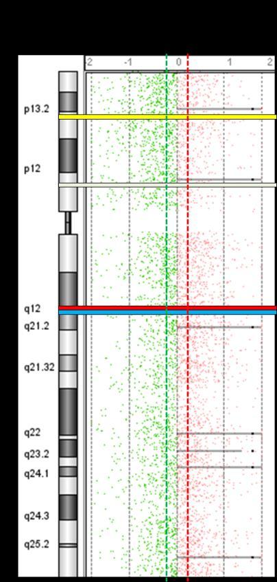
25 Figure 1. Distribution of mean HER2 and CEP17 copy numbers in 1013 invasive breast cancers. The cases (colored circles) are plotted based on the CEP17 copy number and HER-2 gene copy number. Vertical line indicates cutoff value for polysomy 17 (CEP17 copy numbers 2.6). Two tilted reference lines indicate HER2:CEP17 ratio of 1.8 (left sided) and 2.2 (right sided). 15
26 Table 1. Comparison of HER2 status and increased CEP17 copy number according to three different criteria in 1013 invasive breast cancers Criteria CEP17 < 2.6 (n=713) CEP (n=300) P-value* HER2 status by HER2/CEP17 ratio < 1.8 (n=789) 609 (77.2%) 180 (22.8%) < (n=17) 9 (52.9%) 8 (47.1%) > 2.2 (n=207) 95 (45.9%) 112 (54.1%) HER2 status by mean HER2 copy number < 4.0 (n=729) 612 (84.0%) 117 (16.0%) < (n=77) 21 (27.3%) 56 (72.7%) > 6.0 (n=207) 80 (38.6%) 127 (61.4%) HERs status by updated ASCO/CAP guidelines in 2013 Non-amplified (n=726) 609 (83.9%) 117 (16.1%) < Equivocal (n=61) 6 (9.8%) 55 (90.2%) Amplified (n=226) 98 (43.4%) 128 (56.6%) * P-value was calculated using Pearson s chi-square test. HER2 amplification is defined as HER2 copy number of 6.0 signals per cell, or HER2/CEP17 ratio of 2. The equivocal category is defined as a ratio of < 2 but copy number of 4 to < 6 signals per cell. A copy number of < 4 signals per cell and ratio of < 2 are defined as HER2 non-amplification. 16
27 2. Additional FISH results using SMS, RARA and TP53 probes in 243 tissue microarray samples The additional FISH results were shown in Table 2. All cases showed CEP 17 copy number of 2.6 in additional experiments. Among SMS, RARA, and TP53, TP53 showed least changes in the gene copy numbers. RARA revealed copy number gains most frequently. Fifty eight (23.9%) of 243 samples showed copy number gain of RARA ( 2.6 and < 6.0 copy numbers), and 12 (4.9%) revealed amplification ( 6 copy numbers). Compared to RARA, SMS and TP53 frequently showed copy number loss (< 1.6 copy numbers) (Figure 3). Seventy-seven (31.7%) of 243 samples had copy number loss of SMS, and 28 (11.5%) of TP53. We used a web-based mining tool, to obtain information on copy number variations of the genes by acgh (36, 37). We used the dataset from The Cancer Genome Atlas (TCGA) containing 943 breast cancer cases (TCGA, Provisional, last cited of July, 2013) (38). Table 3 shows the copy number variations of 4 genes by acgh from TCGA dataset. The general proportions of each group were similar to our results by FISH but losses of TP53 were more frequently observed. On the basis of copy number alterations of HER2 and the 4 reference genes, 17
28 we could build the schematic presentation of distributions and extents of amplicons on chromosome 17 (Figure 4). In 2 of 243 samples, the copy numbers of all 5 probes showed 2.6 (Figure 5). We suspected that they had true polysomy 17. Other samples showed various patterns of copy number alterations. The most frequent pattern was that of a short amplicon including only HER2 and CEP17 areas (57.6%, 140/241). Most amplicons were placed in long arm of chromosome 17 and pericentromeric areas. Only 10 cases had large amplicons including both long and short arms. 18
, but that of TP53 (red signal) was decreased (mean copy number, 1.52). 19")
29 Figure 2. A representative example of copy number loss in TP53. Case #156 had increased CEP17 and HER2 copy numbers with mean copy number of 4.35 and 3.8, respectively. Copy numbers of SMS and RARA showed no alterations (mean copy number; SMS, 2.12; RARA, 2.04), but that of TP53 (red signal) was decreased (mean copy number, 1.52). 19
30 Table 2. Copy number variation of additional reference genes on chromosome 17 in 243 tissue microarray samples by FISH analyses Copy number Status* HER2 RARA SMS TP53 Copy number loss 1 (0.4%) 37 (15.2%) 77 (31.7%) 28 (11.5%) Disomy 15 (6.2%) 136 (56.0%) 154 (63.4%) 190 (78.2%) Copy number gain 133 (54.7%) 58 (23.9%) 11 (4.5%) 25 (10.3%) Amplification 94 (38.7%) 12 (4.9%) 1 (0.4) 0 (0%) Co-amplification with HER2-10/94 (10.6%) 1/94 (1.1%) 0/94 (0%) *Copy number loss, mean copy number < 1.6; disomy, 1.6 mean copy number < 2.6; copy number gain, 2.6 mean copy number 6.0; amplification, mean copy number > 6.0 Of the 94 HER2 amplified cases (> 6.0 HER2 copy numbers), those with amplification in each gene (> 6.0 mean copy numbers) is presented. 20
31 Table 3. Copy number variation of 934 breast cancers by acgh from TCGA dataset Copy number status HER2 RARA SMS TP53 Homozygous deletion 1 (0.1%) 2 (0.2%) 5 (0.5%) 10 (1.0%) Heterozygous deletion 228 (24.4%) 264 (28.0%) 178 (18.9%) 561 (59.5%) Diploid 405 (42.9%) 420 (44.5%) 587 (62.2%) 318 (33.7%) Low level gain 179 (19.0%) 189 (20.0%) 149 (15.8%) 44 (4.7%) High level amplification 121 (12.8%) 59 (6.3%) 15 (1.6%) 1 (0.1%) Co-amplification with HER2* - 59/121(48.8%) 4/121(3.3%) 0/121 (0%) *Of the HER2 amplified cases, the number of cases with amplification in each gene is presented. 21

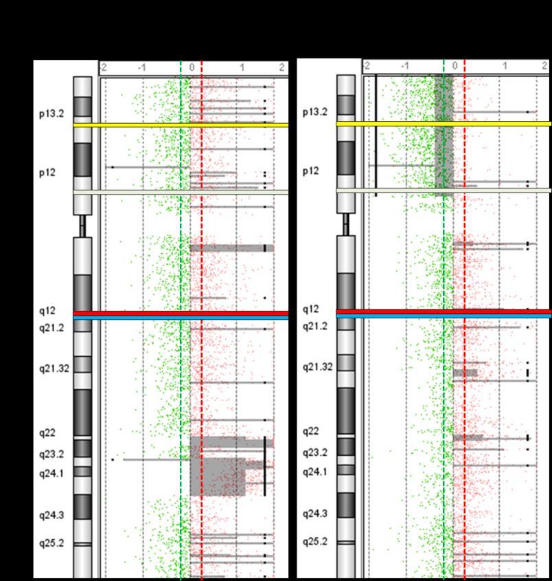
32 Figure 3. Schematic drawing of presumed extent of amplicons on chromosome 17 based on the results of FISH analyses using additional reference genes. The red bar indicates 2 possible true polysomy 17 cases. The numbers in the red and gray bars indicate the number of cases corresponding to each group 22

33 Figure 4. Two cases with suspected true polysomy 17 by FISH analyses. Representative photos of FISH using additional reference probes show increased copy numbers for all examined genes for case #155 (A) and case #250 (B). 23
34 3. Re-graded HER2 status after additional FISH analyses After additional FISH studies, we re-graded the HER2 status based on the gene copy number of the additional reference gene. Two cases in which all the 5 probes had copy number of 2.6 and higher were excluded for re-grading. Table 4 details the HER2 status between different criteria. Among the cases with HER2:CEP17 ratio less than 1.8, HER2 status was upgraded to amplified in 42 cases (27.8%), and to equivocal in 33 (21.8%) of 151. Among the cases with HER2:CEP17 ratios between 1.8 and 2.2, 7 (87.5%) of 8 were upgraded to HER2-amplified. All cases which were diagnosed as amplified based on HER2:CEP17 ratio retained the same status after re-grading. In total, 82 (34.0%) of 241 cases were upgraded after additional FISH analyses. Using 2013 ASCO/CAP guidelines, only two among 101 non-amplified cases were upgraded to amplified, and 23.8% of non-amplified cases were reclassified as equivocal. In equivocal cases, 76.1% were reclassified as amplified. However, all amplified cases retained the same status. We also investigated the relationship between HER2 copy numbers and changes of HER2 status after additional FISH studies (Table 4). When mean HER2 copy number was less than 4, 24 (23.8%) of 101 cases were upgraded from non-amplified to equivocal, and 2 (2.0%) cases were upgraded from non-amplified to amplified. When mean HER2 copy number was between 4 24
35 and 6, 36 (76.6%) of 47 cases were upgraded from equivocal to amplified and 1 (2.1%) case were downgraded from equivocal to non-amplified. All cases which were amplified by mean HER2 copy numbers (> 6) remained the same after re-grading. 25
36 Table 4. HER2 status by FISH analyses using additional reference genes on chromosome 17. Criteria HER2 status by HER2:CEP17 ratio Total HER2 status based on additional reference genes Nonamplified Equivocal Amplified Kappa coefficient* Non-amplified (<1.8) (50.4%) 33 (21.8%) 42 (27.8%) Equivocal (1.8~2.2) 8 a 0 (0%) 1 (12.5%) 7 (87.5%) Amplified (>2.2) 82 c 0 (0%) 0 (0%) 82 (100%) HER2 status by mean HER2 copy number Non-amplified (<4) (74.3%) 24 (23.8%) 2 (2.0%) Equivocal (4~6) 47 1 (2.1%) 10 (21.3%) 36 (76.6%) Amplified (>6) 93 d 0 (0%) 0 (0%) 93 (100%) HER2 status by ASCO/CAP guidelines in 2013 Non-amplified (74.3%) 24 (23.8%) 2 (2.0%) Equivocal 46 1 (2.1%) 10 (21.3%) 35 (76.1%) Amplified 94 e 0 (0%) 0 (0%) 94 (100%) Total (31.5%) 34 (14.1%) b 131 (54.4%) f *P-value is < a vs. b, P<0.001; c vs. f, d vs. f, e vs. f, P<0.001 by Chi-square test 26
37 4. Correlation between re-graded HER2 status by additional reference genes and HER2 immunohistochemistry The correlations between HER2 gene status by various criteria and HER2 IHC are shown in Table 6. Of the 253 cases with increased CEP17 copy numbers ( 2.6), more than half (55.7%) demonstrated negative (0~1+) immunostaining for HER2. Eighty-nine (35.2%) of the 253 cases showed positive (3+) immunostaining. Only 23 (9.1%) of 253 cases showed equivocal (2+) immunostaining. We evaluated the concordance rate between HER2 FISH results by standard methods based on ASCO/CAP guidelines and our new method employing additional reference genes and HER2 protein expression by IHC. We observed a good agreement (κ = 0.624, p < 0.001) between IHC result and conventional FISH results using HER2:CEP17 ratio by 2007 ASCO/CAP guidelines. When updated 2013 guidelines were applied, the kappa value became poorer (κ = 0.485) (Table 5). However, after regrading by additional reference genes, cases with negative immunostaining were upgraded to amplified in substantial portions, and thus, the agreement between FISH and IHC results significantly declined (κ = 0.390, p < 0.001). 27
38 Table 5. Correlation between HER2 FISH and HER2 immunohistochemistry Criteria HER2 Status based on additional FISH analyses* HER2 IHC Negative (0~1+) Equivocal (2+) Positive (3+) Kappa coefficient Non-amplified 66/76 (86.8%) 6/76 (7.9%) 4/76 (5.3%) Equivocal 30/34 (88.2%) 3/34 (8.8%) 1/34 (2.9%) Amplified 37/131 (28.2%) 13/131 (9.9%) 81/131 (61.8%) Total 133/241 (55.2%) 22/241 (9.1%) 86/241 (35.7%) HER2 Status based on ASCO/CAP guideline 2007 using HER2/CEP17 ratio HER2 Status based on ASCO/CAP guideline 2013 Non-amplified 130/161 (80.7%) 16/161 (9.9%) 15/161 (9.8%) Equivocal 3/8 (37.5%) 2/8 (25.0%) 3/8 (37.5%) Amplified 8/82 (9.5%) 5/82 (6.0%) 71/82 (84.5%) Total 141/253 (55.7%) 23/253 (9.1%) 89/253 (35.2%) Non-amplified 90/105 (85.7%) 9/105 (8.6%) 6/105 (5.7%) Equivocal 37/49 (75.5%) 6/49 (12.2%) 6/49 (12.2%) Amplified 14/99 (14.1%) 8/99 (8.1%) 77/99 (77.8%) Total 141/253 (55.7%) 23/253 (9.1%) 89/253 (35.2%) *Re-grading by additional FISH analyses were available in 241 cases. P-value is <
39 5. Comparison between FISH and acgh in 8 cases We performed acgh in select 8 cases to investigate the accuracy of FISH analyses with additional reference genes. Each case showed a different pattern of copy number variation by FISH analyses (Appendix A2). Table 6 shows comparison between the results of FISH and acgh, and Figure 6 shows chromosomal plots for each case. Concordance between gene copy number evaluated by FISH and mean log2 ratio by acgh was poor. Two of 8 cases (#155 and # 250) were suspected to have true polysomy 17 by FISH analyses, but there was no copy number gain of whole chromosome by acgh. Case #115 showed a copy number gain in chromosomal regions belonging to RARA and HER2, and amplification in TP53 gene based on the mean log2 ratio of the probes. In FISH analysis, mean copy number of SMS was 2.84 in #115, but there was no evidence of gain or amplification in SMS probes. #250 also showed mean copy number higher than four in RARA, HER2, SMS and TP53. However, it only showed gain of TP53 probes (Table 6; Figure 6 (E) and (G)). #115 and #250 were classified as non-amplified by conventional HER2:CEP17 FISH and finally identified as non-amplified by acgh. Five of the 8 cases (#15, #89, #104, #116 and #163) were upgraded from non-amplified to amplified after additional FISH studies (Table 6). Two (#116 29
40 and #163) of the five cases revealed amplification in HER2 gene by acgh (Table 6; Figure 6 (D) and (F)). Case #116 displayed amplification in entire long arm of chromosome 17, and case #163 displayed amplification of 17q12 including HER2 and RARA. Case #89 and #155 displayed copy number gain of HER2 (Table 6). In case #15, mean HER2 copy number was less than 4 and mean copy number of RARA, an alternative reference gene, was However, the signals by FISH, and the mean log2 ratio by acgh were not well-correlated. Based on acgh, #15 displayed copy number gain of RARA. The remaining case #251 was upgraded from non-amplified to equivocal by additional FISH. Mean copy number of HER2 was 3.35 but that of alternative reference gene, TP53 was as low as 1.6. Finally, it was re-graded to equivocal by additional FISH studies. However, acgh study revealed that HER2 and other 3 gene were within normal copy number. 30
41 Table 6. Comparison between FISH and acgh in the 8 selected cases Case Method RARA HER2 SMS TP53 Interpretation # 15 # 89 # 104 # 116 # 155 # 163 # 250 # 251 FISH* HER2 non-amplified amplified acgh CNL of HER2; CNG of 17q21.2, 17q22-17q25.3, 17q23.2 and 17q25.1 IHC 1+/3 No HER2 overexpression FISH HER2 non-amplified amplified acgh CNG of HER2; gain of 17q IHC 1+/3 No HER2 overexpression FISH HER2 non-amplified amplified acgh No alteration of HER2; CNG of 17q21.2 and 17q23.2 IHC 0/3 No HER2 overexpressed FISH HER2 non-amplified amplified acgh Amplification of HER2; CNG of 17p and amplification of 17q IHC 1+/3 No HER2 overexpression FISH HER2 non-amplified true polysomy 17? CNG of HER2; acgh Multiple amplicons along 17q including 17q q24.3 IHC 3+/3 HER2 overexpression FISH HER2 non-amplified amplified acgh Amplification of HER2; CNL of 17p and multiple small amplicons including 17q12 (HER2/RARA), 17q21.32 and 17q q23.2 IHC 3+/3 HER2 overexpression FISH HER2 non-amplified true polysomy 17? acgh No alteration of HER2; amplification of p13.2, q23.2 and q25.3 IHC 0/3 No HER2 overexpression FISH HER2 non-amplified equivocal acgh No alteration of HER2; Multiple small amplicons along the 17p and 17q IHC 2+/3 Equivocal of HER2 expression *Mean copy number of each gene is presented. 31
42 Mean log2 ratio of probes belonging to each gene is presented. If the gene is included in the copy number variation region defined by ADM2 algorithm, the mean log2 ratio of the genes belonging to those copy number variation regions are presented. Abbreviations: CNG, copy number gain; CNL, copy number loss 32
43 33
44 34
45 35
 and green (below 0) on the X-axis against each probe according to chromosomal location on the Y-axis; the genomic position of 4")
46 Figure 5. Chromosome plots of 8 cases by acgh. Log 2 ratios of each oligonucleotide probe is plotted in red (above 0) and green (below 0) on the X-axis against each probe according to chromosomal location on the Y-axis; the genomic position of 4 genes are highlighted by colored boxes (yellow: TP53, green: SMS, red: HER2 and blue: RARA). Horizontal red and green dotted lines represent the cutoff between genomic gain and loss (log2 ratio > 36
47 0.25, log2 ratio < -0.25). Semi-transparent filled grey box and thick black vertical line represent copy number variation regions defined by ADM2 algorithm. 37
48 6. Pathologic review of cases with upgraded HER2 status after additional FISH analyses We reviewed the pathologic characteristics of the 35 cases whose HER2 status were upgraded from equivocal (based on 2013 ASCO/CAP guidelines) to non-amplified after additional FISH studies. The results were demonstrated in Appendix A3. Most of the cases (30/35) were invasive ductal carcinomas, not otherwise specified (IDC, NOS) in histologic subtype. Two cases were invasive lobular carcinomas (ILC) and one was invasive micropapillary carcinoma. And the remaining two were metaplastic carcinomas. According to initial HER2 status, these two metaplastic carcinomas were classified as triple-negative breast cancer, but additional FISH results designated them as HER2+ subtype. In terms of histologic grade, there were two IDCs with low histologic grade. And about half (19/35) of the cases had low Ki67 proliferative index (< 20%). Moreover, in these 35 cases, HER2 gene amplification was not accompanied by HER2 protein overexpression. Only 6 cases (17.1%) showed HER2 protein overexpression (3+). Five demonstrated equivocal (2+) immunoreactivity and the remaining 24 (68.6%) showed negative immunoreactivity (0 or 1+) to HER2. 38
49 To sum up, some pathologic features of the cases which were designated to have HER2 amplification after additional FISH were not compatible with those of HER2 amplified breast cancers. 39
50 DISCUSSION Genome-based studies reveal that true polysomy is rare in breast cancers and most of cases, so-called polysomy 17 by ISH, are not a true increased number of whole chromosome but a gain of the pericentromeric or centromeric region, many experts concurred that polysomy 17 could affect the interpretation of results from ISH and lead to underestimation or overestimation of HER2 status. Recently updated 2013 ASCO/CAP guidelines finally focused on this issue and proposed options to avoid such false negative results of polysomy 17. One of such options is to repeat HER2 testing using another gene in chromosome 17 which is not expected to co-amplify with HER2. They recommended such re-testing when HER2:CEP17 ratio is < 2.0, average HER2 copy number is 4 and < 6 (equivocal group), and average CEP17 copy number is > 2.0 (15, 39). We applied this method to 243 IBCs with increased CEP17 copy number ( 2.6) identified by FISH analyses in our institution. Tse and his colleagues also used this method in a relatively large series of 171 IBCs with polysomy 17, defined 2.6 CEP17 (17). In our and their studies, SMS, RARA and TP53 were used as additional reference genes, based on the concept that all of the genes in chromosome 17 would increase in true polysomy 17 and 40
51 any gene that showing eusomy in chromosome 17 could be used as a new surrogate instead of CEP17. Tse and his colleagues revealed that polysomy 17 was very rare (14%) by FISH analyses, and nearly half of the patients with an equivocal range of HER2 gene copy numbers and increased CEP 17 signals had their HER2 status upgraded from non-amplified to amplified using this additional verification method. Comparing the results from this study, our study, based on the same method, showed that only 2 cases would be possible true polysomy 17 by additional FISH analyses, and more than half (63.85%, 30/47) of cases with equivocal HER2 copy number were upgraded from nonamplified to amplified after additional analysis. Use of alternative genes as a new surrogate seemed to be helpful in finding hidden HER2-amplified cases within breast cancers with polysomy 17. Although updated 2013 ASCO/CAP guidelines proposed a possible solution for the dilemma regarding polysomy 17, they did not provide specifics. As we mentioned earlier, updated guidelines simply proposed using other genes on chromosome 17 that are not expected to co-amplify with HER2 should be used. If so, which gene should we select from the numerous genes on chromosome 17? How many new genes do we have to use? What should we do if the HER2 status remained equivocal after alternative FISH? Unfortunately, we were not able to find answers to these questions but our 41
52 study did reveal some hints to these questions. The most important consideration that must be given upon choosing a surrogate for a chromosome is the stability of genomic copy number. If the gene we chose did not display diploidy, the status of HER2 would be false-negative or false-positive just as with using CEP17. RARA, SMS and TP53 have been used in previous studies. Because they are commercially available, they can be easily purchased and used for additional FISH analysis in the clinical setting. However, there was no evidence that they were suitable for reference of chromosome 17. To evaluate the utility of RARA, SMS and TP53 as references, we assessed the copy number variation of these genes in breast cancers. According to copy number variation from TCGA data and our FISH results, we can evaluate the utility of 3 genes as surrogates of chromosome 17. First, RARA is a gene encoding retinoic acid receptorα (RARα) and located at 17q21 (40, 41). It maps very close to HER2 gene (0.65Mb) and is frequently amplified in HER2 amplified breast cancers. RARA is one of the genes that belong to a short HER2 amplicon which spans between 280 and 746kb (40). Paroni et al. demonstrated that 23 32% of HER2 amplified breast cancers showed co-amplification of RARA using quantitative PCR (41). Results from TCGA also demonstrate that 12.8% of breast cancers showed co-amplification of HER2 and RARA, and that 48.8% of HER2 amplified breast cancers 42
53 showed RARA amplification (Table 3). Our study showed that approximately 25% of breast cancers with increased CEP17 copy number showed copy number gain of RARA with HER2, and about 10% showed co-amplification of two genes by FISH (Figure 3 and Table 2). Second, SMS also known as RAI1 (retinoic acid induced 1), is located on 17p11.2, and its chromosomal deletion is related to many congenital anomalies (43). The gene has been so barely studied in breast cancer that its copy number variation in breast cancer is not well-known. Based on acgh results from TCGA dataset, only 1.3% of breast cancer showed high level amplification of SMS, and co-amplification of HER2 and SMS was identified in 3.3% of HER2 amplified breast cancers. However, gene loss was frequently identified. About 20% of breast cancers showed a low-level copy number loss which was regarded as heterozygote deletion by acgh study (Table 6). In our study, 37.1% of the cases demonstrated decreased copy number of SMS. Such decreased copy numbers of SMS can result in overestimation of HER2 status. Marchio and his colleagues also showed that using SMS as reference gene could overestimate HER2 amplification for this reason, and the value of SMS as reference gene was limited in certain conditions (33). Lastly, TP53 is a well-known tumor suppressor gene. It is located at 17p13.1, relatively far from HER2, and the possibility of sharing amplicon 43
54 between HER2 and TP53 is theoretically very low. TP53 is one of the most frequently mutated genes in breast cancer but copy number gain or amplification of TP53 is almost absent (42). However, chromosome 17p where TP53 is located is mainly involved in genetic losses in breast cancers (43). Even though our FISH studies showed that loss of TP53 (11.7%) was not common and that TP53 was the most stable gene that had least copy number alterations (Table 2), acgh results from TCGA dataset show the frequency of genetic loss of TP53 is the most common among the three reference genes. Thus, TP53 also has limitations for use as surrogate. Our study demonstrates that the majority of breast cancers with increased CEP17 copy number could show copy number changes of RARA, SMS and TP53 by FISH. Using any of these 3 genes can produce wrong interpretations when used alone. However, when the 3 genes are used altogether, most (94.2%, 227/241) of the cases had at least one gene with disomy in our study. Therefore, it is recommended to use a couple of genes together for accurate estimation of HER2 in breast cancers with increased CEP17 copy numbers. In practice, performing a couple of FISH analyses for diagnosis of one case may be a burden in terms of cost and time. To simplify application of this method and decrease costs, more studies will be needed in search of a gene on chromosome 17 that barely shows copy number alteration in breast cancer. 44
55 However, our study which used three alternative genes in combination still showed some problems. First, after re-grading using additional reference genes, HER2 equivocal cases were significantly increased compared to those based on 2007 ASCO/CAP guidelines. Moreover, 27% (36/131) of the amplified cases had HER2 copies between 4 and 6 (Table 4). These cases may have low levels of amplification of HER2 or no amplification at all. However, there is no way to establish the accuracy of HER2 status in this subset. Another problem is the decreased concordance rate between HER2 FISH and IHC. Concordance between FISH and IHC became poorer after using additional genes. This discordance might be due to the increase in falsepositive cases. After re-grading, about 30% of breast cancers with 0 or 1+ immunoreactivity were classified as amplified. They might be overestimated due to decreased copy number of the new reference gene. The last is that the cases whose HER2 statuses were upgraded to be amplified by additional FISH studies were not compatible with HER2 amplified breast cancers in pathologic findings. Of the 35 cases with upgraded HER2 status from equivocal to amplified based on 2013 ASCO/CAP guideline, two cases were histologically metaplastic carcinomas, which are usually triple negative breast cancers. Moreover, in these cases, HER2 gene amplification was not accompanied by HER2 protein overexpression. Based 45
56 on these findings, SMS, TP53 and RARA do not seem to be appropriate alternative for CEP17, even if used in combination. To confirm the accuracy of FISH analyses, we performed acgh in 8 cases. There were some limitations in acgh that we performed. First, we performed acgh in a limited number of cases. Second, the acgh platform that we used did not contain a centromeric region, and thus we could not confirm the true status of the centromere. Finally, we used formalin-fixed tissue for acgh. Though many studies revealed that acgh using FFPE was a reliable tool, there still exist chances for errors. In spite of these limitations, we could obtain a few important findings from the results of acgh. First, the results of FISH and acgh were not wellcorrelated. Five of 8 cases were upgraded from non-amplified to amplified by additional FISH. Among them, only 4 cases were identified to have a gain of HER2 by acgh. Moreover, case #15 and #250 that showed copy number gains of HER2 by FISH turned out to be loss by acgh. A small number of HER2 probes that were contained in acgh could be the reason for the discrepancy. There were only 2 probes that covered HER2 region and neither of the probes covered the entire region of HER2. If we used multiple probes that covered the entire gene, the results of acgh could be different. Also, the intratumoral heterogeneity of tumor can be another reason. As the 8 cases 46
57 were selected after additional FISH analyses, the tumor areas examined in FISH and acgh were not the same. Second, we demonstrated that deducing the status of chromosome 17 by FISH of several genes could be incorrect. We made schematic pictures of chromosome 17 based on copy number alteration of 5 genes on it. There were 2 cases of possible true polysomy 17 in the schematic picture. The status of chromosome 17 in both cases turned out to be disomy after performing acgh. They showed many small-sized amplicons throughout the whole chromosome, but not polysomy 17 (appendix A2). In conclusion, FISH does not seem to be an appropriate method for evaluation of chromosomal copy number status in breast cancers with increased CEP17 copy numbers. 47
58 CONCLUSION In conclusion, our study showed that using other reference genes on chromosome 17 as new surrogates for chromosome 17 status may be a way to evaluate HER2 status in breast cancers with increased CEP17 copy number. However SMS, RARA, and TP53 that we used in this study were not suitable as an independent marker. Combined use of these reference genes seemed to compensate the unstable copy number of each alternative gene. However, combined method still has some limitations. Performing FISH using multiple probes together is neither a simple nor affordable way in everyday practice. Also, this method can lead to over-grading of HER2 status when the tumor has loss of the new reference genes. Moreover, the accuracy of this method has not been verified thoroughly. It would be impossible to predict the complex genomic status by additional FISH analyses. Until now, three alternative genes, SMS, RARA and TP53 are not good choice for HER2 analysis. To apply this method in daily practice, further studies to find the most stable genes that barely show copy number alterations will be needed. 48
59 APPENDICES A1. Log2 ratio of each probe that belongs to the region of TP53, SMS, HER2 and RARA Gene Name of probe. Start End # 15 # 89 # 104 # 116 # 155 # 163 # 250 # 251 TP53 A_14_P TP53 A_14_P Average log2 ratio of TP SMS A_16_P SMS A_16_P SMS A_14_ SMS A_16_ SMS A_14_P SMS A_16_ SMS A_14_P SMS A_16_P SMS A_16_P SMS A_14_P
60 Gene probe no. Start End # 15 # 89 # 104 # 116 # 155 # 163 # 250 # 251 SMS A_16_P SMS A_16_P Average log2 ratio of SMS HER2 A_14_P HER2 A_14_P Average log2 ratio of HER RARA A_16_P RARA A_14_P RARA A_14_P RARA A_14_P Average log2 ratio of RARA
61 51
Assessment of Breast Cancer with Borderline HER2 Status Using MIP Microarray
 Assessment of Breast Cancer with Borderline HER2 Status Using MIP Microarray Hui Chen, Aysegul A Sahin, Xinyan Lu, Lei Huo, Rajesh R Singh, Ronald Abraham, Shumaila Virani, Bal Mukund Mishra, Russell Broaddus,
저작권법에따른이용자의권리는위의내용에의하여영향을받지않습니다.
 저작자표시 - 비영리 - 변경금지 2.0 대한민국 이용자는아래의조건을따르는경우에한하여자유롭게 이저작물을복제, 배포, 전송, 전시, 공연및방송할수있습니다. 다음과같은조건을따라야합니다 : 저작자표시. 귀하는원저작자를표시하여야합니다. 비영리. 귀하는이저작물을영리목적으로이용할수없습니다. 변경금지. 귀하는이저작물을개작, 변형또는가공할수없습니다. 귀하는, 이저작물의재이용이나배포의경우,
Guideline. Associated Documents ASCO CAP 2018 GUIDELINES and SUPPLEMENTS -
 Guideline Subject: ASCO CAP 2018 HER2 Testing for Breast Cancer Guidelines - Recommendations for Practice in Australasia Approval Date: December 2018 Review Date: December 2022 Review By: HER2 testing
Kristen E. Muller, DO, Jonathan D. Marotti, MD, Vincent A. Memoli, MD, Wendy A. Wells, MD, and Laura J. Tafe, MD
 AJCP / Original Article Impact of the 2013 ASCO/CAP HER2 Guideline Updates at an Academic Medical Center That Performs Primary HER2 FISH Testing Increase in Equivocal Results and Utility of Reflex Immunohistochemistry
Welcome! HER2 TESTING DIAGNOSTIC ACCURACY 4/11/2016
 HER2 TESTING DIAGNOSTIC ACCURACY Can t We Finally Get It Right? Allen M. Gown, M.D. Medical Director and Chief Pathologist PhenoPath Laboratories Seattle, Washington Clinical Professor of Pathology University
HER2/neu Evaluation of Breast Cancer in 2019
 HER2/neu Evaluation of Breast Cancer in 2019 A.A. Sahin, M.D. Professor of Pathology and Translation Molecular Pathology Section Chief of Breast Pathology ERBB2 (HER2) Background 185-kDa membrane protein
Dr. dr. Primariadewi R, SpPA(K)
") Curriculum Vitae Dr. dr. Primariadewi R, SpPA(K) Education : Medical Doctor from UKRIDA Doctoral Degree from Faculty of Medicine University of Indonesia Pathologist Specialist and Consultant from Faculty
Curriculum Vitae Dr. dr. Primariadewi R, SpPA(K) Education : Medical Doctor from UKRIDA Doctoral Degree from Faculty of Medicine University of Indonesia Pathologist Specialist and Consultant from Faculty
Priti Lal, MD, 1 Paulo A. Salazar, 1 Clifford A. Hudis, MD, 2 Marc Ladanyi, MD, 1 and Beiyun Chen, MD, PhD 1. Abstract
 Anatomic Pathology / DUAL- VS SINGLE-COLOR SCORING IN IMMUNOHISTOCHEMICAL AND FISH HER-2 TESTING HER-2 Testing in Breast Cancer Using Immunohistochemical Analysis and Fluorescence In Situ Hybridization
Optimal algorithm for HER2 testing
 Optimal algorithm for HER2 testing The revised definition of IHC 2+ (equivocal) is invasive breast cancer with Weak to moderate complete membrane staining observed in >10% of tumor cells. (see Figure 1
IT S ABOUT TIME. IQFISH pharmdx Interpretation Guide THREEHOURSTHIRTYMINUTES. HER2 IQFISH pharmdxtm. TOP2A IQFISH pharmdxtm
 I N T E R P R E TAT I O N IQFISH pharmdx Interpretation Guide TM HER2 IQFISH pharmdxtm TOP2A IQFISH pharmdxtm Breast carcinoma (FFPE) stained with HER2 IQFISH pharmdx Breast carcinoma (FFPE) stained with
HER2 FISH pharmdx TM Interpretation Guide - Breast Cancer
 P A T H O L O G Y HER2 FISH pharmdx TM Interpretation Guide - Breast Cancer For In Vitro Diagnostic Use FDA approved as an aid in the assessment of patients for whom Herceptin TM (trastuzumab) treatment
MEDICAL POLICY. Proprietary Information of YourCare Health Plan
 MEDICAL POLICY SUBJECT: HER-2 TESTING IN INVASIVE BREAST OR PAGE: 1 OF: 7 If the member's subscriber contract excludes coverage for a specific service it is not covered under that contract. In such cases,
Three Hours Thirty Minutes
 INTERPRETATION HER2 IQFISH pharmdx TM Interpretation Guide Three Hours Thirty Minutes it s about time Breast carcinoma (FFPE) stained with HER2 IQFISH pharmdx Gastric cancer (FFPE) stained with HER2 IQFISH
Assessment Run B HER2 IHC
 Assessment Run B24 2017 HER2 IHC Material The slide to be stained for HER2 comprised the following 5 materials: IHC: HER2 Score* (0, 1+, 2+, 3+) FISH: HER2 gene/chr 17 ratio** 1. Breast carcinoma, no.
Dako IT S ABOUT TIME. Interpretation Guide. Agilent Pathology Solutions. ALK, ROS1 and RET IQFISH probes (Dako Omnis) MET IQFISH probe (Dako Omnis)
 MET IQFISH probe (Dako Omnis)") INTERPRETATION Dako Agilent Pathology Solutions IQFISH Interpretation Guide ALK, ROS1 and RET IQFISH probes (Dako Omnis) MET IQFISH probe (Dako Omnis) IT S ABOUT TIME For In Vitro Diagnostic Use ALK, ROS1,
INTERPRETATION Dako Agilent Pathology Solutions IQFISH Interpretation Guide ALK, ROS1 and RET IQFISH probes (Dako Omnis) MET IQFISH probe (Dako Omnis) IT S ABOUT TIME For In Vitro Diagnostic Use ALK, ROS1,
HER2 CISH pharmdx TM Kit Interpretation Guide Breast Cancer
 P A T H O L O G Y HER2 CISH pharmdx TM Kit Interpretation Guide Breast Cancer FROM CERTAINTY COMES TRUST For in vitro diagnostic use HER2 CISH pharmdx Kit HER2 CISH pharmdx Kit is intended for dual-color
Comparative Analysis of Methods Used in Breast Cancer HER2 and Sentinel Lymph Node Diagnosis
 SHORT THESIS FOR THE DEGREE OF DOCTOR OF PHILOSOPHY (Ph.D.) Comparative Analysis of Methods Used in Breast Cancer HER2 and Sentinel Lymph Node Diagnosis by Monika Francz MD Supervisor: Zoltán Szöllősi
Product Introduction
 Product Introduction Product Codes: HCL026, HCL027 and HCL028 Contents Introduction to HER2 2 HER2 immunohistochemistry 3 Cell lines as controls 5 HER2 Analyte Control DR IHC 7 HER2 Analyte Control DR
HER-2/neu amplification detected by fluorescence in situ hybridization in fine needle aspirates from primary breast cancer
 Original article Annals of Oncology 13: 1398 1403, 2002 DOI: 10.1093/annonc/mdf217 HER-2/neu amplification detected by fluorescence in situ hybridization in fine needle aspirates from primary breast cancer
HER2 ISH (BRISH or FISH)
") Assessment Run H14 2018 HER2 ISH (BRISH or FISH) Material Table 1. Content of the multi-block used for the NordiQC HER2 ISH assessment, run H14 HER2 IHC* IHC score Dual - SISH** FISH*** FISH*** HER2/chr17
Assessment Run H14 2018 HER2 ISH (BRISH or FISH) Material Table 1. Content of the multi-block used for the NordiQC HER2 ISH assessment, run H14 HER2 IHC* IHC score Dual - SISH** FISH*** FISH*** HER2/chr17
Reviewer's report. Version: 1 Date: 24 May Reviewer: Cathy Moelans. Reviewer's report:
 Reviewer's report Title: Validation of HER2 testing with core needle biopsy specimens from primary breast cancers in terms of interobserver reproducibility and concordance with surgically resected specimens
Journal of Breast Cancer
 ORIGINAL ARTICLE Journal of Breast Cancer J Breast Cancer 2009 December; 12(4): 235-40 DOI: 10.4048/jbc.2009.12.4.235 Comparison of Silver-Enhanced in situ Hybridization and Fluorescence in situ Hybridization
MEDICAL POLICY. Proprietary Information of Excellus Health Plan, Inc. A nonprofit independent licensee of the BlueCross BlueShield Association
 MEDICAL POLICY SUBJECT: HER-2 TESTING IN INVASIVE BREAST OR PAGE: 1 OF: 7 If a product excludes coverage for a service, it is not covered, and medical policy criteria do not apply. If a commercial product,
CANCER. Clinical Validation of Breast Cancer Predictive Markers
 Clinical Validation of Breast Cancer Predictive Markers David Hicks, MD Loralee McMahon, MS, HTL(ASCP) CANCER The human body is composed of billions of highly regulated cells Cancer cells no longer respond
Layered-IHC (L-IHC): A novel and robust approach to multiplexed immunohistochemistry So many markers and so little tissue
: A novel and robust approach to multiplexed immunohistochemistry So many markers and so little tissue") Page 1 The need for multiplex detection of tissue biomarkers. There is a constant and growing demand for increased biomarker analysis in human tissue specimens. Analysis of tissue biomarkers is key to
Page 1 The need for multiplex detection of tissue biomarkers. There is a constant and growing demand for increased biomarker analysis in human tissue specimens. Analysis of tissue biomarkers is key to
Human epidermal growth factor receptor 2 (HER2)
") Utility of Alternate, Noncentromeric Chromosome 17 Reference Probe for Human Epidermal Growth Factor Receptor Fluorescence In Situ Hybridization Testing in Breast Cancer Cases Trupti Pai, MD; Tanuja Shet,
Utility of Alternate, Noncentromeric Chromosome 17 Reference Probe for Human Epidermal Growth Factor Receptor Fluorescence In Situ Hybridization Testing in Breast Cancer Cases Trupti Pai, MD; Tanuja Shet,
Abstract. Optimization strategy of Copy Number Variant calling using Multiplicom solutions APPLICATION NOTE. Introduction
 Optimization strategy of Copy Number Variant calling using Multiplicom solutions Michael Vyverman, PhD; Laura Standaert, PhD and Wouter Bossuyt, PhD Abstract Copy number variations (CNVs) represent a significant
HER2 status assessment in breast cancer. Marc van de Vijver Academic Medical Centre (AMC), Amsterdam
, Amsterdam") HER2 status assessment in breast cancer Marc van de Vijver Academic Medical Centre (AMC), Amsterdam 13e Bossche Mamma Congres 17 th June 2015 Modern cancer therapies are based on sophisticated molecular
HER2 status assessment in breast cancer Marc van de Vijver Academic Medical Centre (AMC), Amsterdam 13e Bossche Mamma Congres 17 th June 2015 Modern cancer therapies are based on sophisticated molecular
Assessment Run B HER2 IHC
 Assessment Run B26 208 HER2 IHC Material The slide to be stained for HER2 comprised the following 5 materials: IHC: HER2 Score* (0, +, 2+, 3+) FISH: HER2 gene/chr 7 ratio**. Breast carcinoma, no. 2+..3
Supplementary Figure 1
 Supplementary Figure 1 Supplementary Fig. 1: Quality assessment of formalin-fixed paraffin-embedded (FFPE)-derived DNA and nuclei. (a) Multiplex PCR analysis of unrepaired and repaired bulk FFPE gdna from
What is HER2 positive breast cancer in 2018? Updated ASCO-CAP guidelines. Giuseppe Viale University of Milan European Institute of Oncology
 What is HER2 positive breast cancer in 2018? Updated ASCO-CAP guidelines Giuseppe Viale University of Milan European Institute of Oncology Mission accomplished! First alarming results Breast Intergroup
Considerable advances in the therapy of breast cancer
 HER-2/neu Status in Breast Cancer Metastases to the Central Nervous System Kelly C. Lear-Kaul, MD; Hye-Ryoung Yoon, MD; Bette K. Kleinschmidt-DeMasters, MD; Loris McGavran, PhD; Meenakshi Singh, MD Context.
Nitta et al. Diagnostic Pathology 2012, 7:60
 Nitta et al. Diagnostic Pathology 2012, 7:60 METHODOLOGY Open Access A gene-protein assay for human epidermal growth factor receptor 2 (HER2): brightfield tricolor visualization of HER2 protein, the HER2
저작권법에따른이용자의권리는위의내용에의하여영향을받지않습니다.
 저작자표시 - 비영리 - 변경금지 2.0 대한민국 이용자는아래의조건을따르는경우에한하여자유롭게 이저작물을복제, 배포, 전송, 전시, 공연및방송할수있습니다. 다음과같은조건을따라야합니다 : 저작자표시. 귀하는원저작자를표시하여야합니다. 비영리. 귀하는이저작물을영리목적으로이용할수없습니다. 변경금지. 귀하는이저작물을개작, 변형또는가공할수없습니다. 귀하는, 이저작물의재이용이나배포의경우,
Instant Quality FISH. The name says it all.
 PRODUCT INFORMATION HER2 IQFISH pharmdx Instant Quality FISH Instant Quality FISH. The name says it all. HER2 IQFISH pharmdx IQ: Instant Quality every time. HER2 IQFISH pharmdx stains of a HER2 non-amplified
HER2 Gene Protein Assay Is Useful to Determine HER2 Status and Evaluate HER2 Heterogeneity in HER2 Equivocal Breast Cancer
 HER2 Gene Protein Assay Is Useful to Determine HER2 Status and Evaluate HER2 Heterogeneity in HER2 Equivocal Breast Cancer Yanjun Hou, MD, PhD, 1 Hiroaki Nitta, PhD, 2 and Zaibo Li, MD, PhD 1 From the
Assessment Run B HER-2 IHC. HER-2/chr17 ratio**
 Assessment Run B2 20 HER-2 IHC Material The slide to be stained for HER-2 comprised the following 5 tissues: IHC HER-2 Score* (0, +, 2+,3+) FISH HER-2/chr7 ratio**. Breast ductal carcinoma 0..3 2. Breast
Cytogenetics 101: Clinical Research and Molecular Genetic Technologies
 Cytogenetics 101: Clinical Research and Molecular Genetic Technologies Topics for Today s Presentation 1 Classical vs Molecular Cytogenetics 2 What acgh? 3 What is FISH? 4 What is NGS? 5 How can these
저작권법에따른이용자의권리는위의내용에의하여영향을받지않습니다.
 저작자표시 - 비영리 - 변경금지 2.0 대한민국 이용자는아래의조건을따르는경우에한하여자유롭게 이저작물을복제, 배포, 전송, 전시, 공연및방송할수있습니다. 다음과같은조건을따라야합니다 : 저작자표시. 귀하는원저작자를표시하여야합니다. 비영리. 귀하는이저작물을영리목적으로이용할수없습니다. 변경금지. 귀하는이저작물을개작, 변형또는가공할수없습니다. 귀하는, 이저작물의재이용이나배포의경우,
Version 2 of these Guidelines were drafted in response to published updated ASCO/CAP HER2 test Guideline Recommendations-
 Introduction: These guidelines represent systematically developed statements to assist in the provision of quality assured HER2 testing in breast and gastric/ gastro-oesophageal carcinoma. They are based
2017 OPTIONS FOR INDIVIDUAL MEASURES: REGISTRY ONLY. MEASURE TYPE: Process
 Measure #449 (NQF 1857): HER2 Negative or Undocumented Breast Cancer Patients Spared Treatment with HER2-Targeted Therapies National Quality Strategy Domain: Efficiency and Cost Reduction 2017 OPTIONS
Comparison of Immunohistochemical and Fluorescence In Situ Hybridization Assessment of HER-2 Status in Routine Practice
 Anatomic Pathology / ASSESSMENT OF HER-2 STATUS Comparison of Immunohistochemical and Fluorescence In Situ Hybridization Assessment of HER-2 Status in Routine Practice Michelle Dolan, MD, 1 and Dale Snover,
Immunotherapy in NSCLC Pathologist role
 Immunotherapy in NSCLC Pathologist role Pimpin Incharoen, M.D. Assistant Professor, Thoracic Pathology Department of Pathology, Ramathibodi Hospital Genetic alterations in NSCLC Khono et al, Trans Lung
Workflow. Connecting the Pieces For Total Patient Care
 Workflow Connecting the Pieces For Total Patient Care Biocare provides a full line of IHC and molecular pathology products for cancer and infectious disease diagnosis. From a full range of equipment: including
Claudin-4 Expression in Triple Negative Breast Cancer: Correlation with Androgen Receptors and Ki-67 Expression
 Claudin-4 Expression in Triple Negative Breast Cancer: Correlation with Androgen Receptors and Ki-67 Expression Mona A. Abd-Elazeem, Marwa A. Abd- Elazeem Pathology department, Faculty of Medicine, Tanta
Next-Generation Immunohistochemistry: Multiplex tissue imaging with mass cytometry
 Nat Met, April 2014 Nat Med, April 2014 Next-Generation Immunohistochemistry: Multiplex tissue imaging with mass cytometry Journal Club Timo Böge Overview Introduction Conventional Immunohistochemistry
Utility of Adequate Core Biopsy Samples from Ultrasound Biopsies Needed for Today s Breast Pathology
 Utility of Adequate Core Biopsy Samples from Ultrasound Biopsies Needed for Today s Breast Pathology Ugur Ozerdem, M.D. 1 Abstract Background: There is a paradigm shift in breast biopsy philosophy. In
Breast Cancer Interpretation Guide
 Breast Cancer Interpretation Guide UCT D O R P NEW ERBB2/ C E P S ht e ZytoLig lor Prob o C l a u 2D D17S12 ng to the i d r o c c a ting for re-tes idelines 2013 ASCO Gu Breast Cancer Interpretation Guide
Department of Pathology, Loyola University Medical Center, Maywood, IL 60153, USA 2
 Hindawi Publishing Corporation Pathology Research International Volume 2012, Article ID 947041, 7 pages doi:10.1155/2012/947041 Clinical Study The Effect of Cold Ischemia Time and/or Formalin Fixation
Breast cancer: Molecular STAGING classification and testing. Korourian A : AP,CP ; MD,PHD(Molecular medicine)
") Breast cancer: Molecular STAGING classification and testing Korourian A : AP,CP ; MD,PHD(Molecular medicine) Breast Cancer Theory: Halsted Operative breast cancer is a local-regional disease The positive
Breast cancer: Molecular STAGING classification and testing Korourian A : AP,CP ; MD,PHD(Molecular medicine) Breast Cancer Theory: Halsted Operative breast cancer is a local-regional disease The positive
microrna Presented for: Presented by: Date:
 microrna Presented for: Presented by: Date: 2 micrornas Non protein coding, endogenous RNAs of 21-22nt length Evolutionarily conserved Regulate gene expression by binding complementary regions at 3 regions
Determination of HER2 Amplification by In Situ Hybridization. When Should Chromosome 17 Also Be Determined?
 Anatomic Pathology / FISH f o r HER2: Wh e n to Use Ch r o m o s o m e 17 Determination of HER2 Amplification by In Situ Hybridization When Should Chromosome 17 Also Be Determined? John M.S. Bartlett,
EARLY ONLINE RELEASE
 EARLY ONLINE RELEASE Note: This article was posted on the Archives Web site as an Early Online Release. Early Online Release articles have been peer reviewed, copyedited, and reviewed by the authors. Additional
HER2/neu Amplification in Breast Cancer Stratification by Tumor Type and Grade
 Anatomic Pathology / HER2/NEU AMPLIFICATION IN BREAST CANCER HER2/neu Amplification in Breast Cancer Stratification by Tumor Type and Grade Elise R. Hoff, MD, Raymond R. Tubbs, DO, Jonathan L. Myles, MD,
Detection of aneuploidy in a single cell using the Ion ReproSeq PGS View Kit
 APPLICATION NOTE Ion PGM System Detection of aneuploidy in a single cell using the Ion ReproSeq PGS View Kit Key findings The Ion PGM System, in concert with the Ion ReproSeq PGS View Kit and Ion Reporter
Supplementary Online Content
 Supplementary Online Content Fumagalli D, Venet D, Ignatiadis M, et al. RNA Sequencing to predict response to neoadjuvant anti-her2 therapy: a secondary analysis of the NeoALTTO randomized clinical trial.
ACMG/CAP Cytogenetics CY
 www.cap.org Cytogenetics Analytes/procedures in bold type are regulated for proficiency testing by the Centers for Medicare & Medicaid Services ACMG/CAP Cytogenetics CY Analyte CY Challenges per Shipment
KEY WORDS: Breast carcinoma, c-erbb2, Fluorescent. Mod Pathol 2001;14(11):
:") HER-2/neu in Breast Cancer: Interobserver Variability and Performance of Immunohistochemistry with 4 Antibodies Compared with Fluorescent In Situ Hybridization Thomas A. Thomson, M.D., Malcolm M. Hayes,
HER-2/neu in Breast Cancer: Interobserver Variability and Performance of Immunohistochemistry with 4 Antibodies Compared with Fluorescent In Situ Hybridization Thomas A. Thomson, M.D., Malcolm M. Hayes,
Quality in Control. ROS1 Analyte Control. Product Codes: HCL022, HCL023 and HCL024
 Quality in Control ROS1 Analyte Control Product Codes: HCL022, HCL023 and HCL024 Contents What is ROS1? 2 The Role of ROS1 in Cancer 3 ROS1 Assessment 3 ROS1 Analyte Control Product Details 4 ROS1 Analyte
HER2 Status and Its Heterogeneity in Gastric Carcinoma of Vietnamese Patient
 Journal of Pathology and Translational Medicine 2017; 51: 396-402 ORIGINAL ARTICLE HER2 Status and Its Heterogeneity in Gastric Carcinoma of Vietnamese Patient Dang Anh Thu Phan Vu Thien Nguyen Thi Ngoc
Transform genomic data into real-life results
 CLINICAL SUMMARY Transform genomic data into real-life results Biomarker testing and targeted therapies can drive improved outcomes in clinical practice New FDA-Approved Broad Companion Diagnostic for
Diagnosis and Treatment of Patients with Primary and Metastatic Breast Cancer. Pathology. AGO e. V. in der DGGG e.v. sowie in der DKG e.v.
 Diagnosis and Treatment of Patients with Primary and Metastatic Breast Cancer Pathology Pathology Versions 2004 2017: Blohmer / Costa / Fehm / Friedrichs / Huober / Kreipe / Lück / Schneeweis / Sinn /
Corporate Medical Policy
 Corporate Medical Policy Microarray-based Gene Expression Testing for Cancers of Unknown File Name: Origination: Last CAP Review: Next CAP Review: Last Review: microarray-based_gene_expression_testing_for_cancers_of_unknown_primary
Data Supplement 1: 2013 Update Rationale and Background Information
 Recommendations for Human Epidermal Growth Factor Receptor 2 Testing in Breast Cancer: American Society of Clinical Oncology /College of American Pathologists Clinical Practice Guideline Update (edited
Template for Reporting Results of Biomarker Testing of Specimens From Patients With Carcinoma of the Breast
 Template for Reporting Results of Biomarker Testing of Specimens From Patients With Carcinoma of the Breast Version: Template Posting Date: January 2018 Includes requirements from the 2017 CAP Accreditation
Understanding the Human Karyotype Colleen Jackson Cook, Ph.D.
 Understanding the Human Karyotype Colleen Jackson Cook, Ph.D. SUPPLEMENTAL READING Nussbaum, RL, McInnes, RR, and Willard HF (2007) Thompson and Thompson Genetics in Medicine, 7th edition. Saunders: Philadelphia.
Central Pathology Review and Tissue MicroArrays. Dr Lisa Storer Children s Brain Tumour Research Centre Nottingham
 Central Pathology Review and Tissue MicroArrays Dr Lisa Storer Children s Brain Tumour Research Centre Nottingham What is a tissue microarray? Tissue microarrays (TMAs) consist of paraffin blocks in which
Surgical Pathology Lab of the Future. Thomas M. Grogan, M.D. Professor of Pathology, University of Arizona Founder, Ventana Medical Systems, Inc.
 Surgical Pathology Lab of the Future Thomas M. Grogan, M.D. Professor of Pathology, University of Arizona Founder, Ventana Medical Systems, Inc. 28 April 2010 Objective Demonstrate how the next generation
Low ER+ Breast Cancer. Is This a Distinct Group? Nika C. Gloyeske, MD, David J. Dabbs, MD, and Rohit Bhargava, MD ABSTRACT
 Low ER+ Breast Cancer Is This a Distinct Group? Nika C. Gloyeske, MD, David J. Dabbs, MD, and Rohit Bhargava, MD From the Magee-Womens Hospital of University of Pittsburgh Medical Center, Pittsburgh, PA.
Comparison of in situ hybridization methods for the assessment of HER-2/neu gene amplification status in breast cancer using a tissue microarray
 reports of practical oncology and radiotherapy 1 7 ( 2 0 1 2 ) 44 49 Available online at www.sciencedirect.com jo u r n al hom epage: http://www.elsevier.com/locate/rpor Original article Comparison of
2019 COLLECTION TYPE: MIPS CLINICAL QUALITY MEASURES (CQMS) MEASURE TYPE: Process High Priority
 MEASURE TYPE: Process High Priority") Quality ID #449 (NQF 1857): HER2 Negative or Undocumented Breast Cancer Patients Spared Treatment with HER2-Targeted Therapies National Quality Strategy Domain: Efficiency and Cost Reduction Meaningful
Quality ID #449 (NQF 1857): HER2 Negative or Undocumented Breast Cancer Patients Spared Treatment with HER2-Targeted Therapies National Quality Strategy Domain: Efficiency and Cost Reduction Meaningful
NPQR Quality Payment Program (QPP) Measures 21_18247_LS.
 Measures 21_18247_LS.") NPQR Quality Payment Program (QPP) Measures 21_18247_LS MEASURE ID: QPP 99 MEASURE TITLE: Breast Cancer Resection Pathology Reporting pt Category (Primary Tumor) and pn Category (Regional Lymph Nodes)
NPQR Quality Payment Program (QPP) Measures 21_18247_LS MEASURE ID: QPP 99 MEASURE TITLE: Breast Cancer Resection Pathology Reporting pt Category (Primary Tumor) and pn Category (Regional Lymph Nodes)
Droplet digital PCR using HER2/EIF2C1 ratio for detection of HER2 amplification in breast cancer tissues
 https://doi.org/10.1007/s12032-018-1210-8 ORIGINAL PAPER Droplet digital PCR using HER2/EIF2C1 ratio for detection of HER2 amplification in breast cancer tissues Anchalee Tantiwetrueangdet 1 Ravat Panvichian
T he HER2/neu type 1 tyrosine kinase growth factor
 710 ORIGINAL ARTICLE HER2 amplification status in breast cancer: a comparison between immunohistochemical staining and fluorescence in situ hybridisation using manual and automated quantitative image analysis
Same Day, Cost-Effective Aneuploidy Detection with Agilent Oligonucleotide array CGH and MDA Single Cell Amplification Method
 Same Day, Cost-Effective Aneuploidy Detection with Agilent Oligonucleotide array CGH and MDA Single Cell Amplification Method Presenter: Dr. Ali Hellani, Founder, Viafet Genomic Center, Dubai Wednesday,
Quality assurance and quality control in pathology in breast disease centers
 Quality assurance and quality control in pathology in breast disease centers Judith Sandbank M.D. Pathology Assaf-Harofeh Medical Center ISRAEL jsandbank@asaf.health.gov.il 1 st IBDC, 28 th January, 2011
Detection of Anaplastic Lymphoma Kinase (ALK) gene in Non-Small Cell lung Cancer (NSCLC) By CISH Technique
 gene in Non-Small Cell lung Cancer (NSCLC) By CISH Technique") Cancer and Clinical Oncology; Vol. 7, No. 1; 2018 ISSN 1927-4858 E-ISSN 1927-4866 Published by Canadian Center of Science and Education Detection of Anaplastic Lymphoma Kinase (ALK) gene in Non-Small Cell
Cancer and Clinical Oncology; Vol. 7, No. 1; 2018 ISSN 1927-4858 E-ISSN 1927-4866 Published by Canadian Center of Science and Education Detection of Anaplastic Lymphoma Kinase (ALK) gene in Non-Small Cell
Evaluation of HER2/neu oncoprotein in serum and tissue samples of women with breast cancer: Correlation with clinicopathological parameters
 Evaluation of HER2/neu oncoprotein in serum and tissue samples of women with breast cancer: Correlation with clinicopathological parameters Mehdi Farzadnia, Naser TayyebiMeibodi, Fatemeh HomayiShandiz,
Pepsin Solution ready-to-use
 SIE HABEN DIE VISION, WIR HABEN DIE SUBSTANZ. Pepsin Solution Single component Pepsin Solution: only one component refrigerator stable Pepsin is a commonly used digestive enzyme for immunohistochemical
Research Article HER2 Protein Overexpression and Gene Amplification in Plasmacytoid Urothelial Carcinoma of the Urinary Bladder
 Disease Markers Volume 2016, Article ID 8463731, 6 pages http://dx.doi.org/10.1155/2016/8463731 Research Article HER2 Protein Overexpression and Gene Amplification in Plasmacytoid Urothelial Carcinoma
Quality Assurance and Quality Control in the Pathology Dept.
 Quality Assurance and Quality Control in the Pathology Dept. Judith Sandbank M.D. Pathology Assaf-Harofeh Medical Center ISRAEL jsandbank@asaf.health.gov.il 2 nd IBDC, 9 th February, 2012 Pathology as
HSL-Advanced Diagnostics 2018 / 19 Test & Service List
 HSL-Advanced Diagnostics 2018 / 19 Test & Service List 2018/19 TEST & SERVICE LIST Haematoxylin & Eosin H&E H&E per slide Routine Immunohistochemistry Immunohistochemical demonstration of an antigen in
A Study to Evaluate the Effect of Neoadjuvant Chemotherapy on Hormonal and Her-2 Receptor Status in Carcinoma Breast
 Original Research Article A Study to Evaluate the Effect of Neoadjuvant Chemotherapy on Hormonal and Her-2 Receptor Status in Carcinoma Breast E. Rajesh Goud 1, M. Muralidhar 2*, M. Srinivasulu 3 1Senior
Journal of Breast Cancer
 Journal of Breast Cancer ORIGINAL ARTICLE J Breast Cancer 2011 December; 14(4): 276-282 Silver-Enhanced In Situ Hybridization as an Alternative to Fluorescence In Situ Hybridization for Assaying HER2 Amplification
Molecular Aspects of Melanocytic Neoplasia. Iwei Yeh MD, PhD University of California, San Francisco
 Molecular Aspects of Melanocytic Neoplasia Iwei Yeh MD, PhD University of California, San Francisco Thanks to: Boris Bastian Timothy McCalmont Philip LeBoit Beth Ruben Jeff North Laura Pincus Thaddeus
Controversies in HER2 Oncogene Testing: What Constitutes a True Positive Result in Patients With Breast Cancer?
 Controversies in HER2 Oncogene Testing: What Constitutes a True Positive Result in Patients With Breast Cancer? Michael F. Press, MD, PhD; Yanling Ma, MD; Susan Groshen, PhD; Guido Sauter, MD, PhD; and
Molecular Probes Introducing 14 new probes
 Molecular Probes Introducing 14 new probes Gene and Chromosome Probes Dual Colour ISH INFORM HER2 Dual ISH DNA Probe Cocktail Assay Product Part Number INFORM HER2 Dual ISH DNA Probe Cocktail 800-4422
CME/SAM ABSTRACT. AJCP / Original Article
 Clinicopathologic Significance of the Intratumoral Heterogeneity of HER2 Gene Amplification in HER2- Positive Breast Cancer Patients Treated With Adjuvant Trastuzumab Hee Jin Lee, MD, PhD, 1 Joo Young
CME/SAM. Abstract. Anatomic Pathology / HER2/neu Results in Breast Cancer
 Anatomic Pathology / HER2/neu Results in Breast Cancer Effect of Ischemic Time, Fixation Time, and Fixative Type on HER2/neu Immunohistochemical and Fluorescence In Situ Hybridization Results in Breast
FAQs for UK Pathology Departments
 FAQs for UK Pathology Departments This is an educational piece written for Healthcare Professionals FAQs for UK Pathology Departments If you would like to discuss any of the listed FAQs further, or have
Structural Variation and Medical Genomics
 Structural Variation and Medical Genomics Andrew King Department of Biomedical Informatics July 8, 2014 You already know about small scale genetic mutations Single nucleotide polymorphism (SNPs) Deletions,
First released in 2007 and updated in 2013, the recommendations
 Special Article Human Epidermal Growth Factor Receptor 2 Testing in Breast Cancer American Society of Clinical Oncology/College of American Pathologists Clinical Practice Guideline Focused Update Antonio
Breast cancer is one of the leading causes of cancerrelated
 Theranostic and Molecular Classification of Breast Cancer Kristine M. Cornejo, MD; Dina Kandil, MD; Ashraf Khan, MD, FRCPath; Ediz F. Cosar, MD Context. Despite advances in breast cancer management, women
Assessment Run C3 2018
 Assessment Run C3 2018 PD-L1 Amended version May 14 th 2018 The third assessment in NordiQC Companion module C3 focused on the accuracy of the PD-L1 IHC assays performed by the participating laboratories
Immunohistochemical classification of breast tumours
 Immunohistochemical classification of breast tumours Workshop in Diagnostic Immunohistochemistry September 19 th - 21 th 2018 Anne-Vibeke Lænkholm Department of Surgical Pathology, Zealand University Hospital,
COMPUTER-AIDED HER-2/neu EVALUATION IN EXTERNAL QUALITY ASSURANCE (EQA) OF BREAST CANCER SCREENING PROGRAMME
 OF BREAST CANCER SCREENING PROGRAMME") COMPUTER-AIDED HER-2/neu EVALUATION IN EXTERNAL QUALITY ASSURANCE (EQA) OF BREAST CANCER SCREENING PROGRAMME Maria Lunardi MD Anatomic Pathology Fracastoro Hospital San Bonifacio, Verona -Italy HER2-neu
COMPUTER-AIDED HER-2/neu EVALUATION IN EXTERNAL QUALITY ASSURANCE (EQA) OF BREAST CANCER SCREENING PROGRAMME Maria Lunardi MD Anatomic Pathology Fracastoro Hospital San Bonifacio, Verona -Italy HER2-neu
Applications of IHC. Determination of the primary site in metastatic tumors of unknown origin
 Applications of IHC Determination of the primary site in metastatic tumors of unknown origin Classification of tumors that appear 'undifferentiated' by standard light microscopy Precise classification
Does chromosome 17 centromere copy number predict polysomy in breast cancer? A fluorescence in situ hybridization and microarray-based CGH analysis
 Journal of Pathology J Pathol 2009; 219: 16 24 Published online in Wiley InterScience (www.interscience.wiley.com) DOI: 10.1002/path.2574 Original Paper Does chromosome 17 centromere copy number predict
SMH (Myosin, smooth muscle heavy chain)
") Material The slide to be stained for SMH comprised: Assessment Run 50 2017 SMH (Myosin, smooth muscle heavy chain) 1.Tonsil, 2. Esophagus, 3. Breast hyperplasia, 4. Breast ductal carcinoma in situ (DCIS),
Material The slide to be stained for SMH comprised: Assessment Run 50 2017 SMH (Myosin, smooth muscle heavy chain) 1.Tonsil, 2. Esophagus, 3. Breast hyperplasia, 4. Breast ductal carcinoma in situ (DCIS),