Structural Characterization of Saturated Branched Chain Fatty Acid Methyl Esters by
|
|
|
- Donald Bishop
- 6 years ago
- Views:
Transcription
1 Structural Characterization of Saturated Branched Chain Fatty Acid Methyl Esters by Collisional Dissociation of Molecular Ions Generated by Electron Ionization Rinat R. Ran-Ressler 1, Peter Lawrence 1, J. Thomas Brenna* Division of Nutritional Sciences Cornell University Savage Hall Ithaca, NY USA Running title: Branched chain fatty acid structural mass spectrometry Revised according to reviewer comments and returned, October 211. Submitted to Journal of Lipid Research, September Contributed equally to this work *<jtb4@cornell.edu> v: (67)
2 Ran-Ressler, Lawrence, Brenna Page 2 Abstract. Saturated branched chain fatty acids (BCFA) are present as complex mixtures in numerous biological samples. The traditional method for structure elucidation, electron ionization (EI) mass spectrometry, sometimes does not unambiguously enable assignment of branching in isomeric BCFA. Zirrolli and Murphy (1993) showed that the molecular ions of four BCFA methyl ester (BCFAME) yield highly characteristic fragments upon collisional dissociation using a triple quadrupole instrument. Here, we confirm and extend these results by analysis using a tabletop 3-D ion trap for activated molecular ion EI-MS/MS (EI-MS/MS) to 3 BCFAME. iso-bcfame produces a prominent ion (3-1% of base peak) for [M-43] (M-C 3 H 7 ), corresponding to the terminal isopropyl moiety in the original iso-bcfame. Anteiso-FAME yield prominent ions (2-1% of base peak) corresponding to losses on both side of the methyl branch, [M-29] and [M- 57], and tend to produce more prominent m/z 115 peaks corresponding to a cyclization product around the ester. Dimethyl and tetramethyl FAME with branches separated by at least one methylene group yield fragment on both sides of the sites of methyl branches that are more than 6 C away from the carboxyl carbon. EI-MS/MS yields uniquely specific ions that enable highly confident structural identification and quantification of BCFAME.
3 Ran-Ressler, Lawrence, Brenna Page 3 Methyl branched chain fatty acids (BCFA) are prevalent in a wide range of biological samples. They are especially rich in the membranes of some bacteria (1) and in animal skin and secretions, for instance, in sebum (2), cerumen (3), and meibomian gland (4) of the human eyelid and the Harderian gland of rodents (5). They are also produced by rumen bacteria and are a substantial constituent of rumen tissue and milk. We recently showed that BCFA constitute 2% of fatty acids in the United States milk supply (6). The most prevalent monomethyl BCFA have branching on the n-2 or n-3 carbons, referred to as iso and anteiso, respectively. Polymethyl BCFA arising in prenol lipids are also common, specifically phytanic and pristanic acids. The routine method of fatty acid analysis is to convert them from their nature lipid class into fatty acid methyl esters (FAME), which have superb characteristics on capillary gas chromatograph columns with respect to peakshape and baseline separation. The usual approach for identifying branching in iso or anteiso branched chain FAME (BCFAME) is to examine the electron ionization (EI) mass spectrum for losses corresponding to branching at the end of the molecule. Although this approach is satisfactory in many cases, peak intensities are often low and sometimes do not enable unambiguous assignment. For this reason, specialized esters that localize charge and enhance structure-specific charge-remote fragmentation such as dimethyloxazoline (DMOX) and picolinyl esters are typically prepared for EI-MS analysis (7, 8). However, preparation of DMOX, picolinyl, or other esters requires derivatization chemistry beyond routine methylation. Specialized esters have chromatographic characteristics that differ from FAME, requiring customized chromatography; peaks change retention times, sometimes invert-
4 Ran-Ressler, Lawrence, Brenna Page 4 ing compared to FAME; establishing correspondence between FAME and other fatty esters can be challenging. High energy collisionally activated dissociation (CAD) in a tandem time-of-flight (TOF-TOF) mass spectrometer initiated by MALDI has been applied to locate branching in a single BCFAME recently (9), however this method has not been further developed. Finally, all methods thus examined tend to yield abundant nondiagnostic product ions. This distributes the signal among several ions which effectively reduces the sensitivity for selected ion monitoring and quantitative analysis. Methods for direct analysis of FAME that yield a few intense peaks are therefore preferred. Zirroli and Murphy presented a method for direct analysis of a limited number of BCFAME by tandem mass spectrometry of the molecular ion using a triple quadrupole mass spectrometer (1). BCFAME were ionized by 7 ev electrons and the molecular ion isolated. CAD from 1-2 ev yield a striking, novel mass spectrum distinct from the MS-1 EI spectrum, devoid of the McLafferty rearrangement (11, 12) peak at m/z 74 that is usually at high abundance and the base peak in many FAME spectra. CAD spectra of the molecular ion yielded a novel series of peaks with intensities depending on CAD energy. CAD of BCFAME yielded intense peaks that unambiguously reveal the locus of branching for three monomethyl BCFAME (methyl 1-methyl-octadecanoate; methyl 13- methylpentadecanoate (anteiso-16:); methyl 14-methylpentadecanoate (iso-16:). A series of fragments also were unique to the tetramethyl BCFAME methyl phytanate (3,7,11,15- tetramethyl 16:).
5 Ran-Ressler, Lawrence, Brenna Page 5 We recently characterized the BCFA of human vernix caseosa, the waxy substance unique to humans that covers the fetus at birth, in comparison to BCFA in meconium from the same infants (13). In vernix, more than 2 monomethyl and dimethyl BCFA with 11 to 26 carbons were found at a total concentration of about 29% of fatty acids (13). Using vernix and lanolin, which contains some BCFA that are not present in vernix, we performed MS/MS on molecular ions of BCFAME using a tabletop internal ionization ion trap mass spectrometer. We report here spectra from BCFAME from these natural mixtures to compare and contrast spectra, and present evidence that the technique applies to a broad range of BCFAME. Experimental Section Instrumentation. Data were generated on a tabletop Varian Star 34CX gas chromatograph equipped with a 178 split / splitless injector coupled to a Varian Saturn 2 3D ion trap (Varian Inc., Walnut Creek CA). BCFAME were separated on a BPX7 capillary column (6m.32 mm.25 µm; SGE Inc., Austin TX). GC conditions were as follows: injector temperature was 25 C in splitless mode with a purge at.85 min after injection; initial oven temperature was 6 C held for 1 min, then ramped to 17 C at 5 C / min and held for 6 min, then ramped up to 2 C at 2.5 C/min and held for 3 min, then ramped up to 222 C at 1 C/min and held for 8.7 min, and then ramped up to 255 C at 5 C/min and held for 1 min. Total run time was 36.8 min. M + ions for BCFAME were isolated for fragmentation in EIMS2 mode. The ionization mode was set to EI auto mode using the default parameters set by the Varian Saturn
6 Ran-Ressler, Lawrence, Brenna Page 6 software V Ion preparation parameters were as follows: isolation window 3. amu; waveform type residence; excitation storage level was calculated using a q value of.215; excitation amplitude was set to.8 volts. Segment set point parameters were: scan rate 1 sec; count threshold 1; emission current.5 µa. All spectra were collected under the identical instrument settings, including collision energy (excitation amplitude) and mass isolation window. In our hands these conditions provided suitable fragments intensities across all BCFAME without the need to customize parameters. The GC elution order for all BCFAME is iso, anteiso, normal under our conditions. Multiply branched FAME tend to elute prior to the iso isomer.samples. Very few BCFA standards are readily available. A limited number of pure BCFA are available commercially at high cost. Vernix caseosa from a previous study (13) and lanolin purchased from a local retail store were methylated using BF 3 /methanol and served as sources of BCFA. Methyl phytanate was obtained from Matreya, LLC (Bellefonte, PA). Identity of n-fame and BCFAME are established by molecular weight and retention time. Saturated FAME appearing at retention times that did not correspond to normal, iso, or anteiso BCFA with internal branching were made by applying rules for interpretation developed for the knowns. Thus, internal and multiple branching spectra are labeled based on the spectra themselves, and since not validated by an independent structurespecific method should be considered tentative identifications. Their spectra are shown to illustrate fragmentation for internal and multiply branched FAME. Spectra are presented in the main paper and in Supplementary information. For convenient reference, Table 1 provides figure numbers for all EI-MS/MS spectra. For sim-
7 Ran-Ressler, Lawrence, Brenna Page 7 plicity of reference specific FAME are discussed according to their fatty acid designations (e.g. n-17:) and the leading methyl (e.g, methyl(n-17:)) is dropped.
8 Ran-Ressler, Lawrence, Brenna Page 8 Results and Discussion We first present the conventional single stage EIMS of isomeric saturated FAME to be contrasted with the MS/MS method. Figure 1 illustrates the single stage EIMS spectra of three isomeric 17:. Careful inspection shows that the three spectra contain no unique ions, thus peak intensities must suffice to distinguish unique isomers. The molecular ions at m/z 284 are present at 5-8% of the m/z 74 (McLafferty rearrangement) peak, with the accompanying m/z 87 peak at about 9%. iso-17: can be distinguished from n-17: and anteiso-17: by the intensity of the m/z 143 peak. n-17: and anteiso- 17: spectra have minor intensity differences in m/z 227 and 255, but are otherwise nearly indistinguishable. Figure 2 presents the EI-MS/MS spectra of the same three isomeric 17: as shown in Figure 1, as well as a dimethyl isomer. Upon collisional dissociation of the m/z 284 peak, n-17: yields a series of ions of uniformly low intensity due to fragmentation between each C-C bond. In contrast, iso-17: yields an unique peak at m/z 241, and very weak fragment ions. The major fragment (m/z 241) arises by loss of the terminal isopropyl group [M-43] (M-C 3 H 7 ). Anteiso-17: yields a peak at m/z 227, corresponding to loss of the terminal isobutyl group [M-57] (M-C 4 H 9 ), and another at m/z 255 for loss of the terminal ethyl group [M-29] (M-C 2 H 5 ). Based on these fragments, we posit that the dimethyl species is 1,13-dimethyl-15:. It has fragments characteristic of branching at the anteiso position, C13, analogous to anteiso-17:. Interpreting peaks at m/z 171 and 199 in the same manner leads to the conclusion that fragments represent loss or retention of the neutral fragment containing the tertiary carbon at position 1, which are at
9 Ran-Ressler, Lawrence, Brenna Page 9 greater intensity than those retaining or losing an addition 14 amu along the carbon chain (m/z 213 or m/z 185, or m/z 157 which is absent). This fragmentation is consistent with that observed for phytanic acid as shown below. Another notable ion is found at m/z 115 in the anteiso and 1,13-dimethyl-15: spectra but is absent in the iso spectrum. As shown below, this ion appears in most anteiso spectra and is absent or at low intensity in iso spectra, and not observed in BCFAME with methyl branching at C3. Formation of FAME products ions has been extensively at low and high collision energies. Isotope labeling shows that it is often accompanied by H or ester group migration (14). The m/z 115 ion corresponds to alkyl radical loss with cleavage between C5-C6 and no rearrangements under our low energy collision conditions. The structure of this common ion cannot be established by the data here, and the fragmentation most consistent with labeling studies makes no obvious prediction as to differences in fragment formation between iso and anteiso isomers (14). However, the appearance of m/z 115 can be rationalized based on cyclization and/or resonance stabilization of a product ion as either a direct cyclization (scheme 1), or perhaps less plausibly as a resonance stabilized diradical cation (scheme 2). The final m/z 115 product may be a resonance stabilized oxane cation.
10 Ran-Ressler, Lawrence, Brenna Page 1 Scheme 1 O O O Collisional activation O O O O O O O O O Scheme 2 m/z 115 Finally, there is no evidence for C-H bond breakage due to collisional activation of the molecular ion. The M-1 and M-2 H-loss peaks are evident in the first stage EI spectrum of Figure 1 and are retained in MS/MS due to imperfect isolation of the molecular ion. All EI-MS/MS spectra are consistent with this result. Figure 3 presents a similar series of three isomeric C2 including one multiply branched FAME. Again, n-2: EI-MS/MS spectra yield an envelope of C-C bond breakage ions and iso-2: presents a prominent fragment representing loss of the tertiary carbon. Phytanic acid (3,7,11,15-tetramethyl 16:) produces an envelope of major peaks corre-
11 Ran-Ressler, Lawrence, Brenna Page 11 sponding to fragmentation around the methyl branch points, with the exception of the branch at C3 (m/z 11) which gave very low signal. These results are similar to those presented previously for this molecule (1). A series of iso and anteiso BCFAME spectra are presented in Figure 4 for C13-C15. The iso BCFAME consistently yield strong peaks for the isopropyl loss fragments, though their intensities and the intensities of the molecular ions vary substantially. The anteiso BCFAME consistently yield strong signals for fragmentation on both sides of the branch point. Intensities are more consistent than for iso BCFAME, with the molecular ion as the base peak in all spectra, and ethyl fragment loss peak of lowest intensity. The m/z 115 peak is below 5% abundance in all the iso spectra, while it was 1-3% abundance in all the anteiso spectra. Though the mechanism for this intensity difference is unclear, it provides confirmatory diagnostic information for structure assignment. The spectra of straight chain and iso FAME are presented in Figure 5 for very long chain (VLC) FAME ( 24 carbons). n-26: and iso-26: yield the analogous fragments observed for shorter chain FAME. n-28: and iso-28: also yield the expected fragments though the abundances are much lower than for n-26: and iso-26: (note change in y-axis scale). These data were obtained in separate analyses and the differences in fragment yields are very likely to be due to differences in collision energy rather than the two carbon difference in structure.
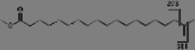
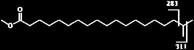
12 Ran-Ressler, Lawrence, Brenna Page 12 Several spectra of anteiso VLC-BCFAME are compared in figure 6. Figure 6A-C are spectra of n-25:, iso-25:, and anteiso-25:, again all showing the expected fragments. Figures 6D-F are spectra for anteiso-27:, anteiso-29:, and anteiso-31:. Fragmentation around the methyl branch point is found for all anteiso spectra. However, lower total concentrations of anteiso-29: and anteiso-31: led to lower signal-to-noise spectra in which the molecular ions are of low abundance. The anteiso-31: spectrum has no peak for m/z 115 and is the only anteiso spectrum in our dataset in which it does not appear, possibly due to the general low abundance of anteiso-31: and inefficient trapping at low masses. Figures 5 and 6 together show that the method yields useful results for large differences in fragmentation efficiency and signal-to-noise. Taken together, these spectra demonstrate greater specificity in locating branch points in isomeric saturated FAME than is available in single stage EIMS spectra. Moreover, the highly abundant ions unique to specific structures facilitate chromatographic resolution by selected ion plots as well as improve quantitative analysis. Table 2 presents expected fragments for monomethyl branches at the iso, anteiso, and mid-chain positions for BCFAME from 12: to 26:. Molecular weights of the methyl esters are located below the FAME designation. Branching at the iso position results in [M-43] as the main loss peak and is recorded two cells below the iso headings. Branching at the anteiso position results in [M-29] and [M-57] as the main loss peaks, both recorded under the anteiso headings. Branching at other positions are recorded under their respective headings as fragments that do, and do not, retain the branching carbon
13 Ran-Ressler, Lawrence, Brenna Page 13 and methyl groups. Thus, fragments are 28 da different in mass. Fragment intensities are expected to be very low for branches within 4 carbons of the carboxyl group. Finally, though it is recorded on the table, by itself m/z 115 is not a reliable diagnostic ion to assign branching position. These data document a convenient and reliable method for assignment of branching in methyl BCFAME. The strong and distinct peak intensities enable assignment with high confidence. Moreover, this approach concentrates signal in a small number of characteristic fragments rather than a distributing signal among many overlapping fragment ions characteristic of methods relying on specialized esters. Quantitative analysis via selected ion monitoring is more sensitive and precise with a small number of high intensity ions. Moreover, the ability to analyze FAME directly obviates the need for special chemistry or chromatography for with specialized esters. Implementation on inexpensive table-top ion traps capable of MS/MS is straightforward and is a very attractive method for BCFAME analysis. Acknowledgements This work was supported by NIH grant R21 HD6464. We thank Robert Murphy for a helpful discussion.
14 Ran-Ressler, Lawrence, Brenna Page 14 References 1. Kaneda, T Iso- and anteiso-fatty acids in bacteria: biosynthesis, function, and taxonomic significance. Microbiol Rev 55: Stewart, M. E Sebaceous gland lipids. Semin Dermatol 11: Harvey, D. J Identification of long-chain fatty acids and alcohols from human cerumen by the use of picolinyl and nicotinate esters. Biomed Environ Mass Spectrom 18: Harvey, D. J., and J. M. Tiffany Identification of meibomian gland lipids by gas chromatography-mass spectrometry: application to the meibomian lipids of the mouse. J Chromatogr 31: Seyama, Y., K. Ohashi, T. Imamura, T. Kasama, and H. Otsuka Branched chain fatty acids in phospholipids of guinea pig Harderian gland. J Biochem 94: Ran-Ressler, R. R., D. Sim, A. M. O Donnell-Megaro, D. E. Bauman, D. M. Barbano, and J. T. Brenna Branched chain fatty acid content of United States retail cow s milk and implications for dietary intake. Lipids 46: Harvey, D. J Picolinyl esters as derivatives for the structural determination of long chain branched and unsaturated fatty acids. Biomed. Mass Spectrom. 9: Yu, Q. T., B. N. Liu, J. Y. Zhang, and Z. H. Huang Location of methyl branchings in fatty acids: fatty acids in uropygial secretion of Shanghai duck by GC-MS of 4,4-dimethyloxazoline derivatives. Lipids 23:
15 Ran-Ressler, Lawrence, Brenna Page Trimpin, S., D. E. Clemmer, and C. N. McEwen. 27. Charge-remote fragmentation of lithiated fatty acids on a TOF-TOF instrument using matrix-ionization. J Am Soc Mass Spectrom 18: Zirrolli, J. A., and R. A. Murphy Low-Energy Tandem Mass Spectrometry of the Molecular Ion Derived from Fatty Acid Methyl Esters: A Novel Method for Analysis of Branched-Chain Fatty Acids. J Am Soc Mass Spectrom 4: Happ, G. P., and D. W. Stewart Rearrangement Peaks in the Mass Spectra of Certain Aliphatic Acids. Journal of the American Chemical Society 74: McLafferty, F. W Mass Spectrometric Analysis. Broad Applicability to Chemical Research. Anal Chem 28: Ran-Ressler, R. R., S. Devapatla, P. Lawrence, and J. T. Brenna. 28. Branched chain fatty acids are constituents of the normal healthy newborn gastrointestinal tract. Pediatr Res 64: Vidavsky, I., R. A. Chorush, P. Longevialle, and F. W. McLafferty Functional Group Migration in Ionized Long-chain Compounds. J Am Chem Soc 116:
16 Ran-Ressler, Lawrence, Brenna Page 16 Figure Legends Figure 1. Electron ionization mass spectra (EIMS) for isomeric C17 FAME. (A) n-17:, (B) iso-17:, (C) anteiso-17:. Structural differences produce differences in ion intensities but no unique major ions. Figure 2. EI-MS/MS of isomeric C17 FAME. (A) n-17: yields exclusively a series of ions reflecting cleavage between C-C bonds; adjacent fragment ions appear as an envelope of intensities, (B) iso-17: yields a major ion at m/z 241 due to loss of the terminal isopropyl group [M-43] in the parent FAME as well as minor ions from C-C bond cleavage, (C) anteiso-17: yields two major ions reflecting bond breakage on both sides of the branch point in the parent FAME, at m/z 227 [M-57] and 255 [M-29], (D) 1,13- dimethyl-15: yields ions reflecting bond breakage around the branch points. A branch at the 1 position is evidence because of the negligible intensity of m/z 185 at the intermediate branch point. Figure 3. EI-MS/MS of isomeric C2 FAME. (A) n-2:. (B) iso-2:, (C) 3,7,11,15- tetramethyl-16: showing most intense fragments corresponding to cleavage at the branch points distal from the ester. The relative intensities decrease for branch point closer to the ester. Figure 4. EI-MS/MS of a series of iso and anteiso C13-C15 BCFAME spectra. (A) iso- 13:, (B) anteiso-13:, (C) iso-14:, (D) anteiso-14:, (E) iso-15:, (F) anteiso-15:. The iso BCFAME yield peaks corresponding to the loss of the terminal isopropyl group. The
17 Ran-Ressler, Lawrence, Brenna Page 17 anteiso BCFAME yield signals for fragmentation corresponding to loss of the terminal isobutyl and ethyl groups. m/z 115 is below 5% abundance in iso spectra, and above 1% abundance in anteiso spectra. Figure 5. EI-MS/MS spectra of very long chain iso FAME. (A, B) Mass spectra of n- 26: and iso-26:. (C,D) Mass spectra of 28: and iso-28:. The loss of terminal isopropyl fragment is the base peak in both iso species. Figure 6. EI-MS/MS spectra of very long chain anteiso FAME. (A) Mass spectrum of n-25:, (B) iso-25:, (C) anteiso-25:. The observed fragments are analogous to those observed for shorter chain BCFAME. (D, E, F) Mass spectra of anteiso-27:, anteiso- 29:, anteiso-31: show similar fragments to shorter chain anteiso BCFAME, though signal-to-noise is lower for these rarer FAME. The m/z 115 ion is observed for most anteiso VLC-BCFAME.
18 Ran-Ressler, Lawrence, Brenna Page 18 Table 1. FAME and BCFAME amiei- MS/MS spectra presented in this work according to Figure number. Acyl chain carbons Other (assigned location of branch point(s)) Normal (n) Iso Anteiso 12 S1A S1B 13 S2A 4A 4B 6,9 (S2B) 14 S3 4C 4D 15 S4A 4E 4F 8,11 (S4B) 16 S5A S5B 9 (S5C) 17 2A 2B 2C 1,13 (2D) 18 S6A S6B (12,14) S6C 19 S7A S7B 2 3A 3B 3,7,11,15 (3C) 21 S8A S8B S8C 22 S9A S9B 24 S1A S1B 25 6A 6B 6C 26 5A 5B 27 6D 28 5C 5D 29 6E 31 6F
19 Ran-Ressler, Lawrence, Brenna Page 19 Table 2. Diagnostic ions expected for branch points upon collisional dissociation of BCFAME activated molecular ions. The molecular weights (molecular masses) are listed below the BCFAME designations (e.g. MW(Me12:) = 214). For iso -BCFAME, the loss of methyl [M-15] (e.g., 12: m/z 199) is absent or weak and is not shown; the loss of isopropyl dominates [M-43] (e.g., 12: m/z 157). Branch Point 12: Iso Anteiso : Iso Anteiso : Iso Anteiso : Iso Anteiso : Iso Anteiso : Iso Anteiso : Iso Anteiso : Iso Anteiso
20 Ran-Ressler, Lawrence, Brenna Page 2 Table 2 (continued) 2: Iso Anteiso : Iso Anteiso : Iso Anteiso : Iso Anteiso : Iso Anteiso : Iso Anteiso : Iso Anteiso : Iso Anteiso
21 Ran-Ressler, Lawrence, Brenna Page 21 Table 2 (continued) 28: Iso Anteiso : Iso Anteiso : Iso Anteiso : Iso Anteiso
22 Ran-Ressler, Lawrence, Brenna Page A EIMS 17: B EIMS Iso 17: C EIMS Anteiso 17: m/z Figure 1


23 Ran-Ressler, Lawrence, Brenna Page 23 1 A 5 17: B Iso 17: C 115 Anteiso 17: D ,13-Dimethyl 15: m /z Figure 2


24 Ran-Ressler, Lawrence, Brenna Page A 2: B Iso 2: C 326 3,7,11,15-tetramethyl 16: m/z Figure 3


25 Ran-Ressler, Lawrence, Brenna Page 25 1 A B Iso 13: Anteiso 13: C Iso 14: D Anteiso 14: E F Iso 15: Anteiso 15: m/z m/z Figure 4

26 Ran-Ressler, Lawrence, Brenna Page A 26: B Iso 26: C D : 1 Iso 28: m/z m/z Figure 5


27 Ran-Ressler, Lawrence, Brenna Page 27 1 A : 1 B Iso 25: C Anteiso 25: D Anteiso 27: E Anteiso 29: F Anteiso 31: m/z Figure 6
methods Electrospray mass spectrometry of human hair wax esters Mark Fitzgerald and Robert C. Murphy 1
 Electrospray mass spectrometry of human hair wax esters methods Mark Fitzgerald and Robert C. Murphy 1 Department of Pharmacology, University of Colorado at Denver and Health Sciences Center, Aurora, CO
Part 2. Monoenoic Fatty Acids
 MASS SPECTRA OF 3-PYRIDYLCARBIOL ESTERS Part 2. Monoenoic Fatty Acids Straight-Chain Monoenoic Fatty Acids The mass spectra of 3-pyridylcarbinol esters of monoenoic fatty acids are distinctive and permit
CHM 424L Organic Laboratory, Dr. Laurie S. Starkey Introduction to Mass Spectrometry
 CM 424L rganic Laboratory, Dr. Laurie S. Starkey Introduction to Mass Spectrometry Mass spectrometry is used to determine a sample's molecular mass and molecular formula. Some structural information can
Chromatography Vacuum Ultraviolet Spectroscopy
 Application Note Differentiation and Determination Differentiation and Determination of Fatty Acid Methyl of Fatty Esters Acid by Gas Methyl Chromatography Esters by Vacuum Gas Ultraviolet Spectroscopy
Time (min) Supplementary Figure 1: Gas decomposition products of irradiated DMC.
 Supplementary Figure 1: Gas decomposition products of irradiated DMC.") 200000 C 2 CH 3 CH 3 DMC 180000 160000 140000 Intensity 120000 100000 80000 60000 40000 C 2 H 6 CH 3 CH 2 CH 3 CH 3 CCH 3 EMC DEC 20000 C 3 H 8 HCCH 3 5 10 15 20 25 Time (min) Supplementary Figure 1: Gas
200000 C 2 CH 3 CH 3 DMC 180000 160000 140000 Intensity 120000 100000 80000 60000 40000 C 2 H 6 CH 3 CH 2 CH 3 CH 3 CCH 3 EMC DEC 20000 C 3 H 8 HCCH 3 5 10 15 20 25 Time (min) Supplementary Figure 1: Gas
Analysis of FAMEs Using Cold EI GC/MS for Enhanced Molecular Ion Selectivity
 APPLICATION NOTE Gas Chromatography/ Mass Spectrometry Author: Adam Patkin PerkinElmer, Inc. Shelton, CT Analysis of FAMEs Using GC/MS for Enhanced Molecular Ion Selectivity Introduction Characterization
Identification of Aromatic Fatty Acid Ethyl Esters
 Chapter 3.2 Identification of Aromatic Fatty Acid Ethyl Esters The only use of gas chromatography is not sufficient to determine which compounds are eluting from the catalytic bed. At the beginning of
MASS SPECTRA OF DERIVATIVES OF ACETYLENIC FATTY ACIDS. PART 2. ENE-YNOIC SYSTEMS
 MASS SPECTRA OF DERIVATIVES OF ACETYLEIC FATTY ACIDS. PART 2. EE-YOIC SYSTEMS As with other documents in this section, this is a subjective account of mass spectrometry of acetylenic fatty acids, detailing
Supporting information
 Supporting information Figure legends Supplementary Table 1. Specific product ions obtained from fragmentation of lithium adducts in the positive ion mode comparing the different positional isomers of
Essential Lipidomics Experiments using the LTQ Orbitrap Hybrid Mass Spectrometer
 Application Note: 367 Essential Lipidomics Experiments using the LTQ rbitrap Hybrid Mass Spectrometer Thomas Moehring 1, Michaela Scigelova 2, Christer S. Ejsing 3, Dominik Schwudke 3, Andrej Shevchenko
MASS SPECTRA OF HALOGENATED FATTY ACIDS
 MASS SPECTRA OF HALOGEATED FATTY ACIDS As with my other documents on mass spectrometry, this is a subjective account that details only those fatty acids relevant to this topic which have been encountered
Mass Spectral Fragmentation Studies of Coumarin-Type Compounds Using GC High-Resolution MS
 The Open Analytical Chemistry Journal, 2011, 5, 27-36 27 Open Access Mass Spectral Fragmentation Studies of Coumarin-Type Compounds Using GC High-Resolution MS Viorica Lopez-Avila * and George Yefchak
Mass Spectrometry based metabolomics
 Mass Spectrometry based metabolomics Metabolomics- A realm of small molecules (
Mass Spectrometry Introduction
 Mass Spectrometry Introduction Chem 744 Spring 2013 What MS is and is not MS is NOT a spectroscopic method. Molecules are not absorbing EM radiation MS is the generation, separation and characterization
1. Sample Introduction to MS Systems:
 MS Overview: 9.10.08 1. Sample Introduction to MS Systems:...2 1.1. Chromatography Interfaces:...3 1.2. Electron impact: Used mainly in Protein MS hard ionization source...4 1.3. Electrospray Ioniztion:
Organic Chemistry Laboratory Fall Lecture 3 Gas Chromatography and Mass Spectrometry June
 344 Organic Chemistry Laboratory Fall 2013 Lecture 3 Gas Chromatography and Mass Spectrometry June 19 2013 Chromatography Chromatography separation of a mixture into individual components Paper, Column,
Rapid, Simple Impurity Characterization with the Xevo TQ Mass Spectrometer
 Robert Plumb, Michael D. Jones, and Marian Twohig Waters Corporation, Milford, MA, USA INTRODUCTION The detection and characterization of impurities and degradation products of an active pharmaceutical
Lecture 10: MS Interpretation Part 4 Postulation of Molecular Structures
 Lecture 10: MS Interpretation Part 4 Postulation of Molecular Structures CU- Boulder CHEM 5181 Mass Spectrometry & Chromatography Prof. Jose-Luis Jimenez Postulation of Molecular Structures There are several
FOCUS: DEVELOPMENT AND APPLICATION OF TOF AND TOF/TOF MS: RESEARCH ARTICLE
 B The Author(s), 2012. This article is published with open access at Springerlink.com J. Am. Soc. Mass Spectrom. (2013) 24:684Y689 DOI: 10.1007/s13361-012-0513-9 FOCUS: DEVELOPMENT AND APPLICATION OF TOF
Rapid differentiation of isomeric lipids by photodissociation mass spectrometry of fatty acid derivatives
 University of Wollongong Research Online Faculty of Science, Medicine and Health - Papers Faculty of Science, Medicine and Health 2013 Rapid differentiation of isomeric lipids by photodissociation mass
The use of mass spectrometry in lipidomics. Outlines
 The use of mass spectrometry in lipidomics Jeevan Prasain jprasain@uab.edu 6-2612 utlines Brief introduction to lipidomics Analytical methodology: MS/MS structure elucidation of phospholipids Phospholipid
Labomtory of Mars Spectmnetry, Shanghai Institute of Materia Medica, Chinese Academy of Sciences, 319 Yueyang Road, Shanghai , China
 Combined in-beam electron impact-b/e-linked scan mass spectrometry of oxazoline derivatives for the structure determination of long-chain unsaturated fatty acids' Y. M. Yang, J. Y. Zhang, and Z. H. Huang'
LIFE CarbOnFarm Progress report Annex 7.1 Deliverables
 Report for C. 2 Action: first year The data are related to the field soil samples from project sites of Piemonte (Tetto Frati and Grugliasco) and Campania, (Castel Volturno and Prima Luce) after the application
Trans Fat Determination in the Industrially Processed Edible Oils By Transmission FT-IR Spectroscopy By
 Trans Fat Determination in the Industrially Processed Edible Oils By Transmission FT-IR Spectroscopy By Dr. Syed Tufail Hussain Sherazi E-mail: tufail_sherazi@yahoo.com National Center of Excellence in
Applying a Novel Glycan Tagging Reagent, RapiFluor-MS, and an Integrated UPLC-FLR/QTof MS System for Low Abundant N-Glycan Analysis
 Applying a Novel Glycan Tagging Reagent, RapiFluor-MS, and an Integrated UPLC-FLR/QTof MS System for Low Abundant N-Glycan Analysis Ying Qing Yu Waters Corporation, Milford, MA, USA APPLICATION BENEFITS
Lecture 3. Tandem MS & Protein Sequencing
 Lecture 3 Tandem MS & Protein Sequencing Nancy Allbritton, M.D., Ph.D. Department of Physiology & Biophysics 824-9137 (office) nlallbri@uci.edu Office- Rm D349 Medical Science D Bldg. Tandem MS Steps:
Chromatographic and Mass Spectral Studies on Methoxymethcathinones Related to 3,4-Methylenedioxymethamphetamine
 Chromatographic and Mass Spectral Studies on Methoxymethcathinones Related to 3,4-Methylenedioxymethamphetamine Tamer Awad, C. Randall Clark*, and Jack DeRuiter Department of Pharmacal Sciences, School
Ion fragmentation of small molecules in mass spectrometry
 Ion fragmentation of small molecules in mass spectrometry Jeevan Prasain jprasain@uab.edu 6-2612 Nomenclature: the main names and acronyms used in mass spectrometry Molecular ion: Ion formed by addition
Supplementary Material
 Supplementary Material Mechanisms for the formation of secondary organic aerosol components from the gasphase ozonolysis of α-pinene, Yan Ma, Andrew T. Russell and George Marston*. Mass spectral data of
Measuring Lipid Composition LC-MS/MS
 Project: Measuring Lipid Composition LC-MS/MS Verification of expected lipid composition in nanomedical controlled release systems by liquid chromatography tandem mass spectrometry AUTHORED BY: DATE: Sven
Relative Quantitation of Human Polymorphonuclear Leukocyte Cell Membrane GPEtn Lipids
 Relative Quantitation of Human Polymorphonuclear Leukocyte Cell Membrane GPEtn Lipids Using the QTRAP System with mtraq Reagents Karin A. Zemski-Berry 1, John M. Hevko 2, and Robert C. Murphy 1 1 Department
Determination of New Isomer of Palmitoleic Acid in Mucuna Pruriens Seed Oil
 Determination of New Isomer of Palmitoleic Acid in Mucuna Pruriens Seed Oil A.Rastogi (Corresponding author) Department of Chemistry, National Institute of Technology, Raipur-492001 INDIA arti.rastogi20@gmail.com
Rapid and Robust Detection of THC and Its Metabolites in Blood
 Rapid and Robust Detection of THC and Its Metabolites in Blood Application Note Forensics/Doping Control Author Stephan Baumann Agilent Technologies, Inc. Santa Clara CA 95051 USA Abstract A robust method
FATTY ACID PROFILING BY GAS CHROMATOGRAPHY FOR THE SHERLOCK MIS
 FATTY ACID PROFILING BY GAS CHROMATOGRAPHY FOR THE SHERLOCK MIS Traditional gas chromatography of complex mixtures of compounds requires precision on the part of the chromatography equipment and considerable
MASS SPECTROMETRY BASED METABOLOMICS. Pavel Aronov. ABRF2010 Metabolomics Research Group March 21, 2010
 MASS SPECTROMETRY BASED METABOLOMICS Pavel Aronov ABRF2010 Metabolomics Research Group March 21, 2010 Types of Experiments in Metabolomics targeted non targeted Number of analyzed metabolites is limited
Glycerolipid Analysis. LC/MS/MS Analytical Services
 Glycerolipid Analysis LC/MS/MS Analytical Services Molecular Characterization and Quantitation of Glycerophospholipids in Commercial Lecithins by High Performance Liquid Chromatography with Mass Spectrometric
Determination of Bath Salts (Pyrovalerone Analogs) in Biological Samples
 in Biological Samples") Determination of Bath Salts (Pyrovalerone Analogs) in Biological Samples Application Note Forensic Toxicology Authors Joe Crifasi Saint Louis University Forensic Toxicology Laboratory Saint Louis, Mo.
Determination of Bath Salts (Pyrovalerone Analogs) in Biological Samples Application Note Forensic Toxicology Authors Joe Crifasi Saint Louis University Forensic Toxicology Laboratory Saint Louis, Mo.
LOCALISATION, IDENTIFICATION AND SEPARATION OF MOLECULES. Gilles Frache Materials Characterization Day October 14 th 2016
 LOCALISATION, IDENTIFICATION AND SEPARATION OF MOLECULES Gilles Frache Materials Characterization Day October 14 th 2016 1 MOLECULAR ANALYSES Which focus? LOCALIZATION of molecules by Mass Spectrometry
Increased Identification Coverage and Throughput for Complex Lipidomes
 Increased Identification Coverage and Throughput for Complex Lipidomes Reiko Kiyonami, David Peake, Yingying Huang, Thermo Fisher Scientific, San Jose, CA USA Application Note 607 Key Words Q Exactive
SEPARATION OF BRANCHED PFOS ISOMERS BY UPLC WITH MS/MS DETECTION
 SEPARATION OF BRANCHED PFOS ISOMERS BY UPLC WITH MS/MS DETECTION Marian Twohig 1, Nicholas Ellor 1, Keith Worrall 2, Tim Jenkins 2, Gordon Kearney 2 1 Waters Corporation, 1 Cummings Center, Beverly, MA
Rapid Analysis of 37 FAMEs with the Agilent 8860 Gas Chromatograph
 Application Note Food Rapid Analysis of 37 FAMEs with the Agilent 88 Gas Chromatograph Author Youjuan Zhang Agilent Technologies (Shanghai) Co. Ltd., Shanghai 131 P. R. China Abstract An Agilent 88 GC
Ultra Performance Liquid Chromatography Coupled to Orthogonal Quadrupole TOF MS(MS) for Metabolite Identification
 for Metabolite Identification") 22 SEPARATION SCIENCE REDEFINED MAY 2005 Ultra Performance Liquid Chromatography Coupled to Orthogonal Quadrupole TOF MS(MS) for Metabolite Identification In the drug discovery process the detection and
22 SEPARATION SCIENCE REDEFINED MAY 2005 Ultra Performance Liquid Chromatography Coupled to Orthogonal Quadrupole TOF MS(MS) for Metabolite Identification In the drug discovery process the detection and
Improved method for the quantification of lysophospholipids including enol ether
 Supplemental Material Improved method for the quantification of lysophospholipids including enol ether species by liquid chromatography-tandem mass spectrometry James G. Bollinger *, Hiromi Ii*, Martin
GC-MS/MS Analysis of Benzodiazepines Using Analyte Protectants
 GC-MS/MS Analysis of Benzodiazepines Using Analyte Protectants Jeremy Matthews, 1 Alex Chen, 2 and Flavio Bedini 1 1 Thermo Fisher Scientific, Singapore; 2 Alpha Analytical Pte Ltd, Singapore Overview
Gas chromatographic behavior of fatty acid derivatives for mass spectrometry on low-polarity capillary columns
 Please note that this is an author-produced PDF of an article accepted for publication following peer review. The definitive publisher-authenticated version is available on the publisher Web site European
Ion Source. Mass Analyzer. Detector. intensity. mass/charge
 Proteomics Informatics Overview of spectrometry (Week 2) Ion Source Analyzer Detector Peptide Fragmentation Ion Source Analyzer 1 Fragmentation Analyzer 2 Detector b y Liquid Chromatography (LC)-MS/MS
C. Randall Clark* and Jack DeRuiter. F. Taylor Noggle. Abstract. Introduction
 Chromatographic and Mass Spectrometry Methods for the Differentiation of N-Methyl-1-(3,4-methylenedioxyphenyO^-butanamine from Regioisomeric Derivatives C. Randall Clark* and Jack DeRuiter Department of
Parent and Neutral Loss Monitoring on a Quadrupole Ion Trap Mass Spectrometer: Screening of Acylcarnitines in Complex Mixtures
 Anal. Chem. 2002, 74, 5799-5806 Parent and Neutral Loss Monitoring on a Quadrupole Ion Trap Mass Spectrometer: Screening of Acylcarnitines in Complex Mixtures Joseph E. McClellan, Scott T. Quarmby, and
Analysis and Quantitation of Cocaine on Currency Using GC-MS/MS. No. GCMS No. SSI-GCMS-1501
 Gas Chromatograph Mass Spectrometer No. GCMS-1501 Analysis and Quantitation of Cocaine on Currency Using GC-MS/MS Shilpi Chopra, Ph.D., Laura Chambers Introduction Cocaine (CAS # 50-36-2), a white crystalline
How to Use TOF and Q-TOF Mass Spectrometers
 How to Use TOF and Q-TOF Mass Spectrometers October 2011 What do TOF and Q-TOF offer? TOF Fast scanning of full spectrum High resolution full scan spectra Accurate mass measurements Q-TOF Fast scanning
[application note] DIRECT TISSUE IMAGING AND CHARACTERIZATION OF PHOSPHOLIPIDS USING A MALDI SYNAPT HDMS SYSTEM
![[application note] DIRECT TISSUE IMAGING AND CHARACTERIZATION OF PHOSPHOLIPIDS USING A MALDI SYNAPT HDMS SYSTEM](/thumbs/93/111965378.jpg "[application note] DIRECT TISSUE IMAGING AND CHARACTERIZATION OF PHOSPHOLIPIDS USING A MALDI SYNAPT HDMS SYSTEM") DIRECT TISSUE IMAGING AND CHARACTERIZATION OF PHOSPHOLIPIDS USING A MALDI SYNAPT HDMS SYSTEM Emmanuelle Claude, Marten Snel, Thérèse McKenna, and James Langridge INTRODUCTION The last decade has seen a
DIRECT TISSUE IMAGING AND CHARACTERIZATION OF PHOSPHOLIPIDS USING A MALDI SYNAPT HDMS SYSTEM Emmanuelle Claude, Marten Snel, Thérèse McKenna, and James Langridge INTRODUCTION The last decade has seen a
Detection of Cannabinoids in Oral Fluid with the Agilent 7010 GC-MS/MS System
 Application Note Forensics, Workplace Drug Testing Detection of Cannabinoids in Oral Fluid with the Agilent 7010 GC-MS/MS System Authors Fred Feyerherm and Anthony Macherone Agilent Technologies, Inc.
Introduction to Peptide Sequencing
 Introduction to Peptide equencing Quadrupole Ion Traps tructural Biophysics Course December 3, 2014 12/8/14 Introduction to Peptide equencing - athan Yates 1 Why are ion traps used to sequence peptides?
Quadrupole and Ion Trap Mass Analysers and an introduction to Resolution
 Quadrupole and Ion Trap Mass Analysers and an introduction to Resolution A simple definition of a Mass Spectrometer A Mass Spectrometer is an analytical instrument that can separate charged molecules according
OMCL Network of the Council of Europe QUALITY MANAGEMENT DOCUMENT
 OMCL Network of the Council of Europe QUALITY MANAGEMENT DOCUMENT PA/PH/OMCL (10) 86 2R QUALIFICATION OF EQUIPMENT ANNEX 7: QUALIFICATION OF MASS SPECTROMETERS Full document title and reference Document
Determination of red blood cell fatty acid profiles in clinical research
 Application Note Clinical Research Determination of red blood cell fatty acid profiles in clinical research Chemical ionization gas chromatography tandem mass spectrometry Authors Yvonne Schober 1, Hans
Automated Sample Preparation for Profiling Fatty Acids in Blood and Plasma using the Agilent 7693 ALS
 Automated Sample Preparation for Profiling Fatty Acids in Blood and Plasma using the Agilent 7693 ALS Application Note Clinical Research Authors Frank David and Bart Tienpont, Research Institute for Chromatography,
The detergent-solubilized and gel filtration purified rhodopsin was partitioned against
 Supplement Jastrzebska et al. Materials and Methods The detergent-solubilized and gel filtration purified rhodopsin was partitioned against H 2 O/MeOH/CHCl 3, and the bottom layer was removed, dried down,
Characterization of sixty alkenes in a cat-cracked gasoline naphtha by gas chromatography
 REFINERY OLEFINES HEXENES ISOMERS ANALYSIS PETROL PROPORTIONS HEPTENES SEPARATION GC-MS Open access accepted manuscript version of Chromatographia 38 (1994) 222-226 Link to publisher Characterization of
[ APPLICATION NOTE ] APPLICATION BENEFITS INTRODUCTION WATERS SOLUTIONS KEYWORDS
![[ APPLICATION NOTE ] APPLICATION BENEFITS INTRODUCTION WATERS SOLUTIONS KEYWORDS](/thumbs/89/100150905.jpg "[ APPLICATION NOTE ] APPLICATION BENEFITS INTRODUCTION WATERS SOLUTIONS KEYWORDS") [ APPLICATI TE ] Ion Mobility-enabled Data-dependent Experiments Distinguishing Co-eluting Isomeric Metabolites Using an IMS-QTof Mass Spectrometer Jayne Kirk, 1 Russell Mortishire Smith, 1 Robert Beecher,
[ APPLICATI TE ] Ion Mobility-enabled Data-dependent Experiments Distinguishing Co-eluting Isomeric Metabolites Using an IMS-QTof Mass Spectrometer Jayne Kirk, 1 Russell Mortishire Smith, 1 Robert Beecher,
Proficiency testing performance of Turkish laboratories on determination of relative composition of fatty acids in sunflower oil
 ORIGINAL ARTICLE J. Chem. Metrol. 11:2 (2017) 40-45 Proficiency testing performance of Turkish laboratories on determination of relative composition of fatty acids in sunflower oil Hasibe Yilmaz 1*, Simay
Tandem mass spectrometry analysis of prostaglandins and isoprostanes
 Tandem mass spectrometry analysis of prostaglandins and isoprostanes Jeevan Prasain jprasain@uab.edu 6-2612 verview Introduction to PGs and their synthesis Mass spectrometry characterization of PGs and
Mass Spectrometry. Actual Instrumentation
 Mass Spectrometry Actual Instrumentation August 2017 See also http://www.uni-bielefeld.de/chemie/analytik/ms f additional infmation 1. MALDI TOF MASS SPECTROMETRY ON THE ULTRAFLEX 2 2. ESI MASS SPECTROMETRY
SUPPLEMENTARY DATA. Materials and Methods
 SUPPLEMENTARY DATA Materials and Methods HPLC-UV of phospholipid classes and HETE isomer determination. Fractionation of platelet lipid classes was undertaken on a Spherisorb S5W 150 x 4.6 mm column (Waters
Lipids Analysis. Lipids
 Lipids Analysis Stephen Barnes 3 5 15 Lipids Lipids are mostly very hydrophobic Most are conjugates of fatty acids of a variety of chain lengths, which have different degrees of unsaturation, cis trans
Lipid Analysis ISOLATION, SEPARATION, IDENTIFICATION AND. Bridgwater, England LIPIDOMIC ANALYSIS. Fourth Edition. Invergowrie, Dundee, Scotland
 Lipid Analysis ISOLATION, SEPARATION, IDENTIFICATION AND LIPIDOMIC ANALYSIS Fourth Edition WILLIAM W.CHRISTIE MRS Lipid Analysis Unit, Scottish Crop Research Institute, Dundee, Scotland Invergowrie, and
Metabolomics Strategies
 Metabolomics Strategies using GC-MS/MS-Technology Hans-Joachim Hübschmann GC/MS Technology Manager, Austin/Singapore Proteomics/Metabolomics Seminars March 2012 Thermo Scientific GC and GC-MS Product Line
Supplementary Information. Effects of Perfluorooctanoic Acid on Metabolic Profiles in Brain and Liver of Mouse by a
 Supplementary Information Effects of Perfluorooctanoic Acid on Metabolic Profiles in Brain and Liver of Mouse by a High-throughput Targeted Metabolomics Approach Nanyang Yu, Si Wei, *, Meiying Li, Jingping
Components of a Mass Spectrometer
 Components of a Mass Spectrometer Sample Introduction Inlet GC LC Direct Insertion (Syringe/Probe) Ionization Ion Separation Ion Detection Ion Source EI,CI,,, MALDI Mass Analyzer Under vacuum TOF, Quadrupole,
Supplementary Information
 Supplementary Information Levulinic esters from the acid-catalysed reactions of sugar and alcohol as part of bio-refinery Xun Hu and Chun-Zhu Li* Fuels and Energy Technology Institute, Curtin University
2. Ionization Sources 3. Mass Analyzers 4. Tandem Mass Spectrometry
 Dr. Sanjeeva Srivastava 1. Fundamental of Mass Spectrometry Role of MS and basic concepts 2. Ionization Sources 3. Mass Analyzers 4. Tandem Mass Spectrometry 2 1 MS basic concepts Mass spectrometry - technique
Desorption Electrospray Ionization Coupled with Ultraviolet Photodissociation for Characterization of Phospholipid Isomers in Tissue Sections
 Desorption Electrospray Ionization Coupled with Ultraviolet Photodissociation for Characterization of Phospholipid Isomers in Tissue Sections Dustin R. Klein, Clara L. Feider, Kyana Y. Garza, John Q. Lin,
DETECTION OF BEESWAX ADULTERATION WITH HYDROCARBONS USING GAS CHROMATOGRAPHY WITH MASS DETECTOR (GC-MS)
") DETECTION OF BEESWAX ADULTERATION WITH HYDROCARBONS USING GAS CHROMATOGRAPHY WITH MASS DETECTOR (GC-MS) Ewa Waś,, Helena Rybak-Chmielewska, Teresa Szczęsna e-mail: ewa.was@man.pulawy.pl Research Institute
DETECTION OF BEESWAX ADULTERATION WITH HYDROCARBONS USING GAS CHROMATOGRAPHY WITH MASS DETECTOR (GC-MS) Ewa Waś,, Helena Rybak-Chmielewska, Teresa Szczęsna e-mail: ewa.was@man.pulawy.pl Research Institute
Biological Mass spectrometry in Protein Chemistry
 Biological Mass spectrometry in Protein Chemistry Tuula Nyman Institute of Biotechnology tuula.nyman@helsinki.fi MASS SPECTROMETRY is an analytical technique that identifies the chemical composition of
High-throughput lipidomic analysis of fatty acid derived eicosanoids and N-acylethanolamines
 High-throughput lipidomic analysis of fatty acid derived eicosanoids and N-acylethanolamines Darren S. Dumlao, Matthew W. Buczynski, Paul C. Norris, Richard Harkewicz and Edward A. Dennis. Biochimica et
Choosing the metabolomics platform
 Choosing the metabolomics platform Stephen Barnes, PhD Department of Pharmacology & Toxicology University of Alabama at Birmingham sbarnes@uab.edu Challenges Unlike DNA, RNA and proteins, the metabolome
Mass Spectral Characterization of p-nonylphenol Isomers Using High-Resolution Capillary GC-MS
 Mass Spectral Characterization of p-nonylphenol Isomers Using High-Resolution Capillary GC-MS Todd F. Wheeler*, John R. Heim, Maria R. LaTorre, and A. Blair Janes Analytical Services and Corporate Technology,
O O H. Robert S. Plumb and Paul D. Rainville Waters Corporation, Milford, MA, U.S. INTRODUCTION EXPERIMENTAL. LC /MS conditions
 Simplifying Qual/Quan Analysis in Discovery DMPK using UPLC and Xevo TQ MS Robert S. Plumb and Paul D. Rainville Waters Corporation, Milford, MA, U.S. INTRODUCTION The determination of the drug metabolism
MALDI Imaging Drug Imaging Detlev Suckau Head of R&D MALDI Bruker Daltonik GmbH. December 19,
 MALDI Imaging Drug Imaging Detlev Suckau Head of R&D MALDI Bruker Daltonik GmbH December 19, 2014 1 The principle of MALDI imaging Spatially resolved mass spectra are recorded Each mass signal represents
What You Can t See Can Hurt You. How MS/MS Specificity Can Bite Your Backside
 What You Can t See Can Hurt You How MS/MS Specificity Can Bite Your Backside Johan van den Heever, Tom Thompson, and Don Noot Agri-Food Laboratories Branch Advances in Trace rganic Residue Analysis early
A Very Unusual Doubly Charged Ion in the Mass Spectrum of a Phosphate (James Little*, T. Huret, Bob Hale)
") A Very Unusual Doubly Charged Ion in the Mass Spectrum of a Phosphate (James Little*, T. Huret, Bob Hale) Doubly charge ions are often noted in the electron impact mass spectra of many polynuclear aromatics.
A Very Unusual Doubly Charged Ion in the Mass Spectrum of a Phosphate (James Little*, T. Huret, Bob Hale) Doubly charge ions are often noted in the electron impact mass spectra of many polynuclear aromatics.
NEW! 200 m GC Columns for Detailed Analysis of cis/trans FAME Isomers
 Order: 00--00 (U.S.) -- (Global) NEW! 00 m GC Columns for Detailed Analysis of cis/trans FAME Isomers Leonard M. Sidisky, R&D Manager; and Michael D. Buchanan, Product Manager mike.buchanan@sial.com Over
New GCMS Applications
 New GCMS Applications Analysis of Trace Fatty Acid Methyl Esters (FAME) in Jet Fuel, Sample Characterization by GCxGC/FID/MSD, Crude Oil Biomarkers Malgorzata Sierocinska Agilent Technologies Waldbronn
[application note] Simultaneous detection and quantification of D 9 THC, 11-OH-D 9 T H C and D 9 THC-COOH in whole blood by GC tandem quadrupole MS
![[application note] Simultaneous detection and quantification of D 9 THC, 11-OH-D 9 T H C and D 9 THC-COOH in whole blood by GC tandem quadrupole MS](/thumbs/75/72512006.jpg "[application note] Simultaneous detection and quantification of D 9 THC, 11-OH-D 9 T H C and D 9 THC-COOH in whole blood by GC tandem quadrupole MS") Simultaneous detection and quantification of D 9 THC, 11-OH-D 9 T H C and D 9 THC-COOH in whole blood by GC tandem quadrupole MS Marie Bresson, Vincent Cirimele, Pascal Kintz, Marion Villain; Laboratoire
Simultaneous detection and quantification of D 9 THC, 11-OH-D 9 T H C and D 9 THC-COOH in whole blood by GC tandem quadrupole MS Marie Bresson, Vincent Cirimele, Pascal Kintz, Marion Villain; Laboratoire
Fundamentals of Soft Ionization and MS Instrumentation
 Fundamentals of Soft Ionization and MS Instrumentation Ana Varela Coelho varela@itqb.unl.pt Mass Spectrometry Lab Analytical Services Unit Index Mass spectrometers and its components Ionization methods:
Using Software Tools to Improve the Detection of Impurities by LC/MS. Application Note. Christine Miller Agilent Technologies.
 Using Software Tools to Improve the Detection of Impurities Application Note Christine Miller Introduction The analysis of raw materials and finished products for or impurities presents a challenge in
Structural Elucidation of N-glycans Originating From Ovarian Cancer Cells Using High-Vacuum MALDI Mass Spectrometry
 PO-CON1347E Structural Elucidation of N-glycans Originating From Ovarian Cancer Cells Using High-Vacuum MALDI Mass Spectrometry ASMS 2013 TP-708 Matthew S. F. Choo 1,3 ; Roberto Castangia 2 ; Matthew E.
Application Note # FTMS-46 solarix XR: Analysis of Complex Mixtures
 Application Note # FTMS-46 solarix XR: Analysis of Complex Mixtures Introduction Natural organic matter (NOM) as well as crude oil and crude oil fractions are very complex mixtures of organic compounds
Determination of N-Nitrososarcosine (NSAR) in tobacco
 in tobacco") JTI-Ökolab Vienna, Austria Determination of N-Nitrososarcosine (NSAR) in tobacco Madeleine Werneth, Jutta Pani, Bernhard Mayer-Helm 2014 CORESTA CONGRESS - ST46 Québec City, Canada 12-16 October 2014 Background
JTI-Ökolab Vienna, Austria Determination of N-Nitrososarcosine (NSAR) in tobacco Madeleine Werneth, Jutta Pani, Bernhard Mayer-Helm 2014 CORESTA CONGRESS - ST46 Québec City, Canada 12-16 October 2014 Background
Amadeo R. Fernández-Alba
 % of compounds % of compounds % of compounds % of compounds Amadeo R. Fernández-Alba LC-Orbitrap QExactive Focus Instrumental LOQ 1% 9% 8% 7% 6% 5% 4% 3% 2% 1% %.1 mg/g.2 mg/g Tomato.5 mg/g ddms2 Target
An Alternative Approach: Top-Down Bioanalysis of Intact Large Molecules Can this be part of the future? Lecture 8, Page 27
 An Alternative Approach: Top-Down Bioanalysis of Intact Large Molecules Can this be part of the future? Lecture 8, Page 27 Top-down HRAM Bioanalysis of Native Proteins/Molecules Relative Abundance 100
Analysis of the fatty acids from Periploca sepium by GC-MS and GC-FID
 Analysis of the fatty acids from Periploca sepium by GC-MS and GC-FID Ling Tong, Lei Zhang, Shuanghui Yu, Xiaohui Chen, Kaishun Bi * Department of Pharmacy, Shenyang Pharmaceutical University, Wenhua Road
Core E Analysis of Neutral Lipids from Human Plasma June 4, 2010 Thomas J. Leiker and Robert M. Barkley
 Core E Analysis of Neutral Lipids from Human Plasma June 4, 2010 Thomas J. Leiker and Robert M. Barkley This protocol describes the extraction and direct measurement of cholesterol esters (CEs) and triacylglycerols
A unique tool for metabolite research Q Exactive GC coupled with IRMS (QE GC-IRMS)
") APPLICATION NOTE 30402 A unique tool for metabolite research Q Exactive GC coupled with IRMS (QE GC-IRMS) Authors Goal Dieter Juchelka and Jens GriepRaming, Thermo Fisher Scientific, Bremen, Germany Isotopically
APPLICATION NOTE 30402 A unique tool for metabolite research Q Exactive GC coupled with IRMS (QE GC-IRMS) Authors Goal Dieter Juchelka and Jens GriepRaming, Thermo Fisher Scientific, Bremen, Germany Isotopically
Considerations of the use of Triple Quadrupoles or Ion Traps in Quantitative Applications
 Considerations of the use of Triple Quadrupoles or Ion Traps in Quantitative Applications Triple Stage Quadrupole API MS / MS Full Scan Products / - IONS AND NEUTRALS FORMED IN API SOURCE Q0 LENS TRANSPORTS
MALDI-TOF. Introduction. Schematic and Theory of MALDI
 MALDI-TOF Proteins and peptides have been characterized by high pressure liquid chromatography (HPLC) or SDS PAGE by generating peptide maps. These peptide maps have been used as fingerprints of protein
Quantification of lovastatin in human plasma by LC/ESI/MS/MS using the Agilent 6410 Triple Quadrupole LC/MS system
 Quantification of lovastatin in human plasma by LC/ESI/MS/MS using the Agilent 641 Triple Quadrupole LC/MS system Application Note Clinical Research Author Siji Joseph Agilent Technologies Bangalore, India
[ APPLICATION NOTE ] High Sensitivity Intact Monoclonal Antibody (mab) HRMS Quantification APPLICATION BENEFITS INTRODUCTION WATERS SOLUTIONS KEYWORDS
![[ APPLICATION NOTE ] High Sensitivity Intact Monoclonal Antibody (mab) HRMS Quantification APPLICATION BENEFITS INTRODUCTION WATERS SOLUTIONS KEYWORDS](/thumbs/79/80328542.jpg "[ APPLICATION NOTE ] High Sensitivity Intact Monoclonal Antibody (mab) HRMS Quantification APPLICATION BENEFITS INTRODUCTION WATERS SOLUTIONS KEYWORDS") Yun Wang Alelyunas, Henry Shion, Mark Wrona Waters Corporation, Milford, MA, USA APPLICATION BENEFITS mab LC-MS method which enables users to achieve highly sensitive bioanalysis of intact trastuzumab
Yun Wang Alelyunas, Henry Shion, Mark Wrona Waters Corporation, Milford, MA, USA APPLICATION BENEFITS mab LC-MS method which enables users to achieve highly sensitive bioanalysis of intact trastuzumab
Characterizing fatty acids with advanced multinuclear NMR methods
 Characterizing fatty acids with advanced multinuclear NMR methods Fatty acids consist of long carbon chains ending with a carboxylic acid on one side and a methyl group on the other. Most naturally occurring
LC/MS/MS SOLUTIONS FOR LIPIDOMICS. Biomarker and Omics Solutions FOR DISCOVERY AND TARGETED LIPIDOMICS
 LC/MS/MS SOLUTIONS FOR LIPIDOMICS Biomarker and Omics Solutions FOR DISCOVERY AND TARGETED LIPIDOMICS Lipids play a key role in many biological processes, such as the formation of cell membranes and signaling