Pelvic Pain? Cause Beyond the Ovary
|
|
|
- Amelia Hunt
- 5 years ago
- Views:
Transcription

1 Pelvic Pain? Cause Beyond the Ovary Catherine Kirkpatrick Consultant Sonographer United Lincolnshire Hospitals Trust
2 Aims Consider not all pelvic pain is ovary or uterus related Explore some non gynae causes of pelvic pain Ultrasound assessment Consideration of inclusion into a standard pelvic assessment?


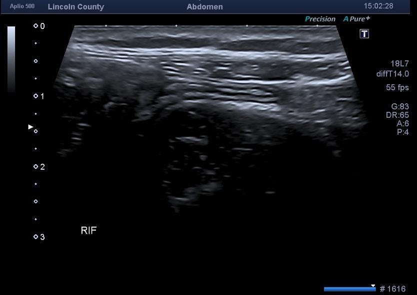
3 The Request USS Pelvis: Pelvic Pain? Ovarian? Cause Put the curvy down and back away!!!! We need to examine the bowel!
4 Bowel Common Bowel Conditions Giving Rise to Pelvic/Iliac Fossa Pain RIF LIF Appendicitis Ileitis (Crohn s) Caecal and Ileocaecal tumours (including lymphoma) Diverticulitis Diverticulosis Distal Colitis Sigmoid tumours
5 Appendicitis Accuracy of clinical evaluation of acute appendicitis is low especially in the young female (Üeberrüeck et al. 2004) Incredibly variable appearances of the normal appendix leads to false positives Operator dependant
6 The Appendix Graded Compression Technique 3.5 Mhz probe starting point to locate the Caecum which is usually gas containing Most often the appendix is located caudally to the ileocaecal valve.however is quite variable!
.")
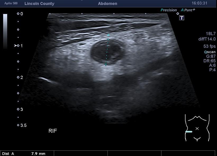
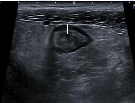
7 Normal Appendix High frequency linear probe Compressible thin walled Compared with the terminal ileum, there is no peristalsis Can be seen in up to 70% of patients (according to the literature..).. Purposely excluded and normal measurement of an appendix although some literature use 4mm
8 Appendicitis Increased diameter, non compressible Top Tips Take with a pinch of salt: unreliable unless other features are also present. Top Tips Be careful not slip off the appendix rather that compress Also in focal appendicitis the appendix may not be enlarged and unless the length of the organ is completely imaged this could be missed

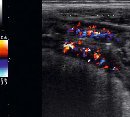
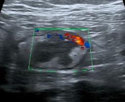
9 Appendicoliths Appendicitis Creeping fat/echogenic inflamed mesenteric fat which may be hyperaemic Localised effusion/free fluid Enlarged/reactive lymphadenopathy
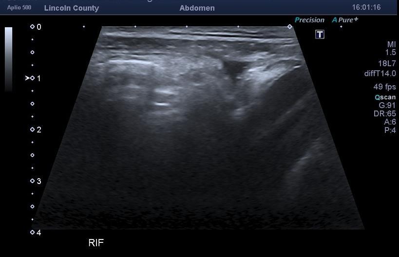
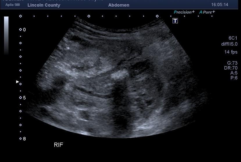

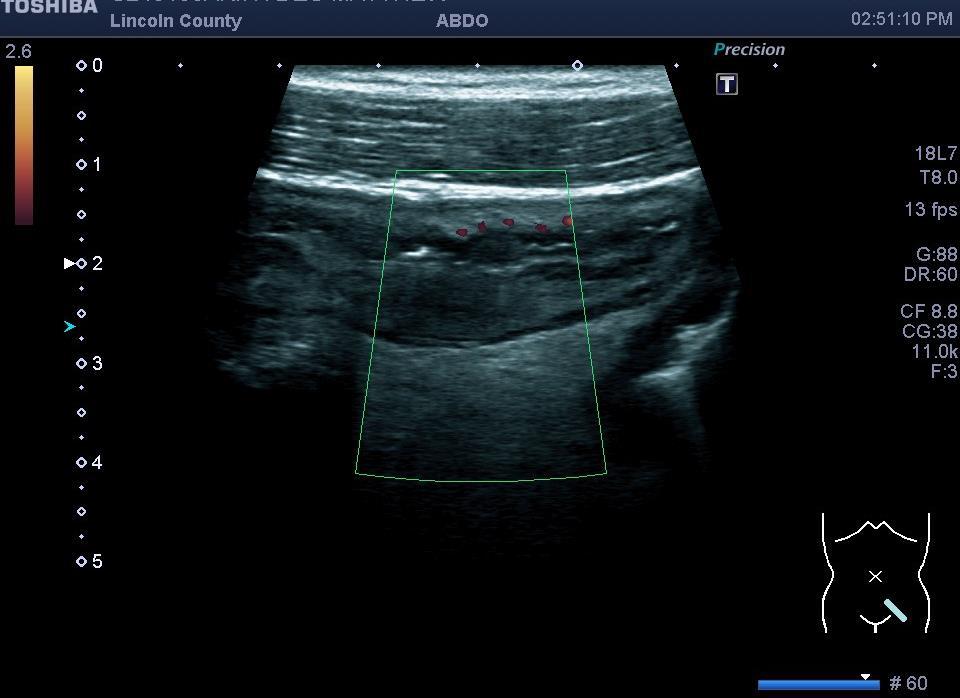
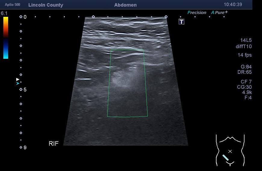
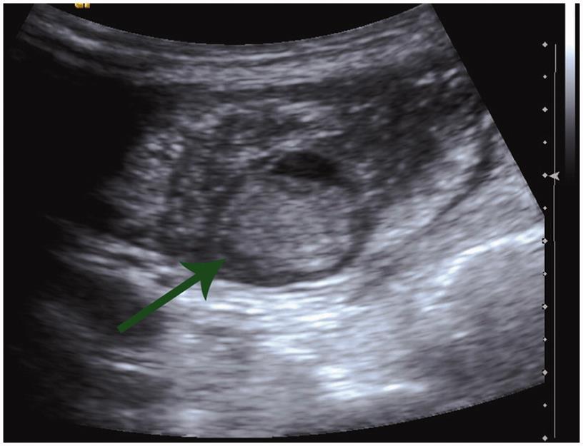
10 Appendicitis
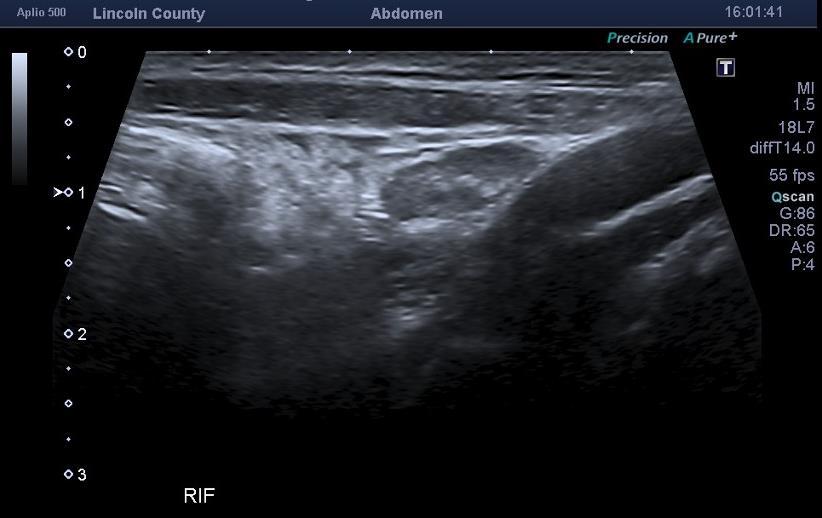
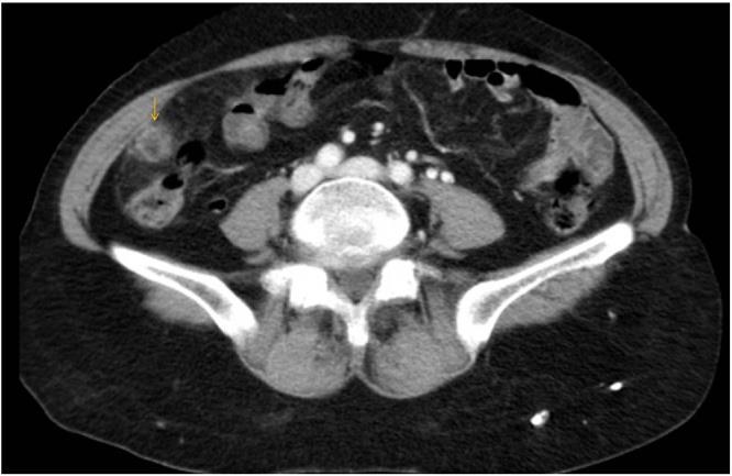
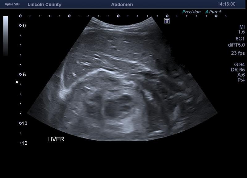
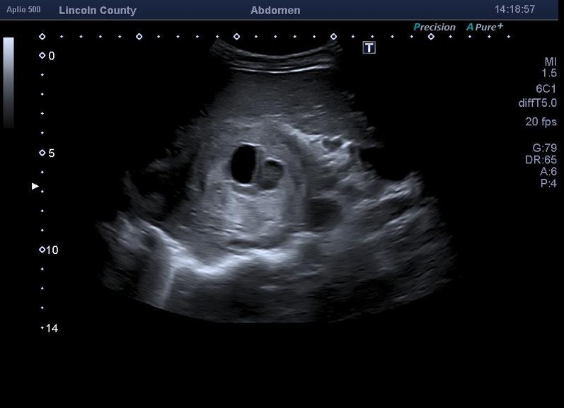
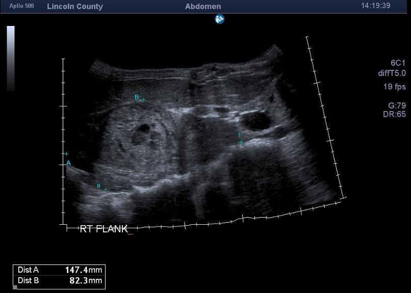
11 Appendicitis - Chronic
 Sigmoid Colon @ Left iliac Fossa Ileocaecal junction @ Right")

12 Overview of gut and mesentery Colon Picture Frame (High Frequency linear) Sigmoid Left iliac Fossa Ileocaecal Right Iliac Fossa Terminal ileum and appendix Jejunum Crohn s Disease Stomach/Duodenum/Oesophagus SMA and central small bowel mesentery Mow the lawn Picture Frame
 Colon (25%) Extensive small bowel involvement (5%) Anorectal, oral,")

13 Bowel Inflammation Can affect any part of the GI tract Ileocaecal (45%) Terminal Ileum (20%) Colon (25%) Extensive small bowel involvement (5%) Anorectal, oral, gastroduodenal (5%) Crohn s Disease Chronic relapsing inflammatory condition Unknown aetiology Increasing incidence Western prevalence Slight Female Predominance
 Acknowledgement: Dr R Beable - Consultant GI Radiologist")
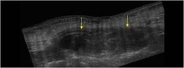
14 Diagnostic Features Crohn s Disease Mural thickening Mesenteric fat wrapping Ulceration - Apthous -Fissures - Cobblestoning Skip lesions Fistulation Mesenteric Lymph Nodes Mesenteric plethora (Comb Sign) Acknowledgement: Dr R Beable - Consultant GI Radiologist Plymouth

15 Crohn s Disease Acknowledgement: Dr R Beable - Consultant GI Radiologist Plymouth
16

17 Crohn s Disease


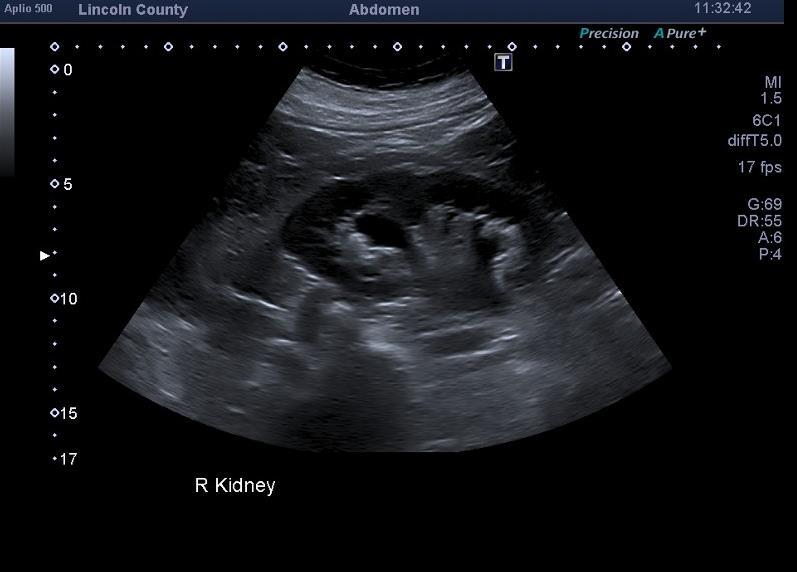
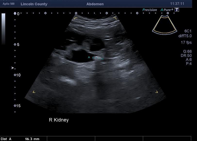
18 Large Bowel Ischemic Colitis Sigmoid Diverticulosis- faecolith Images Marconi & Bianchi

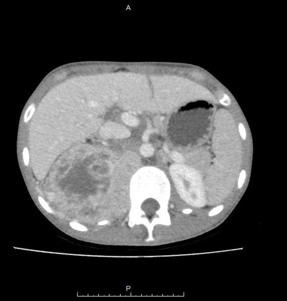
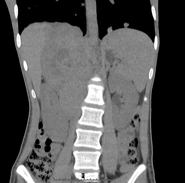
19 Caecal Tumour
20 Epiploic Appendagitis Acute epiploic appendagitis is a selflimited inflammation of the appendices epiploicae Presentation :? Ovarian torsion /Acute pelvic pain not typical of appendicitis
21 Epiploic Appendagitis Epiploic (or omental) appendages are peritoneal pouches filled with adipose tissue that arise from the serosal surface of the colon They are attached by a vascular stalk They frequently arise in association with colonic diverticula Composed of adipose tissue and blood vessels, the appendages typically have a length of cm Typically only seen on imaging when inflamed


22 Epiploic Appendagitis
23 Omental Infarction Omental infarction is a rare cause of acute abdomen resulting from vascular compromise of the greater omentum This condition has a non-specific clinical presentation and is usually managed conservatively. The term along with epiploic appendagitis is grouped under the broader umbrella term intraperitoneal focal fat infarction.
24 Omental Infarction

25 Rectus Sheath Haematoma Vague and non specific pelvic pain May be some Tachycardia Hypotension Fall in Hbg levels Direct tears to the rectus muscles Damage to the inferior epigastric artery or its branches Minor trauma to those on anti-coag Tx

26 Rectus Sheath Haematoma X
27 Hernia Inguinal hernias are more difficult to detect in women clinically Women often have occult, non palpable inguinal herniae causing pelvic pain Indirect inguinal hernia is the most common hernia in women Pelvic floor herniae may be seen on MRI

28 Hernia


29 Pyelonephritis Urological Causes Interstitial cystitis
30 Calculi

31 X
32 Chronic Nerve Damage Ilioinguinal neuralgia entrapment From previous surgery such as hernia repair/ C- Section Pudendal Neuralgia Excessive compression and repeated minor trauma e.g. from keen cyclists, horse-riding Damage following childbirth Previous pelvic surgery or Pelvic trauma/#
33 Psychosocial Issues Chronic pelvic pain (CPP), a common condition particularly in reproductive-aged women, causes disability and distress, and significantly compromises quality of life and affects healthcare costs. Depression /Anxiety/ Low mood Drug/Alcohol Addictions Previous abuse patients Ultrasound has value in exclusion
34 18 y/o female Final Case Dec 16 :RIF pain 2/12? Tubo-ovarian cause Ultrasound: The uterus, endometrium and both ovaries appear normal. The urinary bladder was empty therefore could not be assessed. No adnexal mass or free fluid seen. Nov 17: Significant W/L and continued RIF pain. Ultrasound for SB US? SB Crohn s No Significant abnormal results in all blood tests or urinalysis performed between Dec 16 & Dec 17
35

36

37 Conclusions Pelvic Pain isn t always a gynaecological issue We can t assess everything it possibly could be! But we can be aware. And perhaps check the possibility of other causes in some cases In our practice pelvic pain with a negative gynae scan usually has the urinary tract assessed..& Thoughts Thinking: the process of considering or reasoning about something using thought or rational judgement; intelligent.
38 References Collins, DC. The length and position of the vermiform appendix: a study of 4,680 specimens. Ann Surg 1932; 96: Google Scholar, Crossref, Medline Rettenbacher, T, Hollerweger, A, Macheiner, P. Outer diameter of the vermiform appendix as a sign of acute appendicitis: Evaluation at US. Radiology 2001; 218: Google Scholar, Crossref, Medline Park, NH, Park, CS, Lee, EJ. Ultrasonographic findings identifying the faecalimpacted appendix: differential findings with acute appendicitis. BJR 2007; 80: Google Scholar, Crossref, Medline Lim, HK, Lee, WJ, Lee, SJ. Focal appendicitis confined to the tip: diagnosis at US. Radiology 1996; 200: Google Scholar, Crossref, Medline Rodgers, PM, Verma, R. Transabdominal ultrasound for bowel evaluation. Radiol Clin North Am 2013; 51: Google Scholar, Crossref, Medline
39 References Marconi G & Bianchi G Ultrasound of the Gastrointestinal Tract. Springer. London

40
Top Tips for Gynaecological Ultrasound. Catherine Kirkpatrick Consultant Sonographer Dublin Oct 2018
 Top Tips for Gynaecological Ultrasound Catherine Kirkpatrick Consultant Sonographer Dublin Oct 2018 We can all scan a pelvis so what can we do to improve? Uterus, endometrium and ovaries, got it covered!
The Role of Ultrasound in the Assessment of Inflammatory Bowel Disease
 The Role of Ultrasound in the Assessment of Inflammatory Bowel Disease Dr. Richard A. Beable Consultant Gastrointestinal Radiologist Queen Alexandra Hospital Portsmouth Hospitals NHS Trust Topics for Discussion
Pictorial review of bowel ultrasound: Common and unsuspected pathologies
 Pictorial review of bowel ultrasound: Common and unsuspected pathologies Poster No.: C-1668 Congress: ECR 2013 Type: Educational Exhibit Authors: A. Law, A. Ali, G. Hutchison; Bolton/UK Keywords: Ultrasound-Colour
Medical application of transabdominal ultrasound in gastrointestinal diseases
 Medical application of transabdominal ultrasound in gastrointestinal diseases Hsiu-Po Wang Department of Emergency Medicine National Taiwan University Hospital Real-time ultrasound has become a standard
FHS Appendicitis US Protocol
 FHS Appendicitis US Protocol Reviewed By: Shireen Khan, MD; Sarah Farley, MD; Anna Ellermeier, MD Last Reviewed: May 2018 Contact: (866) 761-4200 **NOTE for all examinations: 1. If documenting possible
The peritoneum. Prof. Oluwadiya KS, MBBS, FMCS(Orthop) Website:
 Website:") The peritoneum Prof. Oluwadiya KS, MBBS, FMCS(Orthop) Website: http://oluwadiya.com The peritoneum Serous membrane that lines the abdominopelvic cavity and invests the viscera The largest serous membrane
The peritoneum Prof. Oluwadiya KS, MBBS, FMCS(Orthop) Website: http://oluwadiya.com The peritoneum Serous membrane that lines the abdominopelvic cavity and invests the viscera The largest serous membrane
Pitfalls in the CT diagnosis of appendicitis
 The British Journal of Radiology, 77 (2004), 792 799 DOI: 10.1259/bjr/95663370 E 2004 The British Institute of Radiology Pictorial review Pitfalls in the CT diagnosis of appendicitis 1 C D LEVINE, 2 O
Mimics of Appendicitis: Alternative Nonsurgical Diagnoses with Sonography and CT
 van reda Vriesman and Puylaert Mimics of ppendicitis bdominal Imaging Pictorial Essay Downloaded from www.ajronline.org by 46.3.193.220 on 12/27/17 from IP address 46.3.193.220. Copyright RRS. For personal
APPENDICITIS AND ITS APPEARANCES ON CT
 APPENDICITIS AND ITS APPEARANCES ON CT APPENDICITIS Results from acute inflammation of the appendix. Most common abdominal surgical emergencies. Diagnosis usually clinical based on physical exam and lab
elical CT plays an important role
 bdominal Imaging Yu et al. Helical CT of cute RLQ Pain Pictorial Essay Jinxing Yu 1 nn S. Fulcher Mary nn Turner Robert. Halvorsen Yu J, Fulcher S, Turner M, Halvorsen R Helical CT Evaluation of cute Right
Summary and conclusions
 Summary and conclusions 7 Chapter 7 68 Summary and conclusions Chapter 1 provides a general introduction to this thesis focused on the use of ultrasound (US) in children with abdominal problems. The literature
SIMPLE GUIDE FOR SONOLOGICAL EVALUATION OF APPENDICITIS
 SIMPLE GUIDE FOR SONOLOGICAL EVALUATION OF APPENDICITIS A Case Study by Dr. Avni K P Skandhan, India (Consultant Radio Diagnosis, Malabar Institute of Medical Science, Malappuram, Kerala) Email: avniskandhan@gmail.com
Emergency MDCT in case of right lower quadrant pain
 Emergency MDCT in case of right lower quadrant pain Poster No.: C-0563 Congress: ECR 2015 Type: Educational Exhibit Authors: M. Lisitskaya, V. Sinitsyn; Moscow/RU Keywords: Abdomen, Emergency, Gastrointestinal
Sonographic Appearances of Common Gut Pathology in Paediatric Patients: Comparison with Plain Abdominal Radiography
 3668 Radiographer Text 1/4/04 2:57 PM Page 11 The Radiographer vol. 51: 11-17 Sonographic Appearances of Common Gut Pathology in Paediatric Patients: Comparison with Plain Abdominal Radiography Lino Piotto
The jejunum and the Ileum. Prof. Oluwadiya KS
 The jejunum and the Ileum Prof. Oluwadiya KS www.oluwadiya.siteled.com Introduction Introduction The small intestine (SI) comprises of the duodenum, jejunum and the ileum The jejunum is the second part
STRUCTURAL BASIS OF MEDICAL PRACTICE EXAMINATION 3. October 16, 2015
 STRUCTURAL BASIS OF MEDICAL PRACTICE EXAMINATION 3 October 16, 2015 PART l. Answer in the space provided. (12 pts) 1. Identify the structures. (2 pts) A. B. A B C. D. C D 2. Identify the structures. (2
Bushra Arafa Zayed & Hanan Jamal. - Dana AF
 - 10 - Bushra Arafa Zayed & Hanan Jamal - Dana AF - Mohammad Al Muhtaseb Notes: This sheet was written in the same order as the slides, and everything in the slides is mentioned in this sheet. Pictures
Preview from Notesale.co.uk Page 1 of 34
 Abdominal viscera and digestive tract Digestive tract Abdominal viscera comprise majority of the alimentary system o Terminal oesophagus, stomach, pancreas, spleen, liver, gallbladder, kidneys, suprarenal
Importance of ultrasonography and colour doppler in evaluation of bowel pathology in clinically suspected cases
 2015; 1(1): 26-32 Research Article JMR 2015; 1(1): 26-32 January- February 2015, All rights reserved www.medicinearticle.com Importance of ultrasonography and colour doppler in evaluation of bowel pathology
THE INS AND OUTS OF HERNIAS WHERE TO START? WHAT IS A HERNIA? CLINICAL INDICATIONS THE INGUINAL CANAL THE CLINICAL QUESTION 18/09/2018
 THE INS AND OUTS OF HERNIAS Cassandra Harrison BA/BSc, MMRU, AMS WHERE TO START? The Clinical Question Essential anatomy Inguinal hernia Scanning technique Variations WHAT IS A HERNIA? CLINICAL INDICATIONS
ACUTE ABDOMEN IN OLDER CHILDREN. Carlos J. Sivit M.D.
 ACUTE ABDOMEN IN OLDER CHILDREN Carlos J. Sivit M.D. ACUTE ABDOMEN Clinical condition characterized by severe abdominal pain developing over several hours ACUTE ABDOMINAL PAIN Common childhood complaint
ADENOMYOSIS CHRONIC PELVIC PAIN IN WOMEN IMAGING CHRONIC PELVIC PAIN IN WOMEN CHRONIC PELVIC PAIN IN WOMEN ADENOMYOSIS: PATHOLOGY ADENOMYOSIS
 CHRONIC PELVIC PAIN IN WOMEN IMAGING CHRONIC PELVIC PAIN IN WOMEN MOSTAFA ATRI, MD Dipl. Epid. UNIVERSITY OF TORONTO Non-menstrual pain of 6 months Prevalence 15%: 18-50 years of age 10-40% of gynecology
Embryology of the Midgut and Hind gut
 Embryology of the Midgut and Hind gut Prof. Abdulameer Al-Nuaimi E-mail: a.al-nuaimi@sheffield.ac.uk E-mail: abdulameerh@yahoo.com Abdominal organs www.google.co.uk/search? Development of Duodenum The
Acute pelvic pain in female patient: Clinical and Radiological evaluation
 Acute pelvic pain in female patient: Clinical and Radiological evaluation Poster No.: C-0909 Congress: ECR 2014 Type: Authors: Keywords: DOI: Educational Exhibit N. Ramesh 1, T. Simelane 2 ; 1 Portlaoise/IE,
Acute pelvic pain in female patient: Clinical and Radiological evaluation
 Acute pelvic pain in female patient: Clinical and Radiological evaluation Poster No.: C-0909 Congress: ECR 2014 Type: Authors: Keywords: DOI: Educational Exhibit N. Ramesh 1, T. Simelane 2 ; 1 Portlaoise/IE,
Mohamed EL-hemaly Gastro- intestinal surgical center, Mansoura University.
 Mohamed EL-hemaly Gastro- intestinal surgical center, Mansoura University. Chronic transmural inflammatory process of the bowel & affects any part of the gastro -intestinal tract from the mouth to the
ANATOMY OF THE SMALL & LARGE INTESTINES. Semester 1, 2011 A. Mwakikunga
 ANATOMY OF THE SMALL & LARGE INTESTINES Semester 1, 2011 A. Mwakikunga LEARNING OBJECTIVES 1. List the parts and anatomical regions of the small and large intestines 2. State anatomical relations of the
Multislice Computed Tomography Imaging with Clinical Outcome in Inflammatory Appendiceal Masses
 Original Article Multislice Computed Tomography Imaging with Clinical Outcome in Inflammatory Appendiceal Masses ID: IJARS/2016/18502:2116 Radiology Section Varsha Rangankar, Abhijit Pawar, Aditi Dongre,
CT Appearance of Acute Appendagitis
 CT Appearance of Acute Appendagitis Poster No.: C-0673 Congress: ECR 2013 Type: Scientific Exhibit Authors: J. SAAD, F. Marrakchi, Y. M. Abdou ; Monastir, TN/TN, 1 2 2 3 1 3 Monastir/TN, Nejran, Nejran/SA
Primary epiploic appendagitis versus omental infarction : The role of MDCT
 Primary epiploic appendagitis versus omental infarction : The role of MDCT e-poster: EE-125 Congress: ESGAR 2010 Type: Educational Exhibit Topic: Diagnostic / Mesentery and Peritoneum Authors: P. Kraniotis,
Anatomy of the Large Intestine
 Large intestine Anatomy of the Large Intestine 2 Large Intestine Extends from ileocecal valve to anus Length = 1.5-2.5m = 5 feet Regions Cecum = 2.5-3 inch Appendix= 3-5 inch Colon Ascending= 5 inch Transverse=
STRUCTURAL BASIS OF MEDICAL PRACTICE EXAMINATION 3. October 17, 2014
 STRUCTURAL BASIS OF MEDICAL PRACTICE EXAMINATION 3 October 17, 2014 PART l. Answer in the space provided. (12 pts) 1. Identify the structures. (2 pts) A. B. A B C. D. C D 2. Identify the structures. (2
Appendicitis. I. Background & Significance: Algorithm Definitions 1. CASE
 I. Background & Significance: Appendicitis Appendicitis is one of the most common acquired surgical conditions of childhood. Diagnosis of appendicitis remains difficult. Much work has been done on validation
Appendix 5. EFSUMB Newsletter. Gastroenterological Ultrasound
 EFSUMB Newsletter 87 Examinations should encompass the full range of pathological conditions listed below A log book listing the types of examinations undertaken should be kept Training should usually
Evaluation of the of the sensitivity, accuracy and positive predictive value of ultrasonography in the diagnosis of Appendicitis.
 West African Journal of Ultrasound Vol 17 Number 2 (2016) Evaluation of the of the sensitivity, accuracy and positive predictive value of ultrasonography in the diagnosis of Appendicitis. 1 2 3 Oguntola
Sigmoid Diverticulitis: Value of Transrectal Sonography in Addition to Transabdominal Sonography
 lois Hollerweger 1 Thomas Rettenbacher 1 Peter Macheiner 1 Walter Brunner 2 Norbert Gritzmann 1 Received ugust 17, 1999; accepted after revision March 17, 2000. 1 Department of Radiology and Nuclear Medicine,
In the name ofgod. Abdomen 3. Dr. Zahiri
 In the name ofgod Abdomen 3 Dr. Zahiri Peritoneum Peritoneum It is the serous membrane(a type of loose connective tissue and is covered by mesothelium) that lines the abdominal cavity. Extensions of the
THE ORAL CAVITY
 THE ORAL CAVITY WALL OF ABDOMEN (ANTERIOR) The paraumbilical vein drains into the portal vein and then through the liver. This is an important clinical connection. THE ABDOMINAL VISCERA The small
Contents I MEDICAL RADIOLOGY. Diagnostic Imaging. Editors: A. L. Baert, Leuven M. Knauth, Göttingen K. Sartor, Heidelberg
 Contents I MEDICAL RADIOLOGY Diagnostic Imaging Editors: A. L. Baert, Leuven M. Knauth, Göttingen K. Sartor, Heidelberg Contents III G. Maconi G. Bianchi Porro (Eds.) Ultrasound of the Gastrointestinal
Development of pancreas and Small Intestine. ANATOMY DEPARTMENT DR.SANAA AL-AlSHAARAWY DR.ESSAM Eldin Salama
 Development of pancreas and Small Intestine ANATOMY DEPARTMENT DR.SANAA AL-AlSHAARAWY DR.ESSAM Eldin Salama OBJECTIVES At the end of the lecture, the students should be able to : Describe the development
The role of abdominal X-rays in the investigation of suspected acute appendicitis
 Journal of Medicine and Medical Sciences Vol. 2(11) pp. 1216-1220, November 2011 Available online@ http://www.interesjournals.org/jmms Copyright 2011 International Research Journals Full Length Research
Alison Douglass Gillian Lieberman, MD. November. Colon Cancer. Alison Douglass, Harvard Medical School Year III Gillian Lieberman, MD
 November Colon Cancer Alison Douglass, Harvard Medical School Year III Our Patient Mr. K. is a 67 year old man with no prior medical problems other than hemorrhoids which have caused occasional rectal
Plain Radiographs in Non-Traumatic Abdominal Pain. Plain Radiographs in Non-Traumatic Abdominal Pain
 Jake Block, MD Associate Professor Associate Vice-Chairman for Clinical Operations Director, Musculoskeletal and Emergency Radiology Department of Radiology and Radiological Sciences Vanderbilt University
Paediatric surgical emergencies. Mani Thyagarajan BWCH
 Paediatric surgical emergencies Mani Thyagarajan BWCH General points Always discuss Call consultant for help ASAP CT scan is a bad modality in paediatrics Ultrasound? Intussusception? Renal colic? UTI
Abdominal Pain in Pediatric Patients Image Gently
 Abdominal Pain in Pediatric Patients Image Gently Susan D. John, M.D. Baptist Health Emergency Radiology 2017 Disclosure I have no financial relationships with a commercial entity producing healthcarerelated
Case Scenario 1. 1/2/13 History: 64-year-old white female presented with right leg swelling and redness, abdominal pain.
 Case Scenario 1 1/2/13 History: 64-year-old white female presented with right leg swelling and redness, abdominal pain. 1/02/13 CT Abdomen/Pelvis: Abnormal area of nodular mesenteric and left anterior
Nordic Forum - Trauma & Emergency Radiology. Bowel Obstruction: Imaging Update
 Nordic Forum - Trauma & Emergency Radiology Bowel Obstruction: Imaging Update Borut Marincek Institute of Diagnostic Radiology University Hospital Zurich, Switzerland Acute Abdomen Bowel Obstruction Bowel
Pelvic Pain in the Pediatric Patient Susan D. John, M.D.
 Pelvic Pain in the Pediatric Patient Susan D. John, M.D. RSNA 2012 Patients First Objectives After attending this presentation, participants will be able to: Understand the common congenital and acquired
Small Plicae Circularis. Short Closely packed together. Sparse, completely absent at distal part Lymphoid Nodule
 Intestines Differences Between Jejunum and Ileum Types Jejunum Ileum Color Deeper red Paler pink Calibre Bigger Smaller Thickness of wall Thick and Heavy Thin and Lighter Vascularity Highly vascularised
Ultrasound of: Appendicitis Intussusception Pyloric Stenosis
 Ultrasound of: Appendicitis Intussusception Pyloric Stenosis Andrew Phelps MD Assistant Professor Pediatric Radiology UCSF Benioff Children s Hospital No Disclosures Take Home Message Appendicitis occurs
ORIGINAL ARTICLE ROLE OF ULTRASONOGRAPHY IN PRE-OPERATIVE EVALUATION OF RIGHT ILIAC FOSSA MASS
 ROLE OF ULTRASONOGRAPHY IN PRE-OPERATIVE EVALUATION OF RIGHT ILIAC FOSSA MASS Madhushankar L 1, Satish Kumar R 2, Sanjay S.C 3, Laxmikanta. L 4, Hemanth V 5 HOW TO CITE THIS ARTICLE: Madhushankar L, Satish
Interesting Pediatric ultrasound cases. Presented by: Falguni Patel (RDMS, RVT)
") Interesting Pediatric ultrasound cases Presented by: Falguni Patel (RDMS, RVT) Role of ultrasound to rule out Appendicitis Overview: Ultrasound is relatively inexpensive, safe and quick solution to rule
Interesting Pediatric ultrasound cases Presented by: Falguni Patel (RDMS, RVT) Role of ultrasound to rule out Appendicitis Overview: Ultrasound is relatively inexpensive, safe and quick solution to rule
Nongynecological causes of acute and chronicpelvic pain. Amela Sofić UKC Sarajevo Bosnia and Herzegovina
 Nongynecological causes of acute and chronicpelvic pain Amela Sofić UKC Sarajevo Bosnia and Herzegovina One of the most challenging problems in a clinical routine is the pelvic pain It is useful to classify
Case Scenario 1. 1/2/13 History: 64-year-old white female presented with right leg swelling and redness, abdominal pain.
 Case Scenario 1 1/2/13 History: 64-year-old white female presented with right leg swelling and redness, abdominal pain. 1/02/13 CT Abdomen/Pelvis: Abnormal area of nodular mesenteric and left anterior
Midgut. Over its entire length the midgut is supplied by the superior mesenteric artery
 Gi Embryology 3 Midgut the midgut is suspended from the dorsal abdominal wall by a short mesentery and communicates with the yolk sac by way of the vitelline duct or yolk stalk Over its entire length the
IMAGING GUIDELINES - COLORECTAL CANCER
 IMAGING GUIDELINES - COLORECTAL CANCER DIAGNOSIS The majority of colorectal cancers are diagnosed on colonoscopy, with some being diagnosed on Ba enema, ultrasound or CT. STAGING CT chest, abdomen and
Duodenum retroperitoneal
 Duodenum retroperitoneal C shaped Initial region out of stomach into small intestine RETROperitoneal viscus Superior 1 st part duodenal cap ; moves upwards and backwards to lie on the R crura medial to
BLOCK IV: OFFICIAL BODY PARTS LIST FOR ANTERIOR ABDOMINAL WALL AND ABDOMINAL CONTENTS
 BLOCK IV: OFFICIAL BODY PARTS LIST FOR ANTERIOR ABDOMINAL WALL AND ABDOMINAL CONTENTS External oblique muscle Muscular portion Aponeurotic portion Superficial inguinal ring Lateral (inferior) crus Medial
Biology Human Anatomy Abdominal and Pelvic Cavities
 Biology 351 - Human Anatomy Abdominal and Pelvic Cavities You must answer all questions on this exam. Because statistics demonstrate that, on average, between 2-5 questions on every 100-point exam are
Vikram Dogra, M.D. Professor of Radiology, Urology & BME Department of Imaging Sciences University Of Rochester Medical Center
 Ultrasound of the Scrotum Vikram Dogra, M.D. Professor of Radiology, Urology & BME Department of Imaging Sciences University Of Rochester Medical Center Etiologies of Acute Scrotal Pain Epididymitis/Orchitis
Curious case of Misty Mesentery
 Curious case of Misty Mesentery Poster No.: C-1385 Congress: ECR 2015 Type: Authors: Keywords: DOI: Educational Exhibit T. Simelane 1, H. Khosa 2, N. Ramesh 2 ; 1 Dublin/IE, 2 Portlaoise/IE Abdomen, Anatomy,
Anatomy: Know Your Abdomen
 Anatomy: Know Your Abdomen Glossary Abdomen - part of the body below the thorax (chest cavity); separated by the diaphragm. Anterior - towards the front of the body. For example, the umbilicus is anterior
ACUTE ABDOMEN. Dr. M Asadi. Surgical Oncology Research Center MUMS. Assistant Professor of General Surgery
 ACUTE ABDOMEN Dr. M Asadi Assistant Professor of General Surgery Surgical Oncology Research Center MUMS Definition I. The term Acute Abdomen refers to signs & symptoms of abdominal pain and tenderness,
Ultrasound Of The Acute Abdomen a survival guide. Alison McGuinness Consultant sonographer Mid Yorks Hospitals NHS Trust
 Ultrasound Of The Acute Abdomen a survival guide Alison McGuinness Consultant sonographer Mid Yorks Hospitals NHS Trust Definition Rapid onset of severe abdominal pain usually requiring admission to hospital
Case 1307 Mesothelial cysts
 Case 1307 Mesothelial cysts Vinhais S, Monteiro M, Cunha TM INSTITUTO PORTUGUÊS DE ONCOLOGIA de Francisco Gentil de LISBOA Section: Gastro-Intestinal Imaging Published: 2001, Nov. 23 Patient: 44 year(s),
Listed below are some of the words that you might come across concerning diseases and conditions of the bowels.
 Listed below are some of the words that you might come across concerning diseases and conditions of the bowels. Abscess A localised collection of pus in a cavity that is formed by the decay of diseased
Imaging Patients with Acute Abdominal Pain 1
 Note: This copy is for your personal non-commercial use only. To order presentation-ready copies for distribution to your colleagues or clients, contact us at www.rsna.org/rsnarights. Jaap Stoker, MD Adrienne
Emergency radiology of the large-bowel: What radiologists should know
 Emergency radiology of the large-bowel: What radiologists should know Poster No.: C-1659 Congress: ECR 2016 Type: Educational Exhibit Authors: A. Falkowski, D. Boll; Basle/CH Keywords: Colon, Emergency,
SUBJECTS 2nd year, 1st semester I. 1. Primitive gut - limits, derivatives 2. Foregut -limits, evolution, derivatives 3. Midgut -limits, evolution,
 SUBJECTS 2nd year, 1st semester I. 1. Primitive gut - limits, derivatives 2. Foregut -limits, evolution, derivatives 3. Midgut -limits, evolution, derivatives 4. Hindgut- limits, evolution, derivatives
Intraperitoneal cysts in infancy and childhood An overview and sonographic differentiation
 Intraperitoneal cysts in infancy and childhood An overview and sonographic differentiation M. Mearadji International Foundation for Pediatric Imaging Aid Rotterdam, The Netherlands Intraperitoneal cysts
Introduction of Appendiceal CT Impact on Negative Appendectomy and Appendiceal
 ANNALS OF SURGERY Vol. 229, No. 3, 344-349 1999 ULppinc Willams & Wilins, Inc. Introduction of Appendiceal CT Impact on Negative Appendectomy and Appendiceal Perforation Rates Patrick M. Rao, MD,* James
Sonographic Appearance of Normal Appendix in Children. Abstract
 Proceeding S.Z.P.G.M.I. vol: 22(2): pp. 57-62, 2008. Sonographic Appearance of Normal Appendix in Children Abdus Sarni Qazi Department of Radiology, Lahore General Hospital, Lahore. Abstract Objective:
COLORECTAL CANCER FAISALGHANISIDDIQUI MBBS; FCPS; PGDIP-BIOETHICS; MCPS-HPE
 COLORECTAL CANCER FAISALGHANISIDDIQUI MBBS; FCPS; PGDIP-BIOETHICS; MCPS-HPE PROFESSOR OF SURGERY & DIRECTOR, PROFESSIONAL DEVELOPMENT CENTRE J I N N A H S I N D H M E D I C A L U N I V E R S I T Y faisal.siddiqui@jsmu.edu.pk
Table 0: Description of Grading System for Anatomic Severity of Disease in Emergency. Local disease confined to the organ with minimal abnormality
 Table 0: of Grading System for Anatomic Severity of Disease in Emergency Local disease confined to the organ with minimal Local disease confined to the organ with severe Local extension Table 1: Universal
US in non-traumatic acute abdomen. Lalita, M.D. Radiologist Department of radiology Faculty of Medicine ChiangMai university
 US in non-traumatic acute abdomen Lalita, M.D. Radiologist Department of radiology Faculty of Medicine ChiangMai university Sagittal Orientation Transverse (Axial) Orientation Coronal Orientation Intercostal
Exploring Anatomy: the Human Abdomen
 Exploring Anatomy: the Human Abdomen PERITONEUM AND PERITONEAL CAVITY PERITONEUM The peritoneum is a thin serous membrane that lines the abdominal cavity and covers, in variable amounts, the viscera within
Gastrointestinal Tract. Anatomy of GI Tract. Anatomy of GI Tract. (Effective February 2007) (1%-5%)
 (1%-5%)") Gastrointestinal Tract (Effective February 2007) (1%-5%) Anatomy of GI Tract Esophagus bulls-eye or target EG junction seen on sagittal scan posterior to left lobe of liver and anterior to aorta Anatomy
Gastrointestinal Tract (Effective February 2007) (1%-5%) Anatomy of GI Tract Esophagus bulls-eye or target EG junction seen on sagittal scan posterior to left lobe of liver and anterior to aorta Anatomy
Non-calculus causes of renal colic on CT KUB
 Non-calculus causes of renal colic on CT KUB Poster No.: C-1341 Congress: ECR 2010 Type: Scientific Exhibit Topic: Genitourinary Authors: A. Afaq, E. L. Leen; London/UK Keywords: renal colic, CT KUB, appendicitis
Caeco-colic Intussusception Simulating an Appendicular Mass
 Article ID: WMC003206 ISSN 2046-1690 Caeco-colic Intussusception Simulating an Appendicular Mass Corresponding Author: Dr. Matthew O Adelekan, Surgeon, North manchester General Hospital - United Kingdom
Inflammatory Bowel Disease Ischemic bowel disease
 Inflammatory Bowel Disease Ischemic bowel disease Inflammatory Bowel Disease The two disorders that comprise IBD are: ulcerative colitis Crohn disease The distinction between ulcerative colitis and Crohn
World Journal of Colorectal Surgery
 World Journal of Colorectal Surgery Volume 3, Issue 2 2013 Article 18 Revenge of the Christmas Turkey; Unusual Presentation of Colonic Perforation Secondary to Foreign Body. Mashuk Khan Sudeep Thomas Warwick
A Practical Approach to Adnexal Masses
 A Practical Approach to Adnexal Masses Darcy J. Wolfman, MD Section Chief of Genitourinary Imaging American Institute for Radiologic Pathology Clinical Associate Johns Hopkins Community Radiology Division
GI Tract Lynn Ta Jennifer Zhang July 6, 2006 GI TRACT. 1) Other Names: Gastrointestinal tract Digestive tract Alimentary tract
 Other Names: Gastrointestinal tract Digestive tract Alimentary tract") GI Tract Lynn Ta Jennifer Zhang July 6, 2006 GI TRACT 1) Other Names: Gastrointestinal tract Digestive tract Alimentary tract 2) Definition/Location: Digestion and absorption are the primary functions
GI Tract Lynn Ta Jennifer Zhang July 6, 2006 GI TRACT 1) Other Names: Gastrointestinal tract Digestive tract Alimentary tract 2) Definition/Location: Digestion and absorption are the primary functions
Gastrointestinal Disorders. Disorders of the Esophagus 3/7/2013. Congenital Abnormalities. Achalasia. Not an easy repair. Types
 Gastrointestinal Disorders Congenital Abnormalities Disorders of the Esophagus Types Stenosis Atresia Fistula Newborn aspirates while feeding. Pneumonia Not an easy repair Achalasia Lack of relaxation
... Inflammatory disorder of the colon that occurs as a complication of antibiotic treatment.
 Definition Inflammatory disorder of the colon that occurs as a complication of antibiotic treatment. " Epidemiology Humans represent the main reservoir of Clostridium difficile, which is not part of the
Ultrasonography of the acute abdomen: gastrointestinal conditions
 Radiol Clin N Am 41 (2003) 1227 1242 Ultrasonography of the acute abdomen: gastrointestinal conditions Julien B.C.M. Puylaert, PhD, MD Department of Radiology, MCH Westeinde Hospital, The Hague, The Netherlands
STAGING AND FOLLOW-UP STRATEGIES
 ATHENS 4-6 October 2018 European Society of Urogenital Radiology STAGING AND FOLLOW-UP STRATEGIES Ahmet Tuncay Turgut, MD Professor of Radiology Hacettepe University, Faculty of Medicine Ankara 2nd ESUR
Intestinal Obstruction Clinical Presentation & Causes
 Intestinal Obstruction Clinical Presentation & Causes V Chidambaram-Nathan Consultant Transplant and General Surgeon Sheffield Kidney Institute Northern General Hospital Intestinal Obstruction One of the
Loyola University Medical Center Female Pelvic Medicine & Reconstructive Surgery
 Loyola University Medical Center Female Pelvic Medicine & Reconstructive Surgery Medical History Questionnaire Name: Date: Age: D.O.B. Race: What is the nature of your current gynecologic or urologic medical
Pitfalls in CT diagnosis of appendicitis: Pictorial essay
 bs_bs_banner Journal of Medical Imaging and Radiation Oncology 57 (2013) 329 336 RADIOLOGY PICTORIAL ESSAY Pitfalls in CT diagnosis of appendicitis: Pictorial essay Ashkan Shademan and Rafel FR Tappouni
Anatomy of the SMALL INTESTINE. Dr. Noman Ullah Wazir PMC
 Anatomy of the SMALL INTESTINE Dr. Noman Ullah Wazir PMC SMALL INTESTINE The small intestine, consists of the duodenum, jejunum, and illium. It extends from the pylorus to the ileocecal junction were the
Always keep it in the differential
 Acute Appendicitis Lissa C. Sakata and Lindsey Perea 2 Always keep it in the differential Learning Objectives 1. The learner should be able to describe the etiology of acute appendicitis. 2. The learner
MPH Quiz. 1. How many primaries are present based on this pathology report? 2. What rule is this based on?
 MPH Quiz Case 1 Surgical Pathology from hysterectomy performed July 11, 2007 Final Diagnosis: Uterus, resection: Endometrioid adenocarcinoma, Grade 1 involving most of endometrium, myometrial invasion
Contrast-enhanced small bowel ultrasound in the assessment of the small bowel in patients with Crohn s Disease
 Contrast-enhanced small bowel ultrasound in the assessment of the small bowel in patients with Crohn s Disease C.F. Healy 1, D. Ferguson 1, S. Jepson 1, B. Salh 2, F. Donnellan 2, N. Chatur 2, A. C. Harris
Cecal Volvulus: Case Presentation and Review of CT Findings
 August 2011 Cecal Volvulus: Case Presentation and Review of CT Findings Omar Pardesi, Harvard Medical School Year III Our Patient LD: History & Physical HPI: 28 y.o. female presents with diffuse abdominal
A Comparative Ultrasound and Plain Abdominal X-Ray: Evaluation of Non-Classical Clinical Cases of Appendicitis
 A Comparative Ultrasound and Plain Abdominal X-Ray: Evaluation of Non-Classical Clinical Cases of Appendicitis Dorothy Makanjuola, FRCR; Qasim Al-Qasabi, FRCS; Tajuddin Malabarey, FRCR From the Departments
UNDERSTANDING X-RAYS: ABDOMINAL IMAGING THE ABDOMEN
 UNDERSTANDING X-RAYS: ABDOMINAL IMAGING THE ABDOMEN Radiology Enterprises radiologyenterprises@gmail.com www.radiologyenterprises.com STOMACH AND SMALL BOWEL STOMACH AND SMALL BOWEL Swallowed air is a
Dr. Zahiri. In the name of God
 Dr. Zahiri In the name of God small intestine = small bowel is the part of the gastrointestinal tract Boundaries: Pylorus Ileosecal junction Function: digestion and absorption of food It receives bile
Thickened gastrointestinal wall findings on computed tomography: simplifying the diagnosis.
 Thickened gastrointestinal wall findings on computed tomography: simplifying the diagnosis. Poster No.: C-0419 Congress: ECR 2015 Type: Educational Exhibit Authors: W. Mnari, O. Fkih, M. Maatouk, A. Zrig,
CLINICAL PRESENTATION AND RADIOLOGY QUIZ QUESTION
 Donald L. Renfrew, MD Radiology Associates of the Fox Valley, 333 N. Commercial Street, Suite 100, Neenah, WI 54956 6/23/2012 Radiology Quiz of the Week # 78 Page 1 CLINICAL PRESENTATION AND RADIOLOGY