Characterization of the Dental Phenotype in Transgenic Mice with Col 1A1 Targeted Overexpression of p20c/ebpb
|
|
|
- Daniella Fitzgerald
- 5 years ago
- Views:
Transcription

1 University of Connecticut SoDM Masters Theses School of Dental Medicine June 2003 Characterization of the Dental Phenotype in Transgenic Mice with Col 1A1 Targeted Overexpression of p20c/ebpb Todd M. Bennett Follow this and additional works at: Recommended Citation Bennett, Todd M., "Characterization of the Dental Phenotype in Transgenic Mice with Col 1A1 Targeted Overexpression of p20c/ EBPB" (2003). SoDM Masters Theses
2 Characterization of the Dental Phenotype in Transgenic Mice with Col 1A1 Targeted Overexpression of p20c/ebpg Todd M. Bennett DDS University of North Carolina, 1998 A Thesis Submitted in Partial Fulfillment of the Requirements for the Degree of Master of Dental Science at the University of Connecticut 2003
3 APPROVAL PAGE Master of Dental Science Thesis Characterization of the Dental Phenotype in Transgenic Mice With Col 1A 1 Targeted Overexpression of p20c/ebpi$ Presented by Todd M. Bennett, DDS Major Advisor (.._) "Jtohn Harrison, PhD Associate Advisor Associate Advisor Yu-Feng ua DS, Ph University of Connecticut 2003
4 Table of Contents Title Pae Title Page Approval Page Table of Contents List of Figures ii iii v Abstract 1 Background 2 Hypothesis 10 Objectives 10 Research Plan 12 Material and Methods 13 Results 17 Discussion 21 Conclusion 25 Figures 26 References 55 iii
5 List of Figures Figure Page Figure 1: Tooth phenotype photograph 34 Figure 2: Lateral skull radiographs 35 Figure 3: Low power histological section comparing wild-type and transgenic mice incisors 36 Figure 4: High power histological section comparing wild-type and transgenic mouse incisors 37 Figure 5: High power histological section comparing the lingual dentin of wild-type and transgenic mouse incisors 38 Figure 6: High power histological section comparing the lingual and labial dentin of the transgenic mouse incisor 39 Figure 7: Histological section of transgenic incisors 40 Figure 8: Histological section of wild-type and transgenic molar roots 41 Figure 9: Histological section comparing alveolar bone in wild-type and transgenic mice 42 Figure 10: Wild-type molar histological section 43 Figure 11: Transgenic molar histological section 44 Figure 12: Micro-computed tomography analysis 45 Figure 13: Micro-computed tomography analysis of wild-type and transgenic mouse incisors 46 iv
6 Figure 14: Graph of linear measurements for line Figure 15: Graph of area data for line Figure 16: Graph of linear measurements for line Figure 17: Graph of area data for line Figure 18: Northern blot analysis for transgenic mouse mandible 51 Figure 19: Low power micrograph localizing transgene expression in wild-type and transgenic mice 52 Figure 20: Low power micrograph of transgenic mouse incisor 53 Figure 21" High power micrograph localizing transgene expression 54
7
8 Abstract: A dominant negative C/EBP transcription factor loss of function transgenic mouse model has been generated to establish the effects of C/EBP inhibition on osteoblast and odontoblast differentiation and function. These mice express a FLAGtagged, dominant negative C/EBP transgene that has been targeted to cells of the stromal/osteoblast lineage with a 3.6 kb type I collagen promoter fragment (3.6). In earlier studies, transgenic pobcol3.6-flp20c/ebpg mice from this model showed evidence of decreased trabecular bone volume. This suggests a possible positive role for C/EBP transcription factors in osteogenesis. Our goal is to compare the dental morphology and micro-architecture of transgenic mice to their wild-type littermates. Two lines of the transgenic mice were also found to have a dental phenotype. Incisors for transgenic mice showed differing degrees of malocclusion along with overgrowth and/or breakage. Preliminary studies using micro-computed tomography and routine histology demonstrated a site-specific dentin dysplasia. This was characterized by a site-specific reduction in thickness and a disrupted structure for the lingual dentin of mandibular incisors. The molars have not been studied as extensively. However, there appears to be a similar disruption in the radicular dentin as well. Preliminary immunohistochemical studies performed on transgenic mandible samples demonstrated expression of the transgene in osteoblasts and odontoblasts. Observations were made using cephalometric films and routine photography. Dissected hemi-mandibles underwent routine histology to identify the quality and amount of dentin present in transgenic and wild type animals. Mandibles also were
9 subjected to micro-computed tomography to gain an understanding of the total and individual tissue areas occupied within the tooth socket. Immunohistochemistry was used to demonstrate localization of the transgene in alveolar bone and dentin. Our goal was to describe the tooth phenotype of transgenic mice with Col lal promotertargeted expression of p20c/ebpi3. Preliminary data suggests that C/EBP transcription factors play a role in odontoblast differentiation or function and in the regulation of alveolar bone mass. These studies should lead to a better understanding of the mechanisms regulating odontoblast differentiation and function and in the pathways involved in dentin dysplasia and dentinogenesis imperfecta. Background: C/EBP Transcription Factor Family C/EBPs are a sub-family of homologous transcription factors that belongs to the basic leucine zipper family. There are currently six members of the C/EBP family. The majority are expressed in bone, liver, spleen, and adipocyte tissue. The members are related structurally to one another, each consisting of an amino-terminal transactivation region, a central DNA-binding domain, and a carboxy-terminal dimerization interface. Activation of these transcription factors can regulate expression of genes, particularly those involved in energy metabolism and immune or inflammatory responses (1-3).
10 C/EBPB Isoforms Differential translation leads to generation of diverse protein isoforms developed from the C/EBPf5 gene. Either full-length or truncated isoforms are produced by differential translation initiated at multiple in-frame AUGs present in the mrna (11). The product originally called LAP (liver-enriched activator protein) has the N-terminal transactivation domain and is the transcriptional activator (11). The product originally called LIP (liver inhibitory protein), or p20c/ebpi3 containing the C-terminal basic leucine zipper domain required for DNA binding and dimerization. Most members of the C/EBP family can bind the same sequences and heterodimerize with one another (2). Therefore, p20c/ebpf3 can function as a dominant negative inhibitor of C/EBP function. Relationship between C/EBPs and Cellular Differentiation C/EBPs play a key role as regulators of cellular differentiation in granulocytes, hepatocytes, macrophages, and adipocytes (12-15). a role for C/EBPs in late osteoblast differentiation. Previous studies seem to indicate Osteogenesis has an inverse relationship to adipogenesis (7). It may be that this reciprocal relationship between the two lies at the level of cellular differentiation, since osteoblasts and adipocytes are derived from a common progenitor cell (8). Undifferentiated stromal cells can be induced to differentiate into either osteoblasts or adipocytes. Differentiation of
11 progenitor cells into adipocytes involves the activation of C/EBPf and, which causes the cells to commit to becoming adipocytes (16,17,18). Function of C/EBP in Osteogenesis Since C/EBP transcription factors are key regulators of adipocyte differentiation, it was initially thought that inhibition of C/EBP function during the initial differentiation of the stromal/osteoblast progenitor lineage might augment the differentiation of osteoblasts at the expense of adipocytes. In order to investigate this hypothesis, a dominant negative C/EBP (pobcol3.6-flp20c/ebp) was constructed which targets expression to the stromal/osteoblast lineage using a 3.6 kb type I collagen (Col 3.6) promoter fragment. Recent studies from Dr. Rowe s laboratory indicate that the Co13.6 promoter fragment is expressed relatively early in the lineage and is therefore likely to be expressed in pluripotent progenitor cells as well as in mature osteoblasts (8). However, production of transgenic mice did not produce the expected phenotype. some sites. Preliminary studies suggested that reduced adipogenesis may occur at However, instead of seeing an increase in osteogenesis as expected, a decrease in bone volume was observed. Given the finding of osteopenia in the transgenic mice, it was concluded that C/EBP transcription factors play a role not only in adipogenesis but in osteogenesis as well.
12 Tooth Differentiation and Development Tooth buds are formed from several pathways that result from epithelialmesenchymal cell interactions. These buds will then proceed through cap and bell stages, which will lead to the development of tooth crowns. The crowns consist of ameloblasts, which produce enamel, and odontoblasts, which secrete dentin (19,20). Dentinogenesis is a highly regulated process that occurs as odontoblasts undergo differentiation. These odontoblasts differentiate independently in each cusp and will eventually result in the production of dentin (21,22). Mouse incisors differ from their human counterparts in that they continue to erupt throughout the life of the animal. Another difference is that enamel is present only on the labial side. The enamel in this area is constantly worn down during mastication (23). Schmitt et al recently demonstrated that a population of stem cells in the cervical loop at the apical end of the incisor continuously gives rise to new groups of ameloblasts and odontoblasts (24). Odontoblasts and DSPP Dentin is produced by odontoblasts which initially produce a predentin matrix primarily composed of type I collagen. This matrix also includes a number of important non-collagen proteins including bone sialoprotein (BSP), osteocalcin (OC),
13 osteonectin, and osteopontin (OP). Dentin sialophosphoprotein (DSPP) is primarily expressed in the teeth by preameloblasts and odontoblasts, although recently low levels have been observed in the ear and in bone (25). Dental sialoprotein (DSP) and dental phosphoprotein (DPP) are the two major dentin specific proteins and are produced from the DSPP gene by cleavage of the 940 amino acid polypeptide. DPP is a highly phosphorylated protein and has an elevated content of phosphoserine and aspartic acid, which contributes to its role as a regulator of dentin mineralization. DSP consists of a sialic acid-rich glycoprotein, which shares some homology to BSP, OP, and DMP-1 (26, 27). DSPP plays a crucial role in the events that are essential during dentin mineralization. Mutations of this gene have been identified in dentinogenesis imperfecta II and dentin dysplasia II syndromes (25). Each of these disorders is characterized by discoloration of the teeth and a defect in the mineralization of the dentin. In recent studies, knocking out the DSPP gene in mice resulted in a similar phenotype to that seen in human dentin dysplasia, including hypo-mineralization of the dentin, enlarged pulp chambers, and attrition of the molar crowns. Recent studies with a transgenic mouse model with DSPP promoter-targeted expression of TGFf5 demonstrated a dentin dysplasia associated with decreased expression of DSPP (28).
14 Dentin Dysplasia and Dentinogenesis Imperfecta Dentin dysplasia is a disorder that affects dentin mineralization. It was recently discovered that etiology of one form of dentinogenesis imperfecta is due to a Tyr6Asp amino acid substitution in the hydrophobic core sequence of the DSPP gene. This mutation results in the inability of DSPP to gain access to the endoplasmic reticulum (25). This disorder has no correlation with any systemic disease. Clinically, teeth lack normal translucency and are often fragile and break easily. Histologically dentin appears globular and amorphous; tubules can no longer be observed. Dentinogenesis imperfecta (DI) is an autosomal dominant disorder, which primarily affects the mineralization of dentin in teeth. DI may or may not be associated with osteogenesis imperfecta (OI). In general, the more severe forms only affect the dentin while the milder cases tend to be associated with OI. The teeth of these patients are often described as "shell teeth", because dentin mineralization does not occur. It is believed that DSPP is mutated and prevented from converting predentin to dentin, which results in the phenotypic feature associated with dentinogenesis imperfecta. How C/EBP Loss-of-Function Relates to Dental Phenotype While distinct changes were noted for bone in the transgenic mice, our goal was to focus more specifically on the dental phenotype. Mice with dominant negative C/EBP expression were noted to have several manifestations of a dental phenotype.
15 the transgenic mice were often maloccluded. These teeth also showed a greater prevalence to overgrowth and/or breakage. It is believed that the observed overgrowth of the incisors is secondary to the malocclusion and subsequent lack of normal wear, since mouse incisors continue to erupt throughout life. In the presence of a malocclusion between the maxillary and mandibular incisors, the teeth may not contact one another properly. This lack of contact may result in abnormal wear or no wear of the incisor at all. This will result in overgrowth and an unfavorable crown to root ratio, resulting in teeth that are more susceptible to breakage. Other factors, such as a decrease in bone formation, volume, or quality may contribute to the malocclusion as well. Preliminary data revealed that over expression of the pobcol3.6-flp20c/ebpf3 transgene results in localized osteopenia in long bones. Expression of the transgene in osteoblasts of alveolar bone of the mandible has been confirmed by immunohistochemistry. It could be hypothesized therefore that if the transgene is expressed in osteoblasts found in the jaws, they may experience decreased bone formation, resulting in malocclusion. The condylar growth plate was also considered as a possible site for expression of the transgene that could account for the morphological differences between transgenic and wild type mice. In the growth plates of long bones we generally do not see significant expression of the transgene, primarily due to a lack of expression of type I collagen. However, this may not be the case with the condylar growth plate. collagen is expressed in the condylar growth plate. Studies suggest that type I Given this finding, it is possible that transgene expression in the condyle could result in a malocclusion (8). Another possibility for increased breakage of incisors is a primary defect in the tooth itself,
16 such as hypoplastic or poorly mineralized dentin, resulting in fragile teeth. If expression of the transgene can be localized to odontoblasts a defect in the dentin would be plausible. Decreased deposition or poor quality of dentin would have an effect on the teeth as seen with dentin dysplasia and dentinogenesis imperfecta. Significance to Orthodontics In this research we will describe a tooth phenotype observed in transgenic mice with the overexpression of p20c/ebpfs. These transgenic mice show an overall universal decrease in size resulting from a decrease in bone mass. This is because a disruption occurs during osteoblast differentiation. Our model provides evidence of a site-specific defect in the dentin for the transgenic mice. This data suggests that C/EBP transcription factors may play a role in both osteogenesis and odontogenesis. Also, the differential effect of the transgene on site-specific targets with dentin suggests some type of cellular heterogeneity within the tooth. This could be explained by individual differences within the odontoblasts or their progenitor cells. While some progress has been made in identifying genetic factors that are involved in malocclusion (9,10), there is still much to be learned about specific genes, their downstream targets, and how they affect dento-craniofacial morphogenesis and disease. Initial data suggest that members of the C/EBP transcription factor family may have a role in this process, as demonstrated by the misalignment, overgrowth and breakage of teeth when C/EBP function is disturbed. The research proposed will help identify the causes of the dental defects in the C/EBP loss-of-function model, and should help us to better understand the role played by
17 C/EBP transcription factors in normal dento-craniofacial morphogenesis. Ultimately, this may lead to the potential identification of specific genes that are involved in the development of dental disorders that affect odontoblast function, such as dentin dysplasia and dentinogenesis imperfecta. Hypotheses: We hypothesize that: 1. Mice with targeted expression of p20c/ebpi3 transcription factor exhibit a dental phenotype. 2. Targeted expression of p20c/ebpi3 transcription factor results in a defect of terminal odontoblast differentiation or function. 3. The interference with odontoblast differentiation in transgenic mice results in the production of dysplastic dentin. Specific Objectives: Characterize the dental phenotype in transgenic mice with Col3.6-targeted expression of p20c/ebpi3 and compare with their wild-type littermates. Histological Analysis Our data demonstrated that transgenic mice, both lines and 65, have exhibitited a defect in the dentin for the mandibular incisors. These experiments will 10
18 11 be camed out in 4-6 week old transgenic mice along with their wild-type littermate controls. Histological examination will be used to determine the quality of the labial and lingual dentin. Gross observations will also be made concerning the differential thickness for both buccal and lingual dentin. Micro-computed Tomography Micro-CT will allow quantitative analysis to be performed for the mandibular incisors. Once again, transgenic mice along with their wild-type littermates, from both and lines, will undergo scanning between the ages of 4-6 weeks. Quantitative parameters will include linear measurements along with cross sectional analysis for enamel and dentin. Incisors will be scanned in cross section from the incisor to the apex at 18-micron intervals. Quantitative measurements on dentin and enamel thickness and area measurements will be performed at the level of the dentoalveolar junction. Immunohistochemistry Expression of the transgene protein will be visualized in odontoblasts by FLAG immunostaining. This will allow us to determine whether the site-specific effects on dentin morphology might be due to differential expression of the transgene. Expression of the transgene in ameloblasts is not expected, however expression is expected in alveolar bone and dentin of transgenic animals. Histological sections of the mandible will be subjected to staining for the transgene using the M2 FLAG antibody. Wild-type mice will serve as the control.
19 Research Plan: 1. Transgenic mice and wild-type littermates were obtained by mating a wild type CD-1 female with a heterozygous transgenic male. 2. Data were collected from mice at 4-6 weeks of age, unless otherwise noted. 3. The dental phenotype of pobcol3.6-flp20c/ebpi3 transgenic mice and their wild-type littermates was characterized as outlined below. a. Photographs and cephalometric radiographs were obtained from both transgenic and control mice for gross analysis of jaw and tooth morphology. Freshly dissected mandibles from both wild type and transgenic mice were bisected at the mid-symphsis region and placed in 70% ethanol. These specimens then underwent micro-computed tomography at the University of Connecticut Health Center under the direction of Dr. Doug Adams. b. Linear measurements and cross-sectional data for the contents were obtained via this method in order to allow quantitative comparison of incisor micro-architecture. c. Routine histology was performed on mandibular specimens immediately dissected and placed in 70% ethanol. Examination includes both the alveolar bone and teeth. 12
20 4. The pattern of expression of pobcol3.6-flp20c/ebp in the surrounding skeletal and dental was examined using immunohistochemistry. Sections taken from the mandible were subjected to staining using the M2 Flag antibody to determine the pattern and localization of transgene expression. Materials and Methods: Materials Transgenic mice were produced and maintained in the Institutional Transgenic Mouse Facility. Experiments were performed on mice between the ages of 4-8 weeks. Mating a heterozygous transgenic male with a wild type CD-1 female produced experimental wild-type and transgenic mice. Cephalometric and Photography Lateral cephalographs of the skull were obtained via micro-radiography using a Faxitron MX-20. Gross observations were made primarily about the curvature of the incisors for both transgenic and wild type mice. Photographs were obtained via 35mm camera with the mouse placed in the frontal position. This view allows for a better view of maxillary and mandibular incisors. Micro-Computed Tomography Dissected mandibles from both transgenic and wild type mice were bisected at the symphsis and fixed in 70% ethanol. Samples were taken from animals between 13
21 14 the ages of 6-8 weeks. Mandibles were then sent to the University of Connecticut Health Center micro-ct facility, headed by Dr. Doug Adams, to undergo examination in the Scanco micro-ct 20 system. Mandibles were positioned vertically in the scanner. The micro-ct system obtains an image of the dissected mandible and takes optical slices at 18-micron intervals. Analysis and measurements of the mandible were performed using semi-automated computer software. Micro-CT allows 3-dimensional reconstruction of the dissected mandibles for comparison of shape and size. Five measurements were made for each mandible: thickness of enamel, thickness of labial dentin, thickness of lingual dentin, total area of enamel, and area of dentin. Histology Freshly dissected mandibles were bisected at the symphsis and fixed in Para formaldehyde. Each specimen was processed for routine histological analysis of bone and teeth. Specimens were taken from 6-8 week old mice. Samples were immediately fixed in 4% paraformaldehyde, decalcified in EDTA, and embedded in paraffin. These sections were then prepared and stained with hematoxylin and eosin for routine histology. This allowed a comparison of quality and quantity of both skeletal and dental structures for the mice. Immunohistochemistry Mandibular sections underwent immunohistochemical analysis in order to establish localization of transgene expression in the jaws and teeth. Mandibular
22 15 specimens were sectioned and underwent staining of the transgene using the M2 FLAG antibody. Negative controls include sections from wild type mice or sections of transgenic mice without the primary antibody. Mandible from 4-week old mice were fixed in 4% paraformaldehyde, decalcified overnight with 5% nitric acid, embedded in paraffin and cut into 5 tm sections, which were mounted onto charged, pre-cleaned slides. The slides were incubated with xylene and then decreasing (100%, 90%, 70%, 50%) concentrations of ethanol to remove the paraffin and rehydrate the tissue. Endogenous peroxidases were quenched with 30% H202 dissolved in methanol. Bovine serum albumin (BSA, 0.5%) was used as a blocking buffer. The tissues were then unmasked in a solution containing 1 M sodium citrate, 1 M citric acid monohydrate and H202. A polyclonal rabbit anti-rat 1113-HSD2 primary antibody (RAH23) was used at 1.13 gg/ml (25) overnight at 4 C in a moist, covered chamber. The slides were rinsed with PBS, incubated with biotinylated anti-rabbit IgG secondary antibody (1:200) (Vector Laboratories, Burlingame, CA) for 1 h at room temperature in a moist, covered chamber. The slides were then rinsed again and then incubated with horseradish peroxidase-streptavidin (1:100) (Zymed, S. San Francisco, CA) for 45 min. After rinsing, the 3-amino-9-ethyl carbonate (AEC) (Sigma, St. Louis, MO) dissolved in 0.1 M acetate buffer, ph 5.2, and 3% H202 was added as a chromagen. Following immunostaining, all slides were counterstained for 5 s with hematoxylin and mounted with crystal mount (Biomeda Corporation, Foster City, CA).
23 16 Gene Expression Mandibles from both transgenic and wild-type mice were cut into three segments based upon anatomic landmarks. The groups included: incisal (I)- from incisors up to first molar, molar (M)- first molar through last molar, and condylar (C). Separation was completed prior to extraction of RNA. RNA was prepared from mandibles using TRIzol Reagent according to the manufacturer s instructions. After the isopropanol precipitation, the pellet was redissolved in 300 gl of GTC buffer (4.5 M of guanidinium isothiocyanate, 10 mm of 2-mercaptoethanol (13-MSH), 15 mm of sodium N-lauryl sarcosine, and 10mM of sodium citrate, ph 7.0) followed by precipitation with 300 gl isopropanol. The precipitate was washed once with 80% ethanol, drained, and redissolved in 50 gl of water. RNA was separated on a 2.2 M formaldehyde/1% agarose gel and transferred onto GeneScreen (New England Nuclear, Boston, MA, USA) by positive pressure. Membranes were probed with a 900-base pair(bp) PstI fragment of rat Collal (plr2)[39], a 440-bp PstI/EcoRI mouse osteocalcin fragment (p923)[40], and a 1000-bp EcoRI mouse BSP and osteopontin (OP) fragment [41]. Probes were radiolabeled by the random primer method using (-32p)deoxycytosine triphosphate (dctp; 3000 Ci/mmol; New England Nuclear, Boston, MA, USA) obtaining a probe-specific activity of approximately 1 x 10 9 cpm/gg. Filters were hybridized with 3 x 10 6 cpm/ml 32p_ labeled probe at 42C in in 50% formamide, 5x SSPE (lx SSPE M of NaC1, 10 mm of NaH2PO4, and 1 mm of EDTA, ph 7.4), 1.2 x Denhardt s solution, and 0.5% sodium dodecyl sulfate (SDS) [42]. Filters were washed once in 6x SSPE and
24 0.5% SDS for 10 minutes at room temperature, once in 0.1 x SSPE and 0.1% SDS for 10 minutes at 65C. Prior hybridization signals were removed by washing in 0.1 x SSPE and 0.1% SDS for minutes at 80C. Filters were exposed to X-ray film with an intensifying screen at-70c. Statistical Analysis Statistical analysis was performed by using two-tailed t-tests. Results: This study was designed to determine the effects of C/EBP loss-of-function by targeting a dominant negative member of the C/EBP family with a 3.6 kb Collal promoter fragment. Prior studies using the 3.6 kb Collal promoter fragment demonstrated that expression of this transgene in the teeth is restricted to functional odontoblasts and alveolar bone osteoblasts. There are two lines of transgenic mice in particular (50-10 and 65) that demonstrated generalized osteopenia along with several dental manifestations. Several phenotypes were observed in the teeth of these animals such as: misalignment, breakage of upper and lower incisors, and overgrowth (Fig.l). Overgrowth was noted using micro-radiography as an increase in curvature primarily in the lower incisors. This example of overgrowth seems to be the most dramatic phenotype observed. Periodic trimming of the mouse incisors was necessary for some transgenic animals due to the incisors rapid growth rate (2.8mm/week). Also, transgenic mice were fed a diet supplemented with granular 17
25 18 chow to ensure proper nutrition. Frontal photographs showed both breakage and overgrowth of the incisors for the transgenic mice, while wild type incisors appear to be normal in appearance. Lateral skull radiographs reveal a pronounce curvature of the maxillary and mandibular incisor in the transgenic mice (Fig. 2). Line 65 seemed to demonstrate a more severe phenotype throughout. Histology Mandibles were harvested from both transgenic and wild-type mice between the ages of 6-8 weeks, fixed, sectioned, and stained. Enamel was not studied and was washed away in the decalcifying procedure during routine histological processing. However it is believed that enamel for both the wild type and transgenic mice is normal, based on their micro-ct studies. Transgenic mice displayed clear evidence of osteopenia in the mandible, microscopic examination revealed a dramatic lack of supporting alveolar bone. This is visualized quite well in low power images in Fig. 3. Note here that supporting alveolar bone appears to be thin for the incisors and molars in transgenic mice, whereas the bone for wild-type mice appears normal. When viewing the molars through a sagittal section it can be clearly be seen that transgenic animals show little to no alveolar bone surrounding the tooth, while the wild-type appears normal (Fig. 4 and 5). This example also provides an excellent view of differences in the radicular and coronal dentin for transgenic and wild type animals. In Fig. 4 the dentin appears normal throughout the section. In the transgenic section (Fig. 5) it is possible to see the cementoenamel junction along with a clear
26 19 demarcation line between what appears to be healthy and abnormal dentin. The dentin appears normal in the crown and dysplastic in the roots. When mandibular incisors were viewed in this same sagittal section a dramatic site specific disorder was noticed. The labial dentin for both transgenic and wild-type mice appeared to be histologically normal, although line demonstrated a slightly wider labial segment. However, the lingual dentin for the transgenic animals was very thin and appeared to be of poor quality (Fig. 6 and 7). At high power, it was evident that the normal tubular structure of the lingual dentin was replaced by a globular amorphous dentin. In contrast, the labial dentin appears to be of good quality (Fig. 8 and 9). The same poor quality dentin is observed in the molar roots of transgenic mice, where as the wild-type appear normal (Fig. 10). We believe this to be caused by a defect in the mineralization cycle (26). Micro-computed Tomography While histological examination provides important data about the quality and microscopic structure of the mouse incisors, it is difficult if not impossible to quantify these data. To overcome this we have employed micro-ct analysis to obtain linear and cross-sectional data. Micro-CT examination showed no change, as expected, in the enamel widths for transgenic and wild type mice. However, micro-ct studies confirmed what was previously observed by histological examination. A pronounced change in the width of the lingual dentin for transgenic incisors was noted. As shown in Fig. 11, both lines of transgenic mice showed a statistically significant decrease in the lingual dentin. Measurements for the labial dentin thickness showed an
27 20 increase for transgenic mice from line 50-10, however no difference was found in line 65 (Fig.12). A statistically significant reduction for the total dentin area was found for both lines (Fig.13). Moreover, it appeared that the area of the pulp chamber was enlarged in both transgenic lines. CT analysis was performed on 7-day-old mice. Our goal was to determine if the tooth phenotype is present developmentally or if environmental factors such as forces of mastication play a role in the etiology. Micro-CT results at this stage are harder to interpret due to the resolution afforded at 18-micron intervals and the small size of mandibular incisors at this age. However, transgenic incisors appear to display a decrease in thickness for lingual dentin along with an increased area of the pulp chamber (Fig.14). Based on this, we believe the observed phenotype to be developmental rather than environmental. Gene Expression in the Mandible Northern blot studies showed a decrease in DSPP expression for incisor and molar segmental regions. As expected there was no DSPP expression detected for the condylar segment, because there are no teeth in this segment. Incisal, molar and condylar segments all showed an increase in BSP and a decrease in OC expression (Fig 15). This data is preliminary, however it leads to the possibility that DSPP expression may be affected or reduced in odontoblasts for transgenic mice. In order to determine if the tooth phenotype is present prior to weaning micro- Site-
28 specific expression studies are beyond the scope of this study, however we predict that when completed DSPP expression will be reduced on the affected lingual dentin and the molar root surfaces of transgenic animals. Immunohistochemistry to Localize the Transgene Immunohistochemical detection of the FLAG epitope was used to visualize expression of the transgene protein product in the mandible and incisors. Odontoblasts lining both the labial and lingual side of the dentin were stained positive (Fig 16 and 17). Other cells, which stained positive in these sections, were osteoblasts; no staining was noted for ameloblasts or dental pulp chamber. These results suggest that the site-specific dentin dysplasia is not due to differential expression of p20c/ebpi3 in labial versus lingual odontoblasts. Discussion: Our goal was to better understand the role of C/EBP transcription factors in osteoblast and odontoblast differentiation and function. We have demonstrated that mice with targeted expression of FLp20C/EBPg show site-specific dentin-related tooth phenotype. The phenotypes are most prominent in lines and 65; this may be explained by a higher level of transgene expression in these two lines. Observation at a gross level shows a reduced body size and osteopenia that is easily detectable in these transgenic mice. 21
29 22 Evidence of the tooth phenotype is primarily manifested by fragility, overgrowth, and malocclusion of the incisors. Studies using routine histology and micro-ct analysis demonstrated a site-specific dentin dysplasia. This dentin dysplasia is characterized by a reduced thickness and a disorganized tubular structure the lingual dentin on transgenic mouse incisors. Preliminary data for molars shows an almost identical picture for the radicular dentin, however the molars have not been evaluated as extensively as the incisors. This finding was accompanied by a virtual complete lack of alveolar bone for transgenic animals. These findings were then quantified using micro-ct analysis. Both transgenic lines showed a statistically significant decrease in lingual dentin thickness. An increase in the labial dentin thickness was observed in line 50-10, but not in line 65. A measurement of interest for future studies is linear and cross-sectional data of the pulp chamber. Once the tooth phenotype was discovered the next question that arose was its etiology. Was the cause developmental or environmental? Micro-CT was performed on very young (7-day-old) mice to determine if the observed tooth phenotype is present prior to weaning. If masticatory forces were the primary cause of this phenotype we would not expect to see the defect this early. Initial data indicate the dentin defect is present at this early stage of development. This leads us to believe that it is highly probable that the tooth morphology is altered developmentally, rather than through chewing forces.
30 23 Gene expression data indicate a decrease in DSPP expression for both incisor and molar segments of transgenic mandibles. These studies also confirmed an increase in BSP and a decrease in OC expression. Since DSPP is odontoblast specific, this raises the question of whether DSPP expression is reduced specifically in affected odontoblast populations. We believe future studies will show that DSPP expression is reduced specifically in odontoblasts adjacent to dysplastic dentin of the lingual incisors and molar roots. A recent study in which the DSPP gene was ablated in transgenic mice resulted in a similar dentin dysplasia to that observed in the present study. Future studies proposed include in situ hybridization and laser capture micro dissection to specifically examine gene expression in affected and unaffected odontoblast populations. These approaches will also make it possible to determine the contribution of odontoblasts to the up regulation of BSP and inhibition of OC observed in this study. Another issue is whether the dentin dysplasia in the p20c/ebpg overexpression model is entirely due to down regulation of DSPP expression. Mutations of the DSPP gene causes dentin dysplasia and dentinogenesis imperfecta in humans, and DSPP gene knockout mice show evidence of dentin dysplasia, indicating that lack of DSPP is sufficient to cause such a phenotype. However it is interesting that DSPP null mice show dysplastic dentin primarily at the incisal end of the incisor. In our model, the lingual dentin at the apical end of the incisor was clearly involved,
31 24 suggesting that pathways or genes other than DSPP may be involved in the production of abnormal dentin. It is possible that epithelial-mesenchymal interactions may play a role in the heterogenenity of this model. Do signals derived from the epithelium, (e.g. amelogenins, growth factors, etc.) mitigate the effects of the transgene on odontoblast function? We have demonstrated that dentin dysplasia reproducibly occurs in specific regions of the teeth, including the lingual dentin of the incisors and the molar roots. In contrast, it appears that dentin in the coronal portion of the molars and the labial side of the incisors is well organized and possesses a normal tubular structure. We hypothesize that an interaction with an enamel organ may account for the heterogeneity of odontoblast function unmasked by transgene expression of p20c/ebpb. Both coronal and labial dentin are located in close proximity to an enamel organ, and are also under the influence of signals derived from these organs. However, lingual dentin of the incisors and radicular dentin of the molars are not associated with an enamel organ, and are presumably under the influence of signal pathways of adjacent epithelial structures, which do not secrete amelogenins. We therefore propose that the differential effects of p20c/ebpg on lingual vs. labial as well and radicular vs. coronal dentin are due to signals derived from enamel organ epithelial which blocks the inhibitory effects of the transgene on labial and coronal odontoblasts.
32 Conclusion- This research utilized a dominant negative C/EBP loss of function transgenic mouse model. Based upon the reported data from this study we propose that C/EBP transcription factors play a role in the regulation of odontoblast differentiation and function. It is possible that the site-specific phenotype observed in transgenic mice is the result of odontoblast heterogeneity which may involve signals produced by the enamel organ. We believe this mesenchymal-epithelial interaction may mitigate the effects of the transgene on the labial incisor and molar crown dentin. These studies should lead to a better understanding of the specific genes and signal pathways involved in dentinogenesis and dentin disorders, such as dentinogenesis imperfecta and dentin dysplasia. 25
33 Figures 26
34 27 Figure 1: Gross Tooth phenotype photograph. This photograph compares wild-type and transgenic mice from two lines, 65 and Both wild-type lines exhibit a normal occlusion. Transgenic mice present with various forms of malocclusion. Both lines show overgrowth and breakage, however line 65 shows a more severe phenotype. Figure 2: Lateral Skull Radiographs. Radiograph comparing wild-type and transgenic mice from line 65. Note the increase in curvature of the maxillary incisors for the transgenic mouse. Figure 3: Low power histological section comparing wild-type and transgenic mouse incisors. Wild -type incisor dentin appears to have well-organized tubular structure and uniform thickness. Transgenic dentin appears to be well organized on the labial with normal to slightly large labial thickness. The dentin on the lingual is very thin, and even at low power shows poor organization and tubular structure.
35 28 Figure 4" High power histological section comparing wild-type and transgenic mouse incisors. At higher power the dentin for the wild-type incisor is confirmed to be normal in quantity and quality. Note for the transgenic incisor the labial dentin is well organized and the dentinal tubules are readily apparent. The lingual dentin for the transgenic sample is poorly organized and globular in appearance, lacking any type of tubular structure. Figure 5: High power histological section comparing the lingual dentin of wild-type and transgenic mouse incisors. The high power illustration for the wild-type incisor is how normal healthy dentin should appear. This illustration shows how poorly mineralized and disorganized the transgenic incisor dentin becomes. Notice again the poor tubular organization and globular appearance. Figure 6" High power histological section comparing the lingual and labial dentin of the transgenic mouse incisor. The labial dentin in the transgenic population appears to be healthy and normal in appearance. Some samples show an increase in thickness for the labial dentin, however the difference is not significant. The lingual dentin shows poor organization with no evidence of functional tubules.
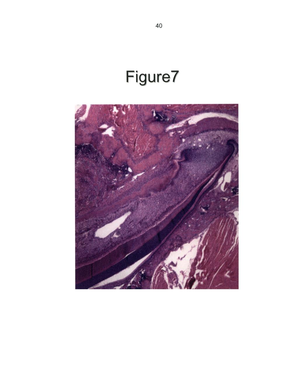
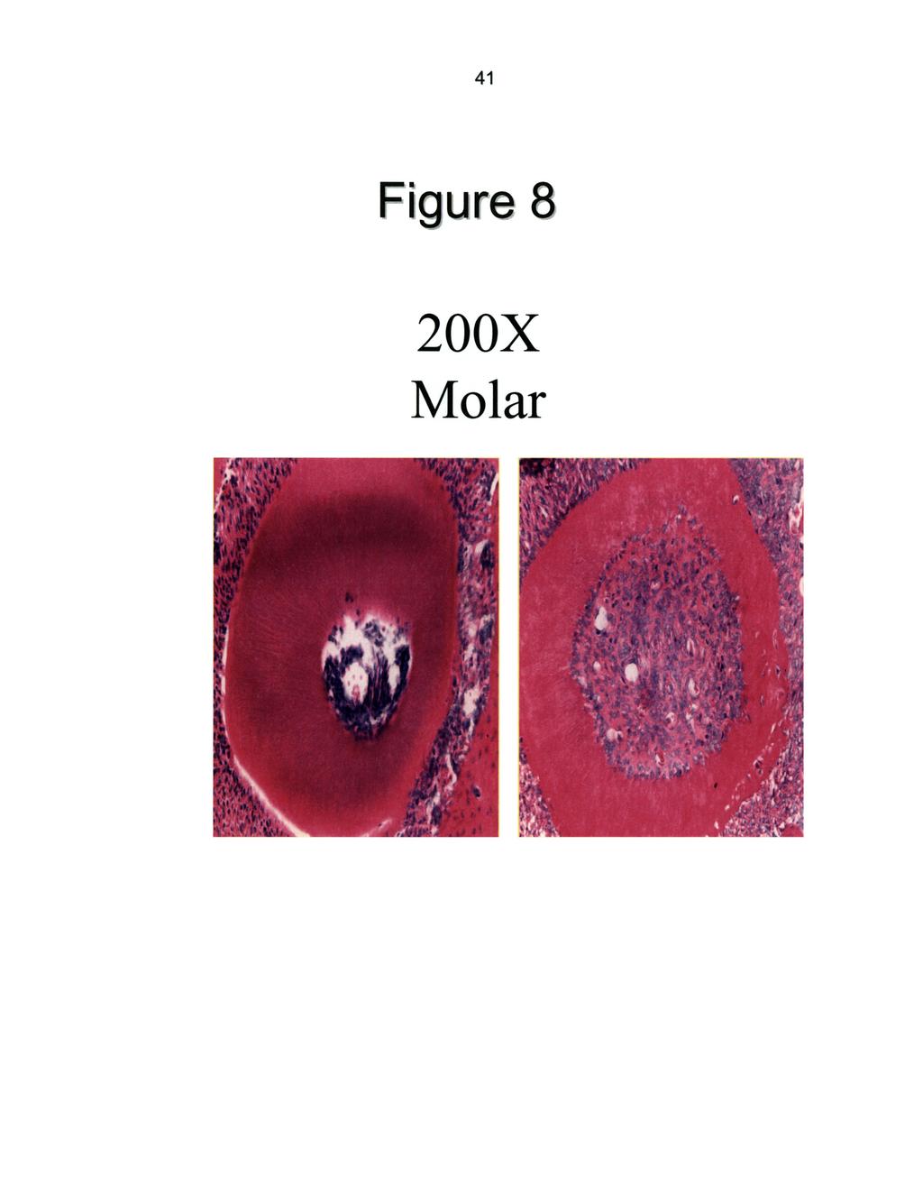
36 29 Figure 7- Histological section of transgenic incisors. Overall view of transgenic incisor with thin lingual dentin. Cervical loop is seen at the roots apex. Figure 8: Histological section of wild-type and transgenic molar roots. Dentin of the molar roots seems to be affected in a similar manner to the lingual incisor in transgenic mice. Molars have not been studied as extensively as incisors so only generalizations can be made at this point. Note the poorly organized dentin and lack of discernible tubular structure. Figure 9" Histological section comparing alveolar bone in wild-type and transgenic mice. Wild-type sample demonstrate normal appearing alveolar bone along with healthy dentin. Transgenic sample displays very little alveolar bone, which is consistent with the osteopenia associated with transgene expression at other skeletal sites.
37 30 Figure 10: Wild-type molar histological section. This section provides a good overall view of a normal periodontium. Bone appears to be high quality and present in sufficient amounts. There is no change in the dentin appearance at the cementoenamel junction (CEJ). Figure 11" Transgenic molar histological section. Overall view demonstrates very little supporting alveolar bone. Inter-radicular bone is nearly absent. Coronal dentin appears to be healthy, however notice a change at the CEJ. A distinct line of demarcation exists at CEJ were once enamel ends the dentin appears to be poor quality. Here, the radicular dentin is also thin when compared to the coronal dentin. Figure 12: Micro-computed tomography analysis. Anatomical landmarks of enamel, dentin, pulp, and bone are show. Linear and area measurements for each region is shown. Figure 13" Micro-computed tomography analysis of wild-type and transgenic mouse incisors. Samples were all sectioned and data collected from the first optical section containing alveolar bone. Enamel and dentin appear to uniform in thickness for the wild-type samples. Enamel appears normal while the labial dentin for the transgenic samples is slightly large. However, the lingual dentin is consistently decreased in thickness for the transgenic mice.
38 31 Figure 14- Graph of linear measurements for line Graph depicting statistical data for linear measurements comparing the thickness in enamel along with labial and lingual dentin in wild-type and transgenic mice from line Significant changes were noted as an increase in labial and decrease lingual dentin thickness in transgenic mouse incisors. Figure 15" Graph of area data for line Graph depicting statistical data for area measurements of enamel and dentin in wild-type and transgenic mice from line Significant changes were noted for a reduction in overall dentin area for transgenic mice incisors. Figure 16: Graph of linear measurements for line 65. Graph depicting statistical data for linear measurements comparing the thickness in enamel along with labial and lingual dentin in wild-type and transgenic mice from line 65. Significant changes were noted as a decrease in the thickness for lingual side dentin in transgenic mice incisors.
39 32 Figure 17: Graph of area data for line 65. Graph depicting statistical data for area measurements of enamel and dentin in wild-type and transgenic mice from line 65. Significant changes were noted for a reduction in overall dentin area for transgenic mice incisors. Figure 18: Northern blot analysis for transgenic mouse mandible. DSPP expression appears to be decreased slightly. BSP is slightly elevated in transgenic samples; while OC expression is decreased. Figure 19: Low power micrograph localizing transgene expression in wild-type and transgenic mice. Immunological staining for the wild-type shows no expression of the transgene. The transgenic sample shows osteoblasts and odontoblasts both staining positive for transgene expression. Figure 20: Low power micrograph of transgenic mouse incisor. Transgenic sample showing low power magnification to depict large area of incisor for view. Note enamel, dentin, pulp chamber, alveolar bone, and cervical loop
40 33 Figure 21" High power micrograph localizing transgene expression. High power magnification of a transgenic sample denotes expression of the transgene in the osteoblasts and odontoblasts.

41
 TG (Line 65)")
42 F gure 2 WT(Line 65) TG (Line 65)
43
44
45
46
47
48
49
50
51

52

53 46 lid-type Figure

54 47 Enamel Labial Dentin Lingual Dentin

55 48 E 0,2- < 0.1- Enamel

56 49 17 Enamel
57 5O 0.4 Enamel Area Dentin Area

58 51! D PP B P EtBr
59
60
61
62 References: 1. Kinoshita, S., S. Akira, and T. Kishimoto, A member ofthe C/EBPfamily, NF-IL6 beta, forms a hetrodimer and transcriptionally synergizes with NF- IL6. Proc Natl Acad Sci U S A, (4): p Osada, S., et al., DNA binding specificity ofthe CCAAT/enhancer-binding protein transcription factorfamily. J Biol Chem, (7): p Williams, S.C., et al., CRP2 (C/EBP beta) contains a bipartite regulatory domain that controls transcriptional activation, DNA binding and cell specificity. Embo J, (13): p Meunier, P., et al., Osteoporosis and the replacement of cell populations of the marrow by adipose tissue. A quantitative study of84 iliac bone biopsies. Clin Orthop, : p Pittenger, M.F., et al., Osteoblastic gene expression during adipogenesis in hematopoietic supporting murine bone marrow stromal cells. J Cell Physiol, : p Yeh, W.C., Cao, Z., Classon, M., McKnight, S.L., Cascade regulation of terminal adipocyte differentiation by three members ofthe C/EBP family of leucine zipper proteins. Genes Dev, (2): p Cao, Z., Umek, R.M., McKnight, S.L., Regulated expressionof three C/EBP isoforms during adipose conversion of3t3-l1 cells. Genes Dev, : p Harrison, J., Kelly, P.L., Pilbeam, C.C., Involvement of CCAAT enhancer binding protein transcription factors in the regulation ofprostaglandin G/H synthase 2 expression by interleukin-1 in osteoblastic MC3T3-E1 cells. J Bone Miner Res, : p Mossey, P.A., The her#ability ofmalocclusion: Part 1-Genetics, priciples and terminology. Br J Orthod, (2): p Mossey, P.A., The heritability ofmalocclusion: Part II-The influence of genetics in malocclusion. Br J Orhtod, (3): p Descombes, P., A liver-enriched transcriptional activator protein, LAP, and a transcriptional inhibitory protein, LIP, are translatedfrom the same RNA. Cell, (3): p Darlington, G.J., Ross, S.E., MacDougald, O.A., The role of C/EBP genes in adipocyte differentiation. J Biol Chem, (46): p Darlington, G.J., Molecular mechanisms of liver development and differentiation. Curr Opin Cell Biol, (6): p Radomska, H.S., et al., CCAAT/enhancer binding protein alpha is a regulatory switch sufficientfor induction ofgranulocytic developmentfrom bipotential myeloidprogenitors. Mol Cell Biol, (7)" p Zhang, D.E., et al., CCAAT enhancer binding protein (C/EBP) and AML1 (CBF alpha2) synergistically activate the macrophage colony-stimulating factor receptor promoter. Mol Cell Biol, (3): p
: p. 1538-52. 17. Lin, F.T., Lane, M.D.")
63 Cao, Z., Umek, R.M., McKnight, S.L., Regulated expression ofthree C/EBP isoforms during adipose conversion of3t3-l1 cells. Genes Dev, (9): p Lin, F.T., Lane, M.D., Antisense CCAAT/enhancer binding protein RNA suppresses coordinate gene expression and triglyceride accumulation during differentiation of3t3-l1 preadipocytes. Genes Dev, i(4): p Lin, F.T., Lane, M.D., CCAAt/enhancer binding protein alpha is sufficient to initiate the 3T3-L1 adipocyte differentiation program. Proc Natl Acad Sci U S A, (19): p Thesleff, I., Sharpe, P., Signalling networks regulating dental development. Mech Dev, i7(2): p Thesleff, I., Mikkola, M., The role ofgrowthfactors in tooth development. Int Rev Cytol, : p Linde, A., Goldberg, M., Dentinogenesis. Crit Rev Oral Biol Med, (5): p Ruch, J.V., Lescot, H., Begue-Kim, C., Odontoblast differentiation. Int J Dev Biol, (1): p Fitzgerald, L.R., Deciduous incisor teeth ofthe mouse. Arch Oral Biol, (3): p Schmitt, R., Ruch, J.V., In vitro synchronization ofembryonic mouse incisor preodontoblasts and preameloblasts: repercussions on terminal differentiation. Eur J Oral Sci, (4): p Sreenath, T., et al., Dentin sialophosphoprotein knockout mouse teeth display widened predentin zone and develop defective dentin mineralization similar to human dentinogenesis imperfecta-iii. J Biol Chem, MacDougall, M., et al., Dentin phosphoprotein and dentin sialoprotein are cleavage products expressedfrom a singletranscript coded by agene on human chromosome 4. Dentin phosphoprotein DNA sequence determination. J Biol Chem, (2): p Feng, J.Q., et al., Genomic organization, chromosomal mapping, and promoter analysis of the mouse dentin sialophosphoprotein (Dspp) gene, which codes for both dentin sialoprotein and dentin phosphoprotein. J Biol Chem, (16): p Thyagarajan, T., et al., Reduced expression ofdentin sialophosphoprotein is associated with dysplastic dentin in mice overexpressing transforming growth factor-beta 1 in teeth. J Biol Chem, i(14)" p
Dentin Formation(Dentinogenesis)
") Lecture four Dr. Wajnaa Oral Histology Dentin Formation(Dentinogenesis) Dentinogenesis begins at the cusp tips after the odontoblasts have differentiated and begin collagen production. Dentinogenesis growth
Lecture four Dr. Wajnaa Oral Histology Dentin Formation(Dentinogenesis) Dentinogenesis begins at the cusp tips after the odontoblasts have differentiated and begin collagen production. Dentinogenesis growth
Fibers and extracellular matrix of hard tissues - Collagen and non-collagen proteins in hard tissues
 Fibers and extracellular matrix of hard tissues - Collagen and non-collagen proteins in hard tissues Dr. Gábor Varga Department of Oral Biology February, 2016 Radiograph of teeth remarkable harmony of
Development of teeth. 5.DM - Pedo
 Development of teeth 5.DM - Pedo Tooth development process of continuous changes in predetermined order starts from dental lamina A band of ectodermal cells growing from the epithelium of the embryonic
Lec. 11 & 12 Dr. Ali H. Murad Dental pulp 1- Coronal pulp
 Lec. 11 & 12 Dr. Ali H. Murad Dental pulp Is the soft connective tissue located in the central portion of each tooth. All pulps have similar morphologic characteristic, such as a soft, gelatinous consistency
06 Tooth Development and Eruption
 + 06 Tooth Development and Eruption Tooth development Root development PDL and alveolar bone development Primary tooth eruption and shedding Permanent tooth eruption Q. Where and how tooth starts to form?
Dental Morphology and Vocabulary
 Dental Morphology and Vocabulary Palate Palate Palate 1 2 Hard Palate Rugae Hard Palate Palate Palate Soft Palate Palate Palate Soft Palate 4 Palate Hard Palate Soft Palate Maxillary Arch (Maxilla) (Uppers)
1. What is the highest and sharpest cusp on the lower first deciduous molar? 2. Which of the following is NOT the correct location of an embrasure?
 1 1. What is the highest and sharpest cusp on the lower first deciduous molar? a. mesiobuccal b. distobuccal c. distolingual d.mesiolingual 2. Which of the following is NOT the correct location of an embrasure?
Morphology of an Anatomic Crown. By: Assistant Professor Dr. Baydaa Ali Al - Rawi
 Morphology of an Anatomic Crown By: Assistant Professor Dr. Baydaa Ali Al - Rawi October 4, 2009 Elevated landmarks Depressed landmarks A) Elevated landmarks : 1. Dental lobe : is one of the primary centers
SalvinOss Xenograft Bone Graft Material In Vivo Testing Summary
 SalvinOss Xenograft Bone Graft Material In Vivo Testing Summary Summary of In Vivo Use Of Bioresorbable Xenograft Bone Graft Materials In The Treatment Of One-Walled Intrabony Defects In A Canine Model
Tooth eruption and movement
 Tooth eruption and movement Dr. Krisztián Nagy Diphydont dentition Deciduous dentition primary dentition Diphydont dentition Permanent dentition secondary dentition Mixed Dentition: Presence of both dentitions
Medical NBDE-II. Dental Board Exams Part I.
 Medical NBDE-II Dental Board Exams Part I http://killexams.com/exam-detail/nbde-ii Question: 149 Anatomically, the term "clinical root" can be defined as which of the following: A. The space in the tooth
Fundamental & Preventive Curvatures of Teeth and Tooth Development. Lecture Three Chapter 15 Continued; Chapter 6 (parts) Dr. Margaret L.
 Dr. Margaret L.") Fundamental & Preventive Curvatures of Teeth and Tooth Development Lecture Three Chapter 15 Continued; Chapter 6 (parts) Dr. Margaret L. Dennis Proximal contact areas Contact areas are on the mesial and
Fundamental & Preventive Curvatures of Teeth and Tooth Development Lecture Three Chapter 15 Continued; Chapter 6 (parts) Dr. Margaret L. Dennis Proximal contact areas Contact areas are on the mesial and
T O O T H A T L A S C O U R S E G U I D E A S S I S T A N T E D I T I O N
 T O O T H A T L A S C O U R S E G U I D E A S S I S T A N T E D I T I O N The information in this guide was prepared by ehuman with contributions from: Cara Miyasaki, RDHEF, MS, Foothill College Kay Murphy,
ODONTOGENESIS- A HIGHLY COMPLEX CELL-CELL INTERACTION PROCESS
 ODONTOGENESIS- A HIGHLY COMPLEX CELL-CELL INTERACTION PROCESS AMBRISH KAUSHAL, MALA KAMBOJ Department of Oral and Maxillofacial Pathology Career Post Graduate Institute of Dental Sciences and Hospital
ANATOMY OF THE PERIODONTIUM. Dr. Fatin Awartani
 ANATOMY OF THE PERIODONTIUM Part II Cementum and Alveolar bone Associate Professor Periodontal division King Saud university Cementum Calcified mesenchymal tissue that forms the outer covering of the anatomic
Periodontal Disease. Radiology of Periodontal Disease. Periodontal Disease. The Role of Radiology in Assessment of Periodontal Disease
 Radiology of Periodontal Disease Steven R. Singer, DDS srs2@columbia.edu 212.305.5674 Periodontal Disease! Includes several disorders of the periodontium! Gingivitis! Marginal Periodontitis! Localized
Ahtiainen et al., http :// /cgi /content /full /jcb /DC1
 Supplemental material JCB Ahtiainen et al., http ://www.jcb.org /cgi /content /full /jcb.201512074 /DC1 THE JOURNAL OF CELL BIOLOGY Figure S1. Distinct distribution of different cell cycle phases in the
Intrusion of Incisors to Facilitate Restoration: The Impact on the Periodontium
 Note: This is a sample Eoster. Your EPoster does not need to use the same format style. For example your title slide does not need to have the title of your EPoster in a box surrounded with a pink border.
(A) PCR primers (arrows) designed to distinguish wild type (P1+P2), targeted (P1+P2) and excised (P1+P3)14-
 PCR primers (arrows) designed to distinguish wild type (P1+P2), targeted (P1+P2) and excised (P1+P3)14-") 1 Supplemental Figure Legends Figure S1. Mammary tumors of ErbB2 KI mice with 14-3-3σ ablation have elevated ErbB2 transcript levels and cell proliferation (A) PCR primers (arrows) designed to distinguish
1 Supplemental Figure Legends Figure S1. Mammary tumors of ErbB2 KI mice with 14-3-3σ ablation have elevated ErbB2 transcript levels and cell proliferation (A) PCR primers (arrows) designed to distinguish
Lecture 2 Maxillary central incisor
 Lecture 2 Maxillary central incisor Generally The deciduous tooth appears in the mouth at 3 18 months of age, with 6 months being the average and is replaced by the permanent tooth around 7 8 years of
Primary Teeth Chapter 18. Dental Anatomy 2016
 Primary Teeth Chapter 18 Dental Anatomy 2016 Primary Teeth - Introduction Synonyms deciduous teeth, baby teeth, temporary teeth, milk teeth. There are 20 primary teeth, designated as A thru T in the Universal
2.79J/3.96J/BE.441/HST522J DENTAL TISSUE REPLACEMENT AND REGENERATION
 Massachusetts Institute of Technology Harvard Medical School Brigham and Women s/massachusetts General Hosp. VA Boston Healthcare System 2.79J/3.96J/BE.441/HST522J DENTAL TISSUE REPLACEMENT AND REGENERATION
CAP STAGE. Ans 1 The following are the stages of tooth development :
 Ans 1 The following are the stages of tooth development : 1. Bud stage 2. Cap stage 3. Bell stage 4. Advanced bell stage 5. Formation of Hertwig s epithelial root sheath BUD STAGE 1. Around the eighth
DENTIN-PULP COMPLEX. Erlina Sih Mahanani. School of Dental sciences Universiti Sains Malaysia. Erlina Sih Mahanani
 DENTIN-PULP COMPLEX School of Dental sciences Universiti Sains Malaysia Introduction Overview anatomy & histology of dentin and pulp. Development of dentin and pulp Structure of dentin and pulp Dentin
The Epithelial-Mesenchymal Interaction Plays a Role in the Maintenance of the Stem Cell Niche of Mouse Incisors via Fgf10 and Fgf9 Signaling
 The Open Biotechnology Journal, 2008, 2, 111-115 111 The Epithelial-Mesenchymal Interaction Plays a Role in the Maintenance of the Stem Cell Niche of Mouse Incisors via Fgf10 and Fgf9 Signaling Tamaki
Oral Embryology and Histology
 Oral Embryology and Histology Chapter 8 Copyright 2018, Elsevier Inc. All Rights Reserved. 1 Learning Objectives Lesson 8.1: Oral Embryology 1. Pronounce, define, and spell the key terms. 2. Define embryology
Index. Note: Page numbers of article titles are in boldface type.
 Index Note: Page numbers of article titles are in boldface type. A Alginate, tooth-shaped, for constructs, encapsulated pulp cells in, 589 590 Antibiotic paste, triple, change in root length and width
#45 Ortho-Tain, Inc PREVENTIVE ERUPTION GUIDANCE -- PREVENTIVE OCCLUSAL DEVELOPMENT
 #45 Ortho-Tain, Inc. 1-800-541-6612 PREVENTIVE ERUPTION GUIDANCE -- PREVENTIVE OCCLUSAL DEVELOPMENT Analysis and Diagnosis of Occlusion: The ideal child of 5 y ears of age that probably has the best chance
DENTIN It a hard vital tissue, surrounds the pulp & underlies the enamel on the crown & the cementum on the roots of the teeth.
 Lec. 7 Dr. Ali H.Murad DENTIN It a hard vital tissue, surrounds the pulp & underlies the enamel on the crown & the cementum on the roots of the teeth. Physical properties: 1-Dentin is pale yellow in color,
The Histology of Dentin
 The Histology of Dentin Pauline Hayes Garrett, D.D.S. Department of Endodontics, Prosthodontics, and Operative Dentistry University of Maryland, Baltimore This material was taken from: Essentials of Oral
BIOLOGICAL FUNCTIONS OF DENTIN SIALOPHSPHOPROTEIN IN MINERALIZED TISSUES. A Dissertation PRIYAM HIMANSHU JANI
 BIOLOGICAL FUNCTIONS OF DENTIN SIALOPHSPHOPROTEIN IN MINERALIZED TISSUES A Dissertation by PRIYAM HIMANSHU JANI Submitted to the Office of Graduate and Professional Studies of Texas A&M University in partial
Dentinogenesis and dentin permeability
 Dentinogenesis and dentin permeability Dr. Gábor Varga February, 2016 Department of Oral Biology Faculty of Dentistry, Semmelweis University Radiograph of teeth dentin is the major component Molar longitudinal
Dental Anatomy and Occlusion
 CHAPTER 53 Dental Anatomy and Occlusion Ma Lou C. Sabino DDS, and Emily G. Smythe, DDS What numerical system is used most commonly in the United States for designating the adult dentition? Pediatric dentition?
6610 NE 181st Street, Suite #1, Kenmore, WA
 660 NE 8st Street, Suite #, Kenmore, WA 9808 www.northshoredentalacademy.com.08.900 READ CHAPTER The Professional Dental Assistant (p.-9) No Key Terms Recall Questions:,,,, and 6 CLASS SYLLABUS DAY READ
Proceedings of the 12th International Congress of the World Equine Veterinary Association WEVA
 www.ivis.org Proceedings of the 12th International Congress of the World Equine Veterinary Association WEVA November 2-5, 2011 Hyderabad, India Reprinted in IVIS with the Permission of WEVA Organizers
Cephalometric Analysis
 Cephalometric Analysis of Maxillary and Mandibular Growth and Dento-Alveolar Change Part III In two previous articles in the PCSO Bulletin s Faculty Files, we discussed the benefits and limitations of
LIST OF ORGANS FOR HISTOPATHOLOGICAL ANALYSIS:!! Neural!!!!!!Respiratory:! Brain : Cerebrum,!!! Lungs and trachea! Olfactory, Cerebellum!!!!Other:!
 LIST OF ORGANS FOR HISTOPATHOLOGICAL ANALYSIS:!! Neural!!!!!!Respiratory:! Brain : Cerebrum,!!! Lungs and trachea! Olfactory, Cerebellum!!!!Other:! Spinal cord and peripheral nerves! Eyes, Inner ear, nasal
and Non-Human MODULE No.17: Structural Variation in Teeth- Human and Non-Human
 SUBJECT Paper No. and Title Module No. and Title Module Tag MODULE No.17: Structural Variation in Teeth- Human and FSC_P11_M17 TABLE OF CONTENTS 1. Learning Outcomes 2. Introduction 3. Structure of Human
Corporate Medical Policy
 Corporate Medical Policy File Name: Origination: Last CAP Review: Next CAP Review: Last Review: orthodontics_for_pediatric_patients 2/2014 10/2017 10/2018 10/2017 Description of Procedure or Service Children
Attachment G. Orthodontic Criteria Index Form Comprehensive D8080. ABBREVIATIONS CRITERIA for Permanent Dentition YES NO
 First Review IL HFS Dental Program Models Second Review Ortho cad Attachment G Orthodontic Criteria Index Form Comprehensive D8080 Ceph Film X-Rays Photos Narrative Patient Name: DOB: ABBREVIATIONS CRITERIA
Hereditary Opalescent Dentin: A Report of Two Cases
 Hereditary Opalescent Dentin: A Report of Two Cases Siddharth Gupta, BDS, MDS; Rahul R. Bhowate, BDS, MDS; Ashok Bhati, BDS, MDS Abstract Aim: The aim of this case report is to present the clinical and
AAO Foundation Awards Final Report
 401 N. Lindbergh Blvd. St. Louis, MO 63141 Tel.: 314.993.1700, #546 Toll Free: 800.424.2841, #546 Fax: 800.708.1364 Cell: 314.283.1983 E-Mail: rhazel@aaortho.org AAO Foundation Awards Final Report In an
Expression of acid base transporters in the kidney collecting duct in Slc2a7 -/-
 Supplemental Material Results. Expression of acid base transporters in the kidney collecting duct in Slc2a7 -/- and Slc2a7 -/- mice. The expression of AE1 in the kidney was examined in Slc26a7 KO mice.
MEDICAL ASSISTANCE BULLETIN COMMONWEALTH OF PENNSYLVANIA DEPARTMENT OF PUBLIC WELFARE
 MEDICAL ASSISTANCE BULLETIN COMMONWEALTH OF PENNSYLVANIA DEPARTMENT OF PUBLIC WELFARE ISSUE DATE EFFECTIVE DATE NUMBER October 21,1996 October 28,1996 03-96-06 SUBJECT BY Information on New Procedures
Institution : College of Dentistry Academic Department : Maxillofacial surgery and Diagnostic sciences
 Institution : College of Dentistry Academic Department : Maxillofacial surgery and Diagnostic sciences [MDS] Program : Bachelor of Dentistry [ BDS ] Course : Oral Biology Course Coordinator : Saleem Shaikh
Radiology. & supporting structures. Lec. 14 Common diseases of teeth Dr. Areej
 Radiology Lec. 14 Common diseases of teeth Dr. Areej & supporting structures A radiograph is only one part of the diagnostic process. Usually one does NOT make a diagnosis solely from a radiograph. A diagnosis
ORAL ANATOMY AND PHYSIOLOGY
 CHAPTER 7 ORAL ANATOMY AND PHYSIOLOGY INTRODUCTION This chapter covers the oral anatomy and physiology of the teeth, the histology of the tissues and supporting structures, and concentrates on the external
Eruption and Shedding of Teeth
 Eruption and Shedding of Teeth Mixed Dentition: Presence of both dentitions Figure from Ten Cate s Oral Histology, Ed., Antonio Nanci, 6 th edition Tooth eruption is the process by which developing teeth
Developing Facial Symmetry Using an Intraoral Device: A Case Report
 Developing Facial Symmetry Using an Intraoral Device: A Case Report by Theodore R. Belfor, D.D.S.; and G. Dave Singh, D.D.Sc., Ph.D., B.D.S. Dr. Theodore Belfor graduated from New York University College
Shell teeth m management from the mixed to the permanent dentition: case report Rosamund Harrison, DMD, MS, MRCD(C) CASE REPORTS.
 CASE REPORTS.") Shell teeth m management from the mixed to the permanent dentition: case report Rosamund Harrison, DMD, MS, MRCD(C) David Kennedy, BDS, MSD, FRCD(C) CASE REPORTS Introduction Relatively few cases of shell
Shell teeth m management from the mixed to the permanent dentition: case report Rosamund Harrison, DMD, MS, MRCD(C) David Kennedy, BDS, MSD, FRCD(C) CASE REPORTS Introduction Relatively few cases of shell
Case Report: Long-Term Outcome of Class II Division 1 Malocclusion Treated with Rapid Palatal Expansion and Cervical Traction
 Case Report Case Report: Long-Term Outcome of Class II Division 1 Malocclusion Treated with Rapid Palatal Expansion and Cervical Traction Roberto M. A. Lima, DDS a ; Anna Leticia Lima, DDS b Abstract:
Osteogenesis imperfecta Report from observation charts
 Orofacial function of persons having Osteogenesis imperfecta Report from observation charts The survey comprises 23 observation charts. Estimated occurrence: 5:100 000 live births. Etiology: The protein
Mesial Step Class I or Class III Dependent upon extent of step seen clinically and patient s growth pattern Refer for early evaluation (by 8 years)
") Orthodontics and Dentofacial Development Overview Development of Dentition Treatment Retention and Relapse Growth of Naso-Maxillary Complex Develops postnatally entirely by intramenbranous ossification
Orthodontics and Dentofacial Development Overview Development of Dentition Treatment Retention and Relapse Growth of Naso-Maxillary Complex Develops postnatally entirely by intramenbranous ossification
Oral Histology. Alveolar bone or process: Functions of alveolar bone: Chemical composition: Development of the alveolar process: Dr.
 Oral Histology Lec.12 Alveolar bone or process: Dr. Nada Al-Ghaban Alveolar bone is a specialized part of the mandibular and maxillary bones that forms the primary support structure for teeth. Although
Course File 243 DDS Physics of Diagnostic Radiology and Oral and Maxillofacial Radiology
 King Saud University College of Dentistry Dept. of Oral Medicine & Diagnostic Sciences Division of Oral & Maxillofacial Radiology Course File 243 DDS Physics of Diagnostic Radiology and Oral and Maxillofacial
SPACE MAINTAINER. Multimedia Health Education. Disclaimer
 Disclaimer This movie is an educational resource only and should not be used to manage your health. All decisions about the management of premature loss of primary teeth and use of space maintainers must
Applied Equine Dental Development
 Published in IVIS with the permission of the AAEP Close this window to return to IVIS Applied Equine Dental Development Kirstie Dacre, BVMS, MSc, Cert EM (Int Med), PhD Author s address: Veterinary Teaching
THE CANADA AND ALBERTA BSE SURVEILLANCE PROGRAM (CABSESP) GUIDELINES FOR AGE VERIFICATION IN CATTLE 1
 GUIDELINES FOR AGE VERIFICATION IN CATTLE 1") AGRICULTURE AND RURAL DEVELOPMENT Food Safety and Animal Health Division, Animal Health Branch 9 th Floor, O.S. Longman Building 6909-116 Street Edmonton, Alberta T6H 4P2 Telephone 780-644-2148 Fax 780-422-5734
AGRICULTURE AND RURAL DEVELOPMENT Food Safety and Animal Health Division, Animal Health Branch 9 th Floor, O.S. Longman Building 6909-116 Street Edmonton, Alberta T6H 4P2 Telephone 780-644-2148 Fax 780-422-5734
CHAPTER 4 ORAL ANATOMY
 CHAPTER 4 ORAL ANATOMY This chapter covers the oral anatomy and physiology of the teeth, the histology of their tissues and supporting structures, and concentrates on the external features of the teeth.
Quantitation of transverse maxillary dimensions using computed tomography: a methodological and reproducibility study
 European Journal of Orthodontics 26 (2004) 209 215 European Journal of Orthodontics vol. 26 no. 2 European Orthodontic Society 2004; all rights reserved. Quantitation of transverse maxillary dimensions
Radiographic assessment of lower third molar prior to surgery: A report of four cases
 Radiographic assessment of lower third molar prior to surgery: A report of four cases V Sreenivas Prasad Department of Oral and Maxillofacial Surgery, College of Dentistry, Gulf Medical University, Ajman,
Odontomes and Odontogenic tumours
 Odontomes and Odontogenic tumours Odontomes Developmental hamartoma Hamartoma: normal tissue in abnormal location Any cells to be neoplastic it must be able to replicate, which is not seen in hamartoma
Structure of an Incisor
 MAMMALIAN TEETH Mammals have different types and shapes of teeth and they are thus termed Heterodonts. Those which have teeth of the same size and shapes are termed as Homodonts. In mammals teeth consist
FRACTURES AND LUXATIONS OF PERMANENT TEETH
 FRACTURES AND LUXATIONS OF PERMANENT TEETH 1. Treatment guidelines and alveolar bone Followup Procedures INFRACTION Clinical findings Radiographic findings Treatment Follow-Up Favorable Outcome Unfavorable
The Premolars. Chapter 17 Permanent Posterior Teeth (p )
") The Premolars Chapter 17 Permanent Posterior Teeth (p. 230-244) General Information Function: u Hold and grind food u Work with molars in mastication. u Even without molars one may be able to chew well
The Premolars Chapter 17 Permanent Posterior Teeth (p. 230-244) General Information Function: u Hold and grind food u Work with molars in mastication. u Even without molars one may be able to chew well
Role of Paired Box9 (PAX9) (rs ) and Muscle Segment Homeobox1 (MSX1) (581C>T) Gene Polymorphisms in Tooth Agenesis
 (rs ) and Muscle Segment Homeobox1 (MSX1) (581C>T) Gene Polymorphisms in Tooth Agenesis") EC Dental Science Special Issue - 2017 Role of Paired Box9 (PAX9) (rs2073245) and Muscle Segment Homeobox1 (MSX1) (581C>T) Gene Polymorphisms in Tooth Agenesis Research Article Dr. Sonam Sethi 1, Dr. Anmol
EC Dental Science Special Issue - 2017 Role of Paired Box9 (PAX9) (rs2073245) and Muscle Segment Homeobox1 (MSX1) (581C>T) Gene Polymorphisms in Tooth Agenesis Research Article Dr. Sonam Sethi 1, Dr. Anmol
Neutrophils contribute to fracture healing by synthesizing fibronectin+ extracellular matrix rapidly after injury
 Neutrophils contribute to fracture healing by synthesizing fibronectin+ extracellular matrix rapidly after injury Bastian OW, Koenderman L, Alblas J, Leenen LPH, Blokhuis TJ. Neutrophils contribute to
Advanced Probing Techniques
 Module 21 Advanced Probing Techniques MODULE OVERVIEW The clinical periodontal assessment is one of the most important functions performed by dental hygienists. This module begins with a review of the
Central Incisor DR.Ahmed Al-Jobory B.D.S.,M.Sc. Conservative Department
 Dental Anatomy Lecture 3 Central Incisor DR.Ahmed Al-Jobory B.D.S.,M.Sc. Conservative Department The permanent maxillary Incisors Maxillary incisor are four in number. The maxillary central incisor is
Orthodontic treatment of midline diastema related to abnormal frenum attachment - A case series.
 Orthodontic treatment of midline diastema related to abnormal frenum attachment - A case series. Running title: Orthodontic treatment of midline diastema. Dr. Amit Dahiya 1, Dr. Minakshi Rana 2, Dr. Arun
OF LINGUAL ORTHODONTICS
 EUROPEAN SOCIETY OF LINGUAL ORTHODONTICS CANDIDATE NUMBER: KDr. KP. kanarelis CASE NUMBER: 1 Year: 2010 WBLO 01 RESUME OF CASE 1 CASE CATEGORY: ADULT MALOCCLUSION NAME : IOANNIS.G BORN: 03.01.1989 SEX:
Concepts of occlusion Balanced occlusion. Monoplane occlusion. Lingualized occlusion. Figure (10-1)
") Any contact between teeth of opposing dental arches; usually, referring to contact between the occlusal surface. The static relationship between the incising or masticatory surfaces of the maxillary or
Any contact between teeth of opposing dental arches; usually, referring to contact between the occlusal surface. The static relationship between the incising or masticatory surfaces of the maxillary or
KJLO. A Sequential Approach for an Asymmetric Extraction Case in. Lingual Orthodontics. Case Report INTRODUCTION DIAGNOSIS
 KJLO Korean Journal of Lingual Orthodontics Case Report A Sequential Approach for an Asymmetric Extraction Case in Lingual Orthodontics Ji-Sung Jang 1, Kee-Joon Lee 2 1 Dream Orthodontic Clinic, Gimhae,
Erupting Compound Odontome - A case report
 IOSR Journal of Dental and Medical Sciences (IOSR-JDMS) e-issn: 2279-0853, p-issn: 2279-0861.Volume 13, Issue 3 Ver. V. (Mar. 2014), PP 26-30 Erupting Compound Odontome - A case report Dr. Sahana Srinath
Supporting Online Material for
 www.sciencemag.org/cgi/content/full/1171320/dc1 Supporting Online Material for A Frazzled/DCC-Dependent Transcriptional Switch Regulates Midline Axon Guidance Long Yang, David S. Garbe, Greg J. Bashaw*
Scientific Treatment Goals for Oral and Facial Harmony
 Scientific Treatment Goals for Oral and Facial Harmony AAO Lecture May 7, 2013 Philadelphia, PA Will A. Andrews, D.D.S. Optimal oral and facial harmony implies a state of maximum health, function and appearance
CASE REPORT. CBCT-Assisted Treatment of the Failing Long Span Bridge with Staged and Immediate Load Implant Restoration
 Computer Aided Implantology Academy Newsletter - Newsletter 20 - July 2009 CASE REPORT CBCT-Assisted Treatment of the Failing Long Span Bridge with Staged and Immediate Load Implant Restoration Case Report
Periodontal ligament
 Periodontal ligament The periodontium The periodontium includes: The gingiva Cementum Periodontal ligament Alveolar bone Def: The periodontal ligament is the dense fibrous connective tissue that occupies
The eternal tooth germ is formed at the apical end of continuously growing teeth*
 The eternal tooth germ is formed at the apical end of continuously growing teeth* Hayato Ohshima 1, Naohiro Nakasone 1, 2, Emi Hashimoto 1, Hideo Sakai 1, Kuniko Nakakura-Ohshima 3 and Hidemitsu Harada
The treatment of destructive periodontal disease, due to specific periodontopathic
 1 1 INTRODUCTION The treatment of destructive periodontal disease, due to specific periodontopathic bacteria, aims at the regeneration of a periodontal attachment composed of new cementum, alveolar bone
Supporting Information
 Supporting Information Franco et al. 10.1073/pnas.1015557108 SI Materials and Methods Drug Administration. PD352901 was dissolved in 0.5% (wt/vol) hydroxyl-propyl-methylcellulose, 0.2% (vol/vol) Tween
Development of occlusion
 Development of occlusion The development of dentition is an important part of craniofacial growth as the formation, eruption, exfoliation and exchange of teeth take place during this period. Term occlusion
Tooth Variations. Suruedee Chinthakanan
 Tooth Variations Suruedee Chinthakanan Tooth variations Dental anomalies Cause : hereditary factor Developmental disturbances of teeth www.ectodermaldysplsia.org Tooth variations Enamel is formed from
The information in this guide was prepared by ehuman with contributions from Dr. Eric Herbranson, Dr. Charles Goodacre, and Dr. Robert Hasel.
 The information in this guide was prepared by ehuman with contributions from Dr. Eric Herbranson, Dr. Charles Goodacre, and Dr. Robert Hasel. 2013 ehuman, Inc. 47865 Fremont Blvd. Fremont, CA 94538 Produced
Alveolar Growth in Japanese Infants: A Comparison between Now and 40 Years ago
 Bull Tokyo Dent Coll (2017) 58(1): 9 18 Original Article doi:10.2209/tdcpublication.2016-0500 Alveolar Growth in Japanese Infants: A Comparison between Now and 40 Years ago Hiroki Imai 1), Tetsuhide Makiguchi
Endodontic Considerations of Equine Incisor and Canine Teeth
 Published in IVIS with the permission of the AAEP Close this window to return to IVIS Endodontic Considerations of Equine Incisor and Canine Teeth David O. Klugh DVM, FAVD/Equine Author s address: Columbia
A Method for Rapid Demineralization of Teeth and Bones
 The Open Dentistry Journal, 2010, 4, 223-229 223 A Method for Rapid Demineralization of Teeth and Bones Open Access Andrew Cho 1, Shigeki Suzuki 1, Junko Hatakeyama 1, Naoto Haruyama 1, and Ashok B. Kulkarni
Arrangement of the artificial teeth:
 Lecture Prosthodontic Dr. Osama Arrangement of the artificial teeth: It s the placement of the teeth on a denture with definite objective in mind or it s the setting of teeth on temporary bases. Rules
Applications in Dermatology, Dentistry and LASIK Eye Surgery using LASERs
 Applications in Dermatology, Dentistry and LASIK Eye Surgery using LASERs http://www.medispainstitute.com/menu_laser_tattoo.html http://www.life123.com/bm.pix/bigstockphoto_close_up_of_eye_surgery_catar_2264267.s600x600.jpg
Monday Morning Pearls of Practice by Bobby Baig
 Dec 19, 2016 Monday Morning Pearls of Practice by Bobby Baig baig@buildyoursmile.com Prosthodontic Associates 2300 Yonge St, suite 905 Toronto, M4P1E4 www.buildyoursmile.com CBCT and Implant Dentistry:
Dental Anatomy High Yield Notes. **Atleast 35 questions comes from these areas of old lectures**
 Dental Anatomy High Yield Notes **Atleast 35 questions comes from these areas of old lectures** This review notes compiled and prepared by my sister for her own study, as a last day review session for
Biology. Dr. Khalida Ibrahim
 Biology Dr. Khalida Ibrahim BONE TISSUE Bone tissue is a specialized form of connective tissue and is the main element of the skeletal tissues. It is composed of cells and an extracellular matrix in which
The morphological studies of root r maxillary primary canines and their Title the position of successive permanen Micro-CT
 The morphological studies of root r maxillary primary canines and their Title the position of successive permanen Micro-CT Author(s) Saka, H; Koyama, T; Tamatsu, Y; Usa Alternative Journal Pediatric dental
You know you would like to stop swearing at the computer after each shot. Troubleshooting oral radiography
 You know you would like to stop swearing at the computer after each shot Troubleshooting oral radiography Goals of oral radiology Achieve diagnostic images of the teeth and surrounding bone. Images should
Gentle-Jumper- Non-compliance Class II corrector
 15 CASE REPORT Gentle-Jumper- Non-compliance Class II corrector Amit Prakash 1,O.P.Mehta 2, Kshitij Gupta 3 Swapnil Pandey 4 Deep Kumar Suryawanshi 4 1 Senior lecturer Bhopal - INDIA 2 Professor Bhopal
TissueSurgeon Hard Tissue Applications
 TissueSurgeon Hard Tissue Applications Laser Based Histology Hard Tissue with TissueSurgeon TissueSurgeon is an excellent tool for easy, fast and flexible sectioning of hard tissue. Native hard tissue
SUPPLEMENTARY INFORMATION
 doi:10.1038/nature10495 WWW.NATURE.COM/NATURE 1 2 WWW.NATURE.COM/NATURE WWW.NATURE.COM/NATURE 3 4 WWW.NATURE.COM/NATURE WWW.NATURE.COM/NATURE 5 6 WWW.NATURE.COM/NATURE WWW.NATURE.COM/NATURE 7 8 WWW.NATURE.COM/NATURE
Index. Note: Page numbers of article titles are in boldface type.
 Index Note: Page numbers of article titles are in boldface type. A Adult performance horse dental care in, 505 519 bit seat reduction in, 514 515 canine teeth disorders, 515 diagnostic nerve blocks in,
Case Report Typical Radiographic Findings of Dentin Dysplasia Type 1b with Dental Fluorosis
 Case Reports in Dentistry Volume 2013, Article ID 902861, 4 pages http://dx.doi.org/10.1155/2013/902861 Case Report Typical Radiographic Findings of Dentin Dysplasia Type 1b with Dental Fluorosis S. Venkata
3D visualization and quantification of bone and teeth mineralization for the study of osteo/dentinogenesis in mice models
 3D visualization and quantification of bone and teeth mineralization for the study of osteo/dentinogenesis in mice models Arnaud Marchadier, Catherine Vidal, Sylvain Ordureau, Roger Lédée, Christophe Léger,