Central Calcifying Epithelial Odontogenic Tumour in the Posterior Maxilla: A Case
|
|
|
- Kathleen Watkins
- 6 years ago
- Views:
Transcription
1 Received Date : 08-Sep-2015 Accepted Date : 25-Oct-2015 Article type Title Page: : case report (ADJ) Central Calcifying Epithelial Odontogenic Tumour in the Posterior Maxilla: A Case Report Authors: Dr Alyssa Zhang,# Dr Sarah Y Chaw,# Dr Anna A Talacko, Dr William J Besly, Associate Professor Neil W Savage, and Professor Paul A Monsour # The first two authors contributed equally to this work. Authors affiliation: School of Dentistry The University of Queensland 288 Herston Road Corner Bramston Terrace & Herston Road Herston QLD 4006 This article has been accepted for publication and undergone full peer review but has not been through the copyediting, typesetting, pagination and proofreading process, which may lead to differences between this version and the Version of Record. Please cite this article as doi: /adj.12384
2 Address for correspondence: Dr Alyssa Zhang and Dr Sarah Y Chaw School of Dentistry The University of Queensland 288 Herston Road Corner Bramston Terrace & Herston Road Herston QLD s: alyssa.zhang@uqconnect.edu.au and sarah.chaw@uqconnect.edu.au Phone: (07) and (07) Short running title: Central CEOT presenting in the posterior maxilla Abbreviations and acronyms: AOT = adenomatoid odontogenic tumour; CBCT = cone beam computed tomography; CCOT = calcifying cystic odontogenic tumour; CEOT = calcifying epithelial odontogenic tumour; COF = central ossifying fibroma; CT = conventional computed tomography; FOV = field of view
3 ABSTRACT: The calcifying epithelial odontogenic tumour (CEOT) or Pindborg tumour is a rare, benign odontogenic tumour. CEOT is usually asymptomatic and an incidental radiological finding, often presenting as a mandibular radiolucency with flecks of calcific material. We report an unusual case of CEOT in the left posterior maxilla of a 46-year-old male that was associated with an unerupted tooth. The tumour in this case caused non-specific sinus symptoms and appeared radiographically similar to an odontoma or ossifying fibroma due to its dense calcific contents. Diagnosis was confirmed histologically following surgical removal of the lesion, which showed classic CEOT histomorphology. We report this case to highlight the unusual clinico-radiologic presentation and illustrate the diagnostic difficulties that can occur with radiolucent and/or radiopaque lesions in the jaws. Introduction: The calcifying epithelial odontogenic tumour (CEOT), or Pindborg tumour, is an uncommon slowlygrowing, expansile tumour composed of odontogenic epithelium without odontogenic mesenchyme. 1, 2 It was first classified as a separate entity by J. J. Pindborg in 1955, but had been reported previously under other pseudonyms. 3 CEOT accounts for roughly one per cent of all odontogenic tumours, has a wide age distribution with a mean of 43.5 years, and has no gender or ethnic predilection. 1, 4, 5 CEOT usually presents as an asymptomatic benign tumour; however, there may be swelling and non-specific pain depending on lesion size and relationship with neighbouring structures, such as the maxillary sinuses. Limited cases of malignant transformation have also been reported. 5-8
4 CEOT may be classified as intra-osseous (central) or extra-osseous (peripheral). 1 Most commonly, it presents as an intra-osseous lesion in the mandibular premolar/molar region. The central lesions are larger and more aggressive than the peripheral lesions, and can grow up to four centimetres in diameter. 2, 9 Peripheral CEOT is comparatively smaller (less than two centimetres in diameter), more localised, and often involves the anterior gingiva, which may lead to misdiagnosis as other epulides. 2, 9 Additionally, peripheral CEOTs have lower recurrence rates due to their less aggressive nature and also their clinical visibility that allows for earlier diagnosis. 2, 9 Radiologically, central CEOT is often described as a pericoronal lesion with the classical feature of radiopaque flecks around the crown of an embedded tooth. 5 However, the radiological features of CEOT can vary greatly and range from a uniform radiolucency to a mixed radiopaque lesion, with either poorly-defined or distinct margins, and may be associated with an unerupted tooth. 2, 5 As the lesion enlarges, it may change from a unilocular to a more multilocular appearance with honey-comb features. 4 As an adjunct to conventional radiology, important three-dimensional information about the lesion can be attained from conventional computed tomography (CT) and cone beam computed tomography (CBCT) scans regarding the true lesion size, the pattern of growth, the presence of calcifications, and the relationship to adjacent structures. 10 Definitive diagnosis of CEOT is based on histological assessment, which in most cases is very 1, 11 distinctive of the tumour. The histopathological features include: 1) Sheets, cords or nests of polyhedral epithelial tumour cells displaying nuclear pleomorphism and eosinophilic cytoplasm, without abnormal mitoses, in mature fibrous connective tissue;
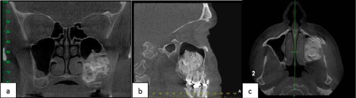
5 2) Acellular homogenous eosinophilic hyaline material containing amyloid, confirmed by Congo Red positivity; 3) Dystrophic calcification within the hyaline material, often arranged in concentric rings (Liesegang rings). We report a case of a large central CEOT occurring in the left posterior maxilla showing an atypical radiologic presentation but with classic CEOT histology. Case Report: A 46-year-old male presented to a dental practitioner reporting non-specific, mild left sinus symptoms. The patient also reported an awareness of left jaw expansion and restriction in jaw movements over the previous 12 month period. A panoramic radiograph (Fig 1) revealed large mixed radiopaque lesion in the left posterior maxilla, measuring approximately 5 x 4 cm. The margins were well-defined and corticated, extending antero-posteriorly from the region of the tooth 25 to the distal margin of the maxillary tuberosity, and infero-superiorly from the apices of the upper left molars to the orbital floor. Further assessment with CBCT (Fig 2) revealed a large, heterogeneous, expansile lesion in the left maxilla, with displacement and elevation of the intact sinus floor. An unerupted molar was displaced superiorly by the mass, but was not grossly eroded. The radiological presentation suggested a benign and non-aggressive process, and an initial differential diagnosis of a complex odontoma or possibly a central ossifying fibroma was made.
6 An excisional biopsy of the lesion was undertaken via an intra-oral approach, and closure of the resultant oro-antral communication was achieved with a buccal mucosal and fat advancement flap. The specimen, comprising of the excised lesion along with the upper left second molar and the embedded tooth, was sent for histopathological assessment. The macroscopic specimen comprised an aggregate of yellow and dark tan, gritty and bony tissue measuring 80 x 60 mm by up to 10 mm, along with two molar teeth. Microscopic examination (Fig 3) showed masses of irregular dystrophic calcification with areas of concentric ring arrangement which were associated with sheets of epithelium with hyperchromatic and mildly pleomorphic nuclei; there was no evidence of atypical mitotic activity. Some dystrophic calcification and epithelium lay within mature fibrous connective tissue. Sheets of acellular eosinophilic material consistent with amyloid, confirmed by Congo Red staining, were present in areas. Besides the molar teeth, no convincing evidence of dental hard material, such as cementum, dentine or enamel matrix was identified. The fragmented nature of the specimen did not allow assessment of the excision margins. The histopathological features, taken in conjunction with the radiological features, were consistent with a diagnosis of calcifying epithelial odontogenic tumour (CEOT). The six-month post-operative panoramic radiograph (Fig 4) and CBCT (Fig 5) showed sinus wall bone repair at the surgical site. A non-homogenous radiopaque mass with what appeared to be retained tooth structure was noted in the left posterior maxilla in the CBCT dataset indicative of remnants of the primary CEOT which was not clear on the conventional panoramic radiograph.
7 The rate of residual tumour growth is currently under review to determine the timing of further surgery via a LeFort I osteotomy approach to the posterior maxilla. Discussion: Due to its asymptomatic nature, CEOT is often an incidental radiologic finding with widely varying radiological features that can cause diagnostic confusion. Radiologically, it may appear similar to a dentigerous cyst, adenomatoid odontogenic tumour (AOT), calcifying cystic odontogenic tumour (CCOT), variants of ameloblastoma, odontoma, or a central ossifying fibroma. From a radiological perspective, the dentigerous cyst, AOT, CCOT, and ameloblastoma were excluded as differential diagnoses as these lesions were not consistent with the radiographic presentation of the current CEOT. Approximately 60% of central CEOTs are associated with the crown of an unerupted tooth or odontoma and may at times resemble a dentigerous cyst; however this is only in the early stages where the CEOT appears more radiolucent, unlike the densely radiopaque lesion presented here. 2, 4, 5 AOTs and CCOTs can both appear as mixed density lesions, but typically present as radiolucencies with scattered radiopaque foci in the anterior jaws. AOTs are often found in the anterior maxilla of females in the second decade, and CCOTs can occur in the anterior maxilla or mandible and are usually associated with root resorption Similarly, early CEOTs that are radiolucent can also be mistaken for ameloblastomas, although ameloblastomas are commonly located in the posterior mandible, and are associated with root resorption and cortical destruction. 15, 16 Due to the intense radiopacity exhibited in the current lesion, an AOT, CCOT and ameloblastoma were radiologically excluded from the differential diagnosis. Importantly though, CEOT can occur in conjunction with a dentigerous cyst, AOT, or an
8 ameloblastoma as a hybrid lesion, which may be noted incidentally on histological examination. 2, Our differential diagnoses for the current case comprised a complex odontoma and central ossifying fibroma (COF). The complex odontoma classically presents as a haphazard radiopaque mass surrounded by a radiolucent halo. 20 Additionally, it may be associated with impacted teeth (often in the molar regions), can cause mild expansion, and is often detected in the second decade. 20, 21 The COF, on the other hand, often presents as a well-defined mixed density lesion in the mandibular molar regions. It causes expansion, and is often diagnosed in females in the third to fourth decades. 22 In the current case, due to the high level of tumour maturity, there was considerable expansion and extensive calcification in the lesion that gave it a densely radiopaque appearance, which exceeded that usually seen in a CEOT and was more suggestive of a complex odontoma or mature central ossifying fibroma. 3-5 Despite the atypical radiological presentation, the lesion was histopathologically diagnostic of CEOT, displaying the distinctive histological features as described earlier in this report. CEOTs are thought to originate from the stratum intermedium or reduced enamel epithelium of odontogenic epithelium; hence their association with embedded teeth. 3, 4, 17, 23 Early histologic lesions resembling odontogenic cysts and tumours, including CEOT, have been found within dental follicles of third molars, and have potential to develop into true odontogenic cysts and tumours. 17, 23, 24 It is therefore possible that the CEOT presented here has developed from the follicle of the unerupted upper left third molar.
9 Both CT and CBCT are useful modalities for three-dimensional assessment of lesions in the dentomaxillofacial region; however there are a number of determining factors for these modalities that affect the imaging choice. CBCT is the modality of choice when optimised assessment of osseous structures is required in the dentomaxillofacial region. 25 Specific design features for the dentomaxillofacial region allow the field of view (FOV) in CBCT machines to be limited. 25 Consequently, for a similar FOV, CBCT produces images of higher resolution while generally using a much lower radiation dose than CT. 25 Many CBCT machines also offer the functional benefit for patients to be orientated in a sitting or standing position in the machine, whilst for all CTs, patients must lie supine within the gantry. 25 On other occasions, CT may be favoured over CBCT, such as for assessment of lesions with suspected soft tissue involvement and CT has the added advantage of much faster scanning times. 25, 26 Based on the factors discussed above, use of CBCT was most appropriate for the current case, as initial presentation of the lesion on the panoramic radiograph warranted assessment of hard tissue pathology in a localised region of the jaw. Treatment for CEOT can range from less aggressive enucleation or curettage to more aggressive hemimandibulectomy or hemimaxillectomy, and is largely dependent on the size and location of the lesion. 2 It has been recommended to treat maxillary lesions more aggressively as they tend to grow faster and are less likely to remain confined. 2 Overall, recurrence rates following surgery range up to 14%, and are usually due to incomplete removal of the tumour. 2 Given the recurrence rate and location of the residual tumour in this case, the area is under close review, with a view to excise the lesion once sufficient healing at the site has occurred to allow for further surgery. To monitor for CEOT recurrence following surgery, a follow-up period of a minimum of 5 years has been recommended. 2
10 Conclusion: CEOT is a rare odontogenic tumour that can attain significant growth in the jaws and is often asymptomatic; however, it may also cause symptoms that mimic dental or sinus pain. It may be confused with radiologically similar lesions such as a dentigerous cyst in its early phases, or an odontoma and central ossifying fibroma in mature lesions. These atypical presentations may lead to CEOT being initially excluded as a differential diagnosis, as seen in this current case. Radiolucent and radiopaque jaw lesions can appear deceptively bland, and be misdiagnosed as less aggressive or less sinister pathology. Three-dimensional imaging can provide valuable clinical information regarding the nature of the lesion and guide its management. Clinicians should be aware of the range of differential diagnoses for odontogenic jaw lesions, and refer as appropriate for diagnosis and management. References: 1. Kramer IRH, Pindborg JJ, Shear M. Histological Classification of Odontogenic Tumours. In: Histological Typing of Odontogenic Tumours. Springer Berlin Heidelberg, 1992: Philipsen HP, Reichart PA. Calcifying epithelial odontogenic tumour: biological profile based on 181 cases from the literature. Oral Oncol 2000;36: Pindborg JJ. The Calcifying Epithelial Odontogenic Tumor. Review of Literature and Report of An Extra-Osseous Case. Acta Odontologica 1966;24: Franklin CD, Pindborg JJ. The calcifying epithelial odontogenic tumor. A review and analysis of 113 cases. Oral Surg Oral Med Oral Pathol 1976;42: Kaplan I, Buchner A, Calderon S, Kaffe I. Radiological and clinical features of calcifying epithelial odontogenic tumour. Dentomaxillofac Radiol 2001;30:22-28.
11 6. Bousdras VA, Bousdras KA, Newman L. Nasal obstruction as the first symptom in a patient with a calcifying epithelial odontogenic tumour (CEOT). Dental update 2009;36: Bridle C, Visram K, Piper K, Ali N. Maxillary calcifying epithelial odontogenic (Pindborg) tumor presenting with abnormal eye signs: case report and literature review. Oral Surg Oral Med Oral Pathol Oral Radiol Endod 2006;102:e Kamath G, Abraham R. Recurrent CEOT of the maxilla. Dent Res J (Isfahan) 2012;9: Patino B, Fernandez-Alba J, Garcia-Rozado A, Martin R, Lopez-Cedrun JL, Sanroman B. Calcifying epithelial odontogenic (pindborg) tumor: a series of 4 distinctive cases and a review of the literature. J Oral Maxillofac Surg 2005;63: Venkateswarlu M, Geetha P, Lakshmi Kavitha N. CT imaging findings of a calcifying epithelial odontogenic tumour. Br J Radiol 2012;85:e Ching AS, Pak MW, Kew J, Metreweli C. CT and MR imaging appearances of an extraosseous calcifying epithelial odontogenic tumor (Pindborg tumor). AJNR Am J Neuroradiol 2000;21: Prakasam M, Tiwari S, Satpathy M, Banda VR. Adenomatoid odontogenic tumour. BMJ Case Rep 2013;2013: 13. Zornosa X, Muller S. Calcifying cystic odontogenic tumor. Head Neck Pathol 2010;4: de Matos FR, Nonaka CFW, Pinto LP, de Souza LB, de Almeida Freitas R. Adenomatoid Odontogenic Tumor: Retrospective Study of 15 Cases with Emphasis on Histopathologic Features. Head and Neck Pathology 2012;6: Bachmann AM, Linfesty RL. Ameloblastoma, Solid/Multicystic Type. Head and Neck Pathology 2009;3: Hsu M-H, Chiang M-L, Chen J-K. Unicystic ameloblastoma. Journal of Dental Sciences 2014;9:
12 17. Azevedo RS, Mosqueda-Taylor A, Carlos R, et al. Calcifying epithelial odontogenic tumor (CEOT): a clinicopathologic and immunohistochemical study and comparison with dental follicles containing CEOT-like areas. Oral Surg Oral Med Oral Pathol Oral Radiol 2013;116: Ismail IM, Al-Talabani NG. Calcifying epithelial odontogenic tumour associated with dentigerous cyst. Int J Oral Maxillofac Surg 1986;15: Seim P, Regezi JA, O'Ryan F. Hybrid ameloblastoma and calcifying epithelial odontogenic tumor: case report. J Oral Maxillofac Surg 2005;63: Amado Cuesta S, Gargallo Albiol J, Berini Aytés L, Gay Escoda C. Review of 61 cases of odontoma. Presentation of an erupted complex odontoma. Medicina oral : órgano oficial de la Sociedad Española de Medicina Oral y de la Academia Iberoamericana de Patología y Medicina Bucal 2003;8: Angiero F, Benedicenti S, Parker S, et al. Clinical and Surgical Management of Odontoma. PHOTOMEDICINE AND LASER SURGERY 2014;32: Chang C-C, Chang JY-F, Hung H-Y, et al. Central Ossifying Fibroma: A Clinicopathologic Study of 28 Cases. Journal of the Formosan Medical Association 2008;107: Mopsik ER, Gabriel SA. Calcifying epithelial odontogenic tumor (Pindborg tumor). Report of two cases. Oral Surg Oral Med Oral Pathol 1971;32: Ficarra G, Hansen LS, Stiesmeyer EH. Intramural calcifying epithelial odontogenic tumor. Int J Oral Maxillofac Surg 1987;16: Scarfe WC, Farman AG. What is Cone-Beam CT and How Does it Work? Dental Clinics of North America 2008;52: Hofmann E, Schmid M, Lell M, Hirschfelder U. Cone beam computed tomography and lowdose multislice computed tomography in orthodontics and dentistry: A comparative evaluation on image quality and radiation exposure. Journal of Orofacial Orthopedics / Fortschritte der Kieferorthopädie 2014;75:


13


14
Peripheral Odontogenic Fibroma: A rare case report
 Annals of Dental Research (2013) Vol 3 (1): 10-14 HATAM Publishers: All Rights Reserved Annals of Dental Research www.hgpub.com www.adres.yolasite.com Case Report Peripheral Odontogenic Fibroma: A rare
Disclosure. Educational Objectives. Terminology. Odontogenic Cysts. Terminology
 Disclosure Lisa J. Koenig BChD, DDS, MS Professor & Program Director, Oral Medicine and Oral Radiology Marquette University School of Dentistry Consultant to Soredex for the Scanora 3D and 3Dx Author/Editor
JOURNAL TAOMFR Mandiblular Ameloblastic fibro-odontoma
 Ameloblastic Fibro-Odontoma of the Mandible with Emphasis on Evaluation Using Cone Beam Computed Tomography Jing-Yi Chen 1, Frank Lei 2, Yu-Fong Chen 3,4 * Divisions of 1 Oral Pathology, 2 Periodotology,
Proceedings of the 36th World Small Animal Veterinary Congress WSAVA
 www.ivis.org Proceedings of the 36th World Small Animal Veterinary Congress WSAVA Oct. 14-17, 2011 Jeju, Korea Next Congress: http://www.ivis.org October 14(Fri) ~ 17(Mon) 2011 ICC Jeju, Korea 2011 WSAVA
Central odontogenic fibroma of the mandible: a case report
 457 Journal of Oral Science, Vol. 51, No. 3, 457-461, 2009 Case Report Central odontogenic fibroma of the mandible: a case report Ioanna Daskala 1), Demos Kalyvas 2), Markos Kolokoudias 2), Dimitris Vlachodimitropoulos
A Radiographic technique for differentiating enamel and dentin in odontogenic tumors
 ISPUB.COM The Internet Journal of Radiology Volume 12 Number 1 A Radiographic technique for differentiating enamel and dentin in odontogenic tumors D Shetty, A Urs, R Kaur Citation D Shetty, A Urs, R Kaur.
PACIFIC JOURNAL OF MEDICAL SCIENCES ISSN:
 PACIFIC JOURNAL OF MEDICAL SCIENCES {Formerly: Medical Sciences Bulletin} ISSN: 2072 1625 Pac. J. Med. Sci. (PJMS) www.pacjmedsci.com. Email: pacjmedsci@gmail.com. ADENOMATOID ODONTOGENIC TUMOR WITH RARE
SQUAMOUS ODONTOGENIC TUMOUR: REPORT OF FIVE CASES FROM NIGERIA AND REVIEW OF LITERATURE
 African Journal of Oral Health Volume 3 Numbers 1&2, 2006:1-5 REFEREED ARTICLE SQUAMOUS ODONTOGENIC TUMOUR: REPORT OF FIVE CASES FROM NIGERIA AND REVIEW OF LITERATURE Adebiyi K.E., Odukoya O., Taiwo, E.O.
An unusual site of Adenomatoid Odontogenic Tumor: A rare case report
 J. Int Oral Health 2010 Case Report All right reserved An unusual site of Adenomatoid Odontogenic Tumor: A rare case report Sapna Panjwani*, Anjana Bagewadi**, Vaishali Keluskar*** *Post Graduate Student
Large Dentigerous Cyst
 Volume 16.2.1 Feb 2016 This Lecture Series qualifies for 0.5 Informal CPD Learning Hours Large Dentigerous Cyst By Dr Hassem Geha A 55 year-old male presented with a painless swelling in the right mandible.
Erupted odontomas: a report of two unusual cases
 ISSN: Printed version: 1806-7727 Electronic version: 1984-5685 RSBO. 2012 Apr-Jun;9(2):199-203 Case Report Article Erupted odontomas: a report of two unusual cases Santosh Patil¹ Farzan Rahman² Shoaib
Erupting Compound Odontome - A case report
 IOSR Journal of Dental and Medical Sciences (IOSR-JDMS) e-issn: 2279-0853, p-issn: 2279-0861.Volume 13, Issue 3 Ver. V. (Mar. 2014), PP 26-30 Erupting Compound Odontome - A case report Dr. Sahana Srinath
Cone Beam Computed Tomography Findings in Calcifying Cystic Odontogenic Tumor Associated with Odontome: A Case Report
 Phulambrikar T., et al. Dent Shiraz Univ Med Sci., December 2015; 16(4): 374-379. Case Report Cone Beam Computed Tomography Findings in Calcifying Cystic Odontogenic Tumor Associated with Odontome: A Case
Adenomatoid Odontogenic Tumour: A Case Report and Review of Literature Sunil S, Nivia M, Devi Gopakumar
 International Journal of Oral & Maxillofacial Pathology. 2012;3(4):52-56 ISSN 2231 2250 Available online at http://www.journalgateway.com or www.ijomp.org Case Report Adenomatoid Odontogenic Tumour: A
Autologous Bone Augmentation in Combination with an Ameloblastoma in the Maxillary Region- A Case Report?
 Archives of Clinical and Medical Case Reports doi: 10.26502/acmcr.96550023 Volume 2, Issue 2 Case Report Autologous Bone Augmentation in Combination with an Ameloblastoma in the Maxillary Region- A Case
Ameloblastomatous Gorlin s cyst
 319 Journal of Oral Science, Vol. 49, No. 4, 319-323, 2007 Case Report Ameloblastomatous Gorlin s cyst Mala Kamboj 1) and Manish Juneja 2) 1) Department of Oral Pathology and Microbiology, U.P. King George
Impeded Eruption of Mandibular Canine
 10.5005/jp-journals-10024-1434 Case Report Gauri S Lele, Darshan Modi Abstract Odontome, tumor of odontogenic origin, is associated with disturbances in the eruption of teeth such as impaction, delayed
Calcifying Odontogenic Cyst with Complex Odontoma: Histological and Immunohistochemical Features
 Case Report Calcifying Odontogenic Cyst with Complex Odontoma: Histological and Immunohistochemical Features Nooshin Mohtasham 1, Amin Rahpeyma 2, Saeedeh Khajeh Ahmadi 3, Mohsen Merati 4 1 Oral and Maxillofacial
A clinicopathologic study on calcifying epithelial odontogenic tumor: with special reference to Langerhans cell variant
 Chen et al. Diagnostic Pathology 2014, 9:37 RESEARCH Open Access A clinicopathologic study on calcifying epithelial odontogenic tumor: with special reference to Langerhans cell variant Yan Chen 1, Ting-Ting
Maxilla and mandible benign lesions: Radiologic Findings and Differential Diagnosis in CT
 Maxilla and mandible benign lesions: Radiologic Findings and Differential Diagnosis in CT Poster No.: C-0964 Congress: ECR 2012 Type: Scientific Exhibit Authors: N. Lopez 1, E. Marcos Naranjo 2, M. D.
Case Report Ameloblastic Fibroodontoma of the Mandible with Normal Karyotype in a Pediatric Patient
 Case Reports in Dentistry Volume 2012, Article ID 969687, 4 pages doi:10.1155/2012/969687 Case Report Ameloblastic Fibroodontoma of the Mandible with Normal Karyotype in a Pediatric Patient Esther Manor,
Cemento-Ossifying Fibroma A Radiographic Diagnostic Dilemma
 Case Report Cemento-Ossifying Fibroma A Radiographic Diagnostic Dilemma Zahra Dalili 1, Somayeh Nemati 2, Fatemeh Shahsavari 3 1 Associate Professor, Department of Maxillofacial Radiology, Guilan University
Odontomes and Odontogenic tumours
 Odontomes and Odontogenic tumours Odontomes Developmental hamartoma Hamartoma: normal tissue in abnormal location Any cells to be neoplastic it must be able to replicate, which is not seen in hamartoma
高雄醫學大學 口腔醫學院 口腔病理影像科 牙科 X 光影像判讀 教學範例
 高雄醫學大學 口腔醫學院 口腔病理影像科 牙科 X 光影像判讀 教學範例 Content Image No. 001 Dentigerous cyst over left upper embedded canine--------------------- 頁 1 Image No. 002---------------------------------------------------------------
Med. J. Malaysia Vol. 42 No. 4 December 1987 CLINICAL STATISTICS OF THE ADENOMATOID ODONTOGENIC TUMOUR IN MALAYSIA
 Med. J. Malaysia Vol. 42 No. 4 December 1987 CLINICAL STATISTICS OF THE ADENOMATOID ODONTOGENIC TUMOUR IN MALAYSIA (1968-1986) CH SIAR* 80S (Mal), MSc (Lond), FDS RCPS (Glasg) K H NG* 80S (Mal), MSc (Lond),
Adenomatoid odontogenic tumor: Case series of 14 with wide range of clinical presentation
 Journal section: Oral Medicine and Pathology Publication Types: Research doi:10.4317/jced.54216 http://dx.doi.org/10.4317/jced.54216 : Case series of 14 with wide range of clinical presentation Fatima
Glandular Odontogenic Cyst Coexisting with a Dentigerous Cyst: Case Report
 SmyrnaMed Case 2018;2(1): 1-5 ISSN (Online): 2564-6869 www.smyrnamed.com Glandular Odontogenic Cyst Coexisting with a Dentigerous Cyst: Case Report Assist.Prof.Dr. Serap Keskin Tunç 1, Dt. Erkan Feslihan
Orthokeratinized Odontogenic Cyst: A Rarity
 aijoc AIJOC Case Report 1 Heena Sonawane, 2 Freny R Karjodkar, 3 Kaustubh Sansare, 4 Nimish Prakash ABSTRACT Orthokeratinized odontogenic cyst (OOC) was first identified as the rare variant of keratocystic
CENTRAL GIANT CELL GRANULOMA PRESENTING AS UNILOCULAR RADIOLUCENCY IN POSTERIOR MANDIBLE A CASE REPORT
 IJCRR Section: Healthcare Sci. Journal Impact Factor 4.016 Case Report CENTRAL GIANT CELL GRANULOMA PRESENTING AS UNILOCULAR RADIOLUCENCY IN POSTERIOR MANDIBLE A CASE REPORT S. Aruleena Shaminey 1, G.
AMELOBLASTIC FIBROMA: A RARE CASE REPORT
 Case Report International Journal of Dental and Health Sciences Volume 04, Issue 03 AMELOBLASTIC FIBROMA: A RARE CASE REPORT Namratha Patil 1 1.Sr lecturer, dept of oral medicine and radiology, KAHES VK
International Journal of Scientific Research and Innovative Technology ISSN: Vol. 4 No. 4; April 2017
 Frequency of Lesions Associated with Impacted Teeth in Patients Referring to the Department of Oral Pathology, Tabriz Faculty of Dentistry from 2004 to 2013 and Its Relationship with Age, Gender, Location
Case Report Intraosseous Follicular Adenomatoid Odontogenic Tumour A Case Report
 International Dentistry Volume 2009, Article ID 597483, 4 pages doi:10.1155/2009/597483 Case eport Intraosseous Follicular Adenomatoid Odontogenic Tumour A Case eport Farhan Durrani 1 and oyana Singh 2
Case Report An Extrafollicular Adenomatoid Odontogenic Tumor Mimicking a Periapical Cyst
 Hindawi Case Reports in Radiology Volume 2018, Article ID 6987050, 5 pages https://doi.org/10.1155/2018/6987050 Case Report An Extrafollicular Adenomatoid Odontogenic Tumor Mimicking a Periapical Cyst
Complex Odontoma in Both the Jaws: A Rare Case Report
 10.5005/jp-journals-10026-1013 Adit Srivastava et al CASE REPORT Complex Odontoma in Both the Jaws: A Rare Case Report Adit Srivastava, AG Annaji, Sanjay B Nyamati, Govind Singh, GC Shivakumar, S Sahana
Successful Conservative Surgical Treatment of Ameloblastic Fibroma in the Posterior Maxilla : A Case Report
 http://dx.doi.org/10.5933/jkapd.2013.40.4.321 ISSN (print) 1226-8496 Successful Conservative Surgical Treatment of Ameloblastic Fibroma in the Posterior Maxilla : A Case Report Youngeun Lee 1, Hyojung
Ameloblastic fibro-odontoma in a 9-year-old boy
 Okui et al. Stomatological Dis Sci 2018;2:7 DOI: 10.20517/2573-0002.2017.23 Stomatological Disease and Science Case Report Open Access Ameloblastic fibro-odontoma in a 9-year-old boy Tatsuo Okui 1, Soichiro
 This article was published in an Elsevier journal. The attached copy is furnished to the author for non-commercial research and education use, including for instruction at the author s institution, sharing
SURGICAL MANAGEMENT OF A COMPLEX ODONTOMA - A CASE REPORT
 Indian J.Sci.Res. 6(2) : 157-161, 2015 Case Report ISSN : 0976-2876 (Print) ISSN : 2250-0138 (Online) SURGICAL MANAGEMENT OF A COMPLEX ODONTOMA - A CASE REPORT a 1b c d NAVDHA CHAUDHARY, BABITA AHLAWAT,
Adenomatoid odontogenic tumor associated with odontoma: a case report and critical review of the literature
 Gomez et al. Head & Face Medicine 2013, 9:20 HEAD & FACE MEDICINE CASE REPORT Adenomatoid odontogenic tumor associated with odontoma: a case report and critical review of the literature Open Access Ricardo
Annals and Essences of Dentistry
 aedj.2014.6.3.2.4 Central Calcifying Cystic Odontogenic Tumor Of Mandible A Case Report 1 Rajasekhar Gali 2 Madan Mohan Reddy 3 Vandana Raghunath 4 Sajan Anand 1 Associate Professor 2 Reader 3 Professor
A case report of cemento-ossifying fibroma presenting as a mass of. the ethmoid sinus
 Received: 27.3.2010 Accepted: 1.8.2010 Case Report A case report of cemento-ossifying fibroma presenting as a mass of the ethmoid sinus Ali Hekmatnia a, Amirhossein Ghazavi* b, Masih Saboori c, Parvin
Removal of a Complex Odontoma Associated With an Impacted Third Molar
 Removal of a Complex Odontoma Associated With an Impacted Third Molar Authored by Mohammad Hosein Kalantar Motamedi, DDS Upon successful completion of this CE activity 1 CE credit hour may be awarded A
Periapical central giant cell granuloma misdiagnosed as odontogenic cyst
 doi: 10.1111/j.1365-2591.2006.01107.x CLINICAL ARTICLE Periapical central giant cell granuloma misdiagnosed as odontogenic cyst T. Lombardi 1, M. Bischof 1,2, R. Nedir 1,2, D. Vergain 1, C. Galgano 3,
Clinical details: Details of scan: CONE BEAM CT REPORT: Name: H. B. Gender: Reason for referral: Referred by:
 Name: H. B. Gender: Male DOB: 11/12/1950 Age: 64 Date taken: 16/11/2015 Date reported: 19/11/2015 Clinical details: Reason for referral: Referred by: Investigate symptoms related to left TMJ. Reconstructed
Open Access Case Presentation of Concomitant and Contiguous Adenomatoid Odontogenic Tumor and Focal Cemento-Ossifying Dysplasia
 Send Orders for Reprints to reprints@benthamscience.ae 340 The Open Dentistry Journal, 2015, 9, (Suppl 2: M14) 340-345 Open Access Case Presentation of Concomitant and Contiguous Adenomatoid Odontogenic
Problem diagnoses. Current issues in Anatomic pathology. Problem Diagnoses in Tumors of the Oral Cavity 5/29/2009
 Current issues in Anatomic pathology Problem Diagnoses in Tumors of the Oral Cavity Richard Jordan DDS PhD FRCPath Professor of Oral Pathology & Pathology Director, UCSF Oral Pathology Diagnostic Laboratory
Case Report Basal Cell Ameloblastoma of Mandible: A Rare Case Report with Review
 Case Reports in Dentistry Volume 2013, Article ID 187820, 4 pages http://dx.doi.org/10.1155/2013/187820 Case Report Basal Cell Ameloblastoma of Mandible: A Rare Case Report with Review Hemant Shakya, 1
Combined Benign Odontogenic Tumors: CT and MR Findings and Histomorphologic Evaluation
 AJNR Am J Neuroradiol 22:867 872, May 2001 Case Report Combined Benign Odontogenic Tumors: CT and MR Findings and Histomorphologic Evaluation Nadine Martin-Duverneuil, Marie-Hélène Roisin-Chausson, Anthony
Glandular odontogenic cyst associated with ameloblastoma: Case report and review of the literature
 Journal section: Oral Medicine and Pathology Publication Types: Case Report doi:10.4317/jced.53775 http://dx.doi.org/10.4317/jced.53775 : Case report and review of the literature Timothée Cousin 1, Samuel
Incidental finding of dentigerous cyst - a case report
 Case Report Incidental finding of dentigerous cyst - a case report Pulivarthi Sushma 1, Sowbhagya M.B 2, Balaji P 3, Mahesh Kumar T.S 4 1 Postgraduate, 2 Reader, 3 Professor and Head of department, 4 Senior
Unusual transmigration of canines report of two cases in a family
 ISSN: Electronic version: 1984-5685 RSBO. 2014 Jan-Mar;11(1):88-92 Case Report Article Unusual transmigration of canines report of two cases in a family Sulabha A. Narsapur 1 Sameer Choudhari 2 Shrishal
Complex and compound odontomas: Analysis of 69 cases and a rare case of erupted compound odontoma
 Original Article Complex and compound odontomas: Analysis of 69 cases and a rare case of erupted compound odontoma C Bereket, N Çakır Özkan, İ Şener, E Bulut, M Tek 1 Department of Oral and Maxillofacial
An Expansile Large Odontogenic Keratocyst Maxilla: A Case Report.
 RESEARCH AND REVIEWS: JOURNAL OF DENTAL SCIENCES An Expansile Large Odontogenic Keratocyst Maxilla: A Case Report. Nasib Chand Khabra 1, Ish Pandhi 1 *, Kiran DN 2, Sunil Alipuria 1, Bhawna Gulati 1, and
Mucous and ciliated cell metaplasia in epithelial linings of odontogenic inflammatory and developmental cysts
 77 Journal of Oral Science, Vol. 47, No. 2, 77-81, 2005 Original Mucous and ciliated cell metaplasia in epithelial linings of odontogenic inflammatory and developmental cysts Yasunori Takeda, Yuko Oikawa,
Management of a Dentigerous Cyst Associated with Inverted and Fused Mesiodens: A Rare Case Report
 Management of a Dentigerous Cyst Associated with Inverted and Fused Mesiodens: A Rare Case Report Kiran Patel 1, Nishtha Patel 2, Karthik Venkataraghavan 3 1 Sr. Lecturer, Department of Oral & Maxillofacial
Ossifying fibromas of the jaw bone: 20 cases
 (2010) 39, 57 63 2010 The British Institute of Radiology http://dmfr.birjournals.org CASE REPORT Ossifying fibromas of the jaw bone: 20 cases Y Liu 1, M You 1, H Wang*,1, Z Yang 1, J Miao 1, K Shimizutani
Pericoronal radiolucency associated with incomplete crown
 Imaging Science in entistry 2013; 43: 295-301 http://dx.doi.org/10.5624/isd.2013.43.4.295 Pericoronal radiolucency associated with incomplete crown Kyung-Soo Nah 1, * 1 epartment of Oral and Maxillofacial
Dentinogenic ghost cell tumor a rare case report with review of literature
 Case Report Dentinogenic ghost cell tumor a rare case report with review of literature Yash Agrawal 1, Giridhar S. Naidu 1, Ramanpal Singh Makkad 1, Ravleen Nagi 1, Supreet Jain 1, Dilip R. Gadewar 1,
The future of health is digital
 Dated: XX/XX/XXXX Name: XXXXXXXX XXXXXXXXXXX Birth Date: XX/XX/XXXX Date of scan: XX/XX/XXXX Examination of the anatomical volume: The following structures are reviewed and evaluated for bilateral symmetry,
Key words: Third molar, Impacted tooth, Tooth Eruption, Molar, Mandible, Unerupted Tooth.
 JOURNAL OF CASE REPORTS 2014;4(2):286-290 OPG and CBCT Finding s of an Ectopic Third Molar in the Sub-condylar Region Tatu Joy E 1, Farakath Khan 1, Shameel Mohammed 2 From the Department of Oral Medicine
Management of Plexiform Ameloblastoma in a 12 year old female: A Case Report
 Article ID: ISSN 2046-1690 Management of Plexiform Ameloblastoma in a 12 year old female: A Case Report Corresponding Author: Dr. Sheeraz Badal, Senior Lecturer, Dept of Oral & Maxillofacial Surgery, MIDSR,
Adenomatoid odontogenic tumor hamartoma or true neoplasm: a case report
 155 Journal of Oral Science, Vol. 51, No. 1, 155-159, 2009 Case Report Adenomatoid odontogenic tumor hamartoma or true neoplasm: a case report Deepti Garg, Sangeeta Palaskar, V. P. Shetty and Anju Bhushan
INFECTED DENTIGEROUS CYST IN IMPACTED CANINE- A case report
 Case Report INFECTED DENTIGEROUS CYST IN IMPACTED CANINE- A case report Gazala Fatima Parveen, M.D. Akheel 1 Department of oral & maxillofacial surgery, MCDRC Lucknow, U.P.,India 1-Department of Oral &
INFLAMMATORY DENTIGEROUS CYST OR INFLAMMATORY CYSTIC LESIONS OF MIXED DENTITION?: A REPORT OF THREE CASES
 Case Report International Journal of Dental and Health Sciences Volume 03, Issue 03 INFLAMMATORY DENTIGEROUS CYST OR INFLAMMATORY CYSTIC LESIONS OF MIXED DENTITION?: A REPORT OF THREE CASES Pritam K Mankapure
Case Report An Unusual Case of Tooth in the Floor of the Orbit: The Libyan Experience
 Case Reports in Dentistry Volume 2012, Article ID 954789, 5 pages doi:10.1155/2012/954789 Case Report An Unusual Case of Tooth in the Floor of the Orbit: The Libyan Experience Y. Naresh Shetty, Irfan Adil
WHO Histological typing of odontogenic tumors, A. Epithelial Odontogenic Tumors
 Cheng-Chung Lin, Prof. in Oral Pathology College of Dental Medicine, KMU 2007 Classification: The following classification is based upon the inductive effect of one dental tissue upon another. In normal
ADENOMATOID ODONTOGENIC TUMOUR: GROSS AND HISTOLOGICAL EXAMINATION OF 45 CASES
 SINGAPORE MEDICAL JOURNAL ADENOMATOID ODONTOGENIC TUMOUR: GROSS AND HISTOLOGICAL EXAMINATION OF 45 CASES CH Siar KHNg P Murugasu SYNOPSIS This paper represents a reappraisal of the gross and histological
IMPACTED CANINE ASSOCIATED WITH COMPOUND ODONTOMA: A CASE REPORT
 Maxilo-facial surgery IMPACTED CANINE ASSOCIATED WITH COMPOUND ODONTOMA: A CASE REPORT Carmen Gabriela STELEA 1, Emilia DÎMBU 2, Alexandra Lorina STELEA 3, Alina BOTEZATU 4 1 Lecturer, Gr. T. Popa University
Complex Odontome: A Review & Case Report Saxena Vasu Siddhartha 1, Karjodkar Freny 2
 Complex Odontome: A Review & Saxena Vasu Siddhartha 1, Karjodkar Freny 2 1 Senior Lecturer, 2 Professor & Head 1 Department of Oral Medicine and Radiology, Career Post Graduate Institute of Dental Sciences
Calcifying Cystic Odontogenic Tumor: Review with Discussion
 DOI 10.7603/s40782-014-0006-9 GST International Journal of Advances in edical Research (JAR) Vol.1.1, ay 2014 Calcifying Cystic Odontogenic Tumor: Review with Discussion Dr. Ritika Jindal BDS, 1 Dr. Ravikiran
Calcifying Cystic Odontogenic Tumor: Case Report
 Article ID: ISSN 2046-1690 Calcifying Cystic Odontogenic Tumor: Case Report Corresponding Author: Prof. Sergio E Cury, DDS PhD, Oral Pathology - UniFOA - University of Volta Redonda, 27.310-060 - Brazil
Case Report Unicystic Ameloblastoma with Mural Proliferation Managed by Conservative Treatment
 Case Reports in Pathology Volume 2016, Article ID 3089540, 4 pages http://dx.doi.org/10.1155/2016/3089540 Case Report Unicystic Ameloblastoma with Mural Proliferation Managed by Conservative Treatment
IMAGING OF CYSTS OF THE JAWS July 2002 N. Serman
 IMAGING OF CYSTS OF THE JAWS July 2002 N. Serman This is an area where radiology plays an important role in assisting with the diagnosis, determining the size of the lesion and the relationship to adjacent
MULTILOCULAR AMELOBLASTOMA OF MANDIBLE-A CASE REPORT
 International Journal of Advancements in Research & Technology, Volume 2, Issue2, February-2013 1 MULTILOCULAR AMELOBLASTOMA OF MANDIBLE-A CASE REPORT DR SANTOSH KUMAR SUBUDHI*, DR SUMIT DASH**, DR.K..PREMANANDA***,
Case Presentation: Figure 1. Palatal lesion. Figure 2. Panoramic xray
 Case Presentation: A 10-year-old girl presented with a hard palatal mass that had been progressively increasing in size for the last 4 months, as reported by her dentist. Two months previously, her dentist
Role of MDCT and VR reconstructions in the diagnosis and characterization of maxillary cystic lesions.
 Role of MDCT and VR reconstructions in the diagnosis and characterization of maxillary cystic lesions. Poster No.: C-0704 Congress: ECR 2011 Type: Scientific Exhibit Authors: M. Coronella 1, P. V. Foti
[ 06-10] Dr. B. Siva Reddy, Dr. B. Ajay Reginald, Dr. D. Sireesha, Dr. Meda Samatha India Abstract: Keywords ARTICLE 20/07/ /09/2018
![[ 06-10] Dr. B. Siva Reddy, Dr. B. Ajay Reginald, Dr. D. Sireesha, Dr. Meda Samatha India Abstract: Keywords ARTICLE 20/07/ /09/2018](/thumbs/87/95905171.jpg "[ 06-10] Dr. B. Siva Reddy, Dr. B. Ajay Reginald, Dr. D. Sireesha, Dr. Meda Samatha India Abstract: Keywords ARTICLE 20/07/ /09/2018") Dentigerous Cyst Associated with an Impacted Mesiodens: A Rare Case Report with Review of Literature [PP: 06-10] Dr. B. Siva Reddy, Dr. B. Ajay Reginald, Dr. D. Sireesha, Dr. Meda Samatha, Department of
Dentigerous Cyst Associated with an Impacted Mesiodens: A Rare Case Report with Review of Literature [PP: 06-10] Dr. B. Siva Reddy, Dr. B. Ajay Reginald, Dr. D. Sireesha, Dr. Meda Samatha, Department of
International Journal of Health Sciences and Research ISSN:
 International Journal of Health Sciences and Research www.ijhsr.org ISSN: 2249-9571 Case Report Orthokeratinized Odontogenic Cyst: A Case Report- A Milder Variant of OKC or an Independent Entity Mariyam
Calcifying Cystic Odontogenic Tumor A Case Report and Review on Diverse Presentation of the Tumor
 : J Dentistry and Otolaryngology Volume 14 Issue 3 Version 1.0 Year 2014 Type: Double Blind Peer Reviewed International Research Journal Publisher: Global Journals Inc. (USA) Online ISSN: 2249-4618 & Print
Case Report Epithelioid osteoblastoma in the periapical region of maxillary molars: a rare case report
 Int J Clin Exp Med 2017;10(6):9664-9668 www.ijcem.com /ISSN:1940-5901/IJCEM0051888 Case Report Epithelioid osteoblastoma in the periapical region of maxillary molars: a rare case report Fachao Lu 1*, Zhengrong
Aggressive Juvenile Ossifying Fibroma of the Anterior Mandible
 Case Report Aggressive Juvenile Ossifying Fibroma of the Anterior Mandible Dr.Ravikumar.R 1, Dr.Raghavendra.K 2, Dr.Santhosh Kumar 3 1 Senior Lecturer, 2 Reader Department of Oral Surgery, Sri Siddhartha
Jaws: Cysts and Odontogenic Neoplasms
 Topic 10: Jaw Cysts General Features of Jaw Cysts Sources of Epithelium in Cysts Radiographic Features of Jaw Cysts Microscopic Features of Jaw Cysts Treatment and Prognosis of Jaw Cysts Classification
Histologic Variants of Calcifying Odontogenic Cyst: A Study of 52 Cases
 ORIGINAL RESEARCH 10.5005/jp-journals-10024-2108 Histologic Variants of Calcifying Odontogenic Cyst: A Study of 52 Cases 1 Soussan Irani, 2 Forough Foroughi ABSTRACT Aim: This study aimed at evaluating
Case Report Adenomatoid Odontogenic Tumor involving Maxillary Sinus -A Rare Case Report
 Case Report Adenomatoid Odontogenic Tumor involving Maxillary Sinus -A Rare Case Report Rahul Sharma, Pushpchander Swami 1, Navneet Singh 2, SM Manjunath 3, Lokender Singh 2, Amandeep Singh 4, Gagandeep
Common/Important Radiolucencies. B. Most Common Location Apex of permanent first molar, rare in primary teeth.
 Cincinnati Dental Association Breakfast at Tiffany s: The Jewels and Gems of Oral Pathology November 17, 2010 John A. Svirsky, DDS, MEd Virginia Commonwealth University 804-828-0547 FAX: 804-828-6234 EMAIL:
SURGICAL'MANAGEMENT'OF' PLEXIFORM'AMELOBLASTOMA:'A' CASE'REPORT'
 HARSH,Ashutosh* PUROHIT,Pragya* PUROHIT,NC** ADLAKHA,Twisha*** SURGICALMANAGEMENTOF PLEXIFORMAMELOBLASTOMA:A CASEREPORT ABSTRACT Ameloblastoma is a benign but locally invasive neoplasm of odontogenicepithelium.patientsusuallypresentlateafterthetumor
Inter-radicular Radiolucencies
 Inter-radicular Radiolucencies Differential Diagnosis Laterally Displaced Radicular Cyst Accessory canals Root fracture Lateral Periodontal Cyst Botryoid variant Odontogenic Keratocyst Incisive Canal Cyst
A Case Report of Odontogenic Keratocyst in Anterior Mandibule Position
 A Case Report of Odontogenic Keratocyst in Anterior Mandibule Position Malihe Moeini 1, Seyed Ehsan Anvar 2, Rasool Barzegari Bafghi 3* 1.Resident of Oral and Maxillofacial Radiology, Faculty of Dentistry,
Soft tissue recurrent ameloblastomas also show some malignant features: A clinicopathological study of a 15-year database
 Journal section: Oral Surgery Publication Types: Research doi:10.4317/medoral.20276 http://dx.doi.org/doi:10.4317/medoral.20276 Soft tissue recurrent ameloblastomas also show some malignant features: A
Ghost Cell Odontogenic Carcinoma Arising from Calcifying Cystic Odontogenic Tumor: A Case Report
 The Korean Journal of Pathology 2012; 46: 478-482 CASE REPORT Ghost Cell Odontogenic Carcinoma Arising from Calcifying Cystic Odontogenic Tumor: A Case Report Zhi-Yu Zhu Zhi-Gang Chu 1 Yu Chen Wei-Ping
IN THE NAME OF GOD. Dr.kheirandish DDS,MSC Oral and maxillofacial pathology
 IN THE NAME OF GOD Dr.kheirandish DDS,MSC Oral and maxillofacial pathology ODONTOGENIC CYSTS AND TUMORS Chapter 15 I. DENTIGEROUS CYST II. III. IV. ERUPTION CYST ODONTOGENIC KERATOCYST Orthokeratinized
Case Report The Onset of a Peripheral Ameloblastoma
 Volume 2012, Article ID 729467, 4 pages doi:10.1155/2012/729467 Case Report The Onset of a Peripheral Ameloblastoma Kellen Cristine Tjioe, 1 José Humberto Damante, 1 anddenisetostesoliveira 2, 3 1 Department
Management of ameloblastic fibro-odontoma in a 6-year-old girl preserving the associated impacted permanent tooth
 331 Journal of Oral Science, Vol. 49, No. 4, 331-335, 2007 Case Report Management of ameloblastic fibro-odontoma in a 6-year-old girl preserving the associated impacted permanent tooth Sílvia R. A. Reis
Adenomatoid Odontogenic Tumor of Maxillary Sinus - A Diagnostic Dilemma: Case Report and Brief Literature Review
 34 Global Journal of Oral Science, 2015, 1, 34-38 Adenomatoid Odontogenic Tumor of Maxillary Sinus - A Diagnostic Dilemma: Case Report and Brief Literature Review Deepak Passi 1, Sarang Sharma 2,*, Shubha
RESEARCH INFORMATION AWARENESS SUPPORT PRIMARY BONE TUMOUR AMELOBLASTOMA (A NON-CANCEROUS TUMOUR) Visit bcrt.org.uk for more information
 Visit bcrt.org.uk for more information") RESEARCH INFORMATION AWARENESS SUPPORT PRIMARY BONE TUMOUR AMELOBLASTOMA (A NON-CANCEROUS TUMOUR) Visit bcrt.org.uk for more information CONTENTS What is it? Who does it affect? Symptoms Types of Ameloblastoma
RESEARCH INFORMATION AWARENESS SUPPORT PRIMARY BONE TUMOUR AMELOBLASTOMA (A NON-CANCEROUS TUMOUR) Visit bcrt.org.uk for more information CONTENTS What is it? Who does it affect? Symptoms Types of Ameloblastoma
Pre-reading - radiolucencies
 Pre-reading - radiolucencies Multiple radiolucencies o Suggests a systemic cause o Most likely: cherubism or KCOT s of nevoid basal cell carcinoma syndrome o Sometimes: florid osseous dysplasia (if limited
Odontogenic Keratocyst Masquerading as a Dentigerous Cyst in the Maxilla: A Case Report of an Unusual Presentation
 European Journal of Therapeutics DOI: 10.5152/EurJTher.2018.496 Case Report Odontogenic Keratocyst Masquerading as a Dentigerous Cyst in the Maxilla: A Case Report of an Unusual Presentation Arvind Karikal
Adenomatoid odontogenic tumor: an unusual histological presentation with Pindborg-like differentiation
 J. Oral Diag. 2018; 03:e20180006 CASE REPORT John Lennon Silva Cunha 1 * Amanda Feitoza da Silva 1 Juliana Batista Melo da- Fonte 2 Raimundo Silva Rocha 1 Maria de Fátima Batista de-melo 2 Ricardo Luiz
Clear cell odontogenic carcinoma mimicking a cystic lesion: a case of misdiagnosis
 CASE REPORT http://dx.doi.org/10.5125/jkaoms.2014.40.4.199 pissn 2234-7550 eissn 2234-5930 Clear cell odontogenic carcinoma mimicking a cystic lesion: a case of misdiagnosis Minkyu Kim 1, Eunae Cho 2,
CBCT Specific Guidelines for South African Practice as Indicated by Current Literature:
 CBCT Specific Guidelines for South African Practice as Indicated by Current Literature: CF Hoogendijk Maxillo- facial and Oral surgery: Trauma: 1. Facial trauma for the confirmation or exclusion of fractures